Genetic regulation of bone mass and susceptibility to...
16
REVIEW Genetic regulation of bone mass and susceptibility to osteoporosis Stuart H. Ralston 1,3 and Benoit de Crombrugghe 2 1 Rheumatic Diseases Unit, Molecular Medicine Centre, Western General Hospital, Edinburgh EH4 2XU, United Kingdom; 2 Department of Molecular Genetics, M.D. Anderson Cancer Center, University of Texas, Houston, Texas 77030, USA Osteoporosis is a common disease with a strong genetic component characterized by reduced bone mass and in- creased risk of fragility fractures. Twin and family stud- ies have shown that the heritability of bone mineral den- sity (BMD) and other determinants of fracture risk— such as ultrasound properties of bone, skeletal geometry, and bone turnover—is high, although heritability of frac- ture is modest. Many different genetic variants of mod- est effect size are likely to contribute to the regulation of these phenotypes by interacting with environmental fac- tors such as diet and exercise. Linkage studies in rare Mendelian bone diseases have identified several previ- ously unknown genes that play key roles in regulating bone mass and bone turnover. In many instances, subtle polymorphisms in these genes have also been found to regulate BMD in the general population. Although there has been extensive progress in identifying the genetic variants that regulate susceptibility to osteoporosis, most of the genes and genetic variants that regulate bone mass and susceptibility to osteoporosis remain to be dis- covered. Osteoporosis is characterized by reduced bone mass, al- terations in the microarchitecture of bone tissue, re- duced bone strength, and an increased risk of fracture (Kanis et al. 1994). Osteoporosis is a common condition that affects up to 30% of women and 12% of men at some point in life. The prevalence of osteoporosis in- creases with age due to an imbalance in the rate at which bone is removed and replaced during the bone remodel- ing cycle, which is an important physiological process that is essential for maintenance of a healthy skeleton. Many factors influence the risk of osteoporosis—includ- ing diet, physical activity, medication use, and coexist- ing diseases—but one of the most important clinical risk factors is a positive family history, emphasizing the im- portance of genetics in the pathogenesis of osteoporosis. In this article, we first review the evidence for a genetic contribution to osteoporosis and related phenotypes, and discuss the mechanisms by which mutations and poly- morphisms in genes that regulate susceptibility to osteo- porosis affect major signaling pathways in bone. Genetic influences on osteoporosis Genetic factors have long been recognized as playing an important role in the pathogenesis of osteoporosis. Evi- dence from twin and family studies suggests that be- tween 50% and 85% of the variance in peak bone mass is genetically determined, depending on skeletal site and the age of the subjects studied (Smith et al. 1973; Pocock et al. 1987; Krall and Dawson-Hughes 1993; Gueguen et al. 1995). Heritability studies have also shown evidence of significant genetic effects on other key determinants of osteoporotic fracture risk, including quantitative ul- trasound properties of bone (Arden et al. 1996), femoral neck geometry (Arden et al. 1996), muscle strength (Arden and Spector 1997), bone turnover markers (Hunter et al. 2001), and body mass index (Kaprio et al. 1995). The role of genetic factors in the pathogenesis of bone loss is less clear. One of the most important deter- minants of bone loss in women is estrogen deficiency at menopause, and previous studies have indicated that age at menopause is genetically determined (Snieder et al. 1998). However, studies on the genetic contribution to age-related bone loss have yielded mixed results. In one study, analysis of axial bone loss in a cohort of male and female twins showed evidence for a genetic contribution to bone loss (Kelly et al. 1993), but in another study of male twins, no such evidence was observed over a 16-yr follow up period (Christian et al. 1989). Further work is clearly required to determine whether bone loss has a heritable component. Conflicting results have also been reported with regard to the heritability of fracture, which is the most important clinical complication of osteopo- rosis. Family history of fracture has been shown in sev- eral studies to be a risk factor for fractures independently of bone mineral density (BMD) (Cummings et al. 1995; Torgerson et al. 1996) and, in keeping with this, several investigators have reported that fracture may also have a heritable component. Studies of post-menopausal [Keywords: Human molecular genetics; linkage; osteoporosis; associa- tion studies] 3 Corresponding author. E-MAIL [email protected]; FAX 44-131-651-1085. Article is online at http://www.genesdev.org/cgi/doi/10.1101/gad.1449506. 2492 GENES & DEVELOPMENT 20:2492–2506 © 2006 by Cold Spring Harbor Laboratory Press ISSN 0890-9369/06; www.genesdev.org Cold Spring Harbor Laboratory Press on August 21, 2018 - Published by genesdev.cshlp.org Downloaded from
-
Upload
trinhthien -
Category
Documents
-
view
218 -
download
0
Transcript of Genetic regulation of bone mass and susceptibility to...
REVIEW
Genetic regulation of bone mass andsusceptibility to osteoporosisStuart H. Ralston1,3 and Benoit de Crombrugghe2
1Rheumatic Diseases Unit, Molecular Medicine Centre, Western General Hospital, Edinburgh EH4 2XU, United Kingdom;2Department of Molecular Genetics, M.D. Anderson Cancer Center, University of Texas, Houston, Texas 77030, USA
Osteoporosis is a common disease with a strong geneticcomponent characterized by reduced bone mass and in-creased risk of fragility fractures. Twin and family stud-ies have shown that the heritability of bone mineral den-sity (BMD) and other determinants of fracture risk—such as ultrasound properties of bone, skeletal geometry,and bone turnover—is high, although heritability of frac-ture is modest. Many different genetic variants of mod-est effect size are likely to contribute to the regulation ofthese phenotypes by interacting with environmental fac-tors such as diet and exercise. Linkage studies in rareMendelian bone diseases have identified several previ-ously unknown genes that play key roles in regulatingbone mass and bone turnover. In many instances, subtlepolymorphisms in these genes have also been found toregulate BMD in the general population. Although therehas been extensive progress in identifying the geneticvariants that regulate susceptibility to osteoporosis,most of the genes and genetic variants that regulate bonemass and susceptibility to osteoporosis remain to be dis-covered.
Osteoporosis is characterized by reduced bone mass, al-terations in the microarchitecture of bone tissue, re-duced bone strength, and an increased risk of fracture(Kanis et al. 1994). Osteoporosis is a common conditionthat affects up to 30% of women and 12% of men atsome point in life. The prevalence of osteoporosis in-creases with age due to an imbalance in the rate at whichbone is removed and replaced during the bone remodel-ing cycle, which is an important physiological processthat is essential for maintenance of a healthy skeleton.Many factors influence the risk of osteoporosis—includ-ing diet, physical activity, medication use, and coexist-ing diseases—but one of the most important clinical riskfactors is a positive family history, emphasizing the im-portance of genetics in the pathogenesis of osteoporosis.In this article, we first review the evidence for a genetic
contribution to osteoporosis and related phenotypes, anddiscuss the mechanisms by which mutations and poly-morphisms in genes that regulate susceptibility to osteo-porosis affect major signaling pathways in bone.
Genetic influences on osteoporosis
Genetic factors have long been recognized as playing animportant role in the pathogenesis of osteoporosis. Evi-dence from twin and family studies suggests that be-tween 50% and 85% of the variance in peak bone mass isgenetically determined, depending on skeletal site andthe age of the subjects studied (Smith et al. 1973; Pococket al. 1987; Krall and Dawson-Hughes 1993; Gueguen etal. 1995). Heritability studies have also shown evidenceof significant genetic effects on other key determinantsof osteoporotic fracture risk, including quantitative ul-trasound properties of bone (Arden et al. 1996), femoralneck geometry (Arden et al. 1996), muscle strength(Arden and Spector 1997), bone turnover markers(Hunter et al. 2001), and body mass index (Kaprio et al.1995). The role of genetic factors in the pathogenesis ofbone loss is less clear. One of the most important deter-minants of bone loss in women is estrogen deficiency atmenopause, and previous studies have indicated that ageat menopause is genetically determined (Snieder et al.1998). However, studies on the genetic contribution toage-related bone loss have yielded mixed results. In onestudy, analysis of axial bone loss in a cohort of male andfemale twins showed evidence for a genetic contributionto bone loss (Kelly et al. 1993), but in another study ofmale twins, no such evidence was observed over a 16-yrfollow up period (Christian et al. 1989). Further work isclearly required to determine whether bone loss has aheritable component. Conflicting results have also beenreported with regard to the heritability of fracture, whichis the most important clinical complication of osteopo-rosis. Family history of fracture has been shown in sev-eral studies to be a risk factor for fractures independentlyof bone mineral density (BMD) (Cummings et al. 1995;Torgerson et al. 1996) and, in keeping with this, severalinvestigators have reported that fracture may also havea heritable component. Studies of post-menopausal
[Keywords: Human molecular genetics; linkage; osteoporosis; associa-tion studies]3Corresponding author.E-MAIL [email protected]; FAX 44-131-651-1085.Article is online at http://www.genesdev.org/cgi/doi/10.1101/gad.1449506.
2492 GENES & DEVELOPMENT 20:2492–2506 © 2006 by Cold Spring Harbor Laboratory Press ISSN 0890-9369/06; www.genesdev.org
Cold Spring Harbor Laboratory Press on August 21, 2018 - Published by genesdev.cshlp.orgDownloaded from

women and their first-degree relatives from the UnitedStates (Deng et al. 2000) showed that the heritability ofwrist fracture was ∼25%, whereas similar studies in acohort of female twins from the United Kingdom sug-gested that the heritability of wrist fracture may be asmuch as 54% (Andrew et al. 2005). Interestingly, theheritable component of wrist fracture in both of thesestudies seemed largely independent of BMD, suggestingthat predisposition may have been mediated through ge-netic influences on bone turnover, and bone geometry ornonskeletal factors such as cognitive function, neuro-muscular control, visual acuity, or other factors relatedto the risk of falling. In contrast to this work, however,another heritability study of elderly twins from Finlandshowed little evidence to suggest that fractures wereheritable (Kannus et al. 1999). These divergent resultsare probably explained by the fact that the heritability offracture decreases with age as environmental factors be-come more important. This was elegantly demonstratedin a large study of Swedish twins that showed that theheritability of hip fracture was high among those underthe age of 65 (∼68%), but dropped off rapidly with age toreach a value of almost zero by the eighth decade (Mi-chaelsson et al. 2005). This illustrates that identifyinggenes that are related to risk factors for osteoporosis suchas BMD does not necessarily mean that these will influ-ence the risk of fracture.
It is currently believed that in the general population,genetic influences on BMD and the other phenotypesmentioned above are polygenic in nature and are medi-ated by the influence of several genetic variants, each ofmodest effect size, and their interaction with environ-mental factors. It is important to note, however, thatsevere osteoporosis and fragility fractures or unusuallyhigh bone mass (HBM) can also be an important featureof rare diseases that are primarily genetic in nature andare inherited in a classical Mendelian manner. Such dis-eases include osteopetrosis, sclerosing bone dysplasias,osteogenesis imperfecta, osteoporosis-pseudogliomasyndrome (OPPS), and osteoporotic syndromes associ-ated with inactivating mutations of the estrogen recep-tor � and aromatase genes (Bilezikian et al. 1998; Jans-sens and Van Hul 2002; Van Wesenbeeck et al. 2003;Balemans et al. 2005). Although these diseases are rare,increasing evidence suggests that subtle polymorphicvariations in genes that are mutated in these disordersalso regulate BMD in the general population.
Identifying osteoporosis genes
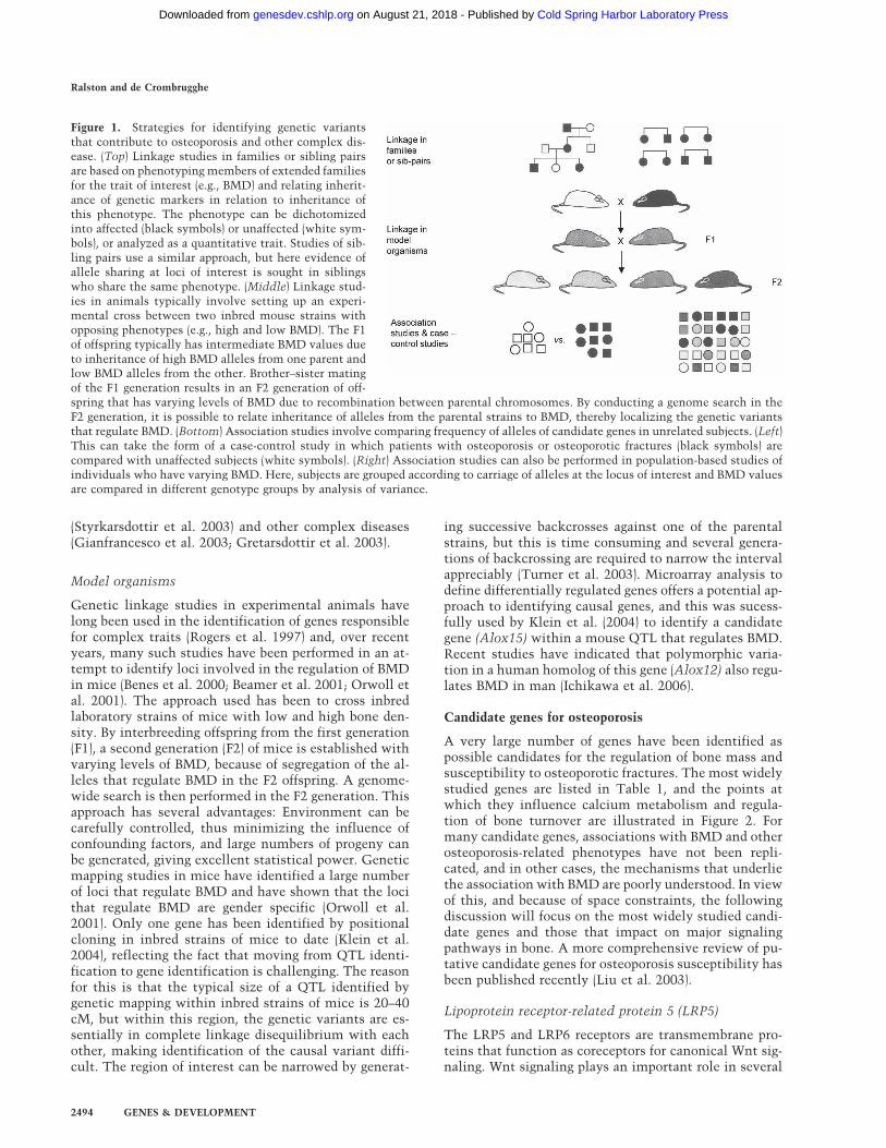
Several approaches have been employed to try to identifythe genes that regulate susceptibility to osteoporosis(Fig. 1), and these are discussed in more detail below.
Association studies
Association studies have been widely used in the field ofosteoporosis genetics. These studies typically involveidentifying polymorphisms of a particular candidate
gene and relating alleles to BMD or osteoporotic frac-tures in a population-based study or a case-control study.Recently, however, it has also become possible to per-form association studies on a genome-wide basis by ana-lyzing a large number of closely spaced single-nucleotidepolymorphisms (SNPs) spread randomly across the ge-nome (Cardon and Abecasis 2003). The rationale forthese studies is that these SNPs will be in linkage dis-equilibrium with causal variants in genes that predis-pose to the disease under study. Classical associationstudies typically involve choosing a candidate gene thatis known to have effects on bone metabolism on thebasis of knowledge about skeletal physiology or bonebiology. Genes that are mutated in rare monogenic bonediseases have also proved to be a rich source of polymor-phisms that regulate BMD in the normal population(Grant et al. 1996; Ferrari et al. 2004; Sobacchi et al.2004; Pettersson et al. 2005). Association studies arerelatively easy to perform and can be powered to detectsmall effects, but spurious results are often reported dueto confounding factors, small sample size, and popula-tion stratification (Ioannidis 2003). In view of this, meta-analysis has been increasingly used to estimate the truesize of the effect of polymorphisms that have been im-plicated in the pathogenesis of complex diseases (Loh-mueller et al. 2003).
Linkage analysis in humans
Linkage studies are a tried and tested way of identifyinggenes responsible for monogenic diseases. Linkage stud-ies have been spectacularly successful in identifying thegenes that are responsible for rare, monogenic bone dis-eases characterized by abnormalities of bone mass suchas osteopetrosis and sclerosing bone dysplasias (Janssensand Van Hul 2002; Balemans et al. 2005). Linkage analy-sis has also been applied to the identification of chromo-somal regions that harbor genes that regulate quantita-tive traits such as bone mass, bone geometry, and ultra-sound properties of bone in the normal population(Koller et al. 2003; Wilson et al. 2003, 2004; Ralston2005). Linkage studies in osteoporosis classically in-volved genotyping a series of polymorphic microsatellitemarkers, typically spread at 10-cM intervals throughoutthe genome, and relating inheritance of alleles to theinheritance of bone mass within the family members.More recently, however, much denser maps of SNP havebeen used for linkage studies of complex disease, andinitial experience suggests that these may be more pow-erful than classical microsatellite-based methods (Saw-cer et al. 2004). A disadvantage of linkage studies in com-plex disease is that they have a relatively low statisticalpower to detect genes that have modest effects on thetrait of interest, even when sample sizes of several thou-sand are used (Altmuller et al. 2001). Reflecting this fact,linkage studies of outbred human populations have so farmet with limited success in terms of identification ofgenes that predispose to osteoporosis, whereas studies ofisolated populations have been more successful in iden-tifying genetic variants that predispose to osteoporosis
Genetics of osteoporosis
GENES & DEVELOPMENT 2493
Cold Spring Harbor Laboratory Press on August 21, 2018 - Published by genesdev.cshlp.orgDownloaded from
(Styrkarsdottir et al. 2003) and other complex diseases(Gianfrancesco et al. 2003; Gretarsdottir et al. 2003).
Model organisms
Genetic linkage studies in experimental animals havelong been used in the identification of genes responsiblefor complex traits (Rogers et al. 1997) and, over recentyears, many such studies have been performed in an at-tempt to identify loci involved in the regulation of BMDin mice (Benes et al. 2000; Beamer et al. 2001; Orwoll etal. 2001). The approach used has been to cross inbredlaboratory strains of mice with low and high bone den-sity. By interbreeding offspring from the first generation(F1), a second generation (F2) of mice is established withvarying levels of BMD, because of segregation of the al-leles that regulate BMD in the F2 offspring. A genome-wide search is then performed in the F2 generation. Thisapproach has several advantages: Environment can becarefully controlled, thus minimizing the influence ofconfounding factors, and large numbers of progeny canbe generated, giving excellent statistical power. Geneticmapping studies in mice have identified a large numberof loci that regulate BMD and have shown that the locithat regulate BMD are gender specific (Orwoll et al.2001). Only one gene has been identified by positionalcloning in inbred strains of mice to date (Klein et al.2004), reflecting the fact that moving from QTL identi-fication to gene identification is challenging. The reasonfor this is that the typical size of a QTL identified bygenetic mapping within inbred strains of mice is 20–40cM, but within this region, the genetic variants are es-sentially in complete linkage disequilibrium with eachother, making identification of the causal variant diffi-cult. The region of interest can be narrowed by generat-
ing successive backcrosses against one of the parentalstrains, but this is time consuming and several genera-tions of backcrossing are required to narrow the intervalappreciably (Turner et al. 2003). Microarray analysis todefine differentially regulated genes offers a potential ap-proach to identifying causal genes, and this was sucess-fully used by Klein et al. (2004) to identify a candidategene (Alox15) within a mouse QTL that regulates BMD.Recent studies have indicated that polymorphic varia-tion in a human homolog of this gene (Alox12) also regu-lates BMD in man (Ichikawa et al. 2006).
Candidate genes for osteoporosis
A very large number of genes have been identified aspossible candidates for the regulation of bone mass andsusceptibility to osteoporotic fractures. The most widelystudied genes are listed in Table 1, and the points atwhich they influence calcium metabolism and regula-tion of bone turnover are illustrated in Figure 2. Formany candidate genes, associations with BMD and otherosteoporosis-related phenotypes have not been repli-cated, and in other cases, the mechanisms that underliethe association with BMD are poorly understood. In viewof this, and because of space constraints, the followingdiscussion will focus on the most widely studied candi-date genes and those that impact on major signalingpathways in bone. A more comprehensive review of pu-tative candidate genes for osteoporosis susceptibility hasbeen published recently (Liu et al. 2003).
Lipoprotein receptor-related protein 5 (LRP5)
The LRP5 and LRP6 receptors are transmembrane pro-teins that function as coreceptors for canonical Wnt sig-naling. Wnt signaling plays an important role in several
Figure 1. Strategies for identifying genetic variantsthat contribute to osteoporosis and other complex dis-ease. (Top) Linkage studies in families or sibling pairsare based on phenotyping members of extended familiesfor the trait of interest (e.g., BMD) and relating inherit-ance of genetic markers in relation to inheritance ofthis phenotype. The phenotype can be dichotomizedinto affected (black symbols) or unaffected (white sym-bols), or analyzed as a quantitative trait. Studies of sib-ling pairs use a similar approach, but here evidence ofallele sharing at loci of interest is sought in siblingswho share the same phenotype. (Middle) Linkage stud-ies in animals typically involve setting up an experi-mental cross between two inbred mouse strains withopposing phenotypes (e.g., high and low BMD). The F1of offspring typically has intermediate BMD values dueto inheritance of high BMD alleles from one parent andlow BMD alleles from the other. Brother–sister matingof the F1 generation results in an F2 generation of off-spring that has varying levels of BMD due to recombination between parental chromosomes. By conducting a genome search in theF2 generation, it is possible to relate inheritance of alleles from the parental strains to BMD, thereby localizing the genetic variantsthat regulate BMD. (Bottom) Association studies involve comparing frequency of alleles of candidate genes in unrelated subjects. (Left)This can take the form of a case-control study in which patients with osteoporosis or osteoporotic fractures (black symbols) arecompared with unaffected subjects (white symbols). (Right) Association studies can also be performed in population-based studies ofindividuals who have varying BMD. Here, subjects are grouped according to carriage of alleles at the locus of interest and BMD valuesare compared in different genotype groups by analysis of variance.
Ralston and de Crombrugghe
2494 GENES & DEVELOPMENT
Cold Spring Harbor Laboratory Press on August 21, 2018 - Published by genesdev.cshlp.orgDownloaded from

key developmental processes, including cell fate deci-sions, limb patterning, and osteoblast and chondrocytedifferentiation, and development of the central nervoussystem and other organs (Johnson et al. 2004). Transduc-tion of the Wnt signal involves both LRP5 and LRP6,which form a receptor complex with Frizzled (Fz) to ac-tivate the transcriptional activity of �-catenin, thedownstream effector of canonical Wnt signaling (Cong etal. 2004). Regulation of signaling through the Wnt–LRP–�-catenin pathway occurs at several levels, and key com-ponents of the pathway are illustrated in Figure 3. Extra-cellular inhibitors of Wnt signaling include the secretedWnt antagonist; Dickkopf (Dkk) proteins, which bind toLRP5 and LRP6 and inhibit Wnt signaling (Bafico et al.2001; Mao and Niehrs 2003); and a family of secretedfrizzled-related proteins (Sfrps), which bind Wnt poly-peptides and function as decoy receptors (Kawano andKypta 2003). Dickkopf proteins can also down-regulateWnt signaling by interacting with the transmembraneprotein Kremen. As a result of this interaction, a com-plex is formed containing Dkk, Kremen, and the LRP5/6proteins that is internalized, thereby down-regulatingWnt signaling (Mao et al. 2002).
The LRP5 pathway was discovered to be a key regula-tor of bone mass following linkage studies in two rarehuman diseases: OPPS, which is a recessively inheritedcondition characterized by severe, early onset osteopo-
rosis and congenital blindness due to vitreous opacity(Gong et al. 1998), and the HBM syndrome that is anasymptomatic autosomal dominant disorder character-ized by increased bone mineral density (Johnson et al.1997). Both of these conditions were mapped to the sameregion of chromosome 11q12 in the late 1990s, and dif-ferent mutations in the LRP5 gene were eventually iden-tified as the cause of both disorders by positional cloningstudies (Gong et al. 2001; Little et al. 2002). The HBMsyndrome was found to be due to a heterozygous mis-sense mutation causing a substitution of valine for gly-cine at codon 171 (G171V) of LRP5 within the first �-pro-peller motif of the molecule (Little et al. 2002). TheOPPS syndrome was found to be due to different homo-zygous missense, nonsense, and frameshift mutationsthroughout the gene (Gong et al. 2001). Since these origi-nal reports, several additional missense mutations ofLRP5 have been identified as a cause of HBM, and all ofthese cluster in or around the first �-propeller motif ofLRP5 (Van Wesenbeeck et al. 2003).
The mutations that cause OPPS produce a truncated ornonfunctional LRP5 protein (Gong et al. 2001), and inaccordance with this, complete inactivation of LRP5 bygene targeting in mice causes a low bone mass pheno-type, which phenocopies the human disease OPPS (Katoet al. 2002). Analysis of bone histomorphometry in LRP5knockout mice has shown that the low bone mass is a
Table 1. Candidate genes for regulation of susceptibility to osteoporosis
Hormones and receptorsBone matrix componentsand degradative enzymes
Androgen receptor � HS2 glycoproteinAromatase Collagen type 1 � Ia
Calcium-sensing receptor OsteocalcinCalcitonin receptor Proline lysine oxidase dehydrogenase-type collagenaseEstrogen receptor �a
Estrogen receptor �
Parathyroid receptor type 1Parathyroid hormoneVitamin D receptora
Cytokines and receptors Osteoblast regulatory factors
Interleukin-1 � and � Alox12 and Alox15a
Interleukin-1 receptor antagonist Bone morphogenic protein 2a
Interleukin-6 Bone morphogenic protein 4a
Tumor necrosis factor receptor 2 Core-binding factor A1a
Tumor necrosis factor Insulin-like growth factor 1Sclerostina
Transforming growth factor �a
Lipoprotein-related receptors 5 and 6a
Peroxisome proliferator-activated receptor �
Miscellaneous Osteoclast-related genes
Apolipoprotein E Cathepsin Ka
Methylene tetrahydrofolate reductase Chloride channel 7a
Klotho Vacuolar proton pump a3 subunita
HLA class II OsteoprotegerinReceptor activator of NF�BReceptor activator of nuclear factor � B ligand
aGenes discussed in this review.
Genetics of osteoporosis
GENES & DEVELOPMENT 2495
Cold Spring Harbor Laboratory Press on August 21, 2018 - Published by genesdev.cshlp.orgDownloaded from
consequence of decreased osteoblast proliferation and re-duced bone matrix deposition rather than an effect ofbone resorption (Kato et al. 2002). The G171V mutationthat was associated with HBM in the family studied byJohnson and colleagues (Johnson et al. 1997; Little et al.2002) was found to cause increased bone mass when ex-pressed in transgenic mice (Babij et al. 2003). In thesestudies, mice were generated that expressed either wild-type human LRP5 or the G171V mutant, and the micethat expressed the mutant had significantly higher BMDthan wild type, despite the fact that levels of transgeneexpression were similar in both mouse strains. Analysisof the skeletal phenotype in the G171V transgenic miceshowed that mineral apposition rate was increased andthe rate of osteoblast apoptosis was reduced, whereaseroded surface (reflecting bone resorption) was unaf-fected. Functional analysis of the HBM-associated muta-tions of LRP5 has shown that the mutations probablycause activation of �-catenin signaling by inhibiting in-teractions between LRP5 and Dkk1. An initial study byBoyden et al. (2002) showed that the G171V mutationdid not result in constitutive activation of LRP5 signal-ing in vitro, but that it impaired Dkk1-mediated inhibi-tion of Wnt-stimulated LRP5 signaling. Another studyreached the same conclusion in showing that severalHBM-associated mutants (G171V, G171R, A214T,A214V, A242T, T253I, and D111Y) were resistant toDkk1 inhibition compared with wild-type LRP5, and hada lower affinity for Dkk1 binding (Ai et al. 2005).
There is evidence that more subtle variations in LRP5could also underlie variations of BMD in the normalpopulation. Several association studies and family-basedstudies have been performed in which various common
polymorphisms of LRP5 have been related to BMD and/or osteoporotic fracture. Most of these studies haveshown evidence of associations between LRP5 allelesand BMD and, interestingly, these associations havebeen particularly strong in men (Ferrari et al. 2004; Kohet al. 2004; Urano et al. 2004; van Meurs et al. 2006).Although many variants have been studied, the mostlikely functional candidate is an alanine-to-valine aminoacid substitution at position 1330 (A1330V). The mecha-nism by which this variant might affect LRP5 signalinghas not been investigated, but evidence of an interactionbetween the LRP5 A1330V variant and a coding poly-morphism of LRP6 (1062V) has also been gained in theRotterdam study (van Meurs et al. 2006), where polymor-phisms of both genes were found to have an additiveeffect on fracture susceptibility. Interactions betweenLRP5 and LRP6 as regulators of BMD have also beenobserved in preclinical studies where skeletal phenotyp-ing of mice with targeted inactivation of both receptorsrevealed allele dose-dependent deficits in BMD and limbformation, suggesting functional redundancy betweenthese two genes in bone and limb development (Holmenet al. 2004).
In conclusion, the data so far indicate that geneticvariation in LRP5 and possibly LRP6 plays an importantrole in regulation of bone mass and osteoporotic frac-tures in humans. Not only do rare mutations in the LRP5gene play a major role in regulating BMD, but moresubtle polymorphisms also seem to regulate BMD in thenormal population. Clearly, other components of thispathway—such as the Wnts, Dkks, Frizzled, Kremen,and Sfrps—now represent prime candidate genes for fur-ther study as potential genetic regulators of bone mass.
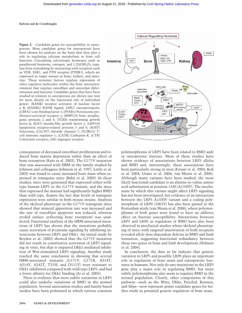
Figure 2. Candidate genes for susceptibility to osteo-porosis. Most candidate genes for osteoporosis havebeen chosen for analysis on the basis that they play arole in regulating calcium metabolism or bone cellfunction. Circulating calciotropic hormones such asparathyroid hormone, estrogen, and 1,25(OH)2D3 regu-late bone remodeling by interacting with receptors suchas VDR, ESR1, and PTH receptor (PTHR1), which areexpressed in target tissues in bone, kidney, and intes-tine. These systemic factors regulate expression ofother regulator molecules within the bone microenvi-ronment that regulate osteoblast and osteoclast differ-entiation and function. Candidate genes that have beenstudied in relation to osteoporosis are shown (see textfor more details on the functional role of individualgenes). (RANK) receptor activator of nuclear factor� B; (RANKL) RANK ligand; (OPG) osteoprotegerin;(CBFA1) core-binding factor 1; (PPAR�) Peroxisome pro-liferator-activated receptor �; (BMP2/4) bone morpho-genic proteins 2 and 4; (TGF�) transforming growthfactor �; (IGF1) insulin-like growth factor 1; (LRP5/6)lipoprotein receptor-related proteins 5 and 6; (SOST)Sclerostin; (CLCN7) chloride channel 7; (TCIRG1) T-cell immune regulator 1; (CATK) Cathepsin K. (CTR)Calcitonin receptor; (AR) angrogen receptor.
Ralston and de Crombrugghe
2496 GENES & DEVELOPMENT
Cold Spring Harbor Laboratory Press on August 21, 2018 - Published by genesdev.cshlp.orgDownloaded from
Transforming growth factor TGF-�1
Members of the TGF-� superfamily of related secretedpolypeptides control critical cellular functions duringembryonic development and also postnatally. Most at-tention has focused on TGF-�1, which is encoded by theTGFB1 gene as a regulator of susceptibility to osteopo-rosis partly because it is particularly abundant in boneand has been shown to have effects on both osteoblastand osteoclast function in vitro (Massagué and Chen2000). The receptor system for TGF� molecules consistsof two different high-affinity receptors, each of whichcontains an extracellular ligand-binding domain, a trans-membrane domain, and an intracellular kinase domain.Upon TGF-� binding, the type II receptor (TBR II) ho-modimer recruits a type I receptor (TBR I) homodimer toform a heterotetrameric receptor complex. Ligand bind-ing to TBR II leads to phosphorylation of a glycine- andserine-rich domain in TBR I, which results in activationof signaling by this receptor. TBR I then phosphorylatesSmad2 and Smad3, two so-called regulatory Smads orR-Smads, each of of which forms a complex with aunique common Smad, Smad4. These complexes trans-locate to the nucleus where they interact with othertranscription factors and also, in the case of Smad3, withspecific DNA sequences to activate a variety of down-stream target genes. Inactivation of components of theTGF-� pathway frequently results in embryonic lethal-ity, but TGF-�1-null mice develop generalized inflam-mation and decreased bone mass with a pronounced re-duction of the number of osteoblasts (Geiser et al. 1998).In addition, Smad3-null mice show osteopenia, which isthought to be due to excessive late-phase osteoblast dif-ferentiation into osteocytes and concomitant apoptosis(Borton et al. 2001).
Polymorphisms of the TGFB1 gene have been exten-sively studied in relation to osteoporosis. One of the ear-liest studies was that of Langdahl et al. (1997), who iden-tified a polymorphism within intron 4 of TGFB1 thatwas associated with severe osteoporosis. Subsequentwork by the same group evaluated the relationship be-tween several polymorphisms in TGFB1 and osteoporo-sis in a case-control study and identified an associationbetween a different polymorphism in intron 5 and BMD(Langdahl et al. 2003). Other research has focused onpolymorphisms in the promoter and first exon of TGFB1in relation to BMD. One of these polymorphisms (a leu-cine-to-proline substitution at codon 10) has been asso-ciated with BMD in some populations (Yamada et al.1999, 2001). Although genetic association studies haveyielded somewhat conflicting results, definitive evi-dence that genetic variation in TGFB1 regulates bonemass in humans comes from the observation that Camu-rati-Engelmann (CED) disease is caused by mutations inTGFB1 (Janssens et al. 2000a; Kinoshita et al. 2000). CEDis a rare autosomal dominant genetic disorder character-ized by hyperostosis and sclerosis, especially affectingthe diaphysis of long bones. Clinically, CED is charac-terized by radiological osteosclerosis, but affected pa-tients also have increased markers of bone resorption,suggesting that bone resorption is elevated as well asbone formation (Hernandez et al. 1997). The causal genefor CED was mapped to chromosome 19q13 by linkageanalysis (Ghadami et al. 2000; Janssens et al. 2000b), andthen mutations in TGFB1 were identified as a cause us-ing a positional candidate approach (Janssens et al.2000a; Kinoshita et al. 2000). Most causal mutations liewithin a region of the gene that encodes the latency-associated peptide (LAP), although two other mutationshave been identified in the signal peptide region, includ-
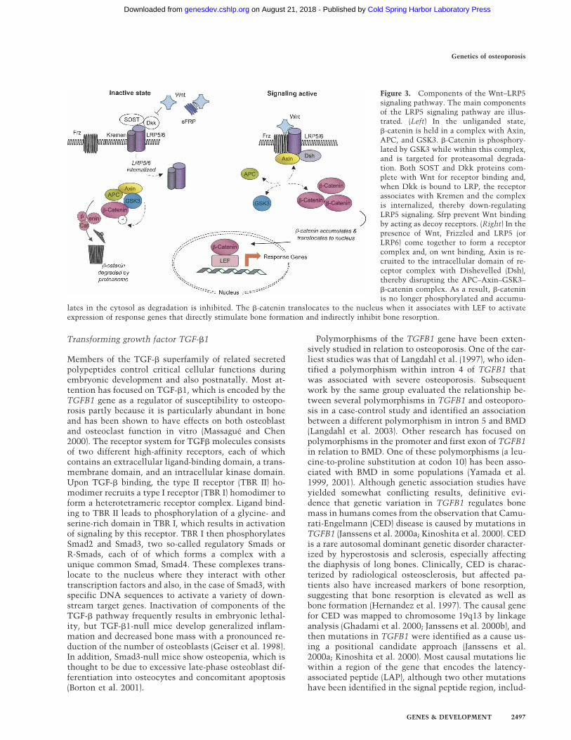
Figure 3. Components of the Wnt–LRP5signaling pathway. The main componentsof the LRP5 signaling pathway are illus-trated. (Left) In the unliganded state,�-catenin is held in a complex with Axin,APC, and GSK3. �-Catenin is phosphory-lated by GSK3 while within this complex,and is targeted for proteasomal degrada-tion. Both SOST and Dkk proteins com-plete with Wnt for receptor binding and,when Dkk is bound to LRP, the receptorassociates with Kremen and the complexis internalized, thereby down-regulatingLRP5 signaling. Sfrp prevent Wnt bindingby acting as decoy receptors. (Right) In thepresence of Wnt, Frizzled and LRP5 (orLRP6) come together to form a receptorcomplex and, on wnt binding, Axin is re-cruited to the intracellular domain of re-ceptor complex with Dishevelled (Dsh),thereby disrupting the APC–Axin–GSK3–�-catenin complex. As a result, �-cateninis no longer phosphorylated and accumu-
lates in the cytosol as degradation is inhibited. The �-catenin translocates to the nucleus when it associates with LEF to activateexpression of response genes that directly stimulate bone formation and indirectly inhibit bone resorption.
Genetics of osteoporosis
GENES & DEVELOPMENT 2497
Cold Spring Harbor Laboratory Press on August 21, 2018 - Published by genesdev.cshlp.orgDownloaded from
ing a duplication of three leucine residues at codon 12and a tyrosine-to-histidine amino acid change at codon81. Following transcription and translation, the matureTGF�1 peptide is cleaved from the LAP but is secretedfrom the cell in an inactive form as a dimer, which isbound noncovalently through disulfide bonds to LAP.These molecules can also form a larger covalent complexwith a latent TGF-�-binding protein (LTBP). Activationof TGF�1, which occurs outside the cell, can occur as theresult of low pH or proteolytic activity and results in therelease of the active TGF�1 dimer from the LAP andother inhibitory molecules so that it can now directlyinteract with the TGF�1 receptors (Janssens et al. 2003).Functional studies have shown that CED-causing muta-tions all act to increase SMAD-dependent TGF�1 signal-ing, but by different mechanisms. Mutations in the LAPdomain appear to inhibit formation of the disulfidebonds that link TGF�1 to the LAP and cause increasedrelease of biologically active TGF�1. However, the othermutations in the signal peptide region prevent TGF�1secretion and result in intracellular accumulation ofTGF-�1 with stimulation of SMAD signaling by an in-tracrine mechanism that is poorly understood.
Bone morphogenic proteins (BMPs)
BMPs are part of the TGF� superfamily of molecules.Signaling through BMP is regulated extracellularly byantagonists such as noggin and chordin (De Robertis andKuroda 2004), and intracellularly by the Smad family ofsignaling proteins that act downstream from specificBMP receptors (Cao and Chen 2005). BMPs are strongcandidate genes for regulation of bone mass, and natu-rally occurring mutations in various components of theBMP pathway result in defects of limb formation(Seemann et al. 2005). Recent studies indicate that poly-morphic variation in members of the BMP family may beinvolved in regulation of bone mass and susceptibility toosteoporosis. A linkage study in Icelanders identified alocus for regulation of BMD on chromosome 20p12(Styrkarsdottir et al. 2003), and subsequent positionalcloning studies showed that the linkage signal was at-tributable in part to genetic variation in the BMP2 gene.A coding polymorphism was identified at codon 37 of theBMP2 protein, resulting in a substitution of alanine for aserine residue (Ser37Ala) that was overrepresented in Ice-landic patients and in Danish patients with osteoporosis(Styrkarsdottir et al. 2003). The mechanisms by whichthis substitution affects BMP signaling to affect bonemass have not yet been investigated. Other workers havereported that common polymorphism, which causes analanine-to-valine substitution at position 152 in theBMP4 protein (Ala152Val), is associated with low BMD(Ramesh et al. 2005). These association studies raise thepossibility that polymorphic variation in members of theBMP family may be involved in osteoporosis, but furtherwork will be required to confirm these observations andto study the mechanism by which the coding variants sofar identified affect bone cell function and modulatebone mass.
Sclerostin
Sclerostin is a cysteine knot-containing protein thatshares homology with the DAN family of BMP antago-nists (Winkler et al. 2003; van Bezooijen et al. 2004).Sclerostin was first identified as a regulator of bone massby genetic mapping studies in the sclerosing bone dys-plasias Sclerosteosis and van Buchem disease. These arerare conditions with a similar phenotype that are inher-ited in an autosomal recessive manner. Both are charac-terized by progressive osteosclerosis mainly affecting theskull, mandible, and tubular bones of the extremities(Beighton et al. 1984). Sclerosteosis differs from vanBuchem disease because it is more severe and is associ-ated with hand malformations such as syndactyly, ab-sent or dysplastic nails, and radial deviation of the ter-minal phalanges (Balemans et al. 2005). Both diseaseswere mapped to the same region of chromosome 17q12-21 in the late 1990s (Van Hul et al. 1998; Balemans et al.1999), and positional cloning studies identified recessiveloss-of-function mutations in Sclerostin (SOST) gene asthe cause of Sclerosteosis in 2001 (Balemans et al. 2001;Brunkow et al. 2001). Interestingly, mutation screeningof SOST in patients with van Buchem disease showed nomutations, but a homozygous 52-Kb deletion was iden-tified 35 Kb downstream from SOST as the cause of thiscondition (Balemans et al. 2002b). Subsequent work hasshown that the deleted region contains highly conservedregulatory elements that play a key role in regulatingSOST expression in bone (Loots et al. 2005). Althoughheterozygous carriers of SOST mutations are asymptom-atic, they have significantly increased BMD, indicatingthat haploinsufficiency of SOST increases bone masswithout apparently causing adverse effects (Gardner etal. 2005).
Polymorphisms of SOST have also been associatedwith BMD in some population-based studies. In the Rot-terdam study (Uitterlinden et al. 2004), polymorphismsin the SOST promoter and the region deleted in vanBuchem disease were both found to be associated withBMD, and evidence was gained to suggest that the effectbecame more pronounced with increasing age. In an-other case-control study, SOST polymorphisms were notfound to be associated with BMD (Balemans et al. 2002a),although this was a relatively small study and partici-pants were of a younger age group than in the Rotterdamstudy.
In view of the structural similarity between SOST andother BMP antagonists, it was initially assumed thatSOST regulates bone mass by affecting BMP signaling(Winkler et al. 2003). In keeping with this view was theobservation that Sclerostin binds to both BMP5 andBMP6, competes with BMP6 for binding to the type IBMP receptor, and antagonizes the effects of BMP6 onSmad signaling (Winkler et al. 2003). Transgenic micethat overexpress SOST are osteopenic and have reducedbone formation, consistent with a model whereby SOSTnegatively regulates osteoblast function (Winkler et al.2003). However, studies by van Bezooijen et al. (2004)showed that SOST was not a classical BMP antagonist,
Ralston and de Crombrugghe
2498 GENES & DEVELOPMENT
Cold Spring Harbor Laboratory Press on August 21, 2018 - Published by genesdev.cshlp.orgDownloaded from
suggesting that it might interact with other signalingpathways to regulate bone formation. The mechanismsby which SOST inhibits bone formation has been clari-fied by recent studies, which have shown that SOST di-rectly interacts with LRP5 and LRP6 and antagonizesWnt signaling both in vitro and in vivo (Li et al. 2005;Semenov et al. 2005). However, other workers have pro-posed that the inhibitory effect of SOST on Wnt signal-ing is indirect and mediated through BMP production(Winkler et al. 2005). While further studies will be re-quired to clarify the mechanisms by which SOST affectsbone formation, it seems possible that the increasedbone mass that results from SOST deficiency is medi-ated, at least in part, through the LRP5–�-catenin path-way.
CBFA1
The CBFA1 gene (also known as Runx2) plays an essen-tial role in regulating osteoblast differentiation, sincemice that are deficient in this transcription factor havecomplete absence of bone (Komori et al. 1997; Otto et al.1997). Moreover, mice with haploinsufficiency ofCBFA1 phenocopy the human syndrome of cleidocranialdysplasia (CCD), a skeletal disorder characterized byshort stature, hypoplasia or aplasia of the clavicles,patent fontanelles, supernumerary teeth, and other de-fects in skeletal patterning and growth (Otto et al. 1997).The human syndrome of CCD is caused by various mis-sense, nonsense, and frameshift mutations of CBFA1(Lee et al. 1997; Mundlos et al. 1997; Quack et al. 1999).Some of these mutations have been shown to interferewith the DNA-binding activity of CBFA1, whereas oth-ers have been found to alter nuclear localization of theprotein or to produce a mutant or truncated protein thatis biologically inactive. In addition to these rare muta-tions, various polymorphisms have been identified inCBFA1 and some of these have been associated withbone mass in population-based studies (Vaughan et al.2002, 2004; Doecke et al. 2006). The best functional can-didates lie within the Runx2 promoter (Doecke et al.2006) or within polyalanine and polyglutamine repeatsin exon 1. The polyalanine and polyglutamine repeatsare of interest since they lie within one of the transacti-vation domains of Runx2. Various polymorphic varia-tions have been identified in this region, including an18-bp deletion that results in a polyalanine repeat of 11residues (11 Ala) compared with the more common re-peat of 17 residues (17 Ala). Various rare length variantswithin the polyglutamine repeat have also been identi-fied, resulting in stretches of between 15 and 30 repeats.The strongest association with BMD has been observedwith an anonymous polymorphism in the Ala repeat re-gion (Vaughan et al. 2002, 2004), although it is thoughtthat this might be due to linkage disequilibrium withpolymorphisms in the promoter that have been shown toaffect Runx2 transcription in reporter assays (Doecke etal. 2006). It is currently unclear whether the length vari-ants in the polyalanine and polyglutamine tracts have
functional importance, but this is an area of ongoing in-vestigation.
Cathepsin K
Mutations in Cathepsin K are responsible for the raresyndrome of Pycnodysostosis, which is a rare recessivebone dysplasia characterized by osteosclerosis and shortstature (Gelb et al. 1996). Various disease-causing muta-tions have been identified that result in production ofeither a truncated or a nonfunctional protein. Studies ofmore common polymorphic variants of Cathepsin K inrelation to BMD regulation in the normal populationhave so far yielded negative results (Giraudeau et al. 2004).
TCIRG1
The TCIRG1 gene encodes the APT6i subunit of the os-teoclast-specific proton pump, and various inactivatingmutations in TCIRGI have been described that are re-sponsible for a subset of patients with recessive osteope-trosis (Frattini et al. 2000). Recent work indicates thatpolymorphisms of TCIRG1 might also contribute toregulation of BMD in the normal population; a study byFrattini (Sobacchi et al. 2004) showed evidence of an as-sociation between a polymorphism affecting an AP1-binding site in the TCIRG1 promoter and BMD in peri-menopausal women. Functional studies need to be per-formed to identify the mechanisms that underlie thisassociation, however, and to replicate the finding inother populations.
CLCN7
The CLCN7 gene encodes a chloride channel that ishighly expressed in osteoclasts and is essential for acidi-fication of the resorption lacuna. Homozygous inactiva-tion mutations in CLCN7 cause a severe form of reces-sive osteopetrosis (Kornak et al. 2001), whereas hetero-zygous missense mutations of CLCN7 cause autosomaldominant osteopetrosis (Balemans et al. 2005). Promptedby this observation, Pettersson et al. (2005) looked forevidence of an association between polymorphisms ofCLCN7 and BMD in normal individuals and found that acommon polymorphism in exon 15 of CLCN7 that re-sults in a methionine-to-valine amino acid change wasassociated with BMD in normal women. Further studieswill be required to determine if this polymorphism isfunctionally important and to replicate the observationin other populations.
Vitamin D receptor (VDR)
Vitamin D, through its principal bioactive form 1,25-dihydroxyvitamin D3[1,25-(OH)2D3] plays a crucial rolein bone metabolism. The action of 1,25-(OH)2D3 is me-diated through a specific hormone-receptor (VDR) thatregulates gene expression by forming a heterodimer withretinoic X receptor (RXR) that binds to vitamin D re-
Genetics of osteoporosis
GENES & DEVELOPMENT 2499
Cold Spring Harbor Laboratory Press on August 21, 2018 - Published by genesdev.cshlp.orgDownloaded from
sponse elements in target genes. Mutations in VDRcause the syndrome of vitamin D-resistant rickets whichis a recessive condition characterized by alopecia, hypo-calcaemia, hypophosphatemia, and severe rickets, and isresistant to treatment with vitamin D and its active me-tabolites (Kristjansson et al. 1993). Targeted inactivationof the VDR gene in mice provides a phenocopy of thehuman syndrome (Bouillon et al. 2003), but when thesemice are fed a diet rich in calcium and phosphorus tonormalize serum calcium and PTH concentrations, theydevelop normally without bone anomalies. It thereforeappears that the bone defects that result from VDR de-ficiency are due to malabsorption of calcium and phos-phate by the intestine rather than absence of 1,25-(OH)2D3 signaling in bone cells. The VDR was the firstcandidate gene to be studied in relation to osteoporosis,and most attention has focused on polymorphisms situ-ated near the 3� flank of VDR recognized by the restric-tion enzymes BsmI, ApaI, and TaqI. A recent retrospec-tive meta-analysis of association studies that genotypedthe BsmI polymorphism concluded that there was evi-dence of an association between spine BMD and theBsmI polymorphism, equivalent to ∼0.15 Z-score unitsbetween the BB genotype and the other genotypes (Thak-kinstian et al. 2004). No association with femoral neckBMD was observed. Another polymorphism affectingexon 2 of VDR has been described that creates an alter-native translational start site, resulting in the productionof two isoforms of the VDR protein, which differ inlength by three amino acids (Gross et al. 1997). This hasbeen associated with BMD in some, but not other, popu-lations, and functional studies of this polymorphismhave yielded inconclusive results (Gross et al. 1998). Afurther polymorphism has been identified in the pro-moter of VDR at a binding site for the transcription fac-tor Cdx-2, which has been associated with BMD in Japa-nese subjects and appears to be functional (Arai et al.2001). This polymorphism has been associated with frac-ture in other populations, but not with BMD (Fang et al.2003). The most comprehensive study of VDR alleles inrelation to osteoporosis-related phenotypes was that ofFang et al. (2005), who conducted a large-scale study ofhaplotype tagging SNP of VDR in 6418 participants ofthe Rotterdam study. As the result of this analysis, theauthors identified haplotype alleles in the promoter re-gion and 3� untranslated region (UTR) that were associ-ated with an increased risk of fracture. For a subgroup ofindividuals who carried risk alleles at both sites, the frac-ture risk was significantly increased by 48% when com-pared with control subjects. Functional studies alsoshowed that the promoter haplotype that increased frac-ture risk was associated with reduced VDR expression inreporter assays, whereas the 3� UTR risk haplotype wasassociated with increased degradation of VDR mRNA.The data would be consistent with a model whereby thecombination of risk haplotypes results in a lower VDRmRNA level due to decreased transcription and in-creased degradation. Interestingly, the risk alleles forfracture identified in this study were not associated withdifferences in BMD. In view of this, the mechanism by
which these polymorphisms predispose to fracture is un-clear, although a possibility, alluded to by the authors,was through an effect on bone geometry. Although thiswas a large and well-conducted study, the risk estimateswere modest, and if correction had been applied for allthe combinations of haplotypes tested (and their inter-actions), the results would not have been significant.
Collagen type I� I
The gene encoding the � I chain of type I collagen (COLIA1)is an important functional candidate for the pathogen-esis of osteoporosis, since type I collagen is the majorprotein of bone and since mutations in this gene causethe syndrome of osteogenesis imperfecta—a rare diseasecharacterized by increased bone fragility and reducedBMD (Boyde et al. 1999). Previous research identifiedassociations between BMD and polymorphisms withinthe proximal promoter of COLIA1 (Garcia-Giralt et al.2002) and within the first intron of COLIA1 (Grant et al.1996). Most research has focused on a polymorphismwithin intron 1, which is situated at a binding site for thetranscription factor Sp1. The COLIA1 Sp1-binding sitepolymorphism has been associated with various osteo-porosis-related phenotypes, including bone density(Grant et al. 1996; Uitterlinden et al. 1998), fragility frac-tures (Grant et al. 1996; Uitterlinden et al. 1998), post-menopausal bone loss (Harris et al. 2000; MacDonald etal. 2001), bone geometry (Qureshi et al. 2001), bone qual-ity (Mann et al. 2001), and bone mineralization (Stewartet al. 2005). Functional analysis has shown that the os-teoporosis-associated T allele (“s”) of the Sp1 polymor-phism is associated with increased DNA–protein bind-ing, increased transcription, and abnormally increasedproduction of the COLIA1 mRNA and protein (Mann etal. 2001). It is thought that the resulting imbalance be-tween the COLIA1 and COLIA2 chains contributes toimpairment of bone strength and reduced bone mass incarriers of the T allele by subtly affecting bone mineral-ization (Stewart et al. 2005). Retrospective meta-analy-ses of published studies have concluded that carriage ofthe T allele is associated with reduced BMD at the lum-bar spine and femoral neck and with vertebral fractures(Efstathiadou et al. 2001; Mann and Ralston 2003). More-over, homozygotes for the T allele of the Sp1 polymor-phism were recently found to be associated with BMDand incident vertebral fractures in a large prospectivemeta-analysis of >20,000 participants of the GENOMOSstudy (Ralston et al. 2006). In the GENOMOS study,however, the BMD association was only observed for ho-mozygotes for the T allele, in contrast to previous stud-ies where heterozygotes also showed a reduction in BMD(Mann and Ralston 2003). The association with vertebralfracture in GENOMOS was similar to previous reportsand equivalent to a 40% increase in fracture risk for eachcopy of the T allele carried. Interestingly, the associationbetween COLIA1 alleles and vertebral fracture reportedin GENOMOS and other studies was not fully explainedon the basis of reduced bone density, implying that theSp1 allele also acts as a marker for bone quality. The
Ralston and de Crombrugghe
2500 GENES & DEVELOPMENT
Cold Spring Harbor Laboratory Press on August 21, 2018 - Published by genesdev.cshlp.orgDownloaded from
promoter polymorphisms of COLIA1 are in strong link-age disequilibrium with the Sp1 polymorphism, and twostudies have now suggested that an extended haplotypedefined by the Sp1 polymorphism and other promoterpolymorphism may exert stronger effects on BMD thanthe individual polymorphisms (Garcia-Giralt et al. 2002;Stewart et al. 2006). Evidence has been presented to sug-gest the promoter polymorphism at position −1663 in-teracts with the transcription factor NMP4, which playsa role in osteoblast differentiation by interacting withSmads (Garcia-Giralt et al. 2005). While a functional in-teraction has been reported between the −1663 and−1997 polymorphisms in regulating COLIA1 transcrip-tion (Garcia-Giralt et al. 2005), it is currently unclearwhether the −1997 polymorphism is also at the site oftranscription factor binding.
Estrogen receptor �
The estrogen receptor �, encoded by the ESR1 gene, isanother important functional candidate for the regula-tion of bone mass. A large number of investigators havelooked for evidence of an association between ESR1 al-leles and BMD, mostly focusing on two polymorphismswithin intron 1 recognized by the XbaI and PvuII restric-tion enzymes, and on a TA repeat in the promoter. Stud-ies of ESR1 alleles in relation to BMD have yielded in-consistent results, possibly because most studies havebeen of small sample size and involved subjects of dif-ferent ages, menopausal status, and ethnic backgrounds(Ioannidis et al. 2002). A meta-analysis of published stud-ies showed evidence of an association between the XbaIpolymorphisms and both BMD and fractures, withhigher BMD values in “XX” homozygotes and a reducedrisk of fractures in association with this genotype (Ioan-nidis et al. 2002). More recently, a prospective meta-analysis of data from >18,000 subjects in the GENOMOSstudy confirmed that “XX” homozygotes had a reducedrisk of fracture (Ioannidis et al. 2004). In this study, noassociation with BMD was observed, indicating thatESR1 influences fracture risk independent of an effect onBMD. The mechanism responsible for this observationremains unclear, but one possibility may be an effect onbone quality, since ESR1 alleles have been associatedwith ultrasound properties of bone and bone loss (Al-bagha et al. 2005). There has been controversy as towhether the associations between BMD and polymor-phisms in the 5� region of the ESR1 gene are driven bythe intron 1 polymorphisms or the TA repeat polymor-phism in the ESR1 promoter. Previous studies haveshown that the PvuII polymorphism lies within consen-sus recognition sites for the AP4 and Myb transcriptionfactors; studies using promoter-reporter assays haveshown that the PvuII polymorphism influences Myb-driven transcription in vitro (Herrington et al. 2002); andother studies have shown that both the XbaI and PvuIIpolymorphisms also influence reporter gene transcrip-tion in vitro (Maruyama et al. 2000). In this regard, it isof interest that the PvuII and XbaI polymorphisms arelocated within a region that is 70%–80% conserved in
the human, mouse, and rat genomes, whereas the TArepeat polymorphism is not conserved to any significantextent across species. These data are consistent with adirect functional effect of the PvuII and XbaI polymor-phisms on ESR1 gene transcription, but it remains pos-sible that the intron 1 polymorphisms are simply in link-age disequilibrium (LD) with causal polymorphismselsewhere in the ESR1 gene.
Concluding remarks
Many advances have been made in understanding therole of genetic factors in osteoporosis since publicationof the first paper in the field in 1994 that showed a strongassociation between VDR alleles and BMD (Morrison etal. 1994). At this time it was believed that osteoporosissusceptibility would be determined by a few genes ofmajor effect and that these would pave the way for spe-cific targeted therapies and provide genetic markers toassess disease risk. It has now become clear that osteo-porosis susceptibility is mediated by a large number ofgenetic variants of modest effect size. Although thereremains a prospect that genotyping for these variantscould help assess the risk of osteoporosis, or complica-tions such as fracture, it is likely that tens, or hundreds,of informative variants would have to be tested to be ofreal diagnostic value. Since some of the genetic variantsthat predispose to osteoporosis seem to do so by mecha-nisms independent of BMD, it could be that these mightbe successfully combined with BMD measurements toimprove risk assessment for complications of osteoporo-sis such as fragility fractures. At the present time, manycommon polymorphic variants of candidate genes havebeen identified that contribute to osteoporosis suscepti-bility in specific studies. Relatively few candidate genepolymorphisms have been validated by large-scale stud-ies, however, and much work remains to be done to iden-tify genetic variants that are consistently associatedwith osteoporosis-related phenotypes and to determinewhether they will represent useful diagnostic tools andmolecular targets for therapeutic manipulation.
References
Ai, M., Holmen, S.L., Van Hul, W., Williams, B.O., and War-man, M.L. 2005. Reduced affinity to and inhibition by DKK1form a common mechanism by which high bone mass-asso-ciated missense mutations in LRP5 affect canonical Wnt sig-naling. Mol. Cell. Biol. 25: 4946–4955.
Albagha, O.M., Pettersson, U., Stewart, A., McGuigan, F.E.,MacDonald, H.M., Reid, D.M., and Ralston, S.H. 2005. As-sociation of oestrogen receptor � gene polymorphisms withpostmenopausal bone loss, bone mass, and quantitative ul-trasound properties of bone. J. Med. Genet. 42: 240–246.
Altmuller, J., Palmer, L.J., Fischer, G., Scherb, H., and Wjst, M.2001. Genomewide scans of complex human diseases: Truelinkage is hard to find. Am. J. Hum. Genet. 69: 936–950.
Andrew, T., Antioniades, L., Scurrah, K.J., MacGregor, A.J., andSpector, T.D. 2005. Risk of wrist fracture in women is heri-table and is influenced by genes that are largely independentof those influencing BMD. J. Bone Miner. Res. 20: 67–74.
Genetics of osteoporosis
GENES & DEVELOPMENT 2501
Cold Spring Harbor Laboratory Press on August 21, 2018 - Published by genesdev.cshlp.orgDownloaded from
Arai, H., Miyamoto, K.I., Yoshida, M., Yamamoto, H., Taketani,Y., Morita, K., Kubota, M., Yoshida, S., Ikeda, M., Watabe, F.,et al. 2001. The polymorphism in the caudal-related ho-meodomain protein Cdx-2 binding element in the humanvitamin D receptor gene. J. Bone Miner. Res. 16: 1256–1264.
Arden, N.K. and Spector, T.D. 1997. Genetic influences onmuscle strength, lean body mass, and bone mineral density:A twin study. J. Bone Miner. Res. 12: 2076–2081.
Arden, N.K., Baker, J., Hogg, C., Baan, K., and Spector, T.D.1996. The heritability of bone mineral density, ultrasound ofthe calcaneus and hip axis length: A study of postmeno-pausal twins. J. Bone Miner. Res. 11: 530–534.
Babij, P., Zhao, W., Small, C., Kharode, Y., Yaworsky, P.J., Boux-sein, M.L., Reddy, P.S., Bodine, P.V., Robinson, J.A., Bhat, B.,et al. 2003. High bone mass in mice expressing a mutantLRP5 gene. J. Bone Miner. Res. 18: 960–974.
Bafico, A., Liu, G., Yaniv, A., Gazit, A., and Aaronson, S.A.2001. Novel mechanism of Wnt signalling inhibition medi-ated by Dickkopf-1 interaction with LRP6/Arrow. Nat. CellBiol. 3: 683–686.
Balemans, W., Van Den Ende, J., Freire Paes-Alves, A., Dikkers,F.G., Willems, P.J., Vanhoenacker, F., Almeida-Melo, N.,Alves, C.F., Stratakis, C.A., Hill, S.C., et al. 1999. Localiza-tion of the gene for sclerosteosis to the van Buchem disease-gene region on chromosome 17q12-q21. Am. J. Hum. Genet.64: 1661–1669.
Balemans, W., Ebeling, M., Patel, N., Van Hul, E., Olson, P.,Dioszegi, M., Lacza, C., Wuyts, W., Van Den Ende, J., Wil-lems, P., et al. 2001. Increased bone density in sclerosteosisis due to the deficiency of a novel secreted protein (SOST).Hum. Mol. Genet. 10: 537–543.
Balemans, W., Foernzler, D., Parsons, C., Ebeling, M., Thomp-son, A., Reid, D.M., Lindpaintner, K., Ralston, S.H., and VanHul, W. 2002a. Lack of association between the SOST geneand bone mineral density in perimenopausal women: Analy-sis of five polymorphisms. Bone 31: 515–519.
Balemans, W., Patel, N., Ebeling, M., Van Hul, E., Wuyts, W.,Lacza, C., Dioszegi, M., Dikkers, F.G., Hildering, P., Wil-lems, P.J., et al. 2002b. Identification of a 52 kb deletiondownstream of the SOST gene in patients with van Buchemdisease. J. Med. Genet. 39: 91–97.
Balemans, W., Van Wesenbeeck, L., and Van Hul, W. 2005. Aclinical and molecular overview of the human osteopetroses.Calcif. Tissue Int. 77: 263–274.
Beamer, W.G., Shultz, K.L., Donahue, L.R., Churchill, G.A.,Sen, S., Wergedal, J.R., Baylink, D.J., and Rosen, C.J. 2001.Quantitative trait loci for femoral and lumbar vertebral bonemineral density in C57BL/6J and C3H/HeJ inbred strains ofmice. J. Bone Miner. Res. 16: 1195–1206.
Beighton, P., Barnard, A., Hamersma, H., and van der Wouden,A. 1984. The syndromic status of sclerosteosis and vanBuchem disease. Clin. Genet. 25: 175–181.
Benes, H., Weinstein, R.S., Zheng, W., Thaden, J.J., Jilka, R.L.,Manolagas, S.C., and Shmookler Reis, R.J. 2000. Chromo-somal mapping of osteopenia-associated quantitative traitloci using closely related mouse strains. J. Bone Miner. Res.15: 626–633.
Bilezikian, J.P., Morishima, A., Bell, J., and Grumbach, M.M.1998. Increased bone mass as a result of estrogen therapy ina man with aromatase deficiency. N. Engl. J. Med. 339: 599–603.
Borton, A.J., Frederick, J.P., Datto, M.B., Wang, X.F., and Wein-stein, R.S. 2001. The loss of Smad3 results in a lower rate ofbone formation and osteopenia through dysregulation of os-teoblast differentiation and apoptosis. J. Bone Miner. Res. 16:
1754–1764.Bouillon, R., Van Cromphaut, S., and Carmeliet, G. 2003. Intes-
tinal calcium absorption: Molecular vitamin D mediatedmechanisms. J. Cell. Biochem. 88: 332–339.
Boyde, A., Travers, R., Glorieux, F.H., and Jones, S.J. 1999. Themineralization density of iliac crest bone from children withosteogenesis imperfecta. Calcif. Tissue Int. 64: 185–190.
Boyden, L.M., Mao, J., Belsky, J., Mitzner, L., Farhi, A., Mitnick,M.A., Wu, D., Insogna, K., and Lifton, R.P. 2002. High bonedensity due to a mutation in LDL-receptor-related protein 5.N. Engl. J. Med. 346: 1513–1521.
Brunkow, M., Gardner, J., Van Ness, J., Paeper, B., Kovacevich,B., Proll, S., Skonier, J., Zhao, L., Sabo, P., Fu, Y., et al. 2001.Bone dysplasia sclerosteosis results from loss of the sost geneproduct, a novel cystine knot-containing protein. Am. J.Hum. Genet. 68: 577–589.
Cao, X. and Chen, D. 2005. The BMP signaling and in vivo boneformation. Gene 357: 1–8.
Cardon, L.R. and Abecasis, G.R. 2003. Using haplotype blocksto map human complex trait loci. Trends Genet. 19: 135–140.
Christian, J.C., Yu, P.L., Slemenda, C.W., and Johnston, C.C.1989. Heritability of bone mass: A longitudinal study in ag-ing male twins. Am. J. Hum. Genet. 44: 429–433.
Cong, F., Schweizer, L., and Varmus, H. 2004. Wnt signalsacross the plasma membrane to activate the �-catenin path-way by forming oligomers containing its receptors, Frizzledand LRP. Development 131: 5103–5115.
Cummings, S.R., Nevitt, M.C., Browner, W.S., Stone, K., Fox,K.M., Ensrud, K.E., Cauley, J., Black, D., and Vogt, T.M.1995. Risk factors for hip fracture in white women. Study ofOsteoporotic Fractures Research Group. N. Engl. J. Med.332: 767–773.
Deng, H.W., Chen, W.M., Recker, S., Stegman, M.R., Li, J.L.,Davies, K.M., Zhou, Y., Deng, H., Heaney, R., and Recker,R.R. 2000. Genetic determination of Colles’ fracture and dif-ferential bone mass in women with and without Colles’ frac-ture. J. Bone Miner. Res. 15: 1243–1252.
De Robertis, E.M. and Kuroda, H. 2004. Dorsal–ventral pattern-ing and neural induction in Xenopus embryos. Annu. Rev.Cell. Dev. Biol. 20: 285–308.
Doecke, J.D., Day, C.J., Stephens, A.S., Carter, S.L., van Daal,A., Kotowicz, M.A., Nicholson, G.C., and Morrison, N.A.2006. Association of functionally different RUNX2 P2 pro-moter alleles with BMD. J. Bone Miner. Res. 21: 265–273.
Efstathiadou, Z., Tsatsoulis, A., and Ioannidis, J.P. 2001. Asso-ciation of collagen I� 1 Sp1 polymorphism with the risk ofprevalent fractures: A meta-analysis. J. Bone Miner. Res. 16:1586–1592.
Fang, Y., van Meurs, J.B., Bergink, A.P., Hofman, A., van Duijn,C.M., van Leeuwen, J.P., Pols, H.A., and Uitterlinden, A.G.2003. Cdx-2 polymorphism in the promoter region of thehuman vitamin D receptor gene determines susceptibility tofracture in the elderly. J. Bone Miner. Res. 18: 1632–1641.
Fang, Y., van Meurs, J.B., d’Alesio, A., Jhamai, M., Zhao, H.,Rivadeneira, F., Hofman, A., van Leeuwen, J.P., Jehan, F.,Pols, H.A., et al. 2005. Promoter and 3�-untranslated-regionhaplotypes in the vitamin D receptor gene predispose to os-teoporotic fracture: The Rotterdam study. Am. J. Hum.Genet. 77: 807–823.
Ferrari, S.L., Deutsch, S., Choudhury, U., Chevalley, T., Bon-jour, J.P., Dermitzakis, E.T., Rizzoli, R., and Antonarakis,S.E. 2004. Polymorphisms in the low-density lipoprotein re-ceptor-related protein 5 (LRP5) gene are associated withvariation in vertebral bone mass, vertebral bone size, and
Ralston and de Crombrugghe
2502 GENES & DEVELOPMENT
Cold Spring Harbor Laboratory Press on August 21, 2018 - Published by genesdev.cshlp.orgDownloaded from
stature in whites. Am. J. Hum. Genet. 74: 866–875.Frattini, A., Orchard, P.J., Sobacchi, C., Giliani, S., Abinun, M.,
Mattsson, J.P., Keeling, D.J., Andersson, A.K., Wallbrandt,P., Zecca, L., et al. 2000. Defects in TCIRG1 subunit of thevacuolar proton pump are responsible for a subset of humanautosomal recessive osteopetrosis. Nat. Genet. 25: 343–346.
Garcia-Giralt, N., Nogues, X., Enjuanes, A., Puig, J., Mel-libovsky, L., Bay-Jensen, A., Carreras, R., Balcells, S., Diez-Perez, A., and Grinberg, D. 2002. Two new single nucleotidepolymorphisms in the COLIA1 upstream regulatory regionand their relationship with bone mineral density. J. BoneMiner. Res. 17: 384–393.
Garcia-Giralt, N., Enjuanes, A., Bustamante, M., Mellibovsky,L., Nogues, X., Carreras, R., Ez-Perez, A., Grinberg, D., andBalcells, S. 2005. In vitro functional assay of alleles and hap-lotypes of two COL1A1-promoter SNPs. Bone 36: 902–908.
Gardner, J.C., van Bezooijen, R.L., Mervis, B., Hamdy, N.A.,Lowik, C.W., Hamersma, H., Beighton, P., and Papapoulos,S.E. 2005. Bone mineral density in sclerosteosis; affected in-dividuals and gene carriers. J. Clin. Endocrinol. Metab. 90:6392–6395.
Geiser, A.G., Zeng, Q.Q., Sato, M., Helvering, L.M., Hirano, T.,and Turner, C.H. 1998. Decreased bone mass and bone elas-ticity in mice lacking the transforming growth factor-�1gene. Bone 23: 87–93.
Gelb, B.D., Shi, G.P., Chapman, H.A., and Desnick, R.J. 1996.Pycnodysostosis, a lysosomal disease caused by cathepsin Kdeficiency. Science 273: 1236–1238.
Ghadami, M., Makita, Y., Yoshida, K., Nishimura, G., Fuku-shima, Y., Wakui, K., Ikegawa, S., Yamada, K., Kondo, S.,Niikawa, N., et al. 2000. Genetic mapping of the Camurati-Engelmann disease locus to chromosome 19q13.1-q13.3.Am. J. Hum. Genet. 66: 143–147.
Gianfrancesco, F., Esposito, T., Ombra, M.N., Forabosco, P.,Maninchedda, G., Fattorini, M., Casula, S., Vaccargiu, S.,Casu, G., Cardia, F., et al. 2003. Identification of a novel geneand a common variant associated with uric acid nephroli-thiasis in a Sardinian genetic isolate. Am. J. Hum. Genet. 72:1479–1491.
Giraudeau, F.S., McGinnis, R.E., Gray, I.C., O’Brien, E.J., Don-caster, K.E., Spurr, N.K., Ralston, S.H., Reid, D.M., andWood, J. 2004. Characterization of common genetic variantsin cathepsin K and testing for association with bone mineraldensity in a large cohort of perimenopausal women fromScotland. J. Bone Miner. Res. 19: 31–41.
Gong, Y., Vikkula, M., Boon, L., Lui, J., Beighton, P., Ramesar,R., Peltonen, L., Somer, H., Hirose, T., Dallapicola, B., et al.1998. Osteoporosis-pseudoglioma syndrome, a disorder af-fecting skeletal strength and vision, is assigned to chromo-some region 11q12-13. Am. J. Hum. Genet. 1996: 146–151.
Gong, Y., Slee, R.B., Fukai, N., Rawadi, G., Roman-Roman, S.,Reginato, A.M., Wang, H., Cundy, T., Glorieux, F.H., Lev,D., et al. 2001. LDL receptor-related protein 5 (LRP5) affectsbone accrual and eye development. Cell 107: 513–523.
Grant, S.F.A., Reid, D.M., Blake, G., Herd, R., Fogelman, I., andRalston, S.H. 1996. Reduced bone density and osteoporosisassociated with a polymorphic Sp1 site in the collagen typeI � 1 gene. Nat. Genet. 14: 203–205.
Gretarsdottir, S., Thorleifsson, G., Reynisdottir, S.T., Man-olescu, A., Jonsdottir, S., Jonsdottir, T., Gudmundsdottir, T.,Bjarnadottir, S.M., Einarsson, O.B., Gudjonsdottir, H.M., etal. 2003. The gene encoding phosphodiesterase 4D confersrisk of ischemic stroke. Nat. Genet. 35: 131–138.
Gross, C., Eccleshall, T.R., Malloy, P.J., Villa, M.L., Marcus, R.,and Feldman, D. 1997. The presence of a polymorphism at
the translation initiation site of the vitamin D receptor geneis associated with low bone mineral density in postmeno-pausal Mexican-American women. J. Bone Miner. Res. 12:1850–1856.
Gross, C., Krishnan, A.V., Malloy, P.J., Eccleshall, T.R., Zhao,X.Y., and Feldman, D. 1998. The vitamin D receptor genestart codon polymorphism: A functional analysis of FokIvariants. J. Bone Miner. Res. 13: 1691–1699.
Gueguen, R., Jouanny, P., Guillemin, F., Kuntz, C., Pourel, J.,and Siest, G. 1995. Segregation analysis and variance com-ponents analysis of bone mineral density in healthy families.J. Bone Miner. Res. 12: 2017–2022.
Harris, S.S., Patel, M.S., Cole, D.E., and Dawson-Hughes, B.2000. Associations of the collagen type I�1 Sp1 polymor-phism with five-year rates of bone loss in older adults. Cal-cif. Tissue Int. 66: 268–271.
Hernandez, M.V., Peris, P., Guanabens, N., Alvarez, L., Mon-egal, A., Pons, F., Ponce, A., and Munoz-Gomez, J. 1997.Biochemical markers of bone turnover in Camurati-Engel-mann disease: A report on four cases in one family. Calcif.Tissue Int. 61: 48–51.
Herrington, D.M., Howard, T.D., Brosnihan, K.B., McDonnell,D.P., Li, X., Hawkins, G.A., Reboussin, D.M., Xu, J., Zheng,S.L., Meyers, D.A., et al. 2002. Common estrogen receptorpolymorphism augments effects of hormone replacementtherapy on E-selectin but not C-reactive protein. Circulation105: 1879–1882.
Holmen, S.L., Giambernardi, T.A., Zylstra, C.R., Buckner-Ber-ghuis, B.D., Resau, J.H., Hess, J.F., Glatt, V., Bouxsein, M.L.,Ai, M., Warman, M.L., et al. 2004. Decreased BMD and limbdeformities in mice carrying mutations in both Lrp5 andLrp6. J. Bone Miner. Res. 19: 2033–2040.
Hunter, D., de Lange, M., Snieder, H., MacGregor, A.J., Swami-nathan, R., Thakker, R.V., and Spector, T.D. 2001. Geneticcontribution to bone metabolism, calcium excretion, andvitamin D and parathyroid hormone regulation. J. BoneMiner. Res. 16: 371–378.
Ichikawa, S., Koller, D.L., Johnson, M.L., Lai, D., Xuei, X., Eden-berg, H.J., Klein, R.F., Orwoll, E.S., Hui, S.L., Foroud, T.M.,et al. 2006. Human ALOX12, but not ALOX15, is associatedwith BMD in white men and women. J. Bone Miner. Res. 21:556–564.
Ioannidis, J.P. 2003. Genetic associations: False or true? TrendsMol. Med. 9: 135–138.
Ioannidis, J.P., Stavrou, I., Trikalinos, T.A., Zois, C., Brandi,M.L., Gennari, L., Albagha, O., Ralston, S.H., and Tsatsoulis,A. 2002. Association of polymorphisms of the estrogen re-ceptor � gene with bone mineral density and fracture risk inwomen: A meta-analysis. J. Bone Miner. Res. 17: 2048–2060.
Ioannidis, J.P., Ralston, S.H., Bennett, S.T., Brandi, M.L., Grin-berg, D., Karassa, F.B., Langdahl, B., van Meurs, J.B.,Mosekilde, L., Scollen, S., et al. 2004. Differential geneticeffects of ESR1 gene polymorphisms on osteoporosis out-comes. JAMA 292: 2105–2114.
Janssens, K. and Van Hul, W. 2002. Molecular genetics of toomuch bone. Hum. Mol. Genet. 11: 2385–2393.
Janssens, K., Gershoni-Baruch, R., Guanabens, N., Migone, N.,Ralston, S., Bonduelle, M., Lissens, W., Van Maldergem, L.,Vanhoenacker, F., Verbruggen, L., et al. 2000a. Mutations inthe gene encoding the latency-associated peptide of TGF-�1cause Camurati-Engelmann disease. Nat. Genet. 26: 273–275.
Janssens, K., Gershoni-Baruch, R., Van Hul, E., Brik, R., Guana-bens, N., Migone, N., Verbruggen, L.A., Ralston, S.H., Bon-duelle, M., Van Maldergem, L., et al. 2000b. Localisation of
Genetics of osteoporosis
GENES & DEVELOPMENT 2503
Cold Spring Harbor Laboratory Press on August 21, 2018 - Published by genesdev.cshlp.orgDownloaded from
the gene causing diaphyseal dysplasia Camurati-Engelmannto chromosome 19q13. J. Med. Genet. 37: 245–249.
Janssens, K., ten Dijke, P., Ralston, S.H., Bergmann, C., and VanHul, W. 2003. Transforming growth factor-�1 mutations inCamurati-Engelmann disease lead to increased signaling byaltering either activation or secretion of the mutant protein.J. Biol. Chem. 278: 7718–7724.
Johnson, M.L., Gong, G., Kimberling, W., Recker, S., Kimmel,D.B., and Recker, R.R. 1997. Linkage of a gene causing highbone mass to human chromosome 11 (11q12-13). Am. J.Hum. Genet. 60: 1326–1332.
Johnson, M.L., Harnish, K., Nusse, R., and Van Hul, W. 2004.LRP5 and Wnt signaling: A union made for bone. J. BoneMiner. Res. 19: 1749–1757.
Kanis, J.A., Melton III, L.J., Christiansen, C., Johnston, C.C., andKhaltaev, N. 1994. The diagnosis of osteoporosis. J. BoneMiner. Res. 9: 1137–1141.
Kannus, P., Palvanen, M., Kaprio, J., Parkkari, J., and Kosken-vuo, M. 1999. Genetic factors and osteoporotic fractures inelderly people: Prospective 25 year follow up of a nationwidecohort of elderly Finnish twins. BMJ 319: 1334–1337.
Kaprio, J., Rimpela, A., Winter, T., Viken, R.J., Rimpela, M., andRose, R.J. 1995. Common genetic influences on BMI and ageat menarche. Hum. Biol. 67: 739–753.
Kato, M., Patel, M.S., Levasseur, R., Lobov, I., Chang, B.H.,Glass, D.A., Hartmann, C., Li, L., Hwang, T.H., Brayton,C.F., et al. 2002. Cbfa1-independent decrease in osteoblastproliferation, osteopenia, and persistent embryonic eye vas-cularization in mice deficient in Lrp5, a Wnt coreceptor. J.Cell Biol. 157: 303–314.
Kawano, Y. and Kypta, R. 2003. Secreted antagonists of the Wntsignalling pathway. J. Cell Sci. 116: 2627–2634.
Kelly, P.J., Nguyen, T., Hopper, J., Pocock, N., Sambrook, P.,and Eisman, J. 1993. Changes in axial bone density with age:A twin study. J. Bone Miner. Res. 8: 11–17.
Kinoshita, A., Saito, T., Tomita, H., Makita, Y., Yoshida, K.,Ghadami, M., Yamada, K., Kondo, S., Ikegawa, S., Nish-imura, G., et al. 2000. Domain-specific mutations in TGFB1result in Camurati-Engelmann disease. Nat. Genet. 26: 19–20.
Klein, R.F., Allard, J., Avnur, Z., Nikolcheva, T., Rotstein, D.,Carlos, A.S., Shea, M., Waters, R.V., Belknap, J.K., Peltz, G.,et al. 2004. Regulation of bone mass in mice by the lipoxy-genase gene Alox15. Science 303: 229–232.
Koh, J.M., Jung, M.H., Hong, J.S., Park, H.J., Chang, J.S., Shin,H.D., Kim, S.Y., and Kim, G.S. 2004. Association betweenbone mineral density and LDL receptor-related protein 5gene polymorphisms in young Korean men. J. Korean Med.Sci. 19: 407–412.
Koller, D.L., White, K.E., Liu, G., Hui, S.L., Conneally, P.M.,Johnston, C.C., Econs, M.J., Foroud, T., and Peacock, M.2003. Linkage of structure at the proximal femur to chromo-somes 3, 7, 8, and 19. J. Bone Miner. Res. 18: 1057–1065.
Komori, T., Yagi, H., Nomura, S., Yamaguchi, A., Sasaki, K.,Deguchi, K., Shimuzu, Y., Bronson, R.T., Gao, Y.-H., Inada,M., et al. 1997. Targeted disruption of Cbfa1 results in acomplete lack of bone formation owing to maturational ar-rest of osteoblasts. Cell 89: 755–764.
Kornak, U., Kasper, D., Bosl, M.R., Kaiser, E., Schweizer, M.,Schulz, A., Friedrich, W., Delling, G., and Jentsch, T.J. 2001.Loss of the ClC-7 chloride channel leads to osteopetrosis inmice and man. Cell 104: 205–215.
Krall, E.A. and Dawson-Hughes, B. 1993. Heritable and life-styledeterminants of bone mineral density. J. Bone Miner. Res. 8:1–9.
Kristjansson, K., Rut, A.R., Hewison, M., O’Riordan, J.L., andHughes, M.R. 1993. Two mutations in the hormone bindingdomain of the vitamin D receptor cause tissue resistance to1,25 dihydroxyvitamin D3. J. Clin. Invest. 92: 12–16.
Langdahl, B.L., Knudsen, J.Y., Jensen, H.K., Gregersen, N., andEriksen, E.F. 1997. A sequence variation: 713-8delC in thetransforming growth factor-�1 gene has higher prevalence inosteoporotic women than in normal women and is associ-ated with very low bone mass in osteoporotic women andincreased bone turnover in both osteoporotic and normalwomen. Bone 20: 289–294.
Langdahl, B.L., Carstens, M., Stenkjaer, L., and Eriksen, E.F.2003. Polymorphisms in the transforming growth factor � 1gene and osteoporosis. Bone 32: 297–310.
Lee, B., Thirunavukkarasu, K., Zhou, L., Pastore, L., Baldini, A.,Hecht, J., Geoffroy, V., Ducy, P., and Karsenty, G. 1997. Mis-sense mutations abolishing DNA binding of the osteoblast-specific transcription factor OSF2/CBFA1 in cleidocranialdysplasia. Nat. Genet. 16: 307–310.
Li, X., Zhang, Y., Kang, H., Liu, W., Liu, P., Zhang, J., Harris,S.E., and Wu, D. 2005. Sclerostin binds to LRP5/6 and an-tagonizes canonical Wnt signaling. J. Biol. Chem. 280:19883–19887.
Little, R.D., Carulli, J.P., Del Mastro, R.G., Dupuis, J., Osborne,M., Folz, C., Manning, S.P., Swain, P.M., Zhao, S.C., Eu-stace, B., et al. 2002. A mutation in the LDL receptor-relatedprotein 5 gene results in the autosomal dominant high-bone-mass trait. Am. J. Hum. Genet. 70: 11–19.
Liu, Y.Z., Liu, Y.J., Recker, R.R., and Deng, H.W. 2003. Molecu-lar studies of identification of genes for osteoporosis: The2002 update. J. Endocrinol. 177: 147–196.
Lohmueller, K.E., Pearce, C.L., Pike, M., Lander, E.S., and Hir-schhorn, J.N. 2003. Meta-analysis of genetic associationstudies supports a contribution of common variants to sus-ceptibility to common disease. Nat. Genet. 33: 177–182.
Loots, G.G., Kneissel, M., Keller, H., Baptist, M., Chang, J., Col-lette, N.M., Ovcharenko, D., Plajzer-Frick, I., and Rubin,E.M. 2005. Genomic deletion of a long-range bone enhancermisregulates sclerostin in van Buchem disease. Genome Res.15: 928–935.
MacDonald, H.M., McGuigan, F.E.A., New, S.A., Campbell,M.K., Golden, M.H., Ralston, S.H., and Reid, D.M. 2001.COL1A1 Sp1 polymorphism predicts perimenopausal andearly postmenopausal spinal bone loss. J. Bone Miner. Res.16: 1634–1641.
Mann, V. and Ralston, S.H. 2003. Meta-analysis of COL1A1 Sp1polymorphism in relation to bone mineral density and os-teoporotic fracture. Bone 32: 711–717.
Mann, V., Hobson, E.E., Li, B., Stewart, T.L., Grant, S.F., Robins,S.P., Aspden, R.M., and Ralston, S.H. 2001. A COL1A1 Sp1binding site polymorphism predisposes to osteoporotic frac-ture by affecting bone density and quality. J. Clin. Invest.107: 899–907.
Mao, B. and Niehrs, C. 2003. Kremen2 modulates Dickkopf2activity during Wnt/LRP6 signaling. Gene 302: 179–183.
Mao, B., Wu, W., Davidson, G., Marhold, J., Li, M., Mechler,B.M., Delius, H., Hoppe, D., Stannek, P., Walter, C., et al.2002. Kremen proteins are Dickkopf receptors that regulateWnt/�-catenin signalling. Nature 417: 664–667.
Maruyama, H., Toji, H., Harrington, C.R., Sasaki, K., Izumi, Y.,Ohnuma, T., Arai, H., Yasuda, M., Tanaka, C., Emson, P.C.,et al. 2000. Lack of an association of estrogen receptor � genepolymorphisms and transcriptional activity with Alzheimerdisease. Arch. Neurol. 57: 236–240.
Massagué, J. and Chen, Y.G. 2000. Controlling TGF-� signaling.
Ralston and de Crombrugghe
2504 GENES & DEVELOPMENT
Cold Spring Harbor Laboratory Press on August 21, 2018 - Published by genesdev.cshlp.orgDownloaded from
Genes & Dev. 14: 627–644.Michaelsson, K., Melhus, H., Ferm, H., Ahlbom, A., and Peder-
sen, N.L. 2005. Genetic liability to fractures in the elderly.Arch. Intern. Med. 165: 1825–1830.
Morrison, N.A., Qi, J.C., Tokita, A., Kelly, P., Crofts, L.,Nguyen, T.V., Sambrook, P.N., and Eisman, J.A. 1994. Pre-diction of bone density from vitamin D receptor alleles. Na-ture 367: 284–287.
Mundlos, S., Otto, F., Mundlos, C., Mulliken, J.B., Aylsworth,A.S., Albright, S., Lindhout, D., Cole, W.G., Henn, W., Knoll,J.H., et al. 1997. Mutations involving the transcription factorCBFA1 cause cleidocranial dysplasia. Cell 89: 773–779.
Orwoll, E.S., Belknap, J.K., and Klein, R.F. 2001. Gender speci-ficity in the genetic determinants of peak bone mass. J. BoneMiner. Res. 16: 1962–1971.
Otto, F., Thornell, A.P., Crompton, T., Denzel, A., Gilmour,K.C., Rosewell, I.R., Stamp, G.W., Beddington, R.S., Mund-los, S., Olsen, B.R., et al. 1997. Cbfa1, a candidate gene forcleidocranial dysplasia syndrome, is essential for osteoblastdifferentiation and bone development.. Cell 89: 765–771.
Pettersson, U., Albagha, O.M., Mirolo, M., Taranta, A., Frattini,A., McGuigan, F.E., Vezzoni, P., Teti, A., Van Hul, W., Reid,D.M., et al. 2005. Polymorphisms of the CLCN7 gene areassociated with BMD in women. J. Bone Miner. Res. 20:1960–1967.
Pocock, N.A., Eisman, J.A., Hopper, J.L., Yeates, M.G., Sam-brook, P.N., and Eberl, S. 1987. Genetic determinants ofbone mass in adults: A twin study. J. Clin. Invest. 80: 706–710.
Quack, I., Vonderstrass, B., Stock, M., Aylsworth, A.S., Becker,A., Brueton, L., Lee, P.J., Majewski, F., Mulliken, J.B., Suri,M., et al. 1999. Mutation analysis of core binding factor A1in patients with cleidocranial dysplasia. Am. J. Hum. Genet.65: 1268–1278.
Qureshi, A.M., McGuigan, F.E.A., Seymour, D.G., Hutchison,J.D., Reid, D.M., and Ralston, S.H. 2001. Association be-tween COLIA1 Sp1 alleles and femoral neck geometry. Cal-cif. Tissue Int. 69: 67–72.
Ralston, S.H. 2005. Genetic determinants of osteoporosis. Curr.Opin. Rheumatol. 17: 475–479.
Ralston, S.H., Uitterlinden, A.G., Brandi, M.L., Balcells, S.,Langdahl, B.L., Lips, P., Lorenc, R., Obermayer-Pietsch, B.,Scollen, S., Bustamante, M., et al. 2006. Large-scale evidencefor the effect of the COLIA1 Sp1 polymorphism on osteopo-rosis outcomes: The GENOMOS study. PLoS Med. 3: e90.
Ramesh, B.L., Wilson, S.G., Dick, I.M., Islam, F.M., Devine, A.,and Prince, R.L. 2005. Bone mass effects of a BMP4 genepolymorphism in postmenopausal women. Bone 36: 555–561.
Rogers, J., Mahaney, M.C., Beamer, W.G., Donahue, L.R., andRosen, C.J. 1997. Beyond one gene-one disease: Alternativestrategies for deciphering genetic determinants of osteopo-rosis. Calcif. Tissue Int. 60: 225–228.
Sawcer, S.J., Maranian, M., Singlehurst, S., Yeo, T., Compston,A., Daly, M.J., De Jager, P.L., Gabriel, S., Hafler, D.A., Ivin-son, A.J., et al. 2004. Enhancing linkage analysis of complexdisorders: An evaluation of high-density genotyping. Hum.Mol. Genet. 13: 1943–1949.
Seemann, P., Schwappacher, R., Kjaer, K.W., Krakow, D.,Lehmann, K., Dawson, K., Stricker, S., Pohl, J., Ploger, F.,Staub, E., et al. 2005. Activating and deactivating mutationsin the receptor interaction site of GDF5 cause symphalan-gism or brachydactyly type A2. J. Clin. Invest. 115: 2373–2381.
Semenov, M., Tamai, K., and He, X. 2005. SOST is a ligand for
LRP5/LRP6 and a Wnt signaling inhibitor. J. Biol. Chem.280: 26770–26775.
Smith, D.M., Nance, W.E., Kang, K.W., Christian, J.C., andJohnston, C.C. 1973. Genetic factors in determining bonemass. J. Clin. Invest. 52: 2800–2808.
Snieder, H., MacGregor, A.J., and Spector, T.D. 1998. Genescontrol the cessation of a woman’s reproductive life: A twinstudy of hysterectomy and age at menopause. J. Clin. Endo-crinol. Metab. 83: 1875–1880.
Sobacchi, C., Vezzoni, P., Reid, D.M., McGuigan, F.E., Frattini,A., Mirolo, M., Albhaga, O.M., Musio, A., Villa, A., and Ral-ston, S.H. 2004. Association between a polymorphism af-fecting an AP1 binding site in the promoter of the TCIRG1gene and bone mass in women. Calcif. Tissue Int. 74: 35–41.
Stewart, T.L., Roschger, P., Misof, B.M., Mann, V., Fratzl, P.,Klaushofer, K., Aspden, R.M., and Ralston, S.H. 2005. Asso-ciation of COLIA1 Sp1 alleles with defective bone noduleformation in vitro and abnormal bone mineralisation invivo. Calcif. Tissue Int. 77: 113–118.
Stewart, T.L., Jin, H., McGuigan, F.E., Albagha, O.M., Garcia-Giralt, N., Bassiti, A., Grinberg, D., Balcells, S., Reid, D.M.,and Ralston, S.H. 2006. Haplotypes defined by promoter andintron 1 polymorphisms of the COLIA1 gene regulate bonemineral density in women. J. Clin. Endocrinol. Metab. (inpress).
Styrkarsdottir, U., Cazier, J.-B., Kong, A., Rolfsson, O., Larsen,H., Bjarnadottir, E., Johannsdottir, V.D., Sigurdardottir, M.S.,Bagger, Y., Christiansen, C., et al. 2003. Linkage of osteopo-rosis to chromosome 20p12 and association to BMP2. PLoSBiol. 1: E69.
Thakkinstian, A., D’Este, C., Eisman, J., Nguyen, T., and Attia,J. 2004. Meta-analysis of molecular association studies: Vi-tamin D receptor gene polymorphisms and BMD as a casestudy. J. Bone Miner. Res. 19: 419–428.
Torgerson, D.J., Campbell, M.K., Thomas, R.E., and Reid, D.M.1996. Prediction of perimenopausal fractures by bone min-eral density and other risk factors. J. Bone Miner. Res. 11:293–297.
Turner, C.H., Sun, Q., Schriefer, J., Pitner, N., Price, R., Boux-sein, M.L., Rosen, C.J., Donahue, L.R., Shultz, K.L., andBeamer, W.G. 2003. Congenic mice reveal sex-specific ge-netic regulation of femoral structure and strength. Calcif.Tissue Int. 73: 297–303.
Uitterlinden, A.G., Burger, H., Huang, Q., Yue, F., McGuigan,F.E.A., Grant, S.F.A., Hofman, A., van Leeuwen, J.P.T.M.,Pols, H.A.P., and Ralston, S.H. 1998. Relation of alleles ofthe collagen type I � 1 gene to bone density and risk ofosteoporotic fractures in postmenopausal women. N. Engl. J.Med. 338: 1016–1022.
Uitterlinden, A.G., Arp, P.P., Paeper, B.W., Charmley, P., Proll,S., Rivadeneira, F., Fang, Y., van Meurs, J.B., Britschgi, T.B.,Latham, J.A., et al. 2004. Polymorphisms in the sclerosteo-sis/van Buchem disease gene (SOST) region are associatedwith bone-mineral density in elderly whites. Am. J. Hum.Genet. 75: 1032–1045.
Urano, T., Shiraki, M., Ezura, Y., Fujita, M., Sekine, E., Hoshino,S., Hosoi, T., Orimo, H., Emi, M., Ouchi, Y., et al. 2004.Association of a single-nucleotide polymorphism in low-density lipoprotein receptor-related protein 5 gene with bonemineral density. J. Bone Miner. Metab. 22: 341–345.
van Bezooijen, R.L., Roelen, B.A., Visser, A., Wee-Pals, L., deWilt, E., Karperien, M., Hamersma, H., Papapoulos, S.E., tenDijke, P., and Lowik, C.W. 2004. Sclerostin is an osteocyte-expressed negative regulator of bone formation, but not aclassical BMP antagonist. J. Exp. Med. 199: 805–814.
Genetics of osteoporosis
GENES & DEVELOPMENT 2505
Cold Spring Harbor Laboratory Press on August 21, 2018 - Published by genesdev.cshlp.orgDownloaded from
Van Hul, W., Balemans, W., Van Hul, E., Dikkers, F.G., Obee,H., Stokroos, R.J., Hildering, P., Vanhoenacker, F., VanCamp, G., and Willems, P.J. 1998. Van Buchem disease (hy-perostosis corticalis generalisata) maps to chromosome17q12-q21. Am. J. Hum. Genet. 62: 391–399.
van Meurs, J.B., Rivadeneira, F., Jhamai, M., Hugens, W., Hof-man, A., van Leeuwen, J.P., Pols, H.A., and Uitterlinden,A.G. 2006. Common genetic variation of the low-densitylipoprotein receptor-related protein 5 and 6 genes deter-mines fracture risk in elderly white men. J. Bone Miner. Res.21: 141–150.
Van Wesenbeeck, L., Cleiren, E., Gram, J., Beals, R.K., Benichou,O., Scopelliti, D., Key, L., Renton, T., Bartels, C., Gong, Y.,et al. 2003. Six novel missense mutations in the LDL recep-tor-related protein 5 (LRP5) gene in different conditions withan increased bone density. Am. J. Hum. Genet. 72: 763–771.
Vaughan, T., Pasco, J.A., Kotowicz, M.A., Nicholson, G.C., andMorrison, N.A. 2002. Alleles of RUNX2/CBFA1 gene areassociated with differences in bone mineral density and riskof fracture. J. Bone Miner. Res. 17: 1527–1534.
Vaughan, T., Reid, D.M., Morrison, N.A., and Ralston, S.H.2004. RUNX2 alleles associated with BMD in Scottishwomen; interaction of RUNX2 alleles with menopausal sta-tus and body mass index. Bone 34: 1029–1036.
Wilson, S.G., Reed, P.W., Bansal, A., Chiano, M., Lindersson,M., Langdown, M., Prince, R.L., Thompson, D., Thompson,E., Bailey, M., et al. 2003. Comparison of genome screens fortwo independent cohorts provides replication of suggestivelinkage of bone mineral density to 3p21 and 1p36. Am. J.Hum. Genet. 72: 144–155.
Wilson, S.G., Reed, P.W., Andrew, T., Barber, M.J., Lindersson,M., Langdown, M., Thompson, D., Thompson, E., Bailey, M.,Chiano, M., et al. 2004. A genome-screen of a large twincohort reveals linkage for quantitative ultrasound of the cal-caneus to 2q33-37 and 4q12-21. J. Bone Miner. Res. 19: 270–277.
Winkler, D.G., Sutherland, M.K., Geoghegan, J.C., Yu, C.,Hayes, T., Skonier, J.E., Shpektor, D., Jonas, M., Kovacevich,B.R., Staehling-Hampton, K., et al. 2003. Osteocyte controlof bone formation via sclerostin, a novel BMP antagonist.EMBO J. 22: 6267–6276.
Winkler, D.G., Sutherland, M.S., Ojala, E., Turcott, E., Geoghe-gan, J.C., Shpektor, D., Skonier, J.E., Yu, C., and Latham, J.A.2005. Sclerostin inhibition of Wnt-3a-induced C3H10T1/2cell differentiation is indirect and mediated by bone morpho-genetic proteins. J. Biol. Chem. 280: 2498–2502.
Yamada, Y., Hosoi, T., Makimoto, F., Tanaka, H., Seino, Y., andIkeda, K. 1999. Transforming growth factor �-1 gene poly-morphism and bone mineral density in Japanese adolescents.Am. J. Med. 106: 477–479.
Yamada, Y., Miyauchi, A., Takagi, Y., Tanaka, M., Mizuno, M.,and Harada, A. 2001. Association of the C-509 → T polymor-phism, alone of in combination with the T869 → C polymor-phism, of the transforming growth factor-�1 gene with bonemineral density and genetic susceptibility to osteoporosis inJapanese women. J. Mol. Med. 79: 149–156.
Ralston and de Crombrugghe
2506 GENES & DEVELOPMENT
Cold Spring Harbor Laboratory Press on August 21, 2018 - Published by genesdev.cshlp.orgDownloaded from
10.1101/gad.1449506Access the most recent version at doi: 20:2006, Genes Dev.
Stuart H. Ralston and Benoit de Crombrugghe Genetic regulation of bone mass and susceptibility to osteoporosis
References
http://genesdev.cshlp.org/content/20/18/2492.full.html#ref-list-1
This article cites 132 articles, 20 of which can be accessed free at:
License
ServiceEmail Alerting
click here.right corner of the article or
Receive free email alerts when new articles cite this article - sign up in the box at the top
Copyright © 2006, Cold Spring Harbor Laboratory Press
Cold Spring Harbor Laboratory Press on August 21, 2018 - Published by genesdev.cshlp.orgDownloaded from