Genetic Polymorphisms of the CASP8Gene Promoter May …mitotool.org/lab/pdf/PLoS ONE 2013 Xiao...
11
Genetic Polymorphisms of the CASP8 Gene Promoter May Not Be Associated with Colorectal Cancer in Han Chinese from Southwest China Mei-Sheng Xiao 1. , Le Chang 2. , Wen-Liang Li 3 , Yong-Sheng Du 2 , Yue Pan 2 , Deng-Feng Zhang 1,4 , Yu Wen 2 , Juan Luo 2 , Xiao-Yan Li 2 *, Yong-Gang Yao 1 * 1 Key Laboratory of Animal Models and Human Disease Mechanisms of the Chinese Academy of Sciences & Yunnan Province, Kunming Institute of Zoology, Kunming, China, 2 Department of Gastroenterology, The First Affiliated Hospital of Kunming Medical University, Kunming, China, 3 Department of Oncology, The First Affiliated Hospital of Kunming Medical University, Kunming, China, 4 University of Chinese Academy of Sciences, Beijing, China Abstract Purpose: Caspase 8 (CASP8) plays a critical role in the apoptotic pathway and aberrant regulation of this pathway causes many diseases including cancers. Genetic variants rs3834129 (CTTACT/2) and rs3769821 (T/C) in the promoter region of the CASP8 gene were documented to be associated with multiple solid cancers and non-Hodgkin’s lymphoma (NHL), respectively, despite of some controversies. We aimed to discern potential association of these two variants and rs113686495 (CTGTCATT/2), as well as CASP8 mRNA and protein expression levels with colorectal cancer (CRC) in Han Chinese. Methods: We genotyped CASP8 genetic variants in 305 CRC patients and 342 healthy individuals from Kunming, Southwest China. Expression levels of CASP8 mRNA and protein were quantified in paired cancerous and paracancerous normal tissues by using real-time quantitative PCR and western blot, respectively. We compared the frequencies of alleles, genotypes, and haplotypes between the cases and controls. Correlation of CASP8 mRNA and protein expression levels in paired cancerous and paracancerous normal tissues from patients with different genotypes and clinical expression were also evaluated. Results: There was no association of the CASP8 genetic variants with CRC in our case-control study. The CASP8 gene mRNA expression levels in cancerous and paracancerous normal tissues were similar and there was no significant difference between subjects with different genotypes and clinical features. However, we found that CASP8 protein level was significantly lower in cancerous tissues than in paired paracancerous normal tissues. Conclusions: Our results suggest that the three CASP8 genetic variants may not be associated with CRC risk in Han Chinese from southwest China. Aberrant CASP8 protein expression may play a role in the pathogenesis of CRC. Citation: Xiao M-S, Chang L, Li W-L, Du Y-S, Pan Y, et al. (2013) Genetic Polymorphisms of the CASP8 Gene Promoter May Not Be Associated with Colorectal Cancer in Han Chinese from Southwest China. PLoS ONE 8(7): e67577. doi:10.1371/journal.pone.0067577 Editor: Jirong Long, Vanderbilt University, United States of America Received March 27, 2013; Accepted May 20, 2013; Published July 2, 2013 Copyright: ß 2013 Xiao et al. This is an open-access article distributed under the terms of the Creative Commons Attribution License, which permits unrestricted use, distribution, and reproduction in any medium, provided the original author and source are credited. Funding: This study was supported by the Top Talents Program of Yunnan Province (2009CI119), National Natural Science Foundation of China (30925021) and Chinese Academy of Sciences. The funders had no role in study design, data collection and analysis, decision to publish, or preparation of the manuscript. Competing Interests: The authors have declared that no competing interests exist. * E-mail: [email protected] (XYL); [email protected] (YGY) . These authors contributed equally to this work. Introduction Colorectal cancer (CRC) is one of the most prevalent cancers around the world, with a 5-year survival rate of 30–65% [1]. Although the majority of CRC (nearly 80%) is sporadic [2,3], about 35% of CRC patients can be attributed to genetic background according to the study of monozygotic twins [3], implying that both genetic and environmental factors have pivotal roles in the pathogenesis of CRC. Many people were exposed to environmental risk factors, such as smoking [4], drinking [4], unhealthy dietary and lifestyles [5,6], but only some of them suffered from CRC, suggesting that genetic variations partially determine the susceptibility to CRC. Apoptosis, also called programmed cell death, is involved in maintaining tissue homeostasis, development and eliminating unwanted cells in multicellular organisms [7]. Dysfunction of this process would result in tumorigenesis [8]. Apoptotic cell death is mediated by a family of highly conserved caspases (cystein- dependent aspartate-specific proteases), which can be divided into ‘‘initiator’’ caspases and ‘‘effector’’ caspases [9]. There are mainly two apoptotic pathways in human: the extrinsic or receptor- mediated pathway and the intrinsic or mitochondrial pathway, both employ caspases cascade [10,11,12]. Caspase 8, encoded by the CASP8 gene (which is located on chromosome 2q33-q34 and has 14 exons), functions as an initiator caspase in the apoptotic pathway and a crucial defensive barrier against malignant proliferation and tumorigenesis [7,8,11,13]. The PLOS ONE | www.plosone.org 1 July 2013 | Volume 8 | Issue 7 | e67577
Transcript of Genetic Polymorphisms of the CASP8Gene Promoter May …mitotool.org/lab/pdf/PLoS ONE 2013 Xiao...
Genetic Polymorphisms of the CASP8 Gene PromoterMay Not Be Associated with Colorectal Cancer in HanChinese from Southwest ChinaMei-Sheng Xiao1., Le Chang2., Wen-Liang Li3, Yong-Sheng Du2, Yue Pan2, Deng-Feng Zhang1,4,
Yu Wen2, Juan Luo2, Xiao-Yan Li2*, Yong-Gang Yao1*
1 Key Laboratory of Animal Models and Human Disease Mechanisms of the Chinese Academy of Sciences & Yunnan Province, Kunming Institute of Zoology, Kunming,
China, 2 Department of Gastroenterology, The First Affiliated Hospital of Kunming Medical University, Kunming, China, 3 Department of Oncology, The First Affiliated
Hospital of Kunming Medical University, Kunming, China, 4 University of Chinese Academy of Sciences, Beijing, China
Abstract
Purpose: Caspase 8 (CASP8) plays a critical role in the apoptotic pathway and aberrant regulation of this pathway causesmany diseases including cancers. Genetic variants rs3834129 (CTTACT/2) and rs3769821 (T/C) in the promoter region of theCASP8 gene were documented to be associated with multiple solid cancers and non-Hodgkin’s lymphoma (NHL),respectively, despite of some controversies. We aimed to discern potential association of these two variants andrs113686495 (CTGTCATT/2), as well as CASP8 mRNA and protein expression levels with colorectal cancer (CRC) in HanChinese.
Methods: We genotyped CASP8 genetic variants in 305 CRC patients and 342 healthy individuals from Kunming, SouthwestChina. Expression levels of CASP8 mRNA and protein were quantified in paired cancerous and paracancerous normal tissuesby using real-time quantitative PCR and western blot, respectively. We compared the frequencies of alleles, genotypes, andhaplotypes between the cases and controls. Correlation of CASP8 mRNA and protein expression levels in paired cancerousand paracancerous normal tissues from patients with different genotypes and clinical expression were also evaluated.
Results: There was no association of the CASP8 genetic variants with CRC in our case-control study. The CASP8 gene mRNAexpression levels in cancerous and paracancerous normal tissues were similar and there was no significant differencebetween subjects with different genotypes and clinical features. However, we found that CASP8 protein level wassignificantly lower in cancerous tissues than in paired paracancerous normal tissues.
Conclusions: Our results suggest that the three CASP8 genetic variants may not be associated with CRC risk in Han Chinesefrom southwest China. Aberrant CASP8 protein expression may play a role in the pathogenesis of CRC.
Citation: Xiao M-S, Chang L, Li W-L, Du Y-S, Pan Y, et al. (2013) Genetic Polymorphisms of the CASP8 Gene Promoter May Not Be Associated with ColorectalCancer in Han Chinese from Southwest China. PLoS ONE 8(7): e67577. doi:10.1371/journal.pone.0067577
Editor: Jirong Long, Vanderbilt University, United States of America
Received March 27, 2013; Accepted May 20, 2013; Published July 2, 2013
Copyright: � 2013 Xiao et al. This is an open-access article distributed under the terms of the Creative Commons Attribution License, which permits unrestricteduse, distribution, and reproduction in any medium, provided the original author and source are credited.
Funding: This study was supported by the Top Talents Program of Yunnan Province (2009CI119), National Natural Science Foundation of China (30925021) andChinese Academy of Sciences. The funders had no role in study design, data collection and analysis, decision to publish, or preparation of the manuscript.
Competing Interests: The authors have declared that no competing interests exist.
* E-mail: [email protected] (XYL); [email protected] (YGY)
. These authors contributed equally to this work.
Introduction
Colorectal cancer (CRC) is one of the most prevalent cancers
around the world, with a 5-year survival rate of 30–65% [1].
Although the majority of CRC (nearly 80%) is sporadic [2,3],
about 35% of CRC patients can be attributed to genetic
background according to the study of monozygotic twins [3],
implying that both genetic and environmental factors have pivotal
roles in the pathogenesis of CRC. Many people were exposed to
environmental risk factors, such as smoking [4], drinking [4],
unhealthy dietary and lifestyles [5,6], but only some of them
suffered from CRC, suggesting that genetic variations partially
determine the susceptibility to CRC.
Apoptosis, also called programmed cell death, is involved in
maintaining tissue homeostasis, development and eliminating
unwanted cells in multicellular organisms [7]. Dysfunction of this
process would result in tumorigenesis [8]. Apoptotic cell death is
mediated by a family of highly conserved caspases (cystein-
dependent aspartate-specific proteases), which can be divided into
‘‘initiator’’ caspases and ‘‘effector’’ caspases [9]. There are mainly
two apoptotic pathways in human: the extrinsic or receptor-
mediated pathway and the intrinsic or mitochondrial pathway,
both employ caspases cascade [10,11,12].
Caspase 8, encoded by the CASP8 gene (which is located on
chromosome 2q33-q34 and has 14 exons), functions as an initiator
caspase in the apoptotic pathway and a crucial defensive barrier
against malignant proliferation and tumorigenesis [7,8,11,13]. The
PLOS ONE | www.plosone.org 1 July 2013 | Volume 8 | Issue 7 | e67577

indel polymorphism, rs3834129 (CTTACT/2, written as 6 bp/
del in the following text), in the promoter region of the CASP8
gene was reported to be associated with susceptibility to a wide
range of cancers including CRC in Chinese populations [14].
Although this variant was subsequently reported to be associated
with the risk of coal workers and bladder cancers in Chinese
populations [15,16], the positive association was not replicated in
subsequent large scale case-control studies in European and
American populations [17,18]. Genotypes TC and CC of another
single nucleotide polymorphism (SNP), rs3769821 (T/C), which is
also located in the promoter region of the CASP8 gene, was found
to influence genetic susceptibility to non-Hodgkin’s lymphoma
(NHL) in a pooled analysis of three populations from the United
States of America and Australia [19]. However, this positive result
was not confirmed in our recent genetic analysis for Han Chinese
with NHL and luciferase assay [20].
To discern whether rs3834129 and rs3769821 contribute to
genetic susceptibility to CRC in Han Chinese from southwest
China, we genotyped these two variants, together with
rs113686495 (CTGTCATT/2, written as 8 bp/del in the
following text; which is located at 50 bp downstream of
rs3769821). We further quantified mRNA expression level of the
CASP8 gene in both cancerous and paracancerous normal tissues
of CRC patients with different genotypes to identify potential
effect of different alleles on gene expression. In addition, we
compared mRNA levels in CRC patients with different clinical
characteristics. The CASP8 protein level was measured in paired
cancerous and paracancerous normal tissues from a total of 39
patients, to compare with the pattern of mRNA expression. Our
results showed that CASP8 genetic variants and mRNA expression
level were unlikely to be associated with CRC in Han Chinese.
However, we found that cancerous tissues had a significantly lower
level of CASP8 protein than paracancerous normal tissues,
suggesting that potential post-transcriptional regulation of this
gene plays an active role in the development of CRC.
Materials and Methods
Ethics StatementThis study was approved by the institutional review board of the
Kunming Institute of Zoology. Written informed consents
conforming to the tenets of the Declaration of Helsinki were
obtained from each participant prior to the study.
Study Population and Tissue SamplesCancerous tissues were collected from 305 Han Chinese with
CRC. These patients underwent surgery at the First Affiliated
Hospital of Kunming Medical University from 2010 to 2011. All
patients were histopathogically confirmed to be CRC and received
no medical treatment (except for 12 patients) prior to the surgery
to remove the tumor. The stage of cancer was classified according
to the 7th edition of AJCC Cancer Staging Handbook [21]. Blood
samples were obtained from 342 healthy individuals (including 133
reported in our previous study [20]). Demographic information
and histopathological characteristics of CRC patients and healthy
controls were shown in Table 1. We also collected paracancerous
normal tissues for some patients; this normal tissue was located in a
region five centimeters away form the boundary of cancerous
tissue, and pathological examination of this tissue showed no sign
of malignant cells.
Genotyping the Promoter Genetic Variants of the CASP8Gene
Genomic DNA was extracted from cancerous tissues of CRC
patients and blood samples of healthy controls by using standard
phenol/chloroform method. We followed the same approach and
condition described in our previous study to genotype the three
genetic variants in the CASP8 promoter region [20]. In brief, the
6 bp/del and 8 bp/del polymorphisms were determined by
polymerase chain reaction (PCR) and polyacrylamide gel electro-
phoresis (PAGE). SNP rs3769821 was genotyped by PCR-RFLP
assay.
Quantitative RT–PCR for CASP8 mRNA ExpressionTotal RNA was isolated from paired cancerous and para-
cancerous normal tissues of 99 CRC patients who received no
radiotherapy and/or chemotherapy treatment before the surgery
by using TRIZOL (Invitrogen, Carlsbad, CA) according to
Table 1. Demographic information and histopathologicalcharacteristics of Han Chinese patients with and withoutcolorectal cancer.
Caseb Control
Characteristics No. (%) No. (%)
Gender
Male 155 (50.8) 182 (53.2)
Female 150 (49.2) 160 (46.8)
Age
#50 years old 50 (16.4) 227 (66.4)
.50 years old 255 (83.6) 115 (33.6)
Tumor location
colon 136 (45.3) –
Rectum 162 (54.0) –
Colon and Rectum 2 (0.7) –
Differentiation
Good 27 (9.0) –
Moderate 234 (78.0) –
Poor 35 (11.7) –
ND 4 (1.3) –
T stagea
T0 2 (0.7) –
T1 3 (1.0) –
T2 49 (16.3) –
T3 83 (27.7) –
T4 163 (54.3) –
N stagea
N0 190 (63.3) –
N1 69 (23.0) –
N2 41 (13.7) –
M stagea
M0 258 (86.0) –
M1 42 (14.0) –
aThe stage of cancer was classified following the 7th edition of AJCC CancerStaging Handbook [21].bAmong all 305 cases, five cases lacked histopathological data.doi:10.1371/journal.pone.0067577.t001
CASP8 Polymorphisms May Not Associated with CRC
PLOS ONE | www.plosone.org 2 July 2013 | Volume 8 | Issue 7 | e67577

manufacturer’s instruction. One microgram of total RNA was
used to synthesize single-strand cDNA using an oligo (dT) 18-mer
as primer and MMLV Reverse Transcriptase (Promega, Madison,
WI) in a final reaction volume of 25 mL. Primers CASP8-F (59-
GCAGAGGGAACCTGGTACAT-39) and CASP8-R (59-
TCATCCTTGTTGCTTACTTCATAG-39) were used to detect
CASP8 mRNA transcripts A (GenBank accession number
NM_001228.4), B (NM_033355.3), C (NM_033356.3), F
(NM_001080124.1) and G (NM_001080125.1). Real-time PCR
was performed on the IQ2 Real-Time PCR system (Bio-Rad,
Hercules, CA) with the SYBR Premix Ex Taq II (Tli RNaseH Plus;
TaKaRa, Otsu, Shiga) and the following amplification condition:
an initial denaturation at 94uC for 3 min, followed by 35 cycles of
94uC for 15 s, 50uC for 15 s, and 72uC for 20 s, and a final
extension cycle at 72uC for 5 min. The GAPDH (Glyceraldehyde
3-phosphate dehydrogenase) gene was amplified for normaliza-
tion. We used primer pair 59- CAACTACATGGTTTA-
CATGTTC -39/59-GCCAGTGGACTCCACGAC-39 and same
thermal cycling parameters (except for a change of annealing
temperature to 55uC) as that of the CASP8 gene to amplify the
GAPDH gene. Each sample was performed in two duplicates.
Western Blot Analysis for CASP8 ProteinTissues were washed with cold ACK buffer to eliminate red
blood cells and were mashed in lysis buffer supplied with protease
inhibitors by using the Pellet Pestle (Sigma-Aldrich, St. Louis,
MO). Protein concentrations were determined by the BCA assay
according to the manufacturer’s instruction (Beyotime, Haimen,
Jiangsu) using bovine serum albumin as a standard. Twenty-five
micrograms of total protein were separated on 15% SDS-PAGE
and transferred to a PVDF membrane (Roche Diagnosis,
Indianapolis, IN). After blocking with 5% non-fat milk for 2 h at
room temperature, membrane was blotted with mouse anti-
caspase-8 antibody (1:4000, Cell Signaling Technology, Danvers,
MA) at 4uC overnight. Membrane was washed with TBST three
times for 10 min each, followed by incubation with goat anti-
mouse IgG secondary antibody (1:10000, KPL, Gaithersburg,
MD) for 1 h at room temperature. Membrane was washed with
TBST as described above and developed using the Immobilon
Western Chemiluminescent HRP substrate (Millipore, Billerica,
MA). The b-actin was quantified in each sample following same
procedure using mouse anti-b-actin antibody (1:100000, Zhong-
shan Goden Bridge Biotechnology Co., Ltd, Beijing) for normal-
ization. The density of each protein band was calculated using the
Image J software (NIH, Bethesda, MD).
Statistical AnalysisStatistical analysis was performed using the R program (Version
2.11.1, Vienna, Austria) and a P value less than 0.05 was
considered as statistically significant. Power calculations were
performed by using the Quanto software [22]. Deviation from the
Hardy–Weinberg equilibrium (HWE) was assessed for each
variant by using the x2-test (df = 1). Allele frequencies of the three
variants between cases and controls were also compared by using
the x2-test. The pairwise D’ value across variants rs3834129,
rs3769821 and rs113686495 in cases and controls were deter-
mined by Haploview v4.2 [23]. Haplotypes and their frequencies
were estimated based on the Bayesian method by using Phase 2.1
[24]. Unconditional logistic regression analysis was employed to
calculate the odds ratio (OR) and 95% confidence intervals (CI),
for estimating potential association of different genotypes of
rs3834129, rs3769821, and rs113686495 with CRC. Genotypes
6 bp/6 bp of rs3834129, TT of rs3769821, and del/del of
rs113686495 and the main haplotype were used as the reference
adjusted for age (#50 and .50 years old) and gender. Paired t-test
was used to determine the difference of the CASP8 gene expression
levels between two groups. ANOVA was used to compare the
mean level of the CASP8 gene expression among groups more than
two.
Results
Lack of Association between Three Genetic Variants ofthe CASP8 Promoter and CRC
Our sample size (305 patients versus 342 controls) under
matched case-control design with a log-additive inheritance mode
had sufficient statistical power for the association study. Among
the three variants analyzed in the control population, minor allele
frequency (MAF) ranged from 21.1% to 26.7%. Considering MAF
of 0.211, the statistical power to detect an odds ratio (OR) value of
1.5 for risk allele was expected to be 85%, whereas the power for
MAF of 0.267 was expected to be 90%.
The allele frequencies of rs3834129, rs3769821, and
rs113686495 of the CASP8 gene promoter in case and control
groups were listed in Table 2. Genotype distribution of all these
variants was not deviated from HWE. No statistically significant
difference was observed between the cases and controls for each
allele of rs3834129, rs3769821, and rs113686495. Note that there
were some slight differences between the allele frequencies of
rs3834129 in our samples and the CRC samples from Sun et al.
[14] (Table 2), which might reflect regional difference. There was
no association of certain genotype of the three variants with CRC
(Table 3).
Table 2. Allele frequencies of rs3834129, rs3769821 and rs113686495 in Han Chinese patients with and without colorectal cancer.
Sample N rs3834129 P* rs3769821 P* rs113686495 P*
6 bp, n (%) del, n (%) T, n (%) C, n (%) del, n (%) 8 bp, n (%)
Casea 305 481 (78.85) 129 (21.15) 0.98 436 (71.48) 174 (28.72) 0.52 446 (73.11) 164 (26.89) 0.60
Controlb 342 539 (78.80) 145 (21.20) 501 (73.25) 183 (26.75) 510 (74.56) 174 (25.44)
Casec 918 1,490 (81.15) 346 (18.85) – – – – – – –
Controlc 890 1,360 (76.40) 420 (23.60) – – – –
aOne patient failed to be genotyped and was excluded.bIncluding 133 control samples that were reported in Xiao et al. [20].cCases and controls were taken from Sun et al. [14].*Two-sided Chi-square test with Yate’s correction.doi:10.1371/journal.pone.0067577.t002
CASP8 Polymorphisms May Not Associated with CRC
PLOS ONE | www.plosone.org 3 July 2013 | Volume 8 | Issue 7 | e67577

Since variants rs3769821 and rs113686495 were in strong
linkage disequilibrium in both case and control populations
(Figure 1), we inferred haplotypes based on variants rs3834129
and rs3769821 only and assessed potential association between
haplotype and CRC risk. Similarly, we observed no association of
haplotype with CRC (Table 4). Taken together, our results
suggested that genetic variants in the CASP8 gene promoter region
did not likely to confer major risk to CRC in Han Chinese from
southwest China.
CASP8 mRNA Expression Levels in CRC Tissues and theCorresponding Normal Tissues are Similar
To examine whether CASP8 expression levels differ between
patients and within matched normal and tumor samples and then
to address whether there is any correlation with genotype, we
analyzed the CASP8 mRNA expression level in paired cancerous
and paracancerous normal tissues from 99 patients who received
no treatment prior to surgery. Similar to genotyping result, there
was no statistically significant difference of the CASP8 mRNA level
in either cancerous or paracancerous normal tissues in patients
with different genotypes (Figure 2). Note that mRNA expression in
tissues with genotypes del/del of rs3834129, CC of rs3769821, and
8 bp/8 bp of rs113686495 showed a relatively lower value than
those of the other genotypes (Figure 2), and this might be
attributed to smaller number of patients with these genotypes if
were not caused by the cis-regulation since these SNPs are in the
promoter region of the CASP8 gene.
To detect potential effect of the CASP8 gene on the pathogenesis
and clinical characteristics of CRC, we compared the CASP8
mRNA expression levels in cancerous tissues and paracancerous
normal tissues in all patients but observed no significant difference
(P = 0.102; Figure 3a). Similarly, the CASP8 mRNA expression in
cancerous tissues from patients with different clinical characteris-
tics showed no significant difference (Figures 3b, c, d, e, f).
Cancerous and paracancerous normal tissues from patients at
different stages of cancer development and progression had a
similar level of CASP8 mRNA expression (Figure 4), although
cancerous tissues from patients at T4 stage had a marginally
significant higher mRNA expression than paired paracancerous
normal tissues (P = 0.045; Figure 4c). Apparently, the CASP8 gene
mRNA expression level was not tightly associated with CRC in
our patients.
CASP8 Protein Level was Significantly Decreased inCancerous Tissues Comparing with Paired ParacancerousNormal Tissues
Many genes were regulated via a post-transcriptional mech-
anism, e.g. upregulation of placental growth factor by vascular
endothelial growth factor in endothelial cells [25]. To investigate
whether the CASP8 gene has a role in CRC development
through a post-transcriptional regulation, we selected paired
cancerous and paracancerous normal tissues from 39 patients
with different CASP8 promoter genotypes, mRNA expression
and quantified protein expression level (Table S1). We observed
a significant lower level of CASP8 protein in cancerous tissues
than paracancerous tissues (P = 0.005), although the CASP8
mRNA levels in these samples were similar (P = 0.464, Figure 5).
Discussion
Caspase 8 (CASP8) is a well-known procurer of death signal
in the apoptotic pathway that was involved in death receptor
stimulation, permeabilisation of the outer mitochondrial mem-
brane and release of pro-apoptotic proteins into the cytosol
Ta
ble
3.
Ge
no
typ
es
of
the
thre
eC
ASP
8g
en
ep
rom
ote
rva
rian
tsin
Han
Ch
ine
sew
ith
and
wit
ho
ut
colo
rect
alca
nce
r.
Ge
no
typ
ers
38
34
12
9G
en
oty
pe
rs3
76
98
21
Ge
no
typ
ers
11
36
86
49
5
Ca
ses,
n(%
)C
on
tro
ls,
n(%
)O
R(9
5%
CI)
P*
Ca
ses,
n(%
)C
on
tro
ls,
n(%
)O
R(9
5%
CI)
P*
Ca
ses,
n(%
)C
on
tro
ls,
n(%
)O
R(9
5%
CI)
P*
n=
30
5n
=3
42
n=
30
5n
=3
42
n=
30
5n
=3
42
6b
p/6
bp
18
7(6
1.3
1)
21
2(6
1.9
9)
refe
ren
ceT
T1
59
(52
.13
)1
80
(52
.63
)re
fere
nce
de
l/d
el
16
2(5
3.1
1)
18
7(5
4.6
8)
refe
ren
ce
6b
p/d
el
10
7(3
5.0
8)
11
5(3
3.6
3)
1.1
4(0
.78
–1
.68
)0
.49
TC
11
8(3
8.6
9)
14
1(4
1.2
3)
0.9
0(0
.62
–1
.31
)0
.58
de
l/8
bp
12
2(4
0.0
0)
13
6(3
9.7
7)
0.9
6(0
.66
–1
.40
)0
.83
de
l/d
el
11
(3.6
1)
15
(4.3
9)
0.8
8(0
.34
–2
.23
)0
.79
CC
28
(9.1
8)
21
(6.1
4)
1.7
4(0
.86
–3
.57
)0
.13
8b
p/8
bp
21
(6.8
9)
19
(5.5
6)
1.2
3(0
.58
–2
.66
)0
.59
,/d
ela
11
8(3
8.6
9)
13
0(3
8.0
1)
1.1
1(0
.77
–1
.61
)0
.57
,/C
b1
46
(47
.86
)1
62
(47
.37
)0
.99
(0.7
0–
1.4
3)
0.9
9,
/de
lc1
43
(46
.89
)1
55
(45
.32
)0
.99
(0.6
9–
1.4
2)
0.9
6
aIn
clu
din
gg
en
oty
pe
s6
bp
/de
lan
dd
el/
de
l.b
Incl
ud
ing
ge
no
typ
es
TC
and
CC
.cIn
clu
din
gg
en
oty
pe
sd
el/
8b
pan
d8
bp
/8b
p.
*Un
con
dit
ion
allo
gis
tic
reg
ress
ion
anal
ysis
adju
ste
dfo
rg
en
de
ran
dag
e(#
50
and
.5
0ye
ars
old
).d
oi:1
0.1
37
1/j
ou
rnal
.po
ne
.00
67
57
7.t
00
3
CASP8 Polymorphisms May Not Associated with CRC
PLOS ONE | www.plosone.org 4 July 2013 | Volume 8 | Issue 7 | e67577

from mitochondria [26]. It acts as a crucial defensive barrier
against malignant proliferation and tumorigenesis [7,10,27].
Previous studies have presented conflicting results regarding the
potential role of genetic variants in the CASP8 gene promoter
region in tumorigenesis [14,15,16,17,18,20,28,29,30]. In a
pioneer study, genetic variant (rs3834129) in the CASP8
promoter region was associated with a wide range of solid
cancers including lung, esophageal, gastric, colorectal, cervical,
and breast cancers in Chinese populations by affecting the Sp1
transcriptional factor binding site and the CASP8 gene
expression [14]. This initial finding was verified in subsequent
case-control association studies in independent Chinese samples
with bladder cancer [16] and coal workers’ pneumoconiosis
[15]. However, several other reports failed to replicate the
association between this 6 bp/del polymorphism and multiple
cancers in different populations [18,30,31,32]. These inconsis-
tent results might be explained by different genetic backgrounds
of populations in different studies [33], as the frequency of 6-bp
del allele was significantly different between Asian and
Caucasian populations (22.4% vs. 49.1%) [32,34]. In a recent
study, Lan and coworkers [19] provided evidence that another
SNP rs3769821 in the CASP8 promoter region was significantly
associated with genetic risk of NHL. However, we failed to
replicate this result in Han Chinese with NHL and cellular
luciferase assay showed no difference of the promoter region
containing different alleles [20]. Hitherto, whether the CASP8
gene expression could affect the pathogenesis of CRC is still
controversial [35,36,37]. There is a necessity to reappraise the
potential effect of promoter variants and expression of the
CASP8 gene on CRC.
In this study, by analyzing CRC patients from Kunming,
Yunnan Province, we intended to answer whether the CASP8
gene was actively involved in the development of CRC. We first
screened three genetic variants in the CASP8 gene promoter
region, in which two have been reported to be associated with
solid tumors (rs3834129; [14]) and NHL (rs3769821; [19]). We
then quantified the CASP8 mRNA level in paired cancerous and
paracancerous normal tissues to further discern potential
correlation between the CASP8 genotypes and mRNA expres-
sion. We found no association of the CASP8 promoter variants
with CRC in the case and control samples (Tables 2 and 3).
Moreover, we did not find any statistically significant difference
of the CASP8 mRNA level in patients with different genotypes
(Figure 2). These negative results were consistent with our
previous luciferase assay [20], in which we observed no
difference for vectors with different alleles in the CASP8
promoter.
The available clinical data for patients offered us an
opportunity to evaluate the correlation between the CASP8
gene expression and development of CRC. By grouping patients
according tumor stage, metastasis, and differentiation status, we
found no significant difference of the CASP8 mRNA expression
levels between cancerous and paracancerous normal tissues and
between patients with different clinical features (Figures 3 and
4), suggesting that the CASP8 mRNA expression might not play
a crucial role in CRC. Similar to our finding, Pan et al. [36]
also did not identify any significant difference of the CASP8
mRNA levels in normal mucosa, polyps, and CRC. However,
in contrary to two previously reported studies that the CASP8
expression was upregulated during colorectal carcinogenesis
[35,37], we showed that the CASP8 protein level in cancerous
Figure 1. Graphical representation of pairwise D’ and r2 values across three genetic variants in the CASP8 promoter in patients withcolorectal cancer (a) and healthy individuals (b). The numbers within the squares displayed for the D’ and r2 scores (x100) for pairwise linkagedisequilibrium (LD) with a red-to-white gradient reflecting higher to lower LD scores.doi:10.1371/journal.pone.0067577.g001
Table 4. Haplotypes of the CASP8 gene promoter variants inHan Chinese with colorectal cancer and healthy individuals.
HaplotypeaCase,n (%)
Control,n (%)
OR(95%, CI) P*
H1 318 (55.54%) 363 (54.84%) reference
H2 163 (23.31%) 176 (23.96%) 1.05 (0.77–1.42) 0.77
H3+ H4 118+11 (21.14%) 138+7 (21.19%) 1.07 (0.78–1.49) 0.67
aOrder of SNP: s3834129-rs3769821; H1: 6 bp-T, H2: 6 bp-C, H3: del-T, H4: del-C.*Unconditional logistic regression analysis adjusted for gender and age (#50and .50 years old).doi:10.1371/journal.pone.0067577.t004
CASP8 Polymorphisms May Not Associated with CRC
PLOS ONE | www.plosone.org 5 July 2013 | Volume 8 | Issue 7 | e67577

tissues was significantly lower than that of paired paracancerous
normal tissues (Figure 5). The incoherent pattern of the CASP8
mRNA and protein expression levels in this study suggested that
a post-transcriptional regulation, such as excessive ubiquitin of
CASP8 protein in tumor tissue and/or miRNA regulation, may
play a crucial role in the development of CRC. Further
characterization should be performed in the future to solidify
this speculation.
Our study had several limitations. First, the sample size (305
cases and 342 controls) for the association analysis was relatively
small and only three variants in the promoter region of the
CASP8 gene were genotyped, which might not have sufficient
power to detect the effects assuming a relatively low odds ratio.
Further studies with larger number of samples and more genetic
variants will be essential to verify our conclusion. Second, we
only analyzed CASP8 mRNA and protein expression in a subset
of CRC patients. As mentioned above, we observed a reduced
expression within certain genotypes (Figure 2). More patients
should be analyzed to definitely exclude potential bias caused by
small numbers of patients in these groups. Third, we did not
pick up all CASP8 transcripts/precursors in this study. Our
measurement for CASP8 mRNA transcripts A, B, C, G and G
in one assay could not distinguish which transcript plays an
important role in the tissue of interest. Finally, we did not
screen mutation in the coding region of the CASP8 gene. It
remains unknown whether specific CASP8 mutant would affect
CRC development. Kim and coworkers [38] reported that the
presence of mutations in the CASP8 gene coding region could
cause dysfunction of the apoptotic pathway, which may finally
contribute to the development of CRC.
Collectively, we speculated that the CASP8 gene might initiate
CRC development and progression, if any, via regulation of
protein level and/or coding region functional mutation(s), instead
of mRNA expression.
ConclusionsWe found that genetic variants rs3834129, rs3769821, and
rs113686495 in the CASP8 promoter region were not associated
with genetic susceptibility to CRC in Han Chinese from
Southwest China. Further analyses of the CASP8 gene expres-
sion in cancerous and paracancerous tissues showed no
correlation of mRNA expression level with different genotypes
and progress of CRC. However, we found that CASP8 protein
was significant lower in cancerous tissues than in paired
paracancerous normal tissues, albeit both samples had similar
level of mRNA expression. These results suggested that aberrant
expression and/or malfunction of the CASP8 protein would
play an active role in CRC development and progression.
Independent population with larger sample size and functional
assays are needed to further confirm our findings.
Figure 2. Relative CASP8 mRNA expression in cancerous and paired paracancerous normal tissues from patients with differentgenotypes of variants rs3834129 (a), rs3769821 (b), and rs113686495 (c). Among 99 untreated patients measured for the CASP8 mRNAexpression, one patient failed to be genotyped and was excluded.doi:10.1371/journal.pone.0067577.g002
CASP8 Polymorphisms May Not Associated with CRC
PLOS ONE | www.plosone.org 6 July 2013 | Volume 8 | Issue 7 | e67577
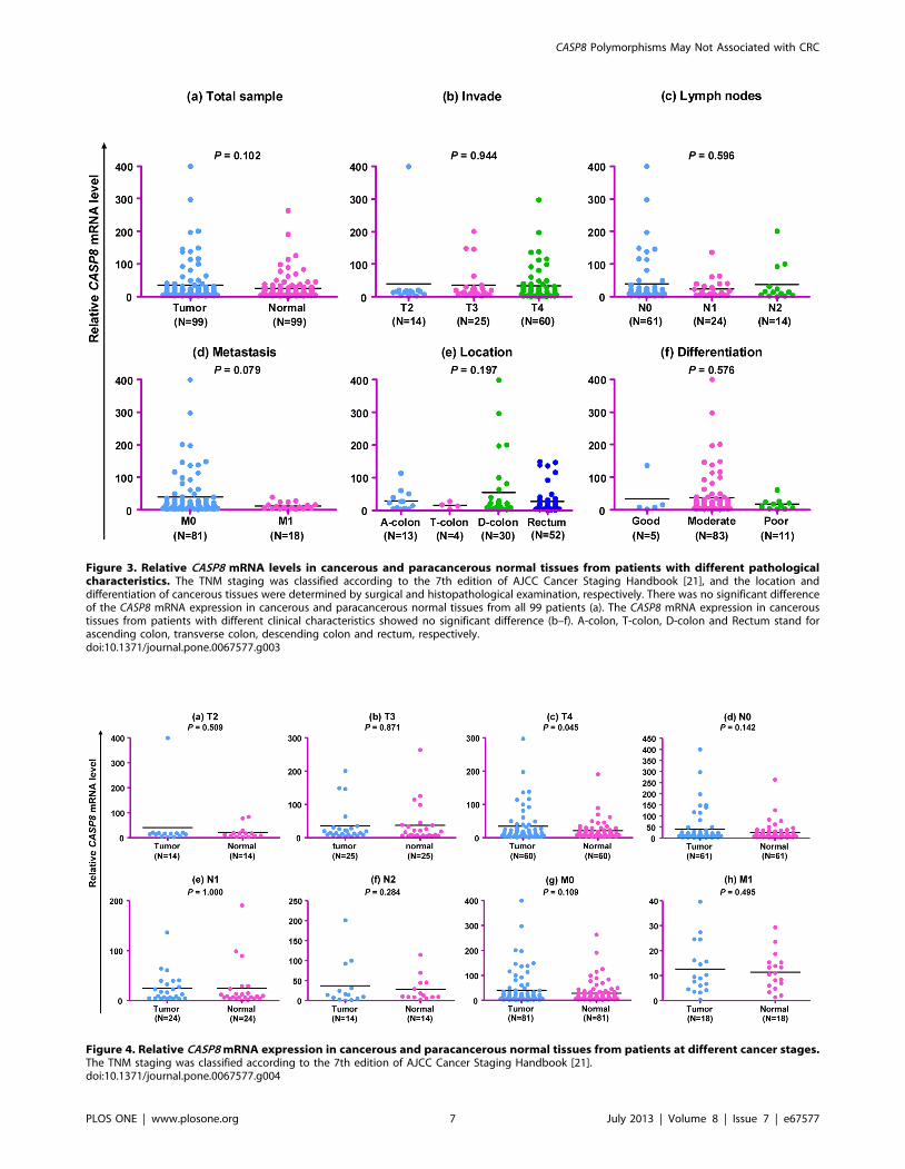
Figure 3. Relative CASP8 mRNA levels in cancerous and paracancerous normal tissues from patients with different pathologicalcharacteristics. The TNM staging was classified according to the 7th edition of AJCC Cancer Staging Handbook [21], and the location anddifferentiation of cancerous tissues were determined by surgical and histopathological examination, respectively. There was no significant differenceof the CASP8 mRNA expression in cancerous and paracancerous normal tissues from all 99 patients (a). The CASP8 mRNA expression in canceroustissues from patients with different clinical characteristics showed no significant difference (b–f). A-colon, T-colon, D-colon and Rectum stand forascending colon, transverse colon, descending colon and rectum, respectively.doi:10.1371/journal.pone.0067577.g003
Figure 4. Relative CASP8 mRNA expression in cancerous and paracancerous normal tissues from patients at different cancer stages.The TNM staging was classified according to the 7th edition of AJCC Cancer Staging Handbook [21].doi:10.1371/journal.pone.0067577.g004
CASP8 Polymorphisms May Not Associated with CRC
PLOS ONE | www.plosone.org 7 July 2013 | Volume 8 | Issue 7 | e67577

Supporting Information
Table S1 Genotype and pathological information for 39patients that were analyzed for CASP8 protein expres-sion.
(DOC)
Acknowledgments
We thank patients for donating tissues. We also thank the two anonymous
referees for their constructive comments.
Figure 5. Incoherent expression pattern of the CASP8 mRNA and protein in paired cancerous tissue (T) and paracancerous normaltissue (N) from 39 patients. The genotype and clinical information for these patients were listed in Table S1.doi:10.1371/journal.pone.0067577.g005
CASP8 Polymorphisms May Not Associated with CRC
PLOS ONE | www.plosone.org 8 July 2013 | Volume 8 | Issue 7 | e67577

Author Contributions
Conceived and designed the experiments: MSX XYL YGY. Performed the
experiments: MSX LC YSD YP. Analyzed the data: MSX DFZ YGY.
Contributed reagents/materials/analysis tools: LC WLL YSD YP DFZ
YW JL XYL YGY. Wrote the paper: MSX YGY.
References
1. Savas S, Younghusband HB (2010) dbCPCO: a database of genetic markerstested for their predictive and prognostic value in colorectal cancer. Human
Mutation 31: 901–907.
2. Cheah PY (1990) Hypotheses for the etiology of colorectal cancer–an overview.Nutr Cancer 14: 5–13.
3. Lichtenstein P, Holm NV, Verkasalo PK, Iliadou A, Kaprio J, et al. (2000)
Environmental and heritable factors in the causation of cancer–analyses ofcohorts of twins from Sweden, Denmark, and Finland. N Engl J Med 343: 78–
85.
4. Tsong WH, Koh W-P, Yuan J-M, Wang R, Sun C-L, et al. (2007) Cigarettesand alcohol in relation to colorectal cancer: the Singapore Chinese Health
Study. Br J Cancer 96: 821–827.
5. Hoshiyama Y, Sekine T, Sasaba T (1993) A case-control study of colorectalcancer and its relation to diet, cigarettes, and alcohol consumption in Saitama
Prefecture, Japan. Tohoku J Exp Med 171: 153–165.
6. Huxley RR, Ansary-Moghaddam A, Clifton P, Czernichow S, Parr CL, et al.(2009) The impact of dietary and lifestyle risk factors on risk of colorectal cancer:
a quantitative overview of the epidemiological evidence. Int J Cancer 125: 171–
180.
7. Raff M (1998) Cell suicide for beginners. Nature 396: 119–122.
8. Thompson CB (1995) Apoptosis in the pathogenesis and treatment of disease.
Science 267: 1456–1462.
9. Chen M, Wang J (2002) Initiator caspases in apoptosis signaling pathways.
Apoptosis 7: 313–319.
10. Nunez G, Benedict MA, Hu Y, Inohara N (1998) Caspases: the proteases of theapoptotic pathway. Oncogene 17: 3237–3245.
11. Hengartner MO (2000) The biochemistry of apoptosis. Nature 407: 770–776.
12. Nicholson DW, Thornberry NA (1997) Caspases: killer proteases. Trends
Biochem Sci 22: 299–306.
13. Li H, Zhu H, Xu CJ, Yuan J (1998) Cleavage of BID by caspase 8 mediates themitochondrial damage in the Fas pathway of apoptosis. Cell 94: 491–501.
14. Sun T, Gao Y, Tan W, Ma S, Shi Y, et al. (2007) A six-nucleotide insertion-
deletion polymorphism in the CASP8 promoter is associated with susceptibilityto multiple cancers. Nature Genetics 39: 605–613.
15. Ni C, Ye Y, Wang M, Qian H, Song Z, et al. (2009) A six-nucleotide insertion-
deletion polymorphism in the CASP8 promoter is associated with risk of coalworkers’ pneumoconiosis. J Toxicol Environ Health A 72: 712–716.
16. Wang M, Zhang Z, Tian Y, Shao J (2009) A six-nucleotide insertion-deletion
polymorphism in the CASP8 promoter associated with risk and progression ofbladder cancer. Clin Cancer Res 15: 2567–2572.
17. Frank B, Rigas SH, Bermejo JL, Wiestler M, Wagner K, et al. (2008) The
CASP8–652 6N del promoter polymorphism and breast cancer risk: amulticenter study. Breast Cancer Res Treat 111: 139–144.
18. Haiman CA, Garcia RR, Kolonel LN, Henderson BE, Wu AH, et al. (2008) A
promoter polymorphism in the CASP8 gene is not associated with cancer risk.Nature Genetics 40: 259–260; author reply 260–261.
19. Lan Q, Morton LM, Armstrong B, Hartge P, Menashe I, et al. (2009) Genetic
variation in caspase genes and risk of non-Hodgkin lymphoma: a pooled analysisof 3 population-based case-control studies. Blood 114: 264–267.
20. Xiao M-S, Zhang D-F, Zeng Y, Cheng Y-F, Yao Y-G (2011) Polymorphisms in
the promoter region of the CASP8 gene are not associated with non-Hodgkin’slymphoma in Chinese patients. Ann Hematol 90: 1137–1144.
21. Edge SB, Byrd DR, Compton CC, Fritz AG, Greene FL, et al. (2010) AJCC
Cancer Staging Handbook.22. Gauderman WJ (2002) Sample size requirements for matched case-control
studies of gene-environment interaction. Stat Med 21: 35–50.23. Barrett JC, Fry B, Maller J, Daly MJ (2005) Haploview: analysis and
visualization of LD and haplotype maps. Bioinformatics 21: 263–265.
24. Stephens M, Donnelly P (2003) A comparison of bayesian methods for haplotypereconstruction from population genotype data. Am J Hum Genet 73: 1162–
1169.25. Yao Y-G, Yang HS, Cao Z, Danielsson J, Duh EJ (2005) Upregulation of
placental growth factor by vascular endothelial growth factor via a post-
transcriptional mechanism. FEBS Lett 579: 1227–1234.26. Kantari C, Walczak H (2011) Caspase-8 and Bid: Caught in the act between
death receptors and mitochondria. Biochimica et Biophysica Acta (BBA) -Molecular Cell Research 1813: 558–563.
27. Jin ZY, El-Deiry WS (2005) Overview of cell death signaling pathways. CancerBiology & Therapy 4: 139–163.
28. De Vecchi G, Verderio P, Pizzamiglio S, Manoukian S, Barile M, et al. (2009)
Evidences for association of the CASP8–652 6N del promoter polymorphismwith age at diagnosis in familial breast cancer cases. Breast Cancer Research and
Treatment 113: 607–608.29. Lan Q, Zheng T, Chanock S, Zhang Y, Shen M, et al. (2007) Genetic variants in
caspase genes and susceptibility to non-Hodgkin lymphoma. Carcinogenesis 28:
823–827.30. Pittman AM, Broderick P, Sullivan K, Fielding S, Webb E, et al. (2008) CASP8
variants D302H and -652 6N ins/del do not influence the risk of colorectalcancer in the United Kingdom population. Br J Cancer 98: 1434–1436.
31. Gangwar R, Mandhani A, Mittal RD (2009) Caspase 9 and caspase 8 gene
polymorphisms and susceptibility to bladder cancer in north Indian population.Ann Surg Oncol 16: 2028–2034.
32. Umar M, Upadhyay R, Kumar S, Ghoshal UC, Mittal B (2011) CASP8–652 6Ndel and CASP8 IVS12–19G.A gene polymorphisms and susceptibility/
prognosis of ESCC: a case control study in northern Indian population. J SurgOncol 103: 716–723.
33. Sun T, Gao Y, Tan W, Ma S, Shi Y, et al. (2008) Reply to’’ A promoter
polymorphism in the CASP8 gene is not associated with cancer risk’’. NatureGenetics 40: 260–261.
34. Yin M, Yan J, Wei S, Wei Q (2010) CASP8 polymorphisms contribute to cancersusceptibility: evidence from a meta-analysis of 23 publications with 55
individual studies. Carcinogenesis 31: 850–857.
35. Heijink DM, Kleibeuker JH, Jalving M, Boersma-van Ek W, Koornstra JJ, et al.(2007) Independent induction of caspase-8 and cFLIP expression during
colorectal carcinogenesis in sporadic and HNPCC adenomas and carcinomas.Cell Oncol 29: 409–419.
36. Pan YF, Zhang MW, Jin MJ, Zhang SC, Chen K, et al. (2011) Expression ofcaspase apoptosis pathway genes mRNA in colorectal cancer and polypus.
Zhejiang Da Xue Xue Bao Yi Xue Ban 40: 245–251.
37. Xu B, Zhou Z-G, Li Y, Wang L, Yang L, et al. (2008) Clinicopathologicalsignificance of caspase-8 and caspase-10 expression in rectal cancer. Oncology
74: 229–236.38. Kim HS, Lee JW, Soung YH, Park WS, Kim SY, et al. (2003) Inactivating
mutations of caspase-8 gene in colorectal carcinomas. Gastroenterology 125:
708–715.
CASP8 Polymorphisms May Not Associated with CRC
PLOS ONE | www.plosone.org 9 July 2013 | Volume 8 | Issue 7 | e67577

Supplementary file: Xiao et al. 2013. PLoS ONE 8(7): e67577
Table S1. Genotype and pathological information for 39 patients that were analyzed for CASP8 protein expression
Sample
No.
Patient
ID
Genotype Tumor location Clinical features
rs3834129 rs3769821 rs113686495 Differentiation TNM a
1 5 6bp/del C/C 8bp/8bp Rectum Moderate T2N0M0
2 18 6bp/6bp C/C 8bp/8bp Rectum Moderate T4N0M0
3 22 6bp/6bp T/C 8bp/del Descending colon Moderate T4N1M1
4 29 6bp/del T/T del/del Descending colon Moderate T4N0M0
5 42 6bp/6bp T/T del/del Rectum Moderate T3N0M0
6 48 del/del C/C 8bp/del Rectum Moderate T3N0M0
7 51 6bp/6bp T/C 8bp/del Rectum Good T4N0M1
8 62 6bp/del T/T del/del Rectum Moderate T3N0M0
9 63 del/del T/T del/del Rectum Moderate T4N2M0
10 84 6bp/6bp T/C 8bp/del Ascending colon Moderate T4N0M0
11 100 6bp/6bp T/C 8bp/del Rectum Moderate T3N0M0
12 102 6bp/6bp T/C 8bp/del Descending colon Moderate T3N2M0
13 107 6bp/6bp T/T del/del Rectum Moderate T4N0M0
14 111 6bp/del C/C 8bp/8bp Ascending colon Moderate T4N1M0
15 32 6bp/del T/T del/del Descending colon Moderate T3N0M0
16 33 6bp/del T/T del/del Rectum Moderate T2N0M0
17 35 6bp/6bp T/T 8bp/del Rectum Moderate T4N1M0
18 36 6bp/del T/C 8bp/del Rectum Moderate T4N2M1
19 37 6bp/6bp C/C 8bp/del Ascending colon Moderate T4N1M1
20 38 6bp/6bp T/T del/del Rectum poor T4N2M0
21 39 6bp/6bp T/T del/del Descending colon Moderate T4N2M0
22 40 6bp/del T/T del/del Rectum Moderate T4N0M0
23 41 6bp/6bp T/C 8bp/del Ascending colon poor T4N1M0
24 43 6bp/6bp T/C 8bp/del Descending colon Moderate T4N2M0
25 44 6bp/del T/T del/del Descending colon Moderate T4N0M0
26 45 6bp/6bp T/T del/del Descending colon poor T3N0M0
27 52 6bp/del T/C 8bp/del Rectum Moderate T4N2M0
28 53 6bp/del T/T del/del Rectum Moderate T4N0M0
29 54 6bp/6bp T/T del/del Rectum Moderate T4N1M0
30 55 6bp/6bp T/C 8bp/del Rectum Moderate T4N1M0
31 58 6bp/del T/T del/del Rectum Moderate T4N0M0
32 59 6bp/del C/C 8bp/8bp Rectum Moderate T4N2M1

Supplementary file: Xiao et al. 2013. PLoS ONE 8(7): e67577
33 72 6bp/6bp T/C 8bp/del Descending colon Moderate T3N0M0
34 67 6bp/del T/T del/del Descending colon Moderate T3N0M0
35 68 6bp/6bp T/T del/del Rectum Moderate T4N2M0
36 69 6bp/6bp T/T del/del Rectum Moderate T4N0M0
37 70 6bp/6bp T/T del/del Rectum Moderate T2N0M0
38 71 6bp/del T/T del/del Rectum Moderate T3N2M0
39 65 6bp/6bp T/C 8bp/del Descending colon Moderate T4N1M1
a The stage of cancer was classified following the 7th edition of AJCC Cancer Staging Handbook [21].