Genetic differentiation of the nucleocapsid protein of Korean isolates of porcine epidemic diarrhoea...
3
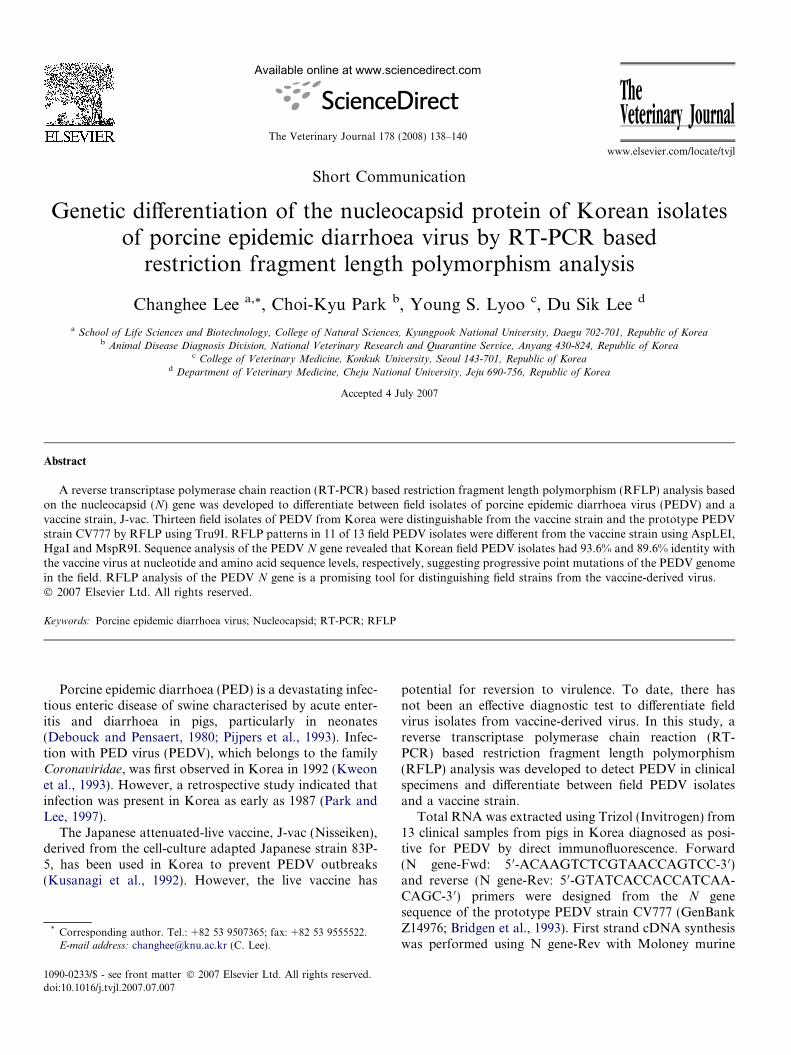
Short Communication Genetic differentiation of the nucleocapsid protein of Korean isolates of porcine epidemic diarrhoea virus by RT-PCR based restriction fragment length polymorphism analysis Changhee Lee a, * , Choi-Kyu Park b , Young S. Lyoo c , Du Sik Lee d a School of Life Sciences and Biotechnology, College of Natural Sciences, Kyungpook National University, Daegu 702-701, Republic of Korea b Animal Disease Diagnosis Division, National Veterinary Research and Quarantine Service, Anyang 430-824, Republic of Korea c College of Veterinary Medicine, Konkuk University, Seoul 143-701, Republic of Korea d Department of Veterinary Medicine, Cheju National University, Jeju 690-756, Republic of Korea Accepted 4 July 2007 Abstract A reverse transcriptase polymerase chain reaction (RT-PCR) based restriction fragment length polymorphism (RFLP) analysis based on the nucleocapsid (N) gene was developed to differentiate between field isolates of porcine epidemic diarrhoea virus (PEDV) and a vaccine strain, J-vac. Thirteen field isolates of PEDV from Korea were distinguishable from the vaccine strain and the prototype PEDV strain CV777 by RFLP using Tru9I. RFLP patterns in 11 of 13 field PEDV isolates were different from the vaccine strain using AspLEI, HgaI and MspR9I. Sequence analysis of the PEDV N gene revealed that Korean field PEDV isolates had 93.6% and 89.6% identity with the vaccine virus at nucleotide and amino acid sequence levels, respectively, suggesting progressive point mutations of the PEDV genome in the field. RFLP analysis of the PEDV N gene is a promising tool for distinguishing field strains from the vaccine-derived virus. Ó 2007 Elsevier Ltd. All rights reserved. Keywords: Porcine epidemic diarrhoea virus; Nucleocapsid; RT-PCR; RFLP Porcine epidemic diarrhoea (PED) is a devastating infec- tious enteric disease of swine characterised by acute enter- itis and diarrhoea in pigs, particularly in neonates (Debouck and Pensaert, 1980; Pijpers et al., 1993). Infec- tion with PED virus (PEDV), which belongs to the family Coronaviridae, was first observed in Korea in 1992 (Kweon et al., 1993). However, a retrospective study indicated that infection was present in Korea as early as 1987 (Park and Lee, 1997). The Japanese attenuated-live vaccine, J-vac (Nisseiken), derived from the cell-culture adapted Japanese strain 83P- 5, has been used in Korea to prevent PEDV outbreaks (Kusanagi et al., 1992). However, the live vaccine has potential for reversion to virulence. To date, there has not been an effective diagnostic test to differentiate field virus isolates from vaccine-derived virus. In this study, a reverse transcriptase polymerase chain reaction (RT- PCR) based restriction fragment length polymorphism (RFLP) analysis was developed to detect PEDV in clinical specimens and differentiate between field PEDV isolates and a vaccine strain. Total RNA was extracted using Trizol (Invitrogen) from 13 clinical samples from pigs in Korea diagnosed as posi- tive for PEDV by direct immunofluorescence. Forward (N gene-Fwd: 5 0 -ACAAGTCTCGTAACCAGTCC-3 0 ) and reverse (N gene-Rev: 5 0 -GTATCACCACCATCAA- CAGC-3 0 ) primers were designed from the N gene sequence of the prototype PEDV strain CV777 (GenBank Z14976; Bridgen et al., 1993). First strand cDNA synthesis was performed using N gene-Rev with Moloney murine 1090-0233/$ - see front matter Ó 2007 Elsevier Ltd. All rights reserved. doi:10.1016/j.tvjl.2007.07.007 * Corresponding author. Tel.: +82 53 9507365; fax: +82 53 9555522. E-mail address: [email protected] (C. Lee). www.elsevier.com/locate/tvjl Available online at www.sciencedirect.com The Veterinary Journal 178 (2008) 138–140 The Veterinary Journal
-
Upload
changhee-lee -
Category
Documents
-
view
212 -
download
0
Transcript of Genetic differentiation of the nucleocapsid protein of Korean isolates of porcine epidemic diarrhoea...
Available online at www.sciencedirect.com
www.elsevier.com/locate/tvjl
The Veterinary Journal 178 (2008) 138–140
TheVeterinary Journal
Short Communication
Genetic differentiation of the nucleocapsid protein of Korean isolatesof porcine epidemic diarrhoea virus by RT-PCR based
restriction fragment length polymorphism analysis
Changhee Lee a,*, Choi-Kyu Park b, Young S. Lyoo c, Du Sik Lee d
a School of Life Sciences and Biotechnology, College of Natural Sciences, Kyungpook National University, Daegu 702-701, Republic of Koreab Animal Disease Diagnosis Division, National Veterinary Research and Quarantine Service, Anyang 430-824, Republic of Korea
c College of Veterinary Medicine, Konkuk University, Seoul 143-701, Republic of Koread Department of Veterinary Medicine, Cheju National University, Jeju 690-756, Republic of Korea
Accepted 4 July 2007
Abstract
A reverse transcriptase polymerase chain reaction (RT-PCR) based restriction fragment length polymorphism (RFLP) analysis basedon the nucleocapsid (N) gene was developed to differentiate between field isolates of porcine epidemic diarrhoea virus (PEDV) and avaccine strain, J-vac. Thirteen field isolates of PEDV from Korea were distinguishable from the vaccine strain and the prototype PEDVstrain CV777 by RFLP using Tru9I. RFLP patterns in 11 of 13 field PEDV isolates were different from the vaccine strain using AspLEI,HgaI and MspR9I. Sequence analysis of the PEDV N gene revealed that Korean field PEDV isolates had 93.6% and 89.6% identity withthe vaccine virus at nucleotide and amino acid sequence levels, respectively, suggesting progressive point mutations of the PEDV genomein the field. RFLP analysis of the PEDV N gene is a promising tool for distinguishing field strains from the vaccine-derived virus.� 2007 Elsevier Ltd. All rights reserved.
Keywords: Porcine epidemic diarrhoea virus; Nucleocapsid; RT-PCR; RFLP
Porcine epidemic diarrhoea (PED) is a devastating infec-tious enteric disease of swine characterised by acute enter-itis and diarrhoea in pigs, particularly in neonates(Debouck and Pensaert, 1980; Pijpers et al., 1993). Infec-tion with PED virus (PEDV), which belongs to the familyCoronaviridae, was first observed in Korea in 1992 (Kweonet al., 1993). However, a retrospective study indicated thatinfection was present in Korea as early as 1987 (Park andLee, 1997).
The Japanese attenuated-live vaccine, J-vac (Nisseiken),derived from the cell-culture adapted Japanese strain 83P-5, has been used in Korea to prevent PEDV outbreaks(Kusanagi et al., 1992). However, the live vaccine has
1090-0233/$ - see front matter � 2007 Elsevier Ltd. All rights reserved.
doi:10.1016/j.tvjl.2007.07.007
* Corresponding author. Tel.: +82 53 9507365; fax: +82 53 9555522.E-mail address: [email protected] (C. Lee).
potential for reversion to virulence. To date, there hasnot been an effective diagnostic test to differentiate fieldvirus isolates from vaccine-derived virus. In this study, areverse transcriptase polymerase chain reaction (RT-PCR) based restriction fragment length polymorphism(RFLP) analysis was developed to detect PEDV in clinicalspecimens and differentiate between field PEDV isolatesand a vaccine strain.
Total RNA was extracted using Trizol (Invitrogen) from13 clinical samples from pigs in Korea diagnosed as posi-tive for PEDV by direct immunofluorescence. Forward(N gene-Fwd: 5 0-ACAAGTCTCGTAACCAGTCC-3 0)and reverse (N gene-Rev: 5 0-GTATCACCACCATCAA-CAGC-3 0) primers were designed from the N genesequence of the prototype PEDV strain CV777 (GenBankZ14976; Bridgen et al., 1993). First strand cDNA synthesiswas performed using N gene-Rev with Moloney murine

C. Lee et al. / The Veterinary Journal 178 (2008) 138–140 139
leukaemia virus reverse transcriptase (Invitrogen). The par-tial N gene was amplified using N gene-Fwd and N-geneRev under the following conditions: initial denaturationat 95 �C for 5 min; 30 cycles of denaturation at 95 �C for45 s, annealing at 52 �C for 45 s and extension at 72 �Cfor 1 min; followed by final extension at 72 �C for10 min. PCR products were separated by 1% agarose gelelectrophoresis and visualised using a gel documentationsystem (Bio-Rad).
A specific DNA band of 691 base pairs (bp) was ampli-fied by RT-PCR from PEDV-infected cells (Fig. 1a, lane 2).The same PCR product could be detected when RT-PCRwas applied to field clinical samples, a Japanese vaccinestrain and PEDV CV777 (Fig. 1b). No PCR products wereamplified from cells infected with another porcine corona-virus (transmissible gastroenteritis virus), cells infectedwith porcine rotavirus, uninfected cells (Fig. 1a, lanes 3–5) or clinical samples diagnosed with other viral infections(data not shown).
1 2 3 4 5 1 2 3 4 5
PE
DV
TG
EV
PR
V
Moc
k-in
fect
ed
M
600 - -N
M 97-1
2897
-138
97-1
51
Fig. 1. Agarose gel electrophoresis of RT-PCR products of the N gene of PEDmarker; lane 2, PEDV; lane 3, transmissible gastroenteritis virus (TGEV);amplification of PEDV N gene from clinical samples. Lane 1, 100 bp DNA ma16, PEDV CV777. 1% agarose gel electrophoresis.
Table 1RFLP patterns of N gene PCR products of 13 field isolates, vaccine virus and
PEDV isolate DNA fragment sizes (bp) digested with restr
AspLEI HgaI
97-128 NCb 343, 34897-138 NC 343, 34897-151 335, 356 NC97-160 NC 343, 34898-14 335, 356 NC98-19 335, 356 NC98-53 NC 343, 34898-81 335, 356 NC98-125 335, 356 NC99-32 335, 356 NC99-98 335, 356 NC99-158 335, 356 NC99-188 335, 356 NCVaccine NC 343, 348CV777 59, 632 343, 348
a Digested fragment sizes (bp) were predicted on the basis of nucleotide seqb Not cleaved (691 bp).
To evaluate the genetic characteristics of field PEDVisolates, PCR products were further analysed by RFLP.After preliminary examination of a number of candidaterestriction enzymes (REs) selected on the basis of the N
gene sequence of CV777, AspLEI, HgaI, MspR9I andTru9I (New England Biolabs) were studied further. PCRproducts were purified, digested with each RE for 1 h at37 �C and separated by electrophoresis on a 1.5% agarosegel. Band sizes generated by RE digestion were predictedfrom the nucleotide sequence of the amplified 691 bp N
gene (Table 1).When digested with AspLEI, PCR products from
CV777 produced two fragments of 59 and 632 bp and nineKorean field isolates produced two fragments of 335 and356 bp, whereas the 691 bp fragment was not digested infour field isolates or the vaccine strain (Fig. 2a). PCR prod-ucts of four field isolates digested with HgaI generated thesame RFLP pattern as that of CV777 and the vaccine virus,whereas nine other field isolates were not digested with
6 7 8 9 10 11 12 13 14 15 16
-N
97-1
6098
-14
98-1
998
-53
98-8
198
-125
99-3
299
-98
99-9
899
-188
J-V
ac
CV
-777
V. (a) Specificity of RT-PCR for detection of PEDV. Lane 1, 100 bp DNAlane 4, porcine rotavirus (PRV); lane 5, Uninfected cells. (b) RT-PCRrker; lanes 2–14, field PEDV isolates; lane 15, Japanese vaccine virus; lane
reference strain (CV777) of PEDV
iction enzymea
MspR9I Tru9I
72, 93, 134, 392 97, 135, 162, 29772, 93, 134, 392 97, 135, 162, 29759, 72, 93, 134, 152, 333 97, 135, 162, 29793, 206, 392 97, 135, 162, 29759, 93, 206, 333 162, 232, 29759, 72, 93, 134, 152, 333 97, 135, 162, 29793, 206, 392 97, 135, 162, 29759, 72, 93, 134, 152, 333 97, 135, 162, 29759, 72, 93, 134, 152, 333 97, 135, 162, 29759, 93, 206, 333 97, 135, 162, 29759, 72, 93, 134, 152, 333 97, 135, 162, 29759, 72, 93, 134, 152, 333 97, 135, 162, 29759, 72, 93, 134, 152, 333 97, 135, 162, 29793, 206, 392 97, 162, 432206, 485 97, 162, 432
uencing data.
1 2 3 4 5 6 7 8 9 10 11 12 13 14 15 16
M 97-1
2897
-138
97-1
5197
-160
98-1
4
98-1
998
-53
98-8
1
98-1
2599
-32
99-9
899
-98
99-1
88J-
Vac
CV
-777
600 -
600 -
600 -
600 -
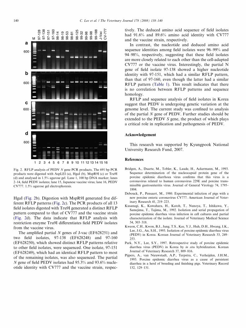
Fig. 2. RFLP analysis of PEDV N gene PCR products. The 691 bp PCRproducts were digested with AspLEI (a), HgaI (b), MspR9I (c) or Tru9I(d) and analysed in 1.5% agarose gel. Lane 1, 100 bp DNA marker; lanes2–14, field PEDV isolates; lane 15, Japanese vaccine virus; lane 16, PEDVCV777. 1.5% agarose gel electrophoresis.
140 C. Lee et al. / The Veterinary Journal 178 (2008) 138–140
HgaI (Fig. 2b). Digestion with MspR9I generated five dif-ferent RFLP patterns (Fig. 2c). The PCR products of all 13field isolates digested with Tru9I generated a distinct RFLPpattern compared to that of CV777 and the vaccine strain(Fig. 2d). The data indicate that RFLP analysis withrestriction enzyme Tru9I differentiates field PEDV isolatesfrom the vaccine virus.
The amplified partial N genes of J-vac (EF628251) andtwo field isolates, 97-138 (EF628248) and 97-160(EF628250), which showed distinct RFLP patterns relativeto other field isolates, were sequenced. One isolate, 97-151(EF628249), which had an identical RFLP pattern to mostof the remaining isolates, was also sequenced. The partialN gene of field PEDV isolates had 95.3% and 93.6% nucle-otide identity with CV777 and the vaccine strain, respec-
tively. The deduced amino acid sequence of field isolateshad 91.6% and 89.6% amino acid identity with CV777and the vaccine strain, respectively.
In contrast, the nucleotide and deduced amino acidsequence identities among field isolates were 96–99% and94–98%, respectively, suggesting that these field isolatesare more closely related to each other than the cell-adaptedCV777 or the vaccine virus. Interestingly, the partial Ngene of field isolate 97-138 showed a higher nucleotideidentity with 97-151, which had a similar RFLP pattern,than that of 97-160, even though the latter had a similarRFLP pattern (Table 1). This result indicates that thereis no correlation between RFLP patterns and sequencehomology.
RFLP and sequence analysis of field isolates in Koreasuggest that PEDV is undergoing genetic variation at thegenome level. The current study was confined to analysisof the partial N gene of PEDV. Further studies should beextended to the PEDV S gene, the product of which playsa critical role in replication and pathogenesis of PEDV.
Acknowledgement
This research was supported by Kyungpook NationalUniversity Research Fund, 2007.
References
Bridgen, A., Duarte, M., Tobler, K., Laude, H., Ackermann, M., 1993.Sequence determination of the nucleocapsid protein gene of theporcine epidemic diarrhoea virus confirms that this virus is acoronavirus related to human coronavirus 229E and porcine trans-missible gastroenteritis virus. Journal of General Virology 74, 1795–1804.
Debouck, P., Pensaert, M., 1980. Experimental infection of pigs with anew porcine enteric coronavirus CV777. American Journal of Veter-inary Research 41, 219–223.
Kusanagi, K., Kuwahara, H., Katoh, T., Nunoya, T., Ishikawa, Y.,Samejima, T., Tajima, M., 1992. Isolation and serial propagation ofporcine epidemic diarrhea virus infection in cell cultures and partialcharacterization of the isolate. Journal of Veterinary Medical Science54, 303–318.
Kweon, C.H., Kwon, B.J., Jung, T.S., Kee, Y.J., Huh, D.H., Hwang, I.K.,Lee, J.G., An, S.H., 1993. Isolation of porcine epidemic diarrhea virus(PEDV) in Korea. Korean Journal of Veterinary Research 33, 249–253.
Park, N.Y., Lee, S.Y., 1997. Retrospective study of porcine epidemicdiarrhea virus (PEDV) in Korea by in situ hybridization. KoreanJournal of Veterinary Research 37, 809–816.
Pijpers, A., van Nieuwstadt, A.P., Terpstra, C., Verheijden, J.H.M.,1993. Porcine epidemic diarrhea virus as a cause of persistentdiarrhea in a herd of breeding and finishing pigs. Veterinary Record132, 129–131.