Genetic control of resistance to murine malaria
12
Journal of Cellular Biochemistry 2491-102 (1984) Molecular Biology of Host-Parasite Interactions 123-134 Genetic Control of Resistance to Murine Mary Stevenson, Suzanne Lemieux, and Emil Skamene The Montreal General Hospital Research Institute, Montreal, Quebec, H3G lA4, Canada (M.S., E.S.) and institut-Armand Frappier, Laval, Quebec H7N 429, Canada (S. L.) Strain variation in the level of resistance to malaria was investigated in inbred mice after infection with Plasmodium chabaudi. Following intraperitoneal infec- tion with the typing dose of parasitized erythrocytes, mice of 11 inbred strains could be separated using survival time as the criterium into resistant and suscepti- ble groups. Genetic analysis of F1 hybrid and backcross progeny derived from one of the most resistant (BIO.A) and from the most susceptible (AiJ) strains as parents suggested that host resistance in this strain combination was genetically controlled by a dominant, non-H-2-linked, autosomal gene or closely linked genes. Analysis of the mechanisms of resistance to P chabaudi showed (1) phenotypic expression of the resistance gene was apparent within 6 days of infection as a significant difference between resistant and susceptible mice in the level of paras- itemia; (2) the level of host NK cell activity was not related to the level of host resistance to malaria; (3) compared with susceptible A/J mice, resistant BIO.A hosts had an augmented erythropoietic response during the course of malaria as well as during phenylhydrazine-induced anemia and (4) treatment with BCG or P acnes resulted in an equal degree of protection, measured by parasitemia and survival, in both resistant and susceptible mice. Key words: malaria, inbred mice, genetic control of resistance It has been well established among human populations that certain genetically determined traits impart to the host a high level of resistance against infection with malaria. These traits, which include absence of the Duffy blood group antigen, sickle cell anemia, thalassemia, and glucose-6-phosphate dehydrogenase deficiency, are phenotypically expressed as biochemical abnormalities in host erythrocytes [I]. Variations in the outcome of malarial infection are also apparent among inbred strains of mice [2-61. As in the human system, these differences in susceptibility have been shown to be genetically determined in experimental models following infection with either Plasmodium berghei or P chabaudi. In the murine models, however, the mechanism(s) of resistance is unknown. It has been shown recently by our laboratory that the phenotypic expression of the superior resistance to infection with P chabaudi Mary Stevenson is an MRC Scholar. Received March 3, 1983; revised and accepted October 1, 1983. 0 1984 Alan R. Liss, Inc.
-
Upload
mary-stevenson -
Category
Documents
-
view
217 -
download
2
Transcript of Genetic control of resistance to murine malaria

Journal of Cellular Biochemistry 2491-102 (1984) Molecular Biology of Host-Parasite Interactions 123-134
Genetic Control of Resistance to Murine
Mary Stevenson, Suzanne Lemieux, and Emil Skamene The Montreal General Hospital Research Institute, Montreal, Quebec, H3G lA4, Canada (M.S., E.S.) and institut-Armand Frappier, Laval, Quebec H7N 429, Canada (S. L.)
Strain variation in the level of resistance to malaria was investigated in inbred mice after infection with Plasmodium chabaudi. Following intraperitoneal infec- tion with the typing dose of parasitized erythrocytes, mice of 11 inbred strains could be separated using survival time as the criterium into resistant and suscepti- ble groups. Genetic analysis of F1 hybrid and backcross progeny derived from one of the most resistant (BIO.A) and from the most susceptible (AiJ) strains as parents suggested that host resistance in this strain combination was genetically controlled by a dominant, non-H-2-linked, autosomal gene or closely linked genes. Analysis of the mechanisms of resistance to P chabaudi showed (1) phenotypic expression of the resistance gene was apparent within 6 days of infection as a significant difference between resistant and susceptible mice in the level of paras- itemia; (2) the level of host NK cell activity was not related to the level of host resistance to malaria; (3) compared with susceptible A/J mice, resistant BIO.A hosts had an augmented erythropoietic response during the course of malaria as well as during phenylhydrazine-induced anemia and (4) treatment with BCG or P acnes resulted in an equal degree of protection, measured by parasitemia and survival, in both resistant and susceptible mice.
Key words: malaria, inbred mice, genetic control of resistance
It has been well established among human populations that certain genetically determined traits impart to the host a high level of resistance against infection with malaria. These traits, which include absence of the Duffy blood group antigen, sickle cell anemia, thalassemia, and glucose-6-phosphate dehydrogenase deficiency, are phenotypically expressed as biochemical abnormalities in host erythrocytes [I].
Variations in the outcome of malarial infection are also apparent among inbred strains of mice [2-61. As in the human system, these differences in susceptibility have been shown to be genetically determined in experimental models following infection with either Plasmodium berghei or P chabaudi. In the murine models, however, the mechanism(s) of resistance is unknown. It has been shown recently by our laboratory that the phenotypic expression of the superior resistance to infection with P chabaudi
Mary Stevenson is an MRC Scholar.
Received March 3, 1983; revised and accepted October 1, 1983.
0 1984 Alan R. Liss, Inc.
92:JCB Stevenson, Lemieux, and Skamene
that is controlled by a non-H-2-linked, dominant, autosomal gene or closely linked genes is apparent early in the course of the infection and results in survival of the resistant host [6]. In the present study, we have evaluated several parameters of host response to malarial parasites in resistant and susceptible mice to determine the mechanism of genetically determined resistance.
MATERIALS AND METHODS Mice
Age- and sex-matched mice 8-12 wk old were used in all experiments. BlO.A, A/J(A), F1 hybrid (B1O.A X A, A X DBA/2J, and A X BALB/c), F2, and backcross mice were bred in our laboratory. The following inbred strains were purchased from the Jackson Laboratory, Bar Harbor, Maine: C3H/HeJ, SJL/J, AKR/J, DBA/lJ, CBA/J, C57BL/6J, C57L/J, and DBA/2J. BALB/c mice were purchased from Cana- dian Breeders, St Constant, Quebec, Canada. Beige mice were obtained from Dr John Roder, Queens University, Kingston, Ontario.
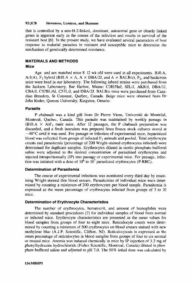
Parasite P chabaudi was a kind gift from Dr Pierre Viens, Universitt de MontrCal,
Montreal, Quebec, Canada. This parasite was maintained by weekly passage in (BIO.A X A)Fl male mice. After 12 passages, the P chabaudi preparation was discarded, and a fresh inoculum was prepared from frozen stock cultures stored at -40°C until it was used. For passage or infection of experimental mice, heparinized blood was collected from groups of infected F1 animals and pooled. Total erythrocyte counts and parasitemia (percentage of 200 Wright-stained erythrocytes infected) were determined for duplicate samples. Erythrocytes diluted in sterile phosphate-buffered saline were adjusted to the desired concentration of parasitized erythrocytes and injected intraperitoneally (IP) into passage or experimental mice. For passage, infec- tion was initiated with a dose of lo6 to lo7 parasitized erythrocytes (P-RBC).
Determination of Parasitemia
The course of experimental infections was monitored every third day by exam- ining Wright-stained thin blood smears. Parasitemias of individual mice were deter- mined by counting a minimum of 200 erythrocytes per blood sample. Parasitemia is expressed as the mean percentage of erythrocytes infected from groups of 3 to 10 mice.
Determination of Erythrocyte Characteristics
The number of erythrocytes, hematocrit, and amount of hemoglobin were determined by standard procedures [7] for individual samples of blood from normal or infected mice. Erythrocyte characteristics are presented as the mean values for blood samples from groups of four to eight mice. Reticulocyte counts were deter- mined by counting a minimum of 500 erythrocytes on blood smears stained with new methylene blue (A. J.P. Scientific, Clifton, NJ). Reticulocytosis is expressed as the mean percentage of reticulocytes in blood samples from groups of four to six normal or treated mice. Anemia was induced chemically in mice by IP injection of 3.2 mg of phenylhydrazine hydrochloride (Fisher Scientific, Montreal, Canada) diluted in phos- phate-buffered saline and adjusted to pH 7.0. The 50% lethal dose was calculated by
l24MBHPI

Resistance to Murine Malaria JCB:93
TABLE I. Strain Survey of Resistance to P chabaudi*
Mean survival
Level of time Mouse strain resistancea (day#
C57BL/6 Resistant > 14 C57L Resistant > 14 DBAl2 Resistant > 14 CBA Resistant > 14 BI0.A Resistant > 14
AIJ BALB/c C 3 HI H e J AKR SJL DBA/ 1 (B1O.A X A)FI (A X DBA/2)Fj (A X BALB/c)F,
Susceptible Susceptible Susceptible Susceptible Susceptible Susceptible Resistant Resistant Susceptible
8.3 & 0.3 8.6 & 0.5 9.7 & 0.6 9.8 i 0.5 9.5 i 0.8 9.2 & 0.6 > 14 > 14 8.3 i 0.3
*Groups of ten mice in two experiments were injected IP with lo6 erythrocytes parasitized with P chabaudi. aSusceptibiIity was defined as a mouse strain in which < 50% of animals survived the typing dose at day 10 of infection. bInfected mice died at times indicated or survived for more than 14 days. Results are expressed as means k standard error of the mean.
the method of Reed and Muench [8] and was determined after injection of doses of phenylhydrazine-hydrochloride ranging from 0.8 % to 8.0 mg into groups of four mice. Mice were observed daily for 10 days, and mortality was recorded.
BCG and P acnes Mice were injected intravenously (IV) with 5 X lo6 colony-forming units (cfu)
of living Mycobacterium bovis, strain BCG (TMC 1029, Phipps substrain, Trudeau Mycobacterial Collection) available from the National Jewish Hospital and Research Center (Denver, CO) or with killed Propionibacterium acnes (50 mg/kg, Wellcome Research Laboratory, Beckenham, England). Seven days later, groups of BCG- treated, P acnes-treated, or normal, control mice were infected IP with 1 x lo6 erythrocytes parasitized with P chabaudi.
Results We recently examined the level of resistance in mice to infection with the
murine malarial species P chabaudi [6] . After intraperitoneal infection with a typing dose of lo6 P-RBC, 11 strains could be characterized as resistant or susceptible. A given mouse strain was considered susceptible if less than 50% survived 10 days after infection with this dose (Table I). Strains C57BL/6, C57L, DBA/2, CBA, and B1O.A mice were characterized as resistant and continued to survive beyond day 14. Infection with P chabaudi, in contrast, was lethal to 100% of strain A mice and > 50% of BALB/c, C3H/HeJ, AKR, SJL, and DBA/I mice with a mean survival time of less
MBHPI: 125
9 4 JCB
than 10 days. These results suggested to us that the level of resistance to infection with P chabaudi was dependent upon the genetic background of the host. The H-2 complex did not appear to be important in determining the level of resistance, since strain A mice (H-2a) were highly susceptible, whereas strain B1O.A mice which share the u- haplotype were among the most resistant.
Next we studied the level of resistance of F1 hybrids to determine if resistance to malaria is inherited as a dominant trait. Resistance was examined in two hybrid combinations resulting from crosses between susceptible strain A mice and resistant BIO.A or DBA/2 mice and an F1 hybrid derived from two susceptible parents (Table I). In the former crosses, 100% of the (B1O.A X A) F1 or (A X DBA/2) F I mice were resistant to the typing dose of lo6 parasitized erythrocytes and survived beyond day 14. The level of resistance of (A X BALB/c) F1 mice was low with a mean survival time (8.3 f 0.3 days), which was similar to the times of the susceptible parents. Thus, the level of resistance to infection with P chabaudi appears to be inherited as a dominant trait.
Using lo6 P-RBC as a standard typing dose, susceptible animals succumbed within 10 days of infection, while resistant hosts survived indefinitely. However, when susceptible A mice were infected with a range of doses of parasitized erythro- cytes ( lo4 - lo9), it was apparent that the infective dose correlated with the time until death. That is, the length of the mean survival time of susceptible A mice decreased from 13 days after infection with lo4 parasitized cells to less than 6 days after infection with lo9 P-RBC. Furthermore, the infection was lethal to 100% of A mice at all doses. Except at a dose of lo9 P-RBC, 100% of resistant BIO.A mice survived. Thus, the 50% lethal dose for susceptible A hosts is apparently < lo4, while for resistant B1O.A hosts, it is > lo9 P chabaudi-parasitized erythrocytes.
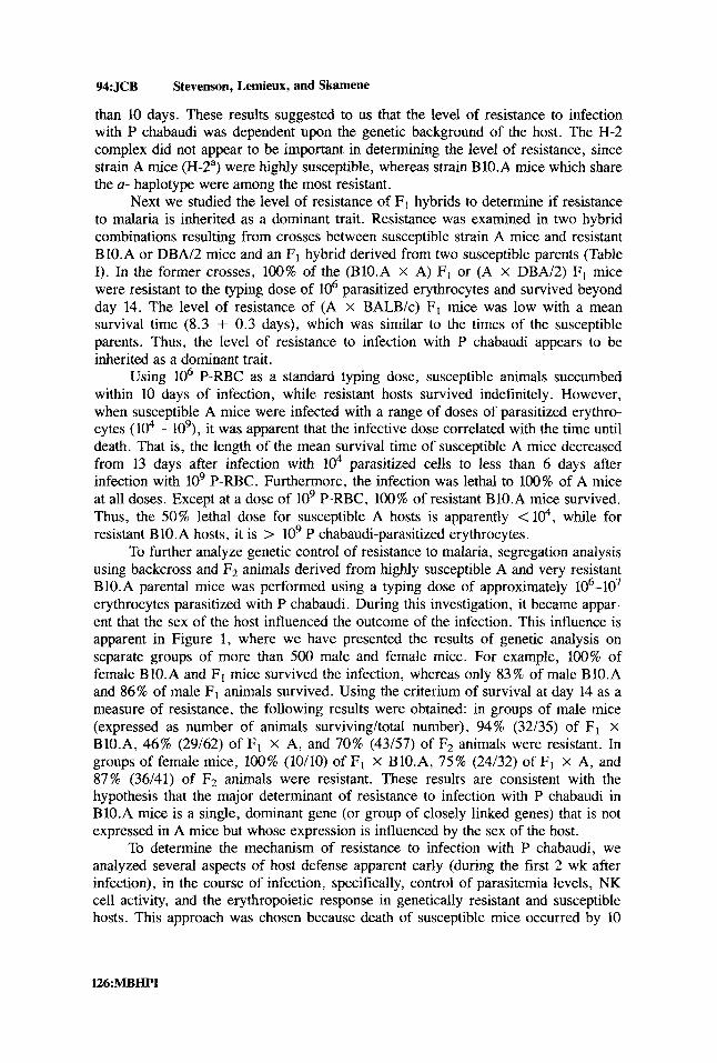
To further analyze genetic control of resistance to malaria, segregation analysis using backcross and F2 animals derived from highly susceptible A and very resistant BIO.A parental mice was performed using a typing dose of approximately 106-107 erythrocytes parasitized with P chabaudi. During this investigation, it became appar- ent that the sex of the host influenced the outcome of the infection. This influence is apparent in Figure 1, where we have presented the results of genetic analysis on separate groups of more than 500 male and female mice. For example, 100% of female BIO.A and Fl mice survived the infection, whereas only 83% of male B1O.A and 86% of male Fl animals survived. Using the criterium of survival at day 14 as a measure of resistance, the following results were obtained: in groups of male mice (expressed as number of animals surviving/total number), 94% (32/35) of F1 x BlO.A, 46% (29/62) of F1 X A, and 70% (43/57) of F2 animals were resistant. In groups of female mice, 100% (10/10) of F1 X BIO.A, 75% (24/32) of F1 x A, and 87% (36/41) of F2 animals were resistant. These results are consistent with the hypothesis that the major determinant of resistance to infection with P chabaudi in BIO.A mice is a single, dominant gene (or group of closely linked genes) that is not expressed in A mice but whose expression is influenced by the sex of the host.
To determine the mechanism of resistance to infection with P chabaudi, we analyzed several aspects of host defense apparent early (during the first 2 wk after infection), in the course of infection, specifically, control of parasitemia levels, NK cell activity, and the erythropoietic response in genetically resistant and susceptible hosts. This approach was chosen because death of susceptible mice occurred by 10
Stevenson, Lemieux, and Skamene
l26:MBHPI
Resistance to Murine Malaria JCB:95
Males Females
" A B1O.A F1 F1XBlO.A FlXA F2
0 Expected &3 Observed
Fig. 1. Segregation analysis of resistance to P chabaudi in male and female mice. Groups of 10 to 100 mice of strains A and BIO.A and their F,, FZ, and backcross progeny were injected IP with lo6 to lo7 P-RBC. The outcome of infection (either death or recovery) was followed in individual animals (expected or observed percentage of survival for genetic control by a single, dominant gene[s]).
days, suggesting that the mechanism of genetically determined resistance is phenotyp- ically expressed in the early stages of infection before the development of specific immunity.
First, we measured the parasitemia levels in resistant and susceptible mice after infection with a low dose (lo6 cells) and a high dose (lo8 cells) of P-RBC (Fig. 2). A significant difference was evident between resistant B1O.A and susceptible A mice at days 6 and 10 after infection with lo6 P-RBC. The peak level of parasitemia in A mice was approximately 50% after infection with either dose. When this critical level of parasitemia was reached, the infection terminated in death of 100% of susceptible A hosts. In contrast, the peak parasitemia level in resistant B1O.A mice was dependent upon the infective dose. At the low dose, the parasitemia level peaked on day 10 at approximately 35% in B1O.A mice. At the high dose, parasitemia approached the 50% level characteristic of susceptible A mice. Despite such a high parasite load, BIO.A mice survived the infection, and the level of parasitemia was decreasing by 14 days after infection. These results observed with BIO.A mice were confirmed when DBA/2 mice were used as the resistant strain.
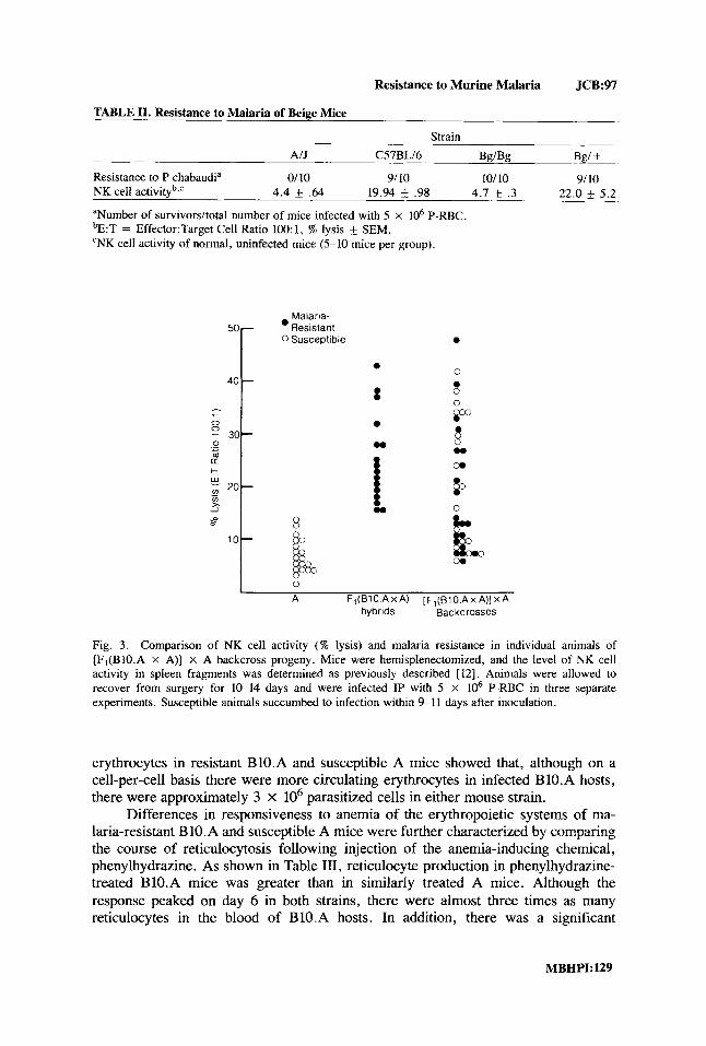
Since a low level of NK cell activity has recently been shown to be associated with susceptibility of both humans and rodents to malaria 14, 9-11], we also examined the relationship between these two genetically determined host responses in two separate experimental models of NK-deficient animals [ 121. First, the level of resist- ance to infection with P chabaudi of NK-deficient bg/bg mice on the C57BL/6 background was found to be identical with that of normal bg/+ littermates (Table 11). In addition, examination of the two traits in backcross [F1 (BIO.A x A) x A]
MBHPI: 127

%:JCB Stevenson, Lemieux, and Skamene
a
0 U m
106 P-RBC 60 -
BIO.A
20 i!k.: 10 3 6 10 14
b 108 P-RBC
6o r
B1O.A
3 6 10 14
Days Post infection ................................................... ...............................................................................................................
C
6o r
20 a 4 10
3 6 10 14
d
6o 50 c 20
10
3 6 10 14
Days Post Infection
Fig. 2 Levels of early parasitemia in resistant and susceptible mouse strains. Groups of five male mice of strain A (0) or B1O.A (O[a and b]) or DBA/2J ( o [ c and d]) were infected IP with lo6 (a, c) or 10' (b, d) P-RBC. At different times after infection, the percentages of P-RBC were determined as described. The data are presented as mean k standard error of the mean. The daggers represent death. Similar results were obtained with female mice.
progeny derived from B1O.A (malaria-resistant, high NK cell activity) and A (ma- laria-susceptible, low NK cell activity) parental mice showed that the levels of NK cell activity and resistance to malaria segregated independently (Fig. 3).
The response of host erythropoietic system to the stress of malaria-induced anemia may also be related to superior resistance to infection with P chabaudi. There was no difference between normal, uninfected A and B1O.A mice in several erythro- cyte characteristics, including numbers of circulating cells, percent hematocrit, and hemoglobin volume [6,13]. However, there were significant differences between the two strains following infection with the typing dose of P chabaudi (Table 111). In general, there was an inverse relationship between the level of parasitemia and the erythrocyte traits: a high level of parasitemia was associated with a low hematocrit and low numbers of erythrocytes. A comparison of the absolute numbers of infected
128:MBHPI
Resistance to Murine Malaria JCB:97
TABLE 11. Resistance to Malaria of Beige Mice
Strain AIJ C57BLI6 W B g Bgl +
Resistance to P chabaudia 0/10 9/ 10 101 10 9/10 NK cell activityb,c 4.4 * .64 19.94 .98 4.7 * .3 22.0 f 5.2
aNumber of survivorsltotal number of mice infected with 5 x lo6 P-RBC. bE:T = Effector:Target Cell Ratio 100: 1, % lysis & SEM. ‘NK cell activity of normal, uninfected mice (5-10 mice per group).
50
40
- - 0 0 - 30- 0 a - m + E
20-
x _I
s 10
-
-
-
Malaria- Resistant
0 Susceptible
w
1
A Fi(B10 A x A) [F,(BlO.A x A)] X A hybrids Backcrosses
Fig. 3. Comparison of NK cell activity ( W lysis) and malaria resistance in individual animals of [F,(BIO.A X A)] x A backcross progeny. Mice were hemisplenectomized, and the level of NK cell activity in spleen fragments was determined as previously described [ 121. Animals were allowed to recover from surgery for 10-14 days and were infected IP with 5 X lo6 P-RBC in three separate experiments. Susceptible animals succumbed to infection within 9-1 1 days after inoculation.
erythrocytes in resistant BIO.A and susceptible A mice showed that, although on a cell-per-cell basis there were more circulating erythrocytes in infected B 10.A hosts, there were approximately 3 X lo6 parasitized cells in either mouse strain.
Differences in responsiveness to anemia of the erythropoietic systems of ma- laria-resistant B 10.A and susceptible A mice were further characterized by comparing the course of reticulocytosis following injection of the anemia-inducing chemical, phenylhydrazine. As shown in Table 111, reticulocyte production in phenylhydrazine- treated BIO.A mice was greater than in similarly treated A mice. Although the response peaked on day 6 in both strains, there were almost three times as many reticulocytes in the blood of B1O.A hosts. In addition, there was a significant

98:JCB Stevenson, Lemieux, and Skamene
TABLE 111. Erythropoietic Responses of A and B1O.A Mice to Anemia
Malaria-induced anemia Phenylhydrazine-induced No. of anemia
~
Mouse Erythrocytes Hematocrit P-RBC % Reticulo- straina %P-RBC (lo6 cells/mm3) ( % I (lo6 cells/mm3) L D ~ , , ~ cytes
A 56.6 k 2.1* 4.6 f 0.03' 20.7 f 7.2** 2.61 f 0.02*** 4.0 mg 18.1 f 4.5* BIO.A 35.8 f2 .7* 9.3 f0.27* 43.7 1.7** 3.33 f 0.24*** 5 .0mg 46 i 0.8'
aGroups of 5-8 male mice injected IP 7 days previously with lo6 P-RBC or 6 days previously with 3.2 mg phenylhydrazine. Values are means SEM. bDetermined by method of Reed and Muench IS]. *Significant (P < 0.005).
Significant (P < 0.01). ** '** Not significant.
difference between the strains in the dose of phenylhydrazine that killed 50% of the animals.
In spite of genetically determined differences in the level of resistance early (within 10 days) in the course of infection with P chabaudi, BCG or P acnes pretreatment resulted in nonspecific immunity against malaria, in both genetically resistant and susceptible hosts (Fig. 4).
The increased protection was evident by a 50% or greater decrease in parasi- temia levels in both BI0.A and A mice. Moreover, more than 90% of treated A mice survived the usually 100% lethal infection with lo6 P-RBC.
DISCUSSION
During malaria, host response leading to decrease of the peak parasitemia level apparent at the crisis stage of infection has been shown to be dependent upon the development of specific immune responses. However, the existence of certain genetic traits among human populations that inherently protect an individual against infection is well known. These traits, for example, sickle-cell anemia, are phenotypically expressed as biochemical abnormalities in host erythrocytes and are unrelated to immune mechanisms. In spite of being deleterious, however, the genes have survived with high frequency in malarious areas of the world because of their protective ef- fects [I].
We have described another example of innate resistance against mdaria, that is, the high level of natural resistance apparent among certain inbred strains of mice following infection with P chabaudi. While more than 50% of the animals of suscep- tible strains (A, SJL, C3H/HeJ) succumbed within 10 days of infection with lo6 parasitized erythrocytes, resistant strains, such as CBA mice or mice with a C57 background (C57BL/6, C57L or BlO.A), survived beyond 14 days. Segregation analysis of the level of resistance of hybrid, backcross, and Fz progeny derived from the parental combination of one of the most resistant strains, BIO.A and highly susceptible A mice showed that resistance to infection with P chabaudi is genetically controlled by a dominant, autosomal gene or closely linked genes [6]. The trait of resistance to this malarial species is not H-2 linked, because A mice that are H-2"
130:MBHPI

Resistance to Murine Malaria JCB:99
60r A
Normal
rn
m 20
10
a
0 3 7 10 14
NOimal
20 lip 10
0 0 3 7 10 14
50[ 40 c57BL'6
111 10
0 0
Normal A 4 7 10 I ' ',, 14 8 8 , ' .
Days Post Infection
Fig. 4. Level of early parasitemia in BCG or P acnes-treated mice. Groups of five mice were treated with BCG or P acnes as described. Normal and treated animals were infected 7 days later with lo6 P- RBC. At different times after infection, the percentages of P-RBC were determined. The daggers represent death. The data are presented as mean + standard error of the mean.
were very susceptible, while BIO.A mice, also H-2a, were very resistant. Differences in the level of resistance between male and female parental and progeny mice suggested that the expression of the trait is influenced by the sex of the host [6]. In addition to the major gene regulating differences in resistance between A and B 10.A mice, other genes, probably of lesser influence, contribute to the overall level of host resistance to P chabaudi. The existence of other genes is suggested by several lines of evidence: (1) Mice of several strains classified as susceptible by our criteria actually exhibited a spectrum of mean survival times [6]; and (2) a spectrum of levels of resistance rather than a clear-cut distinction into a resistant and a susceptible group was apparent when resistance to P chabaudi was analyzed in AXB/BXA recombinant inbred mice [Stevenson and Skamene, unpublished observations]. However, since A mice seem to possess no resistance alleles at the other putative loci (that is, A strain is 100% susceptible), analysis of the extreme ends of the spectrum (strains A and BIO.A or C57BL/6) should be advantageous for determining a single, major mecha- nism of resistance without the interference of the other minor gene products.
The phenotypic expression of superior resistance was apparent early in the course of infection. Death of susceptible mice, such as A strain mice, occurred by 10 days after infection with the typing dose of lo6 P-RBC. Therefore, we examined several early defense mechanisms in resistant and susceptible hosts in order to determine the mechanism of natural resistance to infection with P chabaudi.
MBHPk131

100.JCB Stevenson, Lemieux, and Skamene
First, there was a significant difference at day 6 following infection with a low dose ( lo6 P-RBC) between genetically resistant and susceptible mice in the magnitude of parasitemia levels. It is during this early patent period that the number of parasi- tized erythrocytes is increasing as a result of parasite multiplication. The early rise occurred in both genetically resistant and susceptible mice. Grun and Weidanz [ 141 have shown that there is no difference in the early rise or parasitemia level between P chabaudi adami-infected immunologically intact mice and B-cell-deficient or athymic nude mice. In our model, there was a difference in parasitemia levels between genetically resistant and susceptible hosts infected with lo6 P-RBC even though the early parasitemia curves were superimposable. These results support our hypothesis that an innate resistance mechanism apparent before the development of specific immunity is responsible for the early differences in parasitemia levels and leads to superior resistance of some strains of mice, such as BIO.A.
The response of the erythropoietic system to the stress of both malaria-induced and phenylhydrazine-induced anemia was also found to be superior in malaria- resistant B1O.A hosts. It has been observed that there is diminished erythrocyte production due to depression of erythropoiesis during malaria [ 11. Such a depression in susceptible A mice, which are inferior to resistant BIO.A mice in the quantity of erythrocytes produced in response to the reticulocytosis-stimulating agent phenylhy- drazine, could result in an almost total diminution of erythropoiesis. Thus, unable to replace destroyed erythrocytes, A mice may succumb to malaria because of severe and lethal anemia.
Since the NK cell has been implicated as an effector cell against Plasmodia [4,9-111, we also examined the relationship between the level of NK cell activity and the level of resistance to malaria. Determination of the levels of the two traits in backcross FIXA progeny derived from B 10.A (malaria-resistant, high NK cell activ- ity) and A (malaria-sensitive, low NK cell activity) parental mice and in NK-deficient beige mice on the resistant C57BL background showed that the level of NK cell activity did not correlate with level of host resistance to malaria [12]. The same conclusion was reached by Wood and Clark [IS] in their studies using 89strontium- treated mice and beige mice infected with another murine malaria species, P vinckei petteri.
Recent evidence from several laboratories strongly implicates the macrophage as the Plasmodia-effector cell. A factor indistinguishable from tumor-necrotizing factor [ 16-19] and products of oxygen metabolism, hydrogen peroxide [20], and reactive oxygen metabolites [21 I, which are produced by macrophages, have been found to be cytotoxic in vivo and in vitro to human and murine species of the parasite. Moreover, macrophages have been shown to be cytotoxic to P yoelii in vitro 1221. Differences between mouse strains in the level of genetically determined resistance to P chabaudi could be due to differences in macrophage response early in the infection. P chabaudi-susceptible A mice are susceptible to infection with a variety of pathogens, for example, Listeria monocytogenes [23] and Rickettsia akari [24]. Susceptibility of A mice in these examples is due to one of several defects in mononuclear phagocytes, including production [25], mobilization 1261, and activation [24].
In addition to defects in macrophage inflammatory responses [26], in macro- phage activation for rickettsicidal [24] and tumoricidal [27] activities, and in hemo- lysis-stimulated erythropoiesis [6], strain A mice are known to be deficient in a variety of other traits including polymorphonuclear inflammatory responses 1281,
132:MBHPI

Resistance to Murine Malaria JCB:lOl
production of eosinophils [29], complement activation [30], interferon induction [3 11, as well as synthesis of C-reactive protein and other acute-phase reactants [32]. Any of these traits may be considered candidate mechanisms of innate resistance to malaria.
It is of interest to point out that recovery from infection with P chabaudi adami, unlike recovery from other species, such as P yoelii, appears to be due to an antibody- independent mechanism of immunity [ 14,331. Additionally, P chabaudi is more susceptible than P berghei or P yoelii to inhibition by nonspecific immunostimulants, such as BCG [34].
We found that treatment of P chabaudi-susceptible A mice with BCG or P acnes resulted in protection against the infection, as measured by both decreased parasitemia levels and survival. This result suggests that genetically susceptible A mice are capable of developing nonspecific immune effector mechanisms necessary for the phase of parasite elimination and recovery in spite of lacking the resistance mecha- nism necessary during the early phase of parasite multiplication. It has been hypoth- esized that the final effector mechanism during P chabaudi infection is the activation of macrophages by T cells, either specifically in response to parasite antigens during infection or nonspecifically by agents such as BGG [ 14,34,35]. An understanding both of the precise mechanism(s) of genetically determined resistance apparent early in the course of infection with P chabaudi and of the final effector mechanisms should provide new strategies for manipulation of the host-Plasmodia relationship in favour of the host.
ACKNOWLEDGMENTS
These studies were supported by grants from the Medical Research Council of Canada (6431 and 7785) and from the USPHS (A1 18693). The authors gratefully acknowledge the expert technical assistance of Yvette Durand, Susan Gauthier, and Mary Luponio, and the secretarial assistance of Jean Walker.
REFERENCES
1. Perrin LH, Mackey LJ, Miescher PA: Semin Hematol 19:70, 1982. 2. Greenberg J, Nadel EM, Coatney GR: J Infect Dis 93:96, 1953. 3. Eling W, vanZon A, Jerusalem C: Z Parasitenkd 54:29, 1977. 4. Eugui EM, Allison AC: Bull WHO 57[Suppl 1]:231, 1979. 5. Nadel EM, Greenberg J, Jay GE, Coatney GR: Genetics 40:620, 1955. 6. Stevenson MM, Lyanga JJ, Skarnene E: Infect Irnmun 38:80, 1982. 7. Brown BA: “Hematology: Principles and Procedures. ” Philadelphia: Lea and Febiger, 1980. 8. Reed LJ, Muench H: Am J Hyg 27:493, 1938. 9. Eugui EM, Allison AC: Parasite Irnrnunol2:227, 1980.
10. Ojo-Amaize EA, Salimona LS, Williams AIO, Akinowolere OAO, S h a h R, Alm GV, Wigzell H:
11. Hunter KW, Folks TM, Sayles PC, Strickland GT: Imrnunol Lett 2:209, 1982. 12. Skarnene E, Stevenson MM, Lernieux S: Parasite Imrnunol 5557, 1983. 13. Crispens CG: “Handbook of the Laboratory Mouse.” Springfield, IL: CC Thomas, 1975. 14. Grun JL, Weidanz WP: Nature 290:143, 1981. 15. Wood PR, Clark IA: Parasite Imrnunol4:319, 1982. 16. Clark IA, Virelizier JL, Carswell EA, Wood PR: Infect Irnmun 32: 1058, 1981. 17. Taverne J, Depledge P, Playfair JHL: Infect Immun 37:927, 1982. 18. Taverne J, Dockrell HM, Playfair JHL: Infect Irnmun 33:83, 1981. 19. Haidaris C, Haynes D, Meltzer MS, Allison AC: J Cell Biochem [Supp1]7A:34, 1983.
J Irnrnunol 127:2296, 1981.
MBHPI:133

102:JCB Stevenson, Lemieux, and Skamene
20. Dockrell HM, Playfair JHL: Infect Irnmun 39:456, 1983. 21. Clark IA, Hunt NH: Infect Irnrnun 39: 1, 1983. 22. Taverne J, Dockrell HM, Playfair JHL: Parasite Irnrnun 4:77, 1982. 23. Skamene E, Kongshavn PAL, Sachs DH: J Infect Dis 139:228, 1979. 24. Meltzer MS, Nacy CA, Stevenson MM, Skarnene E: J Irnrnunol 129:1719, 1982. 25. Sadarangani C, Skamene E, Kongshavn PAL: Infect Imrnun 28:381, 1980. 26. Stevenson MM, Kongshavn PAL, Skamene E: J Imrnunol 127:402, 1981. 27. Boraschi D, Meltzer MS: Cell Irnrnunol45:188, 1979. 28. Gervais F, Stevenson, MM, Skamene, E: J. Irnrnunol (in press). 29. Vadas MA: J Irnrnunol 128:691, 1982. 30. Erickson RP, Tachibana DK, Herzenberg LA, Rosenberg LT: J Irnmunol92:611, 1964. 31. DeMaeyer E, DeMaeyer-Guignard J: In Gresser I (ed): “Interferon 1.” New York: Academic,
32. Siboo R, Kulisek E: J Irnrnunol Methods 23:59, 1978. 33. Roberts DW, Weidanz WP: Am J Trop Med Hyg 28: 1, 1979. 34. Clark IA, Allison AC, Cox FEG: Nature 259:309, 1976. 35. Allison AC, Clark IA: Am J Trop Med Hyg 26:216, 1977.
1979, p 75.
134:MBHPI