Genetic and Molecular Epidemiology Lecture III: Molecular and Genetic Measures Jan 19, 2009 Joe...
31
Genetic and Molecular Epidemiology Lecture III: Molecular and Genetic Measures Jan 19, 2009 Joe Wiemels HD 274 (Mission Bay) 514-0577 [email protected]
-
Upload
cuthbert-williamson -
Category
Documents
-
view
220 -
download
1
Transcript of Genetic and Molecular Epidemiology Lecture III: Molecular and Genetic Measures Jan 19, 2009 Joe...

Genetic and Molecular Epidemiology
Lecture III:
Molecular and Genetic Measures
Jan 19, 2009
Joe Wiemels
HD 274 (Mission Bay)
514-0577
Lecture
Types of genetic markers: SNPs and microsatellites
Assessing genetic markers 1. PCR (polymerase chain reaction) and DNA amplification methods
2. Detection of mutations and polymorphisms: low and high throughput techniques
Microarray techniques: SNPs, gene expression, DNA methylation.
Next Generation Sequencing methods

A disease has a genetic component, what do you do now?
No idea of the gene: whole genome scan of genetic markers: SNPs or microsatellites
Fair idea of the gene: candidate gene SNPs at “medium throughput”
You know what the gene is, but no idea of the genetic alteration: DNA sequencing.

Microsatellite (aka STS, sequence tagged site): highly polymorphic DNA sequence feature (not functionally polymorphic).
A simple repeat sequence that invites slippage-mispair during replication, and hence many polymorphic variations in size in the population.
DNA sequence, showing alternating “ACACACAC”
6-20 or more alleles, so nearly everyone is heterozygote

Single Nucleotide Polymorphism
For usefulness as a genetic marker, it should be common (>5% allele frequency)
Only two variants, so much less information per test than a microsatellite
Whole genome disease scan requires far more tests than microsatellite, but each test is far less expensive

How do we test for genetic variants?
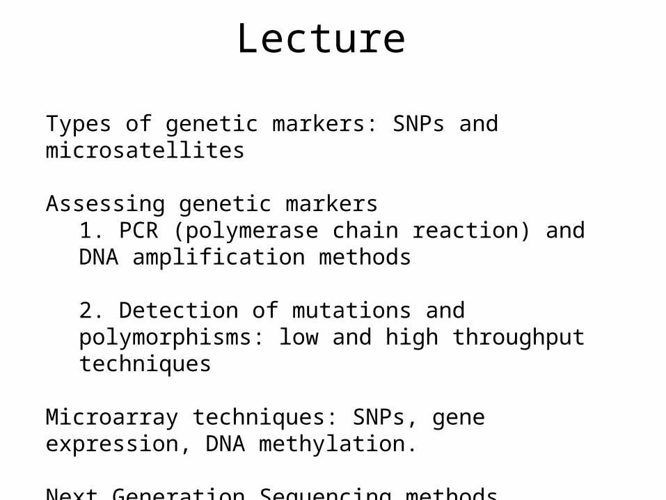
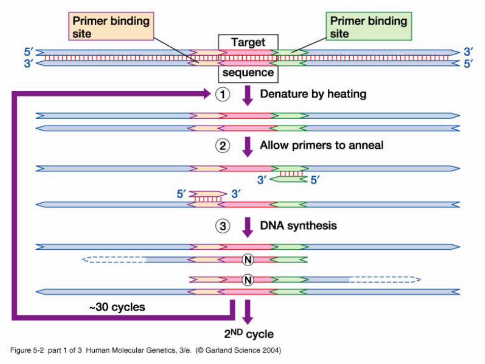
Many Genetic Analyses begin with PCR
Polymerase Chain Reaction (PCR) – specific amplification of a single gene sequence
2 synthetic oligonucleotides can “find” their complementary DNA sequences among 3 billion nucleotide sequence.
Able to faithfully amplify a specific sequence 1030 times.

1. Get genomic DNA from subject (buccal cell demonstration in class)
2. Isolate DNA on Autogen 3000
1. Lyses cells with detergent and digests protein with Proteinase K
2. Removes protein with Phenol
3. Concentrates DNA using ethanol precipitation, rehydrates DNA in buffered water.
Genotyping in MGE – TICR Individuals

Basis of all nucleic acid techniques: DNA hybridization
Long DNA melts around 75-85 degrees C

Tm calculation
Melting temperature of DNA dependent on:
• length of oligonucleotide• content of A,C,G,T• salt content of solution

Hybridization to specific sequence
A 15 base pair sequence would be unique in a random genome of 3 billion bases.
Hybridization is specific around the Tm.
Nearly all genetic applications are dependent on this feature of DNA. The sizes of nucleotides will be adjusted for specificity and efficiency at a specific temperature.
PCR: 17-35 base pairsMicroarrays: 25-80 base pairs

Genotyping in MGE - TICR Individuals (continued)
Purified genomic DNA will be amplified in the region of the polymorphisms, then a “readout” performed
PCR amplification is a standard method, but there are many methods to “read” the polymorphism
Cellular DNA is 3 X 10^9 base pairs, a gamish of sequence but only a few copies of the gene of interest
Two PCR primers (oligonucleotides) will be able to make billions of copies of one small segment, crowding out the rest of the genomic DNA

PCR design for TAS2R38 polymorphism
These probes are used to diagnose the SNP.

PCR protocol:10 ng of DNA mixed with
10 pmoles each PCR primer
1 pmoles each probe
2.5 umoles each dNTP
Reaction buffer (salts including MgCl2)
Taq polymerase (thermostable DNA polymerase)
The temperature of the mixture is cycled 35 times:
60 degrees 30 seconds
72 degrees 30 seconds
94 degrees 15 seconds
05_02.jpg
05_02_2.jpg

05_02_3.jpg

Detection of PCR products using Electrophoresis gel.
-
+
PCR products for a SNP are all the same size; this “gel” is not diagnostic for the SNPs
individuals
PCR product

Taqman allelic discrimination genotyping (for taste receptor TASR32)
There are four oligonucleotides in the reaction mix -- two PCR primers and two “probes” each labeled different color and each matching different SNP allele.

PCR design for TAS2R38 polymorphism
These probes are used to diagnose the SNP.

Taqman Genotyping - Real-time PCR
hets
homozygotes
homozygotes
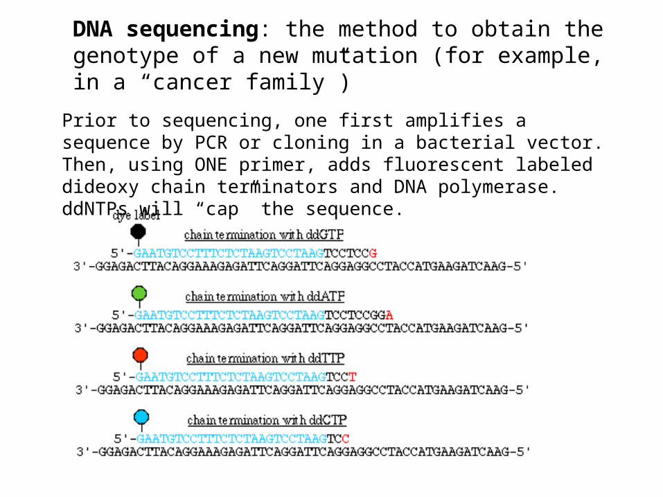
DNA sequencing: the method to obtain the genotype of a new mutation (for example, in a “cancer family”)
Prior to sequencing, one first amplifies a sequence by PCR or cloning in a bacterial vector. Then, using ONE primer, adds fluorescent labeled dideoxy chain terminators and DNA polymerase. ddNTPs will “cap” the sequence.

DNA sequencing
mutation
Useful when you suspect a gene, but don’t know the variant. This one is BRAF gene in leukemia
The products of the sequencing reaction are separated on a gel mixture that can separate fragments by one base pair.
Larger fragments Smaller fragments
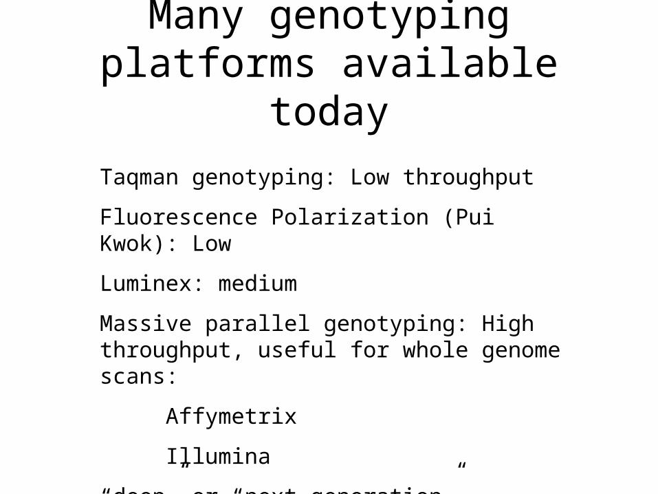
Many genotyping platforms available today
Taqman genotyping: Low throughput
Fluorescence Polarization (Pui Kwok): Low
Luminex: medium
Massive parallel genotyping: High throughput, useful for whole genome scans:
Affymetrix
Illumina
“deep” or “next generation” sequencing: Illumina (Solexa), Applied Biosystems Solid, 454 (Roche)

Illumina GoldenGate technology
for 384-6000 SNPs at a time (medium, not whole
genome)
45,000 beads 96-well plate, each with bead array

Illumina Infinium assay:
up to 1 million SNPs (for whole genome study)
Bead array on slide
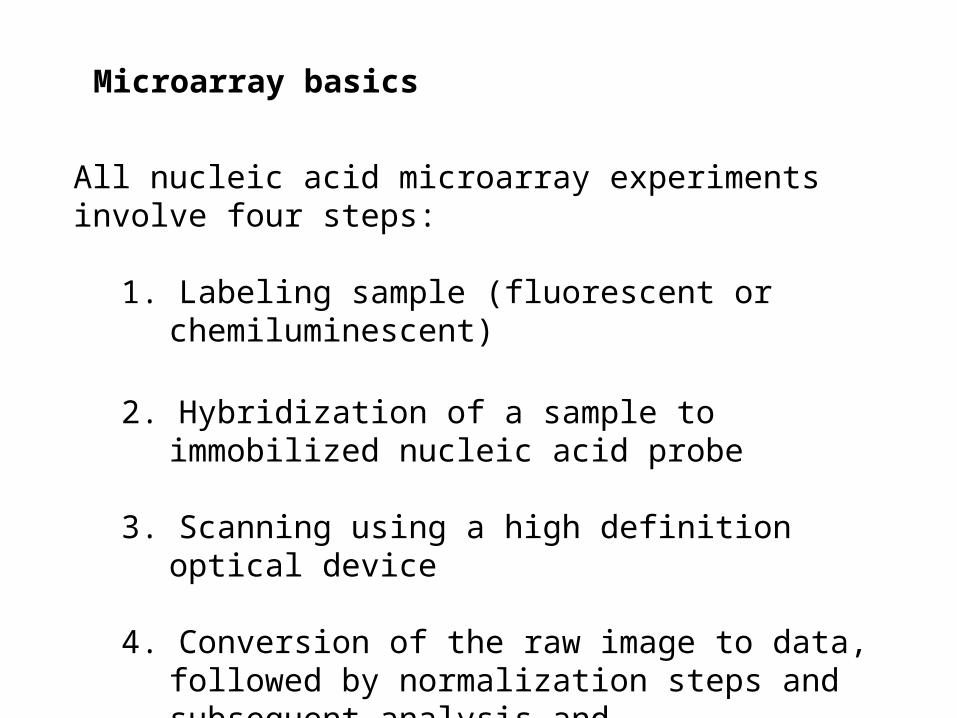
Microarray basics
All nucleic acid microarray experiments involve four steps:
1. Labeling sample (fluorescent or chemiluminescent)
2. Hybridization of a sample to immobilized nucleic acid probe
3. Scanning using a high definition optical device
4. Conversion of the raw image to data, followed by normalization steps and subsequent analysis and interpretation.
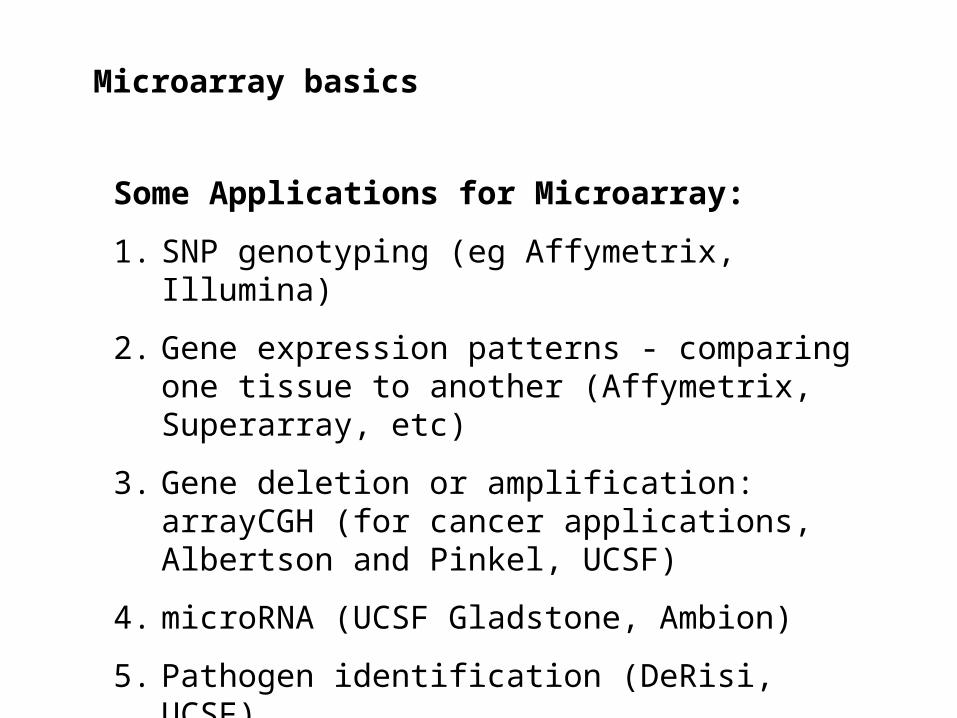
Microarray basics
Some Applications for Microarray:
1. SNP genotyping (eg Affymetrix, Illumina)
2. Gene expression patterns - comparing one tissue to another (Affymetrix, Superarray, etc)
3. Gene deletion or amplification: arrayCGH (for cancer applications, Albertson and Pinkel, UCSF)
4. microRNA (UCSF Gladstone, Ambion)
5. Pathogen identification (DeRisi, UCSF)
6. DNA methylation
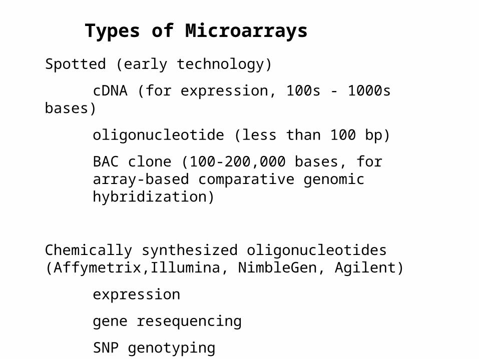
Types of Microarrays
Spotted (early technology)
cDNA (for expression, 100s - 1000s bases)
oligonucleotide (less than 100 bp)
BAC clone (100-200,000 bases, for array-based comparative genomic hybridization)
Chemically synthesized oligonucleotides (Affymetrix,Illumina, NimbleGen, Agilent)
expression
gene resequencing
SNP genotyping
array-based CGH

Spotted microarray for gene expression (oligos or cloned genes)
The microrarray may have immobilized oligonucleotides (eg., virochip, UCSF) or cloned genes

Affymetrix arrays have 25 bp oligonucleotides, very short, but massive parallel probes for redundancy. One color array.