Genet mo
14
Reprogenetics: Preimplantational genetics diagnosis Roberto Coco Fecunditas Instituto de Medicina Reproductiva, Facultad de Medicina, Universidad de Buenos Aires, Buenos Aires, Argentina. Abstract Preimplantational Genetics Diagnosis (PGD) is requested by geneticists and reproductive specialists. Usually genet- icists ask for PGD because one or both members of the couple have an increased genetic risk for having an affected offspring. On the other hand, reproductive specialists ask for embryo aneuploidy screening (PGS) to assures an euploid embryo transfer, with the purpose to achieve an ongoing pregnancy, although the couple have normal karyo- types. As embryonic aneuploidies are responsible for pre and post implantation abortions, it is logical to considerer that the screening of the embryonic aneuploidies prior to embryo transfer could improve the efficiency of the in vitro fertilization procedures. Nevertheless, it is still premature to affirm this until well-designed clinical trials were done, especially in women of advanced age where the rate of embryos with aneuploidies is much greater. Although the in- dications of PGD are similar to conventional prenatal diagnosis (PND), PGD has less ethical objections than the PND. As with the PGD/PGS results only unaffected embryos are transferred, both methods can avoid the decision to interrupt the pregnancy due to a genetic problem; this makes an important difference when compared to conven- tional prenatal diagnosis. Keywords: PGD, PGS, PGSS, embryo biopsy, trophectoderm biopsy. Introduction The term Reprogenetics was proposed to designate the combination of two types of approaches, reproductive technology and genetic methods. This combination emerged with the advent of in vitro fertilization. The most important issue is the use of preimplantational diagnosis in routine in vitro fertilization (IVF) prior to embryo trans- fer. Another important topic is prenatal diagnosis. The purpose of preimplantation genetic diagnosis (PGD) is to avoid the transmission of genetic disease in the offspring of couples with increased risk, similar to conventional prenatal diagnosis (PND). However, with this procedure arises the possibility of its use for tailored babies with cer- tain genetic characteristics. Many ethicists argue that the purpose of PGD is eugenics, but such a purpose should be condemned if it were to be imposed by the state. The in- herent desire of every parent is to give birth to a healthy and happy child. Preimplantation diagnosis has existed since IVF use began, first with morphological observations and then with specific genetic analyses of embryos produced in vitro. PGD has an advantage over PND because it facilitates a pregnancy under conditionsin which a couple would other- wise have a great risk. PGD essentially consists of several steps: 1) ovarian superstimulation, 2) aspiration of ovarian follicles, 3) oocyte retrieval, 4) intracytoplasmic injection of oocytes with processed sperm, 5) in vitro culture of fer- tilized oocytes until D5-6, blastomere biopsy on D3 or trophectoderm biopsy on D5, 6) genetic testing and 6) the transfer of a genetically normal embryo. If the blastocyst is not transferred to a receptive uterus until the 5th or 6th day, it loses the ability to produce an embryo. To preserve this possibility, it must be vitrified for later transfer. The first preimplantation diagnosis was performed in 1989 for sex selection due to an X-linked disease. Cur- rently, there are an estimated 10,000 children who were born after preimplantational biopsies. Preimplantation di- agnosis is requested by geneticists and reproductive spe- cialists. Usually, geneticists request PGD because one or both members of the couple have an increased genetic risk for passing a particular genetic disease to their offspring. Additionally, reproductive specialists request embryo aneuploidy screening (PGS) to assure a euploid embryo transfer, even when the parents have normal karyotypes. As in PGD or PGS, only unaffected embryos are trans- ferred, and both methods can thus avoid the decision to in- terrupt the pregnancy due to a genetic problem; this is an important difference with conventional prenatal diagno- sis. Genetics and Molecular Biology, 37, 1 (suppl), 271-284 (2014) Copyright © 2014, Sociedade Brasileira de Genética. Printed in Brazil www.sbg.org.br Send correspondence to Roberto Coco. Fecunditas Instituto de Medicina Reproductiva, Facultad de Medicina, Universidad de Buenos Aires, Larrea 790,1030 Buenos Aires, Argentina. E-mail: [email protected]. Review Article
Transcript of Genet mo

Reprogenetics: Preimplantational genetics diagnosis
Roberto Coco
Fecunditas Instituto de Medicina Reproductiva, Facultad de Medicina, Universidad de Buenos Aires,
Buenos Aires, Argentina.
Abstract
Preimplantational Genetics Diagnosis (PGD) is requested by geneticists and reproductive specialists. Usually genet-icists ask for PGD because one or both members of the couple have an increased genetic risk for having an affectedoffspring. On the other hand, reproductive specialists ask for embryo aneuploidy screening (PGS) to assures aneuploid embryo transfer, with the purpose to achieve an ongoing pregnancy, although the couple have normal karyo-types. As embryonic aneuploidies are responsible for pre and post implantation abortions, it is logical to considererthat the screening of the embryonic aneuploidies prior to embryo transfer could improve the efficiency of the in vitrofertilization procedures. Nevertheless, it is still premature to affirm this until well-designed clinical trials were done,especially in women of advanced age where the rate of embryos with aneuploidies is much greater. Although the in-dications of PGD are similar to conventional prenatal diagnosis (PND), PGD has less ethical objections than thePND. As with the PGD/PGS results only unaffected embryos are transferred, both methods can avoid the decision tointerrupt the pregnancy due to a genetic problem; this makes an important difference when compared to conven-tional prenatal diagnosis.
Keywords: PGD, PGS, PGSS, embryo biopsy, trophectoderm biopsy.
Introduction
The term Reprogenetics was proposed to designate
the combination of two types of approaches, reproductive
technology and genetic methods. This combination
emerged with the advent of in vitro fertilization. The most
important issue is the use of preimplantational diagnosis
in routine in vitro fertilization (IVF) prior to embryo trans-
fer. Another important topic is prenatal diagnosis. The
purpose of preimplantation genetic diagnosis (PGD) is to
avoid the transmission of genetic disease in the offspring
of couples with increased risk, similar to conventional
prenatal diagnosis (PND). However, with this procedure
arises the possibility of its use for tailored babies with cer-
tain genetic characteristics. Many ethicists argue that the
purpose of PGD is eugenics, but such a purpose should be
condemned if it were to be imposed by the state. The in-
herent desire of every parent is to give birth to a healthy
and happy child.
Preimplantation diagnosis has existed since IVF use
began, first with morphological observations and then with
specific genetic analyses of embryos produced in vitro.
PGD has an advantage over PND because it facilitates a
pregnancy under conditionsin which a couple would other-
wise have a great risk. PGD essentially consists of several
steps: 1) ovarian superstimulation, 2) aspiration of ovarian
follicles, 3) oocyte retrieval, 4) intracytoplasmic injection
of oocytes with processed sperm, 5) in vitro culture of fer-
tilized oocytes until D5-6, blastomere biopsy on D3 or
trophectoderm biopsy on D5, 6) genetic testing and 6) the
transfer of a genetically normal embryo. If the blastocyst is
not transferred to a receptive uterus until the 5th or 6th day,
it loses the ability to produce an embryo. To preserve this
possibility, it must be vitrified for later transfer.
The first preimplantation diagnosis was performed
in 1989 for sex selection due to an X-linked disease. Cur-
rently, there are an estimated 10,000 children who were
born after preimplantational biopsies. Preimplantation di-
agnosis is requested by geneticists and reproductive spe-
cialists. Usually, geneticists request PGD because one or
both members of the couple have an increased genetic risk
for passing a particular genetic disease to their offspring.
Additionally, reproductive specialists request embryo
aneuploidy screening (PGS) to assure a euploid embryo
transfer, even when the parents have normal karyotypes.
As in PGD or PGS, only unaffected embryos are trans-
ferred, and both methods can thus avoid the decision to in-
terrupt the pregnancy due to a genetic problem; this is an
important difference with conventional prenatal diagno-
sis.
Genetics and Molecular Biology, 37, 1 (suppl), 271-284 (2014)
Copyright © 2014, Sociedade Brasileira de Genética. Printed in Brazil
www.sbg.org.br
Send correspondence to Roberto Coco. Fecunditas Instituto deMedicina Reproductiva, Facultad de Medicina, Universidad deBuenos Aires, Larrea 790,1030 Buenos Aires, Argentina. E-mail:[email protected].
Review Article
Indications of PGD
Indications are similar to conventional PND with re-
gard to 1) genetic risks with monogenic or chromosomal
causes, 2) major predisposition to tumors, 3) non-genetic
risks or 4) selection of the best embryos in IVF laboratories.
As PGD or PGS involves both an IVF or intracyto-
plasmic sperm injection (ICSI) procedure and a genetic
study, it is mandatory to predict the number of unaffected
embryos obtainable for transfer prior to realizing the proce-
dure. The number depends on the embryogenic potential of
the fertilized oocytes and the implicated risk according to
the genetic disorder. The embryogenic potential depends
mainly on the woman’s age and the absence of factors that
facilitate the production of incompetent gametes. Gen-
erally, when a woman is younger than 35 years and the
male produces good quality sperm, the embryogenic poten-
tial is approximately 50%. The embryogenic potential de-
creases when a woman’s age increases or when sperm is of
inferior quality. However, the genetic risk depends on the
type of disorder (recessive, dominant, sex-linked) or if the
disorder is chromosomal. Table 1 shows the estimated
number of embryos needed to have the chance to transfer
some unaffected embryos, based on the reasoning of PGD.
Recessive Monogenic Disorders
Examples of recessive disorders are congenital disor-
ders such as cystic fibrosis, Tay-Sachs, and thalassemia,
which involve two mutated chromosomes from each
healthy carrier parent. When the disorder is molecularly
characterized, the mutation may be analyzed in cells re-
moved from a cleavage embryo or blastocyst. Mini-
sequencing is the method of choice. However, when the
mutation is not known, it might be determined by a linkage
study.
In cases where the mutation has not been identified in
one of the parents, the use of polymorphic markers linked to
the gene of interest could help to provide a better diagnosis
and allow to have more transferable embryos; otherwise,
embryos carrying the known mutation would be considered
as affected when they could be healthy carriers. Today,
with the availability of SNParrays, the characterization of
individual mutations is no longer needed.
Dominant Monogenic Disorders
Examples of autosomal dominant disorders are myo-
tonic dystrophy, fascio-scapular-humeral dystrophy, reti-
noblastoma, Von Hippel Lindau, MEN I and II, Hunting-
ton’s disease, osteogenesis imperfecta, and achondroplasia.
When the patient has a “de novo mutation” it is neces-
sary to sequence the entire gene to identify the mutation.
Once the mutation has been characterized, this sequence
can be targeted in the cells removed from the embryo. In
contrast, when there are several affected members in the
family, PGD can also be addressed with polymorphic
markers linked to the respective gene.
Usually, Huntington’s disease develops late in life or
when the offspring are of child-bearing age. Many of them
do not want to perform the genetic study because they do
not want to know their genetic status in advance, but they
want to make sure that their children do not have the muta-
tion. Unlike PND, PGD for Huntington’s disease avoids
disclosure of the status of the carrier of the mutation.
It is well known that people with certain genetic dis-
orders live in communities, such as mute communities for
congenital deafness or persons with achondroplastic dwarf-
ism, and that these couples desire PGD to increase their
likelihood of having similarly affected offspring. This is a
situation in which it is difficult to satisfy the parents be-
cause the medical team cannot help them.
Sex Linked Disorders
X-linked disorders are transmitted by the healthy car-
rier mothers to their sons, while the affected males transmit
the condition to their grandchildren through their healthy
carrier daughters but not through their sons. When the mu-
tation is characterized, it is recommended to perform PGD
by minisequencing the mutation. Some reprogeneticists
272 Coco
Table 1 - Prediction of the number of transferable unaffected embryos according to the reason for PGD.
Disorders Affected embryos Unaffected embryos Ongoing embryos Transferable embryos
Autosomal recessive (AR) 1/4 3/4 1/2 3/8
Autosomal dominant (AD) 1/2 1/2 1/2 1/4
X-linked recessive (XLR) 1/4 3/4 1/2 3/8
HLA 3/4 (no histoidentical) 1/4 (histoidentical) 1/2 1/8
AR+HLA 1/4 y 3/4 3/4 x 1/4 1/2 3/32
AD+HLA 1/2 y 3/4 1/2 x 1/4 1/2 1/16
RLX 1/4 y 3/4 3/4 x 1/4 1/2 3/32
Reciprocal translocation 4/5 1/5 1/2 1/10
Robertsonian translocation 3/4 1/4 1/2 1/8
carry out embryo sexing to avoid the birth of males, in such
cases, but as was mentioned, this is not recommended.
Examples of recessive X-linked diseases are hemo-
philia, Fragile X, and Duchenne muscular dystrophy.
In contrast, dominant X-linked diseases are transmit-
ted by affected women to 50% of their daughters and sons,
but affected males do not transmit it to their sons. Examples
of diseases linked dominant X are Rett syndrome, incon-
tinentia pigmenti, pseudohyperparathyroidism, and vita-
min D-resistant rachitism.
As an example of Y-linked disorders, there are some
AZF region microdeleted in the long arm of the Y-chro-
mosome. In this case, the only option to avoid transmission
to the offspring is female sex selection.
Chromosomal Disorders
This category mainly includes carriers of balanced
chromosomal rearrangements, such as reciprocal translo-
cations, Robertsonian translocations, inversions and some
cryptic deletion-duplication abnormalities. Most of the nu-
merous anomalies in autosomal chromosomes are lethal
and rarely reach adulthood, except Down syndrome.
Women affected with Down syndrome are fertile and have
a 50% risk of transmitting the condition, while affected
males are sterile. In contrast, the numerous chromosomal
abnormalities of the sex chromosomes allow individuals to
reach adulthood, but some cause sterility and other types of
infertility or sterility alone. Women that are 47, XXX are
generally fertile, and they have a 50% of risk to have
daughters with the same condition and males with 47,
XXY. The 47, XYY males who are fertile have no risk of
transmitting the YY condition because one of these chro-
mosomes is excluded from the meiotic sexual body. How-
ever, the XXY males who produce sperm in fact have
mosaic gonads, and only the XY spermatogonial cells un-
dergo meiosis (Sciurano et al., 2009). Prior to the advent of
the Comparative Genomic Hybridization array (aCGH),
these PGDs were addressed by FISH using probes of the
chromosomes involved. Today, the best tool for chromo-
somal PGDs is molecular karyotyping or aCGH. The carri-
ers of reciprocal translocations, both male and female, have
a theoretical risk of 80% to produce abnormal gametes be-
cause the meiotic quadrivalent is segregated. Only alternate
segregation produces balanced gametes; instead, adjacent I,
adjacent 2, 3:1 and 4:0 segregations are all abnormal. The
real risk evaluated in sperm, oocytes and cleavage embryos
on day 3 agrees with the theoretical risk. The carriers of
Robertsonian translocations also have a theoretical risk of
75% because the meiotic trivalent is segregated (alternate,
adjacent 1, adjacent 2 and 3:0), but the real risk observed in
gametes and cleavage embryos is approximately 30%. It is
much lower than the theoretical risk. The same occurs with
the carriers of peri/paracentric chromosome inversions. In
fact, both Robertsonian translocations and chromosome in-
versions are considered to be benevolent rearrangements
compared to reciprocal translocations (Coco et al., 2005).
Cryptic duplications or deletions have a theoretical risk of
50%, but there are not enough published reports to estimate
the empirical risk of carriers of such anomalies.
The frequency of balanced chromosome rearrange-
ments is 0.2% in control population of newborn, while it is
0.6% in infertile couples, 3.2% in those with recurrent
failed IVF procedures, 9.2% in couples with recurrent mis-
carriages, 3.1% in men who require ICSI, and a similar fig-
ure for their partners (Peschka et al., 1999; Stern et al.,
1999; Gekas et al., 2001; Clementini et al., 2005; Moz-
darani et al., 2008). Therefore, it is advisable to always
carry out karyotyping of the couple prior to an IVF/ICSI
procedure.
While the carriers of balanced chromosome rear-
rangements would be ideal candidates for pre-implantation
genetic diagnosis, one has to consider that the only couples
that can take advantage are those in which women respond
very well to ovarian stimulation and are under the age of
40 years.
PGD for Rh Blood Group Typing
PGD can also be indicated in women who are Rh neg-
ative and are highly sensitized with antibodies against Rh
factor. If Rh genotyping in the male shows that he is hetero-
zygous, it is feasible to perform a PGD to avoid possible
erythroblastosis fetalis and intrauterine blood exchange
transfusion. PGD has also been used in women sensitized
by other blood factors, such as the Kell/Cellentano group.
PGD for Human Leukocyte Antigen HLA Typing
It is known that persons affected with certain genetic
or acquired disorders require an HLA-compatible bone
marrow transplant to survive. Due to the existence of public
international banks of bone marrow or umbilical cord
blood, the majority of those in need may find donors. When
it is impossible to find a compatible bone marrow and the
couple is still young and wants to have another child, the
couple might use PGD for HLA typing to have an HLA-
compatible child that can save the life of a sick sibling.
PGD is performed using single tandem repeat polymor-
phisms (STRs) linked to HLA that map to chromosome 6.
The purpose of PGD is to select the embryos with the same
haplotype of the person who needs the transplant. These
types of PGD are a real challenge because the probability of
finding an HLA-compatible unaffected embryo is one in
every 10 embryos studied (see Table 1). Socially, this type
of PGD is known as PGD for having a child that serves as a
“medication baby” as well as for “a la carte” designed ba-
bies, both of which are pejorative terms. If the best option is
the latter one after an exhaustive international donor search,
I consider it correct to attempt to have a child who can save
the life of a sibling that will die if he/she does not receive a
compatible bone marrow transplant.
Reprogenetics 273

Indications of Preimplantation GeneticScreening (PGS)
PGS could have the same indications as PND, if one
assumes that the chromosomal constitution found in polar
body I, in a blastomere removed on day 3, or in several cells
of the trophoblast on day 5 represent the constitution of the
future embryo. However, today, there is evidence that this
is not always true. Taking into account that the majority of
miscarriages in the first trimester are caused by aneuploi-
dies, and the rate of aneuploid oocytes increases with ad-
vancing age and is increased in males with oligoastheno-
teratozoospermia (AOT), the ideal candidates for PGS
would be as follows: 1) advanced maternal age, 2) couples
with recurrent miscarriages, 3) couples with repeated IVF
failures, and 4) severe male infertility. It could also be used
to select a single euploid embryo for transfer, especially to
avoid multiple pregnancies or to restrict the number of em-
bryos to vitrify.
Advanced Maternal Age
Women at an advanced age have a greater chance of
having aneuploid pregnancies because they have increased
rates of producing aneuploid oocytes. Oocytes are always
the same age as the woman. However, in males, sperm are
produced every 65-75 days. Therefore, it might be said that
sperm are not the same age as the male. The prolonged ar-
rest of oocytes at meiotic prophase I mainly contributes to
aneuploidy due to the decline in competence of the cyto-
plasm of the oocyte. The number and distribution of chias-
mata during prophase I as the weak centromeric cohesion
may be the main factor that predisposes aneuploidy that is
inherent to age. In fact, the principal cause of oocyte aneu-
ploidy is the precocious separation of sister chromatids
rather than classic non-disjunction (Chiang et al., 2012). In
the male, the expected sperm aneuploidy rate is between
0.5 and 1% because the sperm is not the age of the male, but
if the sperm is not ejaculated for prolonged periods, it could
have a high rate of DNA fragmentation, which is also re-
sponsible for abnormal fertilization. Competent oocytes
from young women can repair the DNA fragmentation of
the sperm, but the oocytes from older women cannot.
Therefore, women of advanced age have higher probabili-
ties of having abnormal pregnancies that might end in mis-
carriage or in a malformed newborn. Most of these embryos
are lost during pre or post implantation stages, while a mi-
nority come to term. That is why the possibility of miscar-
riage also increases with the age of the woman (see Ta-
ble 2).
It is well recognized that most autosomic aneuploi-
dies in live newborns are de novo or inherent to maternal
age, while most sex chromosome aneuploidies are of pater-
nal origin independent of paternal age, or are associated
with poor sperm quality (Hassold et al., 1984; Jacobs and
Hassold, 1995). The majority of males with a normal
karyotype and a normal spermiogram have a 0.5 to 1%
sperm aneuploidy rate, while the rate for oocytes is much
higher, between 20 and 50%, mainly depending on the age
of the woman (Hultén et al., 2005). However, the rate of
sperm aneuploidy in males with OAT and a normal karyo-
type is much greater than that observed in males with
normozoospermia (Coco, et al., 2000; Colagero et al.,
2001; Rubio, et al., 2001; Burello et al., 2003). Although
women more than 37 years old and males with OAT could
be ideal candidates to benefit from PGS, most of them fail
due to the low probability for producing euploid embryos.
The first randomized controlled clinical trials (RCT) for
PGS in patients with advanced maternal age were promis-
ing due to the lower miscarriage rate and higher take-home
baby rate achieved in the group that underwent PGS (Mun-
né et al., 2006). However, other clinical trials emerged
without differences in the findings between both groups,
with and without PGS, and others showed even worse re-
sults in the studied group with PGS. (Staessen et al., 2004,
2008; Stevens et al., 2004;Jansen et al., 2008; Mastenbroek
et al., 2007; Hardarson et al., 2008; Schoolcraft et al.,
2009). Most of those studies were performed with biopsies
on D3 and FISH, first enumerating five chromosomes and
later 7, 9 and 12 chromosomes. Three arguments were used
to explain these poor performances: a) limitation of the
technique to enumerate all chromosomes, b) lower implan-
tation rates after the removal of one or two blastomeres,
which means a loss of embryonic mass of between 12.5% to
25%, and c) discarding supposed aneuploid embryos that
might have been self-corrected and lead to a normal preg-
nancy. Currently, we have much hope with comparative
genomic hydridization arrays (aCGH), which could solve
the first and second limitations because they allow the
study of all 24 chromosomes, and because the cells ex-
tracted correspond to trophectoderm cells and not to the
stem cells of the inner cellular mass of the blastocyst
(ICM). The third and last limitation mentioned remains un-
answered until we know the rate of false positives and neg-
atives for this method. While we are performing
randomized controlled trials (RCTs) similar to those car-
ried out with PGS by FISH, we will continue, of course,
without knowing the clinical value of PGS.
274 Coco
Table 2 - Pregnancy loss rate and maternal age.
Maternal age Down’s risk All chromosome risks Miscarriages rate
20 1/1667 1/526 8
25 1/1200 1/476 10
30 1/952 1/385 12
35 1/378 1/192 16
40 1/106 1/66 40
45 1/30 1/21 60
The first clinical trial with aCGH in blastocysts was
performed by Yang et al. (2012). The authors, worked with
patients with good prognoses, and these showed an in-
creased pregnancy rate in patients who had received the
best euploid blastocyst compared to those without aCGH
(70.9% vs. 45.8%). The same authors (Yang et al., 2013),
also working with patients with good prognoses, docu-
mented a better implantation rate in a group with aCGH
(65%) vs. a group without aCGH (33%).The authors also
showed that the rate of spontaneous miscarriage was signif-
icantly lower in the group screened with an array (0%) vs.
controls (16.7%).
Schoolcraft and Katz-Jaffe (2013), working with pa-
tients over 35 years old, also showed a better ongoing preg-
nancy rate (60.8%) in the group that had received devi-
trified euploid blastocysts compared to the control group
(40.9%) that had received blastocysts without aCGH. A
clinical trial of quite different design was published by
Scott et al. (2013). These authors carried out the transfer of
the two best embryos, whereby one was biopsied on D3 or
D5 in accordance with the routine protocol for patients with
good prognosis, but without knowing the outcome of an ar-
ray before the embryo transfer. The authors found that
48.2% of the euploid blastocysts achieved pregnancy,
while 93.5% of the aneuploid blastocysts did not achieve
implantation. In contrast, the transfer on D3 showed that
29.2% of the euploid embryos achieved pregnancy, while
98.1% of the aneuploid embryos failed to do so. These re-
sults clearly reinforce the need to continue with this type of
clinical trial in patients indicated for an IVF procedure, es-
pecially those of poor prognosis due to advanced age. The
authors also showed that the implantation rate decreases
nearly 50% when the biopsy is performed on D3 with
respect to non-biopsied controls (30.4% vs. 50.0%). How-
ever, the implantation rate was not modified by the
trophectoderm biopsy (51.0% vs. 54.0%). Therefore, such
biopsy done on D5 has no deleterious effect compared to
the one on D3. Another RCT (Forman et al., 2013) showed
that the cumulative rate of take-home babies was similar
when transferring the best euploid single blastocyst (69%)
or the two best blastocysts without aCGH (72%), with a
47% multiple pregnancy rate in the group transferred with
the two best blastocysts. The authors documented an
aneuploid blastocyst rate of 31%, with 21% of women
younger than 35 years and 56% older than 40 years. Almost
half of the newborns were twins in the transfer group with
two blastocysts. The rate of preterm, low birth weight and
greater number of days required in neonatal intensive care
was also twice that of the group with a single blastocyst
transfer.
Recurrent Pregnancy Loss (RPL)
Usually, RPL is defined as two or more consecutive
pregnancies lost before 20 weeks of gestation. Different
cytogenetic studies of miscarriages in the first trimester of
pregnancy show that aneuploidy rates varied between 50%
and 80% (Strom et al., 1992).
Additionally, it has been documented that couples
with RPL produce more aneuploid embryos than those who
have not had RPL (Pellicer et al., 1999). According to some
authors, PGS does not improve the rate of pregnancy in
RPL, but increases the chance of birth at term (Platteau et
al., 2005).
Recurrent IVF Failure (RIF)
RIF is usually defined as the failure of three or more
IVF attempts with good quality embryo transfer. Some au-
thors argue that these couples produce more embryos with
aneuploidies (Hodes-Wertz et al., 2012). However, there is
no evidence that PGS improves the rate of pregnancy or
live IVF births.
Severe Male Factors
As mentioned above, the rate of aneuploidy in sper-
matozoa from fertile males with a normal spermiogram is
much lower than that observed in oocytes, and aneuploidy
also does not increase with age in men.
The study of the chromosomal complement of the
sperm has been feasible since Rudak et al. (1978) published
the possibility of fertilizing oocytes from hamsters with hu-
man sperm. Later on, the hamster test was replaced by
FISH on semen, a technique that is much less complex than
Rudak’s technique. Although FISH is ideal for enumerat-
ing all chromosomes, it has the limitation of the number of
chromosome probes used per hybridization round, which is
fundamental for the small size of the sperm nucleus, be-
cause the number recommended is no more than three
probes per round of hybridization. Rives et al. (1998) per-
formed FISH for all chromosomes in semen samples from
four semen donors. They found uniformity in the percent-
age of autosome disomies, which varied between 0.1 and
0.5%. This finding encouraged several authors to evaluate
the chromosome complement of spermatozoa by perform-
ing FISH on semen samples with a few chromosome probes
and to estimate the rate of aneuploid sperm using a simple
mathematical calculation that assumes that the remaining
chromosomes (i.e., those not studied) would behave simi-
larly. In a study conducted with 10 voluntary donors of se-
men in our laboratory during the year 2000, we found that
the percentage of sperm aneuploidy ranged from 4.2% to
14.3%, with an average value of 10.1% � 3.8. In contrast,
when we studied patients with oligoasthenoteratozoosper-
mia (OAT), the frequency of aneuploidies varied from 4 to
83% with an average value of 25.8% � 8.4 (n = 53). We also
found that sperm aneuploidies increased with the severity
of OAT. We have extended the study to 10 patients with
OAT who used the ICSI procedure. FISH in semen was per-
formed with the same sample used in the ICSI procedure,
and we found that the percentage of sperm aneuploidies
Reprogenetics 275
was higher in the group that had not achieved pregnancy
(Coco et al., 2000). These findings put in evidence the im-
portance of the genetic risk assessment before the ICSI pro-
cedure to predict the chance of success. Now, with the
possibility of PGD and lower costs, FISH is no longer used
to assess sperm. It should be noted that the chances of se-
lecting an euploid embryo mainly depend of the number of
embryos produced during the procedure. When it is sus-
pected that the couple has a major chromosomal risk due to
advanced maternal age or severe male factors, it is manda-
tory to inform them of the low chance of achieving a preg-
nancy with the PGS procedure, unless the couple produces
many embryos that provide one or two euploid embryos apt
for transfer (Harper and Sengupta, 2012).
Sex Selection (PGSS)
The selection of the gender of the future child is a
wish that a majority of people have when they plan to have
offspring. However, for most people, it is not viewed posi-
tively unless sex-linked diseases or other important con-
cerns exist. Today, sex selection is a feasible reality that can
be satisfied without ethical or legal disadvantages if the se-
lection was carried out when the pregnancy is not estab-
lished. Before the advent of PGD, there were no attempts to
perform a prenatal diagnosis for sex selection, for diseases
of late onset in life or for the predisposition to suffer certain
tumors. While it is reasonable to state that the bioethics sta-
tus of a preimplantation embryo is not equal to that of an
embryo-fetus, there are, nonetheless, those who consider
that already a fertilized oocyte has the status of a person,
and for these, PGD would not be an option. Nonetheless, if
we take into account the number of PGSS procedures per-
formed worldwide as a proportion of the total number of re-
quests for sex selection, it reasonable to assume that many
of these reconsidered their concepts after being informed of
the procedure.
Table 3 provides a list of the different motivations for
PGDs from 54,589 cycles registered during data collection
I-XIV for PGD by the ESHRE Consortium (Traeger Syno-
dinos et al., 2013).
Biopsy Techniques
There are several types of recognized biopsies: polar
bodies, blastomeres on D3, trophectoderm on D5/D6 and,
more recently, attempts to perform blastocentesis in blas-
tocysts on D5/D6, as a new type of noninvasive embryo bi-
opsy based on the presence of cells and DNA in the
blastocoelic cavity. Actually, all of them are invasive and
involve some risk of loss of the embryo. Working with a
simple cell is not easy and may yield no results. In this re-
gard, the biopsy of blastocysts is most suitable. Blasto-
centesis is by now a new hope for less invasiveness. While
none of these methods assures the proper constitution of the
future embryo, they minimize the risk of the disorder that is
being investigated. The best result was obtained via the bi-
opsy of blastomeres on D3 with fresh transfer on the same
day of the biopsy, or transfer on D4/D5. To remove one or
two cells from the preimplantation embryo it is first neces-
sary to perforate the zona pellucida. The perforation of the
zona pellucida can be done by several methods: A) me-
chanically, by cutting through the pellucida with a micro-
pipette; B) chemically, by dissolving part of the pellucida
with an acid solution; or C) by laser, through modulating a
laser beam via the optical system of a microscope. Prior to
biopsy, the preimplantation embryos can be placed in a
suitable medium to loosen the cell junctions at room tem-
perature. Then, the embryos are placed in separate micro-
drops composed of the medium for biopsy under oil and
labeled. It is not convenient to have more than two pre-
embryos in a dish to minimize their time out of the incuba-
tor. Using a micromanipulator/microscope setup, the
oocyte or the pre-embryo that will be biopsied is placed in
the center of the field and focused at a 400X magnification.
The embryo or oocyte is fastened with a micropipette
holder. The zona pellucida is perforated, and the polar bod-
ies (PBI/PBII), or blastomeres are removed gently with an
appropriate micropipette. When the biopsy is performed on
D5, the zona pellucida is perforated on day 3 to facilitate
the hatching of the blastocyst and to easily remove some
cells of the trophectoderm. The cells will have to be col-
lected according to the protocol of the genetic study indi-
cated. If the indication is a FISH study, the removed cells
are fixed on a slide, but if the indication is a PCR assay, the
removed cells are collected in a small tube.
Biopsy of Polar Bodies (PB I/PBII)
The biopsy of the first PB prior to the fertilization of
the oocyte evaluates the result of the first meiotic division.
Because errors can also occur during the second division of
the oocyte, it is necessary to also study the second PB to
avoid misdiagnosis. The second division of the oocyte is
completed when the sperm penetrates the oocyte. There-
fore, the biopsy of the second PB is performed once the
ovum has been fertilized. As PB biopsies do not allow the
evaluation of male meiotic errors and/or errors that occur
after fertilization of the egg, the biopsy of blastomeres is
more preferred because it allows the assessment of both pa-
276 Coco
Table 3 - Different reasons for PGD from the I-XIV ESHRE PGD Consor-
tium data collection.
Reason for PGD Nº PGD cycles Percentage
Monogenic 11.084 20.3%
Chromosomal 8.104 14.8%
Sex selection for monogenic X-linked 1.603 2.9%
Social sexing 765 1.4%
PGS 33.033 60.6%
Total 54.589 100%

rental contributions and/or errors during cleavage. Polar
body biopsy is only useful when women have a major risk
of transmission of monogenetic diseases or aneuploidies
inherent at the maternal age. As was mentioned above, to
avoid misdiagnoses, both polar bodies should always be
biopsied. When the purpose is to evaluate aneuploidies,
both biopsies can be performed simultaneously. Instead,
when the question is a monogenic disease, it is necessary to
perform the biopsy in a sequential way. The biopsy of the
first polar body prior to fertilization only indicates errors
during the first meiotic division and/or whether the oocyte
carries the same maternal mutation, always assuming that
an interchange did not occur in the locus where the muta-
tion maps to. In countries where embryo biopsy is prohib-
ited, the pre-implantation genetic diagnosis can only be
performed by biopsy of polar body I because the second po-
lar body appears after the fertilization of the oocyte. There-
fore, the biopsy of the second polar body would have the
same connotation as the embryo biopsy.
PBI biopsy to assess aneuploidies is not optimal be-
cause there are other possibilities for segregation during the
second division, without assuming that the first meiosis
was normal or abnormal. Figure 1 shows the different pos-
sibilities of the second division after a normal first meiotic
division, where two of three possibilities are abnormal. Fig-
ure 2 shows the different possibilities of the second division
after a non-disjunction occurred during the first division;
one of the six possibilities corresponds to an aneuploid res-
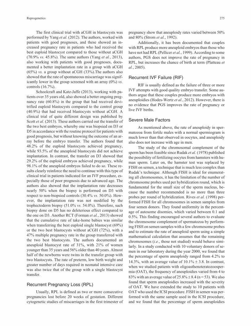
cue. Figure 3 shows the different possibilities during the
second division after a premature separation of sister chro-
matids in the first meiotic division; two of six possibilities
correspond to an aneuploidy rescue.
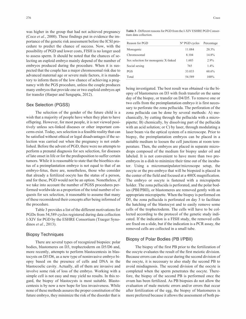
Polar body biopsy is also not ideal for monogenic dis-
eases due to the possibility of crossing over. Figure 4 A
shows a normal segregation without crossing over, while
Figure 4 B shows a segregation after a crossing over event
at the level of a mutated gene that is able to produce normal
and abnormal oocytes.
In spite of the disadvantages indicated according to
the ESHRE Consortium, 16% of the biopsies done corre-
spond to those of polar bodies. Kuliev and Verlinsky (2004)
studied more than 8,000 oocytes from women older than 35
years with FISH for chromosomes 13, 15, 16, 21 and 22
found that more than 50% had aneuploidy. Recently, a pilot
study of the ESHRE PGD Consortium using aCGH in polar
bodies from women over 40 years showed an aneuploidy
rate of 75% (Geraedts, 2013)
Biopsy of Blastomeres in Cleaved Embryos onD3
This is the type of biopsy used to remove one or two
cells in embryos with more than six cells on D3. Embryolo-
gists have acquired great skill in performing this technique,
but there is evidence that it decreases the rate of implanta-
tion. FISH or PCR analysis in a simple cell is a real chal-
lenge for specialists and for patients. It is not easy to work
Reprogenetics 277
Figure 1 - Possibilities of segregation during the second division from a normal oocyte. One of the three possibilities is normal (A).

with a single molecule of DNA for chromosomal and ge-
netic tests. However, this was the methodology used in the
last 20 years. Indeed, 80% of the PGDs recorded by the
ESHRE Consortium were D3 blastomere biopsies. During
that time, fresh transfer was used to avoid cryopreservation.
At first, the protocol mostly used for PGD/PGS was trans-
fer on the same day of the biopsy. Improvement of sequen-
tial culture media allowed the prolonging of in vitro
development until the fifth day, and transferring only those
that reached the blastocyst stage. Biopsy on D3 achieved a
clinical pregnancy rate of 18.7% and a take-home baby rate
of 14.7%, with a misdiagnosis between 5 and 10%, accord-
ing to records I-XIV of the ESHRE Consortium.
Blastocyst Biopsy
The blastocyst is the highest degree of development
that an embryo can reach in vitro, and it is characterized by
three elements: the inner cell mass, the outer cell layer or
trophectoderm and the blastocoel. The blastocyst begins to
form on the fifth day and is completed on the sixth.
A blastocyst usually has more than 100 cells. The ma-
jority will form the placenta, the chorionic villus and other
extraembryonic structures. Only a small percentage of the
ICM will differentiate into the embryo proper after implan-
tation of the embryo in the endometrium. Therefore, the
trophectoderm biopsy procedure is considered equivalent
to the puncture of a chorionic villus, with the same limita-
tions of not corresponding to the constitution of the embryo
per se due to the possibility of mosaicism.
Once the decision to perform a trophectoderm biopsy
is taken, it is preferable to perform the transfer in a deferred
cycle, because not all blastocysts are obtained on D5, and
the genetic studies demand time. This decision is very im-
portant because it allows better organization of the genetic
laboratory assays. At present, there is sufficient evidence
that the deferred transfer to the stimulated cycle has its ad-
vantages in terms of implantation, ongoing pregnancy and
278 Coco
Figure 2 - Possibilities of segregation during second division from an abnormal oocyte by non-disjunction. One of the six possibilities is normal (B).
Reprogenetics 279
Figure 3 - Possibilities of segregation during second division from an abnormal oocyte by early separation of sister chromatids. Two of the six possibili-
ties are normal (B and C).
Figure 4 - Segregation with and without cross-over. (A) Normal segregation without cross-over at the level of the mutated gene. The oocyte will always
have the opposite chromosome constitution to that of polar body I. (B) Normal segregation with a cross-over at the level of the mutated gene. In contrast,
when exchange occurs, the egg may or may not have the mutation.

lower risk of genetic and epigenetic alterations (Papaniko-
laou et al., 2006; Maheshwari et al., 2012; Shapiro et al.,
2012, Roque et al., 2013). In 2011, our team took the deci-
sion to switch from the blastomere biopsy to the trophecto-
derm biopsy procedure with deferred transfer to the
stimulated cycle. The results were much better, and impor-
tantly, the entire team worked in a more comfortable,
friendly, healthy and efficient environment (Coco et al.,
2012).
Genetic Testing
There are three fundamental techniques in a PGD pro-
gram: FISH, PCR and aCGH. Handyside (2013) recently
published the different diagnostic tests available to carry
out PGD/PGS, along with their pros and contras.
Considerations of PGD/PGS
Not all people who want to perform PGD are able to
do so. There are two fundamental requisites: the couple
should be fertile and it should have the genetic character-
ization of the disorder which is intended to be diagnosed.
This last point is very important because couples assume
that they will have a normal healthy child, whereas it is only
possible to offer to minimize the risk for the disease for
which they have a great risk of transmitting to their off-
spring. As there is a small risk of misdiagnosis due to the
existence of mosaicism or limitations of the techniques
used, couples should always be offered the possibility of an
amniocentesis to double check the results. It should be re-
membered that all the other prenatal diagnoses, both the
non-invasive prenatal test (NIPT) and the chorionic villi
sample, as well as PGD are screening methods because
these analyses are based on cells from the trophectoderm.
The procedure should always be explained in detail during
the entire treatment to ensure that couples are informed
about the potential risks in the short, medium and long
term.
Because the majority of patients for PGD are advised
by a geneticist, the patients are usually more informed
about the risk at birth or during pregnancy, from the end of
the first trimester and beginning of the second, which are
completely different from the risk during the preimplan-
tation phase, particularly with regard to the risks of
aneuploidies and abnormal segregations in carriers of chro-
mosomal rearrangements. During preimplantation devel-
opment, the risks are much higher. To benefit patients with
a more predictable PGD, doctors should ensure that the
couple produces a sufficient quantity of embryos to obtain
non-affected embryos for transfer.
With respect to PGS, it can be said that the ideal can-
didates have the following characteristics: women of ad-
vanced maternal age, couples with recurrent miscarriages
and recurrent IVF failures, and men with severe male fac-
tors. However, the usefulness of PGS is still controversial.
For PGS by FISH the results first were very promising, but
later on discouraging and, finally, undesirable because this
method produces effects contrary to those expected. These
became evident when studying biopsies taken on D3. We
have now begun to investigate biopsies taken on D5 as
these provide more cells to study and permit to apply differ-
ent diagnostic methodologies that are more robust than
FISH. The use of blastocyst biopsy is very promising, as
happened at the beginning with FISH with biopsy on D3.
Although it is undisputed that the CGH array is superior to
FISH, there are doubts about the benefit of these arrays in
the group of patients who are more likely to produce aneu-
ploid embryos. Women of advanced age have a higher risk
of producing aneuploid oocytes. Additionally, such women
respond poorly to ovarian stimulation and thus produce
only few blastocysts. If the existence of a male factor is
added, the prognosis is even more discouraging. The mei-
otic risk of the couple based on the woman’s age and the ex-
istence of male factors should be used to estimate the
number of blastocysts required to obtain at least one
euploid blastocyst apt for transfer. For women of advanced
age more oocytes are needed to have a chance of finding
euploid blastocysts, but they produce much fewer oocytes
than young women. An alternative would be the collection
of blastocysts in various cycles of ovarian stimulation in-
stead of only one, though there are insufficient, primarily
anecdotal data to conclude that this may be a solution
(Mondadori et al., 2012). Harton et al. (2013) recently re-
ported that selective transfer of euploid embryos showed
that implantation and pregnancy rates were not signifi-
cantly different between reproductively younger and older
patients of up to 42 years of age. Nevertheless, information
on more well-designed clinical trials is needed to know if
the embryos from older women are really harmed or not by
biopsies, because the poor quality of the embryos might
make them more vulnerable to the procedure.
Therefore, for biopsies on D3 or D5, one must assume
that the result found in the removed cells corresponds to the
condition of the rest of the embryo. However, this is only
true if errors do not occur after fertilization. In contrast,
when an error occurs after fertilization, the probability to
detect it depends on the cleavage stage and the day that the
biopsy is carried out. If the biopsy is performed on D3 and
the error occurred in the first division, there is a 50% possi-
bility of detection; but when it occurred during the second
division, the chance drops to 25%, and during the third divi-
sion, the probability is only 12.5%. If the biopsy is per-
formed on D5 and the errors have occurred in the first three
divisions, there is a chance of detection, but it is also true
that a newly originated trisomy might be detected in one of
the isolated cells of the trophoblast. One trisomic cell
among 5 to 10 removed cells is already detectable with the
current diagnostic tools, and if the trisomy is viable, it
would surely be confined to the placenta, without any im-
plication for the chromosome constitution of the embryo.
280 Coco

Patients should understand that the constitution of the tro-
phectoderm is not always coincident with that of the ICM
because different possibilities of chromosomal constitution
can exist: a) the trophectoderm and the ICM are homoge-
neously normal or abnormal, b) the trophectoderm may be
abnormal but the ICM may be normal, (c) the trophec-
toderm may be normal but the ICM may be abnormal, d)
the trophectoderm may be a mosaic and the ICM may be
normal or abnormal, and e) both the trophectoderm and
ICM may be mosaic.
If clinical trials are done on a group with PGS and an-
other one without PGS and the euploid embryos are always
transferred, one can never know the rate of false positives
and negatives that may PGS produce. This is not a minor
detail, and it is relevant, especially when couples produce
one or two blastocysts, and the array indicates that they are
abnormal. Should we advise discarding them? Obviously,
it is necessary to have better designed clinical trials to ob-
tain the correct answer. If we are to follow the same type of
clinical trials as carried out on PGS by FISH, we are likely
to make the same mistakes, and we are perhaps eliminating
the last chance for older couples to be parents of their ge-
netic children. Today, there is evidence that blastocyst bi-
opsy does not harm the implantation process, so we should
offer it to patients who require IVF/ICSI to participate in a
clinical trial. The patients must accept that the best blas-
tocyst be biopsied without knowing the result of aCGH. I
personally consider that this type of clinical trial will allow
us to know the behavior of the anomalies detected in the
trophectoderm. The classical randomized clinical trials in-
clude two groups of patients, studied and unstudied, with
subsequent re-analysis of the blastocysts that were not
transferred because they had been diagnosed as aneuploids
with the purpose of verifying the existence of mosaics;
these are, in my opinion, very expensive for an unrecover-
able blastocyst. Johnson et al. (2010) reported a correspon-
dence of 96.1% between karyotypes of the trophectoderm
and ICM when they re-examined 51 blastocysts diagnosed
as aneuploids. However other authors (Liu et al., 2012)
who re-examine 13 blastocysts diagnosed as aneuploids
found a high rate of mosaics in the trophectoderm, and in
four cases mosaics were not present in the ICM because the
aCGH was normal. The establishment, characterization and
differentiation of a karyotypically normal human embry-
onic stem cell line from a blastocyst diagnosed as chromo-
some 21 trisomy was recently reported (Mandal et al.,
2013). Such a finding could be due to a self-correction of
the aneuploidy or to the existence of a mosaicism of the
trophectoderm, as well as due to a recent error that occurred
in only one cell among the cells removed during trophec-
toderm biopsy.
Main Conclusions
PGD is an alternative to prenatal diagnosis for pa-
tients with increased genetic risk in their offspring. It is a
prerequisite that the couple should be fertile or that their in-
fertility can be reverted by IVF. Preimplantation diagnosis
has a great advantage over conventional prenatal diagnosis
because it can avoid possible genetic miscarriage. How-
ever, it has the disadvantage of being expensive, and fertile
couples have to undergo a highly complex treatment, as do
infertile couples. When the couple is genetically infertile it
is mandatory to perform PGD to avoid the inherent risk to
the offspring.
Although PGS is indicated for advanced maternal
age, recurrent miscarriages, or for severe male factors, the
benefits of its use are not yet clear, especially for patients
with normal karyotypes. It is true that these patients have a
higher risk of producing aneuploid zygotes, but it is also
true that most homogeneous aneuploidies are lethal, on the
order of 99%, and that the majority die before implantation
or during embryo-fetal development (Hassold and Hunt,
2001). Unlike partial aneuploidies, almost from abnormal
segregation of carriers of balanced rearrangements, the
pregnancy may continue to term and birth a malformed
child. Therefore, the above-mentioned patients should use
PGD, especially if they are infertile.
The experts’ committees of the main scientific societ-
ies for reproductive medicine support the idea that there is
still no evidence that establishes PGS as beneficial for those
groups of patients who would be the ideal candidates for
PGS (Hardarson et al., 2008). As these groups of patients
produce fewer normal embryos for transfer, they should be
informed that the pregnancy rate after PGS could be lower
compared to IVF without PGS.
Among the different types of existing biopsies, it is
possible to conclude that the polar body biopsy has very
low diagnostic efficiency. Blastomere biopsy on D3 de-
creases the rate of implantation, and between 10 and 20% of
the embryos studied may not give results due to the limita-
tions of the assays performed on only one or two DNA mol-
ecules. The rate of misdiagnosis according to the data of the
ESHRE PGD Consortium is between 3-10%.
Thus far, there is no information on the increase of
malformed children after PGD/PGS. However, one cannot
rule out a potential risk for genetic diseases with any of the
IVF procedures. The procedures IVF / ICSI / PGD should
be considered experimental until one can see what happens
to the grandchildren born post assisted reproduction tech-
nologies (ART). Trophectoderm biopsy might be consid-
ered less invasive than blastomeric biopsy. In fact, the most
recent work by Scott et al. (2013) demonstrated that it does
not affect the implantation rate. The removal of more cells
always has the advantage of obtaining a result with genetic
testing, favorable or not, in addition to permitting the real-
ization of any of the available genetic diagnostic methods.
However, PGS has the disadvantage of discarding aneu-
ploid blastocysts that might undergo self-correction or be
confined to the placenta. Improvement in culture media for
embryos, incubators with low concentration of oxygen and
Reprogenetics 281
vitrification permits to implement blastocyst biopsy. This
has the advantage of performing the biopsy in an embryo
that has reached the maximum developmental degree in the
laboratory and can be transferred to the uterus in its optimal
state of development. Also, there is a cost reduction be-
cause only embryos that reach this stage are biopsied and
studied. The reduced invasiveness of the biopsy, the larger
number of cells removed and the programming of the stud-
ies on several weekdays are responsible for its good accep-
tance for teamwork. Our PGD program has used blastocyst
biopsy with transfer in an unstimulated cycle since 2011
(Coco et al., 2012). Such decisions allow older women to
produce several blastocysts in several cycles of ovarian
stimulation, and they minimize the risk of miscarriage
while transferring euploid blastocysts (Mondadori et al.,
2012). Today there is no doubt that the pregnancy rate with
transfer in the unstimulated cycle is much higher than that
in the stimulated cycle (Shapiro et al., 2012; Roque et al.,
2013). Additionally, a recent systematic review and meta-
analysis of 11 studies showed better obstetric and perinatal
outcomes with cryopreserved embryos vs. non-cryopre-
served ones (Davies et al., 2012; Maheshwari et al., 2012).
The improved results are most likely due to the better em-
bryo-endometrial synchronization in a natural or more
physiologically acceptable cycle (Haouzi et al., 2010).
All the biopsies that can be realized in the in vitro
preimplantational stage are invasive, and they have the
value of screening, rather than a diagnostic value. There-
fore, there should always be more benefits than risks. Most
likely, the aspiration of the blastocoel is indeed less inva-
sive. Because all embryo transfers in the future will proba-
bly be done with devitrified blastocysts in non-stimulated
cycles, and because blastocysts with induced collapse are
better vitrified, the aspirated fluid might be kept for PGS
using new generation sequencing with a much lower cost
compared to the current method. If all blastocyst fluids are
chromosomally analyzed after transfer, we would have the
ideal clinical observational essay to determine the rate of
false positives and negatives of the PGS and to know more
about the biological behavior of chromosomal anomalies
during the in vitro preimplantational stage.
There are no doubts that reprogenetic developments
have attained huge achievements during recent years and
that the final beneficiaries are persons/patients who strug-
gle with their difficulty to create families. All this progress
benefits them greatly by providing information based on
which they have to take very important decisions in their
lives.
There are three conditions that have to be fulfilled to
make these decisions sustainable for life: 1) clear and neu-
tral information, 2) a healthy and productive doctor-patient
relationship, and 3) psychological coaching to allow pa-
tients to cope with a genetic condition and to avoid transfer-
ring it to their offspring.
ReferencesBurello N, Vicari E, Shin P, Agarwal A, De Palma A, Grazioso C,
D’Agata R and Calogero EA (2003) Lower sperm aneu-
ploidy frequency is associated with high pregnancy rates in
ICSI programes. Hum Reprod 18:1371-1376.
Chiang T, Schultz RM and Lampson MA (2012) Meiotic origins
of maternal age-related aneuploidy. Biol Reprod 86:1-7.
Clementini E, Palka C, Iezzi I, Stuppia L, Guanciali-Franchi P and
Tiboniln GM (2005) Prevalence of chromosomal abnormal-
ities in 2078 infertile couples referred for assisted reproduc-
tive techniques. Hum Reprod 20:437-442.
Coco R, Sartori C, Coco F, Gallo A and Neuspiller N (2000)
Estimación del riesgo reproductivo en varones con semen
anormal usando FISH para las aneuploidías más frecuentes.
Medicina 60:818.
Coco R, Coco Ludueña F, Urquiza M, Mincman J, Gallo A and
Neuspiller N (2005) Riesgo genético reproductivo en porta-
dores de rearreglos cromosómicos. Reproducción 20:25-36.
Coco R, Mondadori A, Ducatelli ME, Mincman J, Gallo A, Coco
F, Neuspiller S, Gismondi FL and Neuspiller N (2012)
Preimplantation diagnosis in blastocyst biopsy and deferred
cycle transfer. JBRA Assist Reprod 16:268-270.
Colagero AE, De Palma A, Grazioso C, Barone N, Romeo R,
Rappazzo G and D’Agata R (2001) Aneuploidy rate in sper-
matozoa of selected men with abnormal semen parameters.
Hum Reprod 16:1172-1179.
Davies MJ, Moore VM, Willson KJ, Van Essen P, Priest K, Scott
H, Haan EA and Chan A (2012) Reproductive technologies
and the risk of birth defects. N Engl J Med 366:1803-1813.
Forman EJ, Hong KH, Franasiak JM and Scott RT (2013) Obstet-
rical and neonatal outcomes from the BEST trial: Single
Embryo transfer with aneuploidy screening improves out-
comes after in vitro fertilization without compromising de-
livery rates. Am J Obstetr Ginecol 210:e157.
Gekas J, Thepot F, Turleau C, Siffroi JP, Dadoune JP, Briault S,
Rio M, Bourouillou G, Carrr-Pigeon F, Wasels R, et al.
(2001) Chromosomal factors on infertility in candidate cou-
ples for ICSI: An equal risk of constitutional aberrations in
women and men. Hum Reprod 16:82-90.
Geraedts J (2013) Still too early to take aconclusive view on tech-
nique or patients in aneuploidy screening. Focus Reprod
2013(September):16-17.
Handyside AH (2013) 24-chromosome copy number analysis: A
comparison of available technologies. Fertil Steril 100:595-
602.
Hardarson T, Hanson C, Lundin K, Hillensjo T, Nilsson L, Stevic
J, Reismer E, Borg K, Wikland M and Bergh C (2008)
Preimplantation genetic screening in women of advanced
maternal age aused a decrease in clinical pregnancy rate: A
randomized controlled trial. Hum Reprod 23:2806-2812.
Harper JC and Sengupta SB (2012) Preimplantation genetic diag-
nosis: State of the art 2011. Hum Genet 131:175-186.
Harton GL, Munné S, Surrey M, Griffo J, Kaplan B, McCulloh D,
Griffin DK and Wells D (2013) Diminished effect of mater-
nal age on implantation after preimplantation genetic diag-
nosis with array comparative genomic hybridization. Fertil
Steril 100:1695-703
Hassold T, Chiu D and Yamane JA (1984) Parental origin of
autosomal trisomies. Ann Hum Genet 42:129-144.
Hassold T and Hunt P (2001) To err (meiotically) is human: The
genesis of human aneuploidy. Nat Genet 2:280-291.
282 Coco

Haouzi D, Assou S, Dechanet C, Anahory T, Dechaud H, De Vos
J and Hamamah S (2010) Controlled ovarian hyperstimu-
lation for in vitro fertilization alters endometrial receptivity
in humans: Protocol effects. Biol Reprod 82:679-686.
Hodes-Wertz B, Grifo J, Ghadir S, Kaplan B, Laskin CA, Glas-
sner M and Munné S (2012) Idiopathic recurrent miscar-
riage is caused mostly by aneuploid embryos. Fertil Steril
98:675-680.
Hultén M, Baker H and Tankimanova M (2005) Meiosis and mei-
otic errors. In: Online Encyclopedia of Genetics, Genomics
and Bioinformatics. Wiley online library.
Jacobs PA and Hassold T (1995) The origin of numerical abnor-
malities. Adv Genet 33:101-133.
Jansen RP, Bowman MC, de Boer KA, Leigh DA, Lieberman DB
and McArthur SJ (2008) What next for preimplantation ge-
netic screening (PGS)? Experience with blastocyst biopsy
and testing for aneuploidy. Hum Reprod 23:1476-1478.
Johnson DS, Cinnioglu C, Ross R, Filby A, Gemelos G, Hill M,
Ryan A, Smotrich D, Rabinowitz M and Murray MJ (2010)
Comprehensive analysis of karyotypic mosaicism between
trophectoderm and inner cell mass. Mol Hum Reprod
16:944-949.
Kuliev A and Verlinsky Y (2004) Meiotic and mitotic nondis-
junction: Lessons from preimplantation genetic diagnosis.
Hum Reprod Update 10:401-407.
Liu J, Wang W, Sun X, Liu L, Jin H, Li M, Witz C, Williams D,
Griffith J, Skorupski J, et al. (2012) DNA microarray reveals
that high proportions of human blastocysts from women of
advanced maternal age are aneuploid and mosaic. Biol
Reprod 87:148-159.
Maheshwari A, Pandey S, Shetty A, Hamilton M and Bhat-
tacharva S (2012) Obstetric and perinatal outcomes in sin-
gleton pregnancies resulting from the transfer of frozen
thawed vs. fresh embryos generated through in vitro fertil-
ization treatment: A systematic review and meta-analysis.
Fertil Steril 98:368-377.
Mandal A, Matheuw S, Saha D and Viswanathan C (2013) Estab-
lishment, characterization and differentiation of a karyo-
typically normal human stem cell line from a trisomy af-
fected embryo. In Vitro Cell Dev Biol Anim 49:15-26.
Mastenbroek S, Twisk M, van Echten-Arends J, Sikkema-Rad-
datz B, Korevaar JC, Verhoeve HR, Vogel NE, Arts EG, de
Vries JW, Bossuyt PM, et al. (2007) In vitro fertilization
with preimplantation genetic screening. N Engl J Med
357:9-17.
Mondadori AG, Ducatelli ME, Mincman J, Gallo A, Coco F and
Coco R (2012) PGD by aCGH and qf_PCR in a couple with
recurrent aneuploidies. JBRA Assist Reprod 16:290-300.
Mozdarani H, Meybodi AM and Zari-Moradi S (2008) A cyto-
genetic study of couples with recurrent spontaneous miscar-
riages and infertile patients with recurrent IVF/ICSI Failure.
Ind J Hum Genet 14:1-6.
Munné S, Fischer J, Warner A, Chen S, Zouves C and Cohen J
(2006) Preimplantation genetic diagnosis significantly re-
duces pregnancy loss in infertile couples: A multicenter
study. Fertil Steril 85:326-332.
Papanikolaou EG, Camus M, Kolibianakis EM, Van Landuyt L,
Van Steirteghem A and Devroey P (2006) In vitro fertiliza-
tion with single blastocyst-stage vs. single cleavage-stage
embryos. N Engl J Med 354:1139-1146.
Pellicer A, Rubio C, Vidal F, Mínguez Y, Giménez C, Egozcue J,
Remohí J and Simón C (1999) In vitro fertilization plus
preimplantation genetic diagnosis in patients with recurrent
miscarriage: An analysis of chromosome abnormalities in
human preimplantation embryos. Fertil Steril 71:1033-
1039.
Peschka B, Leygraaf J, van der Ven K, Montag M, Schartmann B,
Schubert R, van der Ven H and Schuanitz G (1999) Type
and frequency of chromosome aberrations in 781 couples
undergoing intracytolasmic sperm injection. Hum Reprod
14:2257-2263.
Platteau P, Staessen C, Michiels A, Van Steirteghem A, Liebaers I
and Devroey P (2005) Preimplantion genetics diagnosis for
aneuploidy screening in patients with unexplained recurrent
miscarriages. Fertil Steril 83:393-397.
Rives N, Mazurier S, Bellet D, Joly G and Mace B (1998) Assess-
ment of autosome and gonosome disomy in human sperm
nuclei by chromosome painting. Hum Genet 102:616-623.
Roque M, Lattes K, Serra S, Solà I, Geber S, Carreras R and Checa
MA (2013) Fresh embryo transfer vs. frozen embryo transfer
in in vitro fertilization cycles: A systematic review and
meta-analysis. Fertil Steril 99:152-156.
Rubio C, Gil-Salomon M, Simón C, Vidal F, Rodrigo L, Mínguez
Y, Remohí J and Pellicer A (2001) Incidence of sperm chro-
mosomal abnormalities in a risk population: Relationship
with sperm quality and ICSI outcome. Hum Reprod
16:2084-2092.
Rudak E, Jacobs P and Yanagimachi R (1978) Direct analysis of
the chromosome constitution of human spermatozoa. Nature
274:911-913.
Schoolcraft WB and Katz-Jaffe MG (2013) Comprehensive chro-
mosome screening of trophectoderm with vitrification facil-
itates elective single-embryo transfer for infertile women
with advanced maternal age. Fertil Steril 100:615-619.
Schoolcraft WB, Katz-Jaffe M, Stevens J, Rawlins M and Munné
S (2009) Preimplantation aneuploidy testing for infertile pa-
tients of advanced maternal age: A randomized prospective
trial. Fertil Steril 92:157-162.
Sciurano RB, Luna Hisano CV, Rahn MI, Brugo Olmedo S, Rey
Valzacchi G, Coco R and Solari AJ (2009) Focal esper-
matogenesis originates in euploid germ cells in classical
Klinefelter patients. Hum Reprod 24:2353-2360.
Scott RT, Upham KM, Forman EJ and Zhao T (2013) Cleav-
age-stage biopsy significantly impairs human embryonic
implantation potential while blastocyst biopsy does not: A
randomized and paired clinical trial. Fertil Steril 100:624-
630.
Shapiro B, Daneshmand S, De Leon L, Garner FC, Aguirre M and
Hudson C (2012) Frozen-thawed embryo transfer is associ-
ated with a significantly reduced incidence of ectopic preg-
nancy. Fertil Steril 98:1490-1494.
Staessen C, Platteau P, Van Assche E, Michiels A, Tournaye H,
Camus M, Devroey P, Liebaers I and Steirteghem A (2004)
Comparison of blastocyst transfer with or without preim-
plantation genetic diagnosis for aneuploidy screening in
couples with advanced maternal age: A prospective random-
ized controlled trial. Hum Reprod 19:2849-2858.
Staessen C, Verpoest W, Donoso P, Haentjens P, Van der Elst J,
Liebaers I and Devroey P (2008) Preimplantation genetic
screening does not improve delivery rate in women under
Reprogenetics 283
the age of 36 following single-embryo transfer. Hum Reprod
23:2818-2825.
Stern C, Pertile M, Norris H, Hale L and Baker HWG (1999)
Chromosome translocations in couples with in-vitro fertil-
ization implantation failure. Hum Reprod 14:2097-2101.
Stevens J, Wale P, Surrey ES and Schoolcraft WB (2004) Is
aneuploidy screening for patients aged 35 or over benefi-
cial? A prospective randomized trial. Fertil Steril 82 (Suppl
2):249.
Strom CM, Ginsberg N, Applebaum M, Bozorgi N, Caffarelli M
and Verlinsky Y (1992 Analyses of 95 first-trimester spon-
taneous miscarriages by chorionic villus sampling and
karyotype. J Assisted Reprod Genet 9:458-461.
Traeger-Synodinos J, Coonen E and Goossens V (2013). Data
from the ESHRE PGD Consortium. Hum Reprod 28(Suppl
1):18-19.
Yang Z, Liu J, Collins GS, Salem SA, Liu X, Lyle SS, Peck AC,
Scott Sills E and Salem RD (2012) Selection of single
blastocysts for fresh transfer via standard morphology as-
sessment alone and with arrayCGH for good prognosis IVF
patients: Results from a randomized pilot study. Mol Cyto-
genet 5:24.
Yang Z, Salem SA, Liu X, Kuang Y, Salem RD and Liu J (2013)
Selection of euploid blastocysts for cryopreservation with
array comparative genomic hybridization (aCGH) results in
increased implantation rates in subsequent frozen and
thawed embryo transfer cycles. Mol Cytogenet 6:32.
License information: This is an open-access article distributed under the terms of theCreative Commons Attribution License, which permits unrestricted use, distribution, andreproduction in any medium, provided the original work is properly cited.
284 Coco