Genes & Cancer Cannabidiol Inhibits Growth and Induces ...
9
Genes & Cancer 3(7-8) 512–520 © The Author(s) 2012 Reprints and permission: sagepub.com/journalsPermissions.nav DOI: 10.1177/1947601912466556 http://ganc.sagepub.com Introduction Kaposi sarcoma–associated herpesvirus (KSHV), also termed human herpesvirus 8 (HHV-8), is the etiological agent of Kaposi sarcoma, a vascular neoplasm. 1,2 While KSHV usually establishes a lifelong latent infection in the host, additional factors, including immunosuppression, can facilitate the transformation of infected cells into an active malignancy. 3,4 KSHV produces a set of viral proteins that promote its survival and spread. These proteins can co-opt normal cellular receptors to facilitate viral entry into target cells, mimic cellular receptors to evade the immune system, and foster tumor cell proliferation. 5,6 One such viral protein, KSHV viral G protein–coupled receptor (vGPCR), is believed to be a key contributor to malignant transformation because it can immortalize endo- thelial cells 7,8 and induce Kaposi sarcoma–like lesions in a transgenic animal model. 9,10 vGPCR is constitutively active. In vitro, it can be detected in target cells during lytic infection. 11 It is also expressed in scattered tumor cells within Kaposi sarcoma lesions. 12,13 vGPCR is highly homologous to the human CXC chemokine receptors CXCR1 and CXCR2, 14,15 and ligands of these receptors, including interleukin-8 (IL-8) and growth-regulated protein α (GRO-α), can augment the effects of vGPCR. 16,17 Although some cellular G protein–coupled receptor kinases may inhibit vGPCR to a small degree, 18 and chemokines such as interferon γ–induced protein 10 (IP-10) 19 and stro- mal cell–derived factor 1α (SDF-1α) have been reported to inhibit vGPCR signaling, 20 no effective means to block the effects of vGPCR have advanced to clinical use. Cannabidiol (CBD) was first isolated in 1940. 21 It is a major component of the plant Cannabis sativa, which is also the source of Δ 9 -tetrahydrocannabinol (Δ 9 -THC). 22 Due to its multiple biological activities, CBD has been 1 Division of Experimental Medicine, Beth Israel Deaconess Medical Center, Harvard Medical School, Boston, MA, USA 2 Department of Hematology and Oncology, Beth Israel Deaconess Medical Center, Harvard Medical School, Boston, MA, USA * The first 2 authors contributed equally to this work. Corresponding Author: Jerome E. Groopman, Division of Experimental Medicine, Beth Israel Deaconess Medical Center, Harvard Medical School, 330 Brookline Avenue, RN-325D, Boston, MA 02215, USA (Email: [email protected]). Cannabidiol Inhibits Growth and Induces Programmed Cell Death in Kaposi Sarcoma– Associated Herpesvirus- Infected Endothelium Yehoshua Maor 1,* , Jinlong Yu 1,* , Paula M. Kuzontkoski 1 , Bruce J. Dezube 2 , Xuefeng Zhang 1 , and Jerome E. Groopman 1 Submitted 23-Apr-2012; accepted 11-Oct-2012 Abstract Kaposi sarcoma is the most common neoplasm caused by Kaposi sarcoma–associated herpesvirus (KSHV). It is prevalent among the elderly in the Mediterranean, inhabitants of sub-Saharan Africa, and immunocompromised individuals such as organ transplant recipients and AIDS patients. Current treatments for Kaposi sarcoma can inhibit tumor growth but are not able to eliminate KSHV from the host. When the host’s immune system weakens, KSHV begins to replicate again, and active tumor growth ensues. New therapeutic approaches are needed. Cannabidiol (CBD), a plant-derived cannabinoid, exhibits promising antitumor effects without inducing psychoactive side effects. CBD is emerging as a novel therapeutic for various disorders, including cancer. In this study, we investigated the effects of CBD both on the infection of endothelial cells (ECs) by KSHV and on the growth and apoptosis of KSHV-infected ECs, an in vitro model for the transformation of normal endothelium to Kaposi sarcoma. While CBD did not affect the efficiency with which KSHV infected ECs, it reduced proliferation and induced apoptosis in those infected by the virus. CBD inhibited the expression of KSHV viral G protein–coupled receptor (vGPCR), its agonist, the chemokine growth-regulated protein α (GRO-α), vascular endothelial growth factor receptor 3 (VEGFR-3), and the VEGFR-3 ligand, vascular endothelial growth factor C (VEGF-C).This suggests a potential mechanism by which CBD exerts its effects on KSHV-infected endothelium and supports the further examination of CBD as a novel targeted agent for the treatment of Kaposi sarcoma. Keywords: cannabidiol, Kaposi sarcoma, Kaposi sarcoma–associated herpesvirus, viral G protein–coupled receptor, vascular endothelial growth factor receptor 3 Original Article
Transcript of Genes & Cancer Cannabidiol Inhibits Growth and Induces ...

Genes & Cancer 3(7-8) 512 –520© The Author(s) 2012Reprints and permission: sagepub.com/journalsPermissions.navDOI: 10.1177/1947601912466556http://ganc.sagepub.com
IntroductionKaposi sarcoma–associated herpesvirus (KSHV), also termed human herpesvirus 8 (HHV-8), is the etiological agent of Kaposi sarcoma, a vascular neoplasm.1,2 While KSHV usually establishes a lifelong latent infection in the host, additional factors, including immunosuppression, can facilitate the transformation of infected cells into an active malignancy.3,4 KSHV produces a set of viral proteins that promote its survival and spread. These proteins can co-opt normal cellular receptors to facilitate viral entry into target cells, mimic cellular receptors to evade the immune system, and foster tumor cell proliferation.5,6
One such viral protein, KSHV viral G protein–coupled receptor (vGPCR), is believed to be a key contributor to malignant transformation because it can immortalize endo-thelial cells7,8 and induce Kaposi sarcoma–like lesions in a transgenic animal model.9,10 vGPCR is constitutively active. In vitro, it can be detected in target cells during lytic infection.11 It is also expressed in scattered tumor cells within Kaposi sarcoma lesions.12,13 vGPCR is highly homologous to the human CXC chemokine receptors CXCR1 and CXCR2,14,15 and ligands of these receptors,
including interleukin-8 (IL-8) and growth-regulated protein α (GRO-α), can augment the effects of vGPCR.16,17 Although some cellular G protein–coupled receptor kinases may inhibit vGPCR to a small degree,18 and chemokines such as interferon γ–induced protein 10 (IP-10)19 and stro-mal cell–derived factor 1α (SDF-1α) have been reported to inhibit vGPCR signaling,20 no effective means to block the effects of vGPCR have advanced to clinical use.
Cannabidiol (CBD) was first isolated in 1940.21 It is a major component of the plant Cannabis sativa, which is also the source of Δ9-tetrahydrocannabinol (Δ9-THC).22 Due to its multiple biological activities, CBD has been
1Division of Experimental Medicine, Beth Israel Deaconess Medical Center, Harvard Medical School, Boston, MA, USA2Department of Hematology and Oncology, Beth Israel Deaconess Medical Center, Harvard Medical School, Boston, MA, USA*The first 2 authors contributed equally to this work.
Corresponding Author:Jerome E. Groopman, Division of Experimental Medicine, Beth Israel Deaconess Medical Center, Harvard Medical School, 330 Brookline Avenue, RN-325D, Boston, MA 02215, USA (Email: [email protected]).
Cannabidiol Inhibits Growth and Induces Programmed Cell Death in Kaposi Sarcoma–Associated Herpesvirus- Infected Endothelium
Yehoshua Maor1,*, Jinlong Yu1,*, Paula M. Kuzontkoski1, Bruce J. Dezube2, Xuefeng Zhang1, and Jerome E. Groopman1
Submitted 23-Apr-2012; accepted 11-Oct-2012
AbstractKaposi sarcoma is the most common neoplasm caused by Kaposi sarcoma–associated herpesvirus (KSHV). It is prevalent among the elderly in the Mediterranean, inhabitants of sub-Saharan Africa, and immunocompromised individuals such as organ transplant recipients and AIDS patients. Current treatments for Kaposi sarcoma can inhibit tumor growth but are not able to eliminate KSHV from the host. When the host’s immune system weakens, KSHV begins to replicate again, and active tumor growth ensues. New therapeutic approaches are needed. Cannabidiol (CBD), a plant-derived cannabinoid, exhibits promising antitumor effects without inducing psychoactive side effects. CBD is emerging as a novel therapeutic for various disorders, including cancer. In this study, we investigated the effects of CBD both on the infection of endothelial cells (ECs) by KSHV and on the growth and apoptosis of KSHV-infected ECs, an in vitro model for the transformation of normal endothelium to Kaposi sarcoma. While CBD did not affect the efficiency with which KSHV infected ECs, it reduced proliferation and induced apoptosis in those infected by the virus. CBD inhibited the expression of KSHV viral G protein–coupled receptor (vGPCR), its agonist, the chemokine growth-regulated protein α (GRO-α), vascular endothelial growth factor receptor 3 (VEGFR-3), and the VEGFR-3 ligand, vascular endothelial growth factor C (VEGF-C). This suggests a potential mechanism by which CBD exerts its effects on KSHV-infected endothelium and supports the further examination of CBD as a novel targeted agent for the treatment of Kaposi sarcoma.
Keywords:cannabidiol, Kaposi sarcoma, Kaposi sarcoma–associated herpesvirus, viral G protein–coupled receptor, vascular endothelial growth factor receptor 3
Original Article
Effect of cannabidiol on KSHV-infected endothelial cells / Maor et al. 513
identified as a potential clinical agent. Moreover, CBD affects these activities without the psychoactive side effects that typify Δ9-THC.23 Recent studies have documented the potential antitumorigenic properties of CBD in the treat-ment of various neoplasms, including breast cancer,24 lung cancer,25 bladder cancer,26 glioblastoma,27 and leukemia.28 CBD induces these effects through a variety of mechanisms and signaling pathways. For example, it inhibits the growth and metastasis of breast cancer by downregulating the expression of inhibitor of differentiation 1 (ID-1),24 limits the invasion of lung cancer cells into adjacent tissues by reducing plasminogen activator inhibitor 1 (PAI-1) levels,25 and induces the programmed cell death of bladder cancer cells by affecting a continuous calcium influx.26 In addition, CBD can enhance the inhibitory properties of Δ9-THC on the growth and survival of glioblastoma cell lines29; how-ever, the precise molecular mechanisms by which CBD mediates these effects are poorly understood.30
KSHV-infected endothelial cells are commonly used as an in vitro model for Kaposi sarcoma.7,8,31,32 We used pri-mary human dermal microvascular endothelial cells (HMVECs) to assess the effects of CBD on infection, pro-liferation, and programmed cell death. CBD inhibited pro-liferation and enhanced apoptosis in KSHV-infected HMVECs by blocking the expression of vGPCR, GRO-α, vascular endothelial growth factor receptor 3 (VEGFR-3), and vascular endothelial growth factor C (VEGF-C). These data support the further evaluation of CBD as a novel thera-peutic to treat Kaposi sarcoma.
ResultsCBD does not modulate the infection of HMVECs by KSHV.
CBD can alter vascular permeability33,34 and therefore may affect viral entry into endothelial cells. To investigate whether CBD may enhance KSHV infection of endothelial
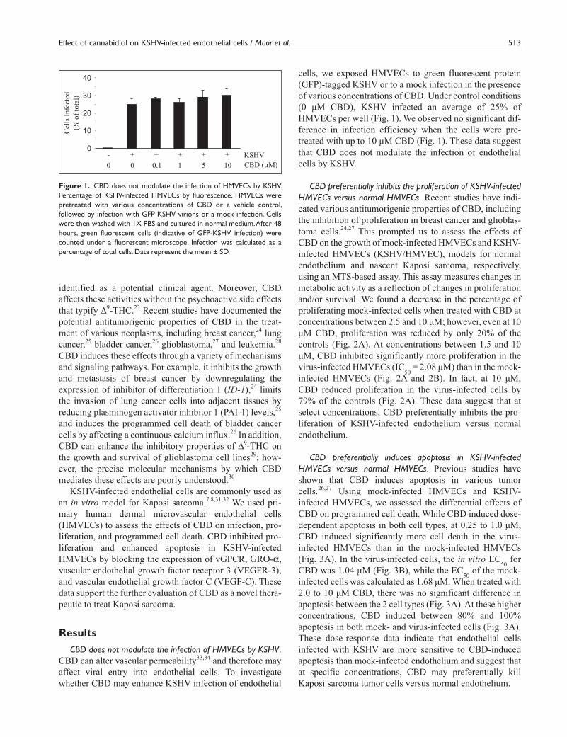
cells, we exposed HMVECs to green fluorescent protein (GFP)-tagged KSHV or to a mock infection in the presence of various concentrations of CBD. Under control conditions (0 µM CBD), KSHV infected an average of 25% of HMVECs per well (Fig. 1). We observed no significant dif-ference in infection efficiency when the cells were pre-treated with up to 10 µM CBD (Fig. 1). These data suggest that CBD does not modulate the infection of endothelial cells by KSHV.
CBD preferentially inhibits the proliferation of KSHV-infected HMVECs versus normal HMVECs. Recent studies have indi-cated various antitumorigenic properties of CBD, including the inhibition of proliferation in breast cancer and glioblas-toma cells.24,27 This prompted us to assess the effects of CBD on the growth of mock-infected HMVECs and KSHV-infected HMVECs (KSHV/HMVEC), models for normal endothelium and nascent Kaposi sarcoma, respectively, using an MTS-based assay. This assay measures changes in metabolic activity as a reflection of changes in proliferation and/or survival. We found a decrease in the percentage of proliferating mock-infected cells when treated with CBD at concentrations between 2.5 and 10 µM; however, even at 10 µM CBD, proliferation was reduced by only 20% of the controls (Fig. 2A). At concentrations between 1.5 and 10 µM, CBD inhibited significantly more proliferation in the virus-infected HMVECs (IC
50 = 2.08 µM) than in the mock-
infected HMVECs (Fig. 2A and 2B). In fact, at 10 µM, CBD reduced proliferation in the virus-infected cells by 79% of the controls (Fig. 2A). These data suggest that at select concentrations, CBD preferentially inhibits the pro-liferation of KSHV-infected endothelium versus normal endothelium.
CBD preferentially induces apoptosis in KSHV-infected HMVECs versus normal HMVECs. Previous studies have shown that CBD induces apoptosis in various tumor cells.26,27 Using mock-infected HMVECs and KSHV-infected HMVECs, we assessed the differential effects of CBD on programmed cell death. While CBD induced dose-dependent apoptosis in both cell types, at 0.25 to 1.0 µM, CBD induced significantly more cell death in the virus-infected HMVECs than in the mock-infected HMVECs (Fig. 3A). In the virus-infected cells, the in vitro EC
50 for
CBD was 1.04 µM (Fig. 3B), while the EC50
of the mock-infected cells was calculated as 1.68 µM. When treated with 2.0 to 10 µM CBD, there was no significant difference in apoptosis between the 2 cell types (Fig. 3A). At these higher concentrations, CBD induced between 80% and 100% apoptosis in both mock- and virus-infected cells (Fig. 3A). These dose-response data indicate that endothelial cells infected with KSHV are more sensitive to CBD-induced apoptosis than mock-infected endothelium and suggest that at specific concentrations, CBD may preferentially kill Kaposi sarcoma tumor cells versus normal endothelium.
0
10
20
30
40C
ells
Infe
cted
(% o
f tot
al)
- KSHV+ ++++CBD (µM)10510.100
Figure 1. CBD does not modulate the infection of HMVECs by KSHV. Percentage of KSHV-infected HMVECs by fluorescence. HMVECs were pretreated with various concentrations of CBD or a vehicle control, followed by infection with GFP-KSHV virions or a mock infection. Cells were then washed with 1X PBS and cultured in normal medium. After 48 hours, green fluorescent cells (indicative of GFP-KSHV infection) were counted under a fluorescent microscope. Infection was calculated as a percentage of total cells. Data represent the mean ± SD.

514 Genes & Cancer / vol 3 no 7-8 (2012)
CBD inhibits the expression of KSHV vGPCR. To explore the mechanism(s) by which CBD inhibits the growth of KSHV/HMVECs and induces their programmed cell death, we examined the effect of CBD on KSHV vGPCR, a protein that immortalizes endothelial cells and promotes Kaposi sarcoma tumorigenesis.7,9 vGPCR facilitates the transfor-mation of KSHV-infected cells and enhances the prolifera-tion of KSHV-infected endothelium.8,10 To confirm that vGPCR is expressed in Kaposi sarcoma, we examined pro-tein levels and localization in Kaposi sarcoma lesions by immunohistochemical analysis. We found vGPCR expres-sion in irregular vascular formations and scattered spindle cells of the Kaposi sarcoma tumor sections (Fig. 4A, panels II and III). Lack of staining in the isotype control–treated section illustrates antibody specificity (Fig. 4A, panel I).
In an earlier publication, we demonstrated that at low doses, Δ9-THC, a psychoactive cannabinoid related to
CBD, enhanced the expression of vGPCR in KSHV-infected HMVECs.35 Here, by Western blot analysis, we examined the effects of CBD on the expression of vGPCR in such cells. We found that CBD altered vGPCR expres-sion in KSHV-infected HMVECs; however, in contrast to Δ9-THC, CBD induced dose-dependent inhibition (Fig. 4B). Moreover, the concentrations at which CBD preferen-tially induced apoptosis and reduced proliferation in the KSHV/HMVECs were those at which vGPCR expression was also inhibited in these cells (Figs. 2, 3, and 4B). This suggests a potential mechanism through which CBD exerts its anticancer effects in KSHV-associated neoplasms.
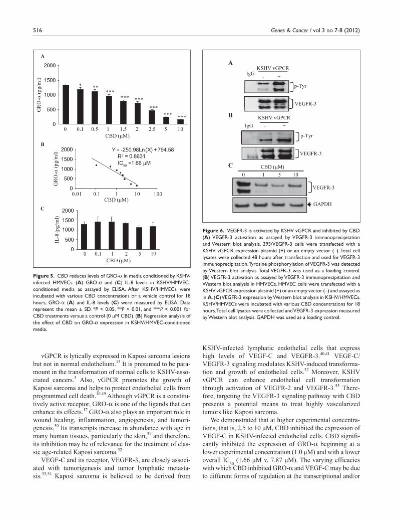
CBD reduces levels of GRO-α in media conditioned by KSHV-infected HMVECs. Although vGPCR is a constitutively active G protein–coupled receptor, IL-8 and GRO-α can enhance its activity.16,17 Therefore, we measured the levels of these cytokines in the conditioned media of KSHV/HMVECs after treatment with various concentrations of CBD or a control using ELISA (Fig. 5). We observed a sig-nificant dose-dependent decrease in GRO-α in the condi-tioned media (Fig. 5A), with an in vitro IC
50 of 1.66 µM
A
0
25
50
75
100
125
0 1 1.5 2 2.5 3 5 10
Prol
ifera
tion
(% o
f co
ntro
l)
HMVEC KSHV/ HMVEC
CBD (µM)
*********
******
**
B
Y = -34.242Ln(X) + 75.082
0
25
50
75
100
125
0.1 1 10 100CBD (µM)
Prol
ifera
tion
(% o
f co
ntro
l)
R2 = 0.767IC50 =2.08 µM
Figure 2. CBD preferentially inhibits the proliferation of KSHV-infected HMVECs. (A) Percentage of proliferating cells as measured by an MTS-based assay. Mock-infected HMVECs (HMVEC) and KSHV-infected HMVECs (KSHV/HMVEC) were incubated for 48 hours with various concentrations of CBD or a vehicle control. The proliferation index of untreated HMVECs was established as 100%, and all other cells and conditions were calculated as a percentage of this control. Data indicate the mean ± SD. **P < 0.01 and ***P < 0.001 indicate the difference in proliferation of KSHV/HMVEC versus HMVEC at each CBD concentration. (B) Regression analysis of the effect of CBD on the proliferation of KSHV-infected HMVECs.
A
B0
25
50
75
100
125
0 0.1 0.25 0.5 0.75 1 2 5 10
Apo
ptot
ic C
ells
(% o
f tot
al )
HMVECKSHV/ HMVEC
CBD (µM)
***
***
******
Y= 21.222Ln(X) + 49.096
R2 = 0.921
0
25
50
75
100
125
0.01 0.1 1 10 100CBD (µM)
Apo
ptot
ic C
ells
(% o
f tot
al )
EC50 =1.04 µM
Figure 3. CBD preferentially induces apoptosis in KSHV-infected HMVECs. (A) Percentage of apoptotic cells as assayed by TUNEL. KSHV-infected HMVECs (KSHV/HMVEC) or mock-infected HMVECs (HMVEC) were treated with various concentrations of CBD or a vehicle control for 24 hours. Data were calculated as apoptotic cells/total cells. Data represent the mean ± SD. ***P < 0.001 indicates the difference in apoptosis levels of KSHV/HMVEC versus HMVEC at each CBD concentration. (B) Regression analysis of the effect of CBD on the apoptosis of KSHV-infected HMVECs.

Effect of cannabidiol on KSHV-infected endothelial cells / Maor et al. 515
(Fig. 5B). In contrast, CBD did not affect a statistically sig-nificant change in IL-8 levels in the conditioned media (Fig. 5C).
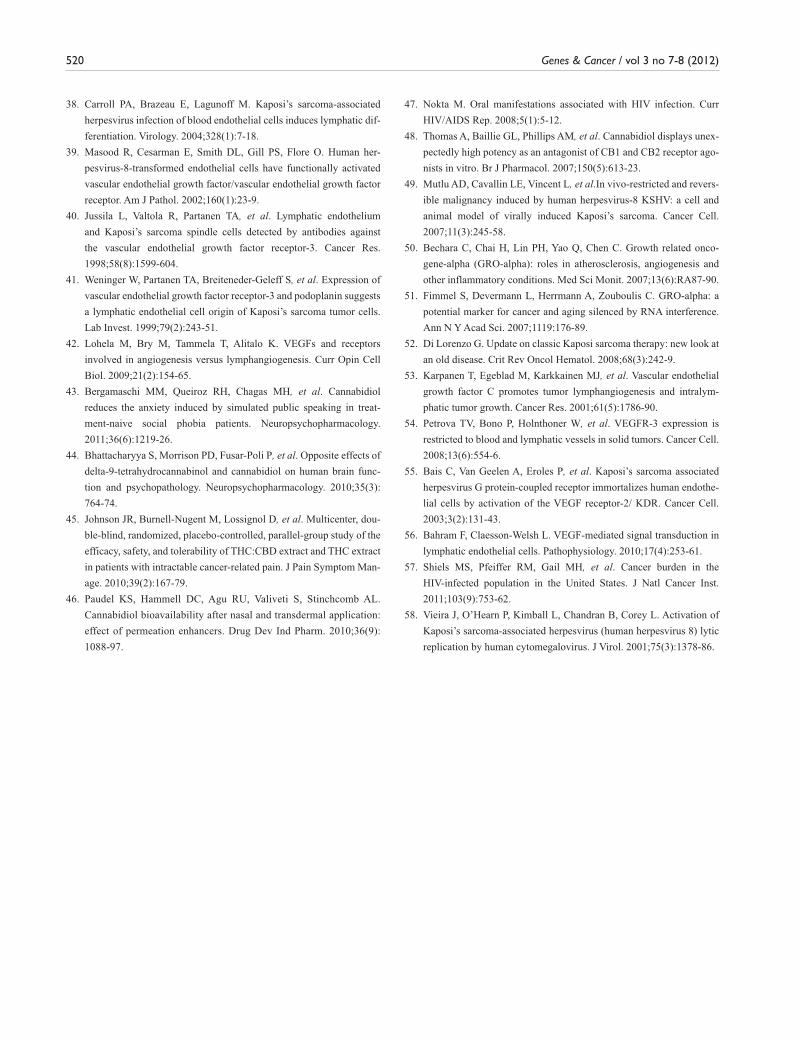
CBD downregulates the expression of VEGFR-3 and VEGF-C. Upon activation, the VEGFR-3 receptor, a key molecule for lymphangiogenesis,36 promotes KSHV-induced infection, growth, and transformation of endothelial cells.37 To exam-ine the activation status of VEGFR-3, we transiently trans-fected a vGPCR expression plasmid or vector control into a stable 293 cell line, manipulated to express high levels of VEGFR-3 (293/VEGFR-3). We immunoprecipitated VEGFR-3 from whole cell lysates and subsequently performed Western blot analysis with a phosphotyrosine antibody (Fig. 6A). We observed significant tyrosine phos-phorylation in the cells transfected with vGPCR (+) as com-pared to those transfected with the vector control (–) (Fig. 6A). To confirm these observations in a primary cell line with endogenous VEGFR-3 expression, we repeated the experiment in HMVECs. Similarly, we observed signifi-cantly greater VEGFR-3 activation in the HMVECs trans-fected with vGPCR as compared to those transfected with the vector control (Fig. 6B). Taken together, these data indi-cate that vGPCR activates VEGFR-3 in 293/VEGFR-3 cells as well as in primary endothelial cells. When we examined total VEGFR-3 levels in KSHV-infected HMVECs, we observed high levels of the protein under control conditions (Fig. 6C, lane 0). This is consistent with existing data that demonstrate the enhancement of VEGFR-3 expression by KSHV38,39 and with high
endogenous expression of VEGFR-3 in Kaposi sarcoma.40,41 Upon treat-ment with various concentrations of CBD, VEGFR-3 levels were signifi-cantly reduced (Fig. 6C), suggesting another mechanism whereby CBD may affect the proliferation and via-bility of Kaposi sarcoma through the VEGFR-3 signaling pathway.
Next, by ELISA, we examined levels of VEGF-C and VEGF-D, the cognate ligands of VEGFR-3,42 in the conditioned media of KSHV/HMVECs after treatment with vari-ous concentrations of CBD (Fig. 7). We observed a significant dose-dependent decrease in VEGF-C lev-els in the conditioned media (Fig. 7A), with an in vitro IC
50 of 7.87 µM
(Fig.7B); however, CBD had no effect on VEGF-D expression (Fig. 7C). These data demonstrate that CBD inhibits the expression of VEGF-C in KSHV/HMVECs, while VEGF-D levels remain very low
with/without CBD treatment. These data are consistent with CBD-induced reduction of VEGFR-3 levels (Fig. 6C), and together, they suggest that CBD may be an inhibitor of VEGFR-3/VEGF-C signaling in Kaposi sarcoma.
DiscussionCBD has been evaluated clinically for the treatment of vari-ous conditions, including anxiety,43 psychosis,44 and pain.45 In contrast to other members of the cannabinoid family, CBD has a strong safety profile and induces no psychotro-pic effects.23,30 Therefore, it has become an attractive agent in the search for new anticancer therapies.23 Our current study demonstrated that CBD preferentially enhanced apoptosis and inhibited the proliferation of KSHV-infected endothelial cells. This selective targeting of KSHV-induced neoplasia suggests that CBD may have a desirable thera-peutic index when used to treat cancer. Moreover, a recent study demonstrated that CBD can be delivered effectively by nasal and transdermal routes,46 which may be particu-larly valuable for the treatment of Kaposi sarcoma oral or skin lesions.47
The molecular mechanisms by which CBD exerts its therapeutic effects are not fully understood, although CBD has been shown to act as an inverse agonist of the cannabi-noid receptors CB1 and CB2.48 We found that CBD reduced the levels of vGPCR, GRO-α, VEGFR-3, and VEGF-C in KSHV-infected endothelium. These proteins are believed to play important roles in the initiation, growth, metastasis, and survival of Kaposi sarcoma.
Figure 4. CBD inhibits the expression of KSHV vGPCR. (A) Representative immunohistochemical analysis of KSHV vGPCR expression in cutaneous Kaposi sarcoma lesions (panels II and III). Kaposi sarcoma lesion stained with an antibody isotype control (panel I). For panels I and II, scale bars = 50 µm. For panel III, scale bar =10 µm. (B) Representative Western blot analysis of KSHV vGPCR levels in whole cell lysates of KSHV/HMVECs after incubation with various concentrations of CBD or a vehicle control for 24 hours. GAPDH was used as a loading control.
516 Genes & Cancer / vol 3 no 7-8 (2012)
vGPCR is lytically expressed in Kaposi sarcoma lesions but not in normal endothelium.15 It is presumed to be para-mount in the transformation of normal cells to KSHV-asso-ciated cancers.5 Also, vGPCR promotes the growth of Kaposi sarcoma and helps to protect endothelial cells from programmed cell death.10,49 Although vGPCR is a constitu-tively active receptor, GRO-α is one of the ligands that can enhance its effects.17 GRO-α also plays an important role in wound healing, inflammation, angiogenesis, and tumori-genesis.50 Its transcripts increase in abundance with age in many human tissues, particularly the skin,51 and therefore, its inhibition may be of relevance for the treatment of clas-sic age-related Kaposi sarcoma.52
VEGF-C and its receptor, VEGFR-3, are closely associ-ated with tumorigenesis and tumor lymphatic metasta-sis.53,54 Kaposi sarcoma is believed to be derived from
KSHV-infected lymphatic endothelial cells that express high levels of VEGF-C and VEGFR-3.40,41 VEGF-C/VEGFR-3 signaling modulates KSHV-induced transforma-tion and growth of endothelial cells.37 Moreover, KSHV vGPCR can enhance endothelial cell transformation through activation of VEGFR-2 and VEGFR-3.55 There-fore, targeting the VEGFR-3 signaling pathway with CBD presents a potential means to treat highly vascularized tumors like Kaposi sarcoma.
We demonstrated that at higher experimental concentra-tions, that is, 2.5 to 10 µM, CBD inhibited the expression of VEGF-C in KSHV-infected endothelial cells. CBD signifi-cantly inhibited the expression of GRO-α beginning at a lower experimental concentration (1.0 µM) and with a lower overall IC
50 (1.66 µM v. 7.87 µM). The varying efficacies
with which CBD inhibited GRO-α and VEGF-C may be due to different forms of regulation at the transcriptional and/or
0
500
1000
1500
2000
IL-8
(pg/
ml)
CBD (µM)
CBD (µM)
CBD (µM)
A
B
C
Y = -250.98Ln(X) + 794.58R2 = 0.8631
0
500
1000
1500
2000
0.01 0.1 1 10 100
GR
O-α
(pg/
ml)
IC50 =1.66 µM
0
500
1000
1500
2000
0
0
0.1 0.5
0.1
1
1 10
1.5 2
2
2.5 5
5
10
GR
O-α
(pg/
ml)
**
****** ***
****
*** ***
Figure 5. CBD reduces levels of GRO-α in media conditioned by KSHV-infected HMVECs. (A) GRO-α and (C) IL-8 levels in KSHV/HMVEC-conditioned media as assayed by ELISA. After KSHV/HMVECs were incubated with various CBD concentrations or a vehicle control for 18 hours, GRO-α (A) and IL-8 levels (C) were measured by ELISA. Data represent the mean ± SD. *P < 0.05, **P < 0.01, and ***P < 0.001 for CBD treatments versus a control (0 µM CBD). (B) Regression analysis of the effect of CBD on GRO-α expression in KSHV/HMVEC-conditioned media.
IgG - +
p-Tyr
VEGFR-3
AKSHV vGPCR
p-Tyr
IgG - +
B KSHV vGPCR
VEGFR-3
C0 1 5 10
CBD (µM)
VEGFR-3
GAPDH
Figure 6. VEGFR-3 is activated by KSHV vGPCR and inhibited by CBD. (A) VEGFR-3 activation as assayed by VEGFR-3 immunoprecipitation and Western blot analysis. 293/VEGFR-3 cells were transfected with a KSHV vGPCR expression plasmid (+) or an empty vector (–). Total cell lysates were collected 48 hours after transfection and used for VEGFR-3 immunoprecipitation. Tyrosine phosphorylation of VEGFR-3 was detected by Western blot analysis. Total VEGFR-3 was used as a loading control. (B) VEGFR-3 activation as assayed by VEGFR-3 immunoprecipitation and Western blot analysis in HMVECs. HMVEC cells were transfected with a KSHV vGPCR expression plasmid (+) or an empty vector (–) and assayed as in A. (C) VEGFR-3 expression by Western blot analysis in KSHV/HMVECs. KSHV/HMVECs were incubated with various CBD concentrations for 18 hours. Total cell lysates were collected and VEGFR-3 expression measured by Western blot analysis. GAPDH was used as a loading control.

Effect of cannabidiol on KSHV-infected endothelial cells / Maor et al. 517
translational levels; however, further study is required to characterize the specific molecular mechanisms responsible. In adults, VEGFR-3 is predominantly expressed by lym-phatic endothelium and tumor vasculature and by a subset of normal endothelial cells, including HMVECs56 (Fig. 6B). As such, CBD could be expected to affect normal endothelial cells at higher concentrations. In contrast, the GRO-α recep-tor, vGPCR, is not expressed by normal endothelial cells.15 Based on these data, we hypothesize that while higher con-centrations of CBD may target the expression of both VEGF-C and GRO-α, lower concentrations of CBD could significantly impact the proliferation and apoptosis of KSHV-infected cells and spare normal endothelium (as shown in Figs. 2A and 3A) by predominantly targeting sig-naling through GRO-α and vGPCR.
With the advent of highly active antiretroviral therapy for the treatment of AIDS in the 1990s, the incidence of AIDS-associated Kaposi sarcoma has decreased.4 How-ever, the number of patients living with AIDS continues to increase, and as they live longer, it is likely that AIDS-associated Kaposi sarcoma will also rise.57 New treatments are needed to prevent or control the disease. Our data support the evaluation of CBD as a novel target-specific therapeutic for KSHV-induced neoplasms, including Kaposi sarcoma. We demonstrated that CBD could preferentially induce apoptosis and attenuate the proliferation of KSHV-infected HMVECs by inhibiting the expression of vGPCR, GRO-α, VEGFR-3, and VEGF-C. Further investigation to elucidate more precisely the mechanisms by which CBD induces its inhibitory effects may help guide the development of CBD as a treatment for KSHV-associated neoplasia.
Materials and MethodsCells. Primary HMVECs (adult) were purchased from
Lonza (Allendale, NJ) and maintained in EBM-2 basal medium, supplemented with EGM-2 MV BulletKit (Lonza). GFP–BCBL-1 cells were a gift from Dr. Jeffrey Vieira (Department of Laboratory Medicine, University of Wash-ington, Seattle, WA). They were maintained in RPMI 1640 medium and supplemented as described previously.58 The 293/VEGFR-3 cell line (Genentech, South San Francisco, CA) was maintained in DMEM with 10% FBS.
Reagents. CBD was purchased from Tocris Bioscience (Ellisville, MO). TPA was purchased from Sigma-Aldrich (St. Louis, MO). Anti–KSHV vGPCR antibody was pur-chased from Cell Sciences (Canton, MA). All other antibod-ies were from Santa Cruz Biotechnology (Santa Cruz, CA).
Plasmids and transfections. The KSHV vGPCR expression plasmid (pcDNA3.1vGPCR) was constructed as follows: KSHV genomic DNA was extracted from KSHV-positive BCBL-1 cells. The vGPCR gene was amplified from the KSHV genomic DNA, using the primers purchased from Invitrogen (Carlsbad, CA) (upstream sequence: 5′-GGAATTCATGGCGGCCGACCATTTCC-3′; down-stream sequence: 5′-GTCTAGACGTGGTGGCGCCGGA-CATG-3′). The purified PCR product was digested with EcoR I and Xba I and inserted into the pcDNA3.1 plasmid digested with same restriction endonucleases to create pcDNA3.1vGPCR. HMVECs and 293/VEGFR-3 cells were transiently transfected with pcDNA3.1vGPCR or a vector control (pcDNA3.1) using Superfect Transfection Reagent from Qiagen (Valencia, CA).
Kaposi sarcoma tissue samples. Tissue samples from Kaposi sarcoma skin lesions of diagnosed HIV-positive patients were obtained after informed consent at Beth Israel
B
A
C
VEG
F-D
(pg/
ml)
0
250
500
750
0 0.1 1 2 5 10CBD (µM)
CBD (µM)
CBD (µM)
IC50 =7.87 µM
Y = -70.49Ln(X) + 419.95R2 = 0.7688
0
250
500
750
0.01 0.1 1 10 100
VEG
F-C
(pg/
ml)
0
250
500
750
0 0.1 1 1.5 2 2.5 5 10
VEG
F-C
(pg/
ml)
*** ******
* * *
Figure 7. CBD inhibits the expression of VEGF-C. (A) VEGF-C and (C) VEGF-D levels in KSHV/HMVEC-conditioned media as assayed by ELISA. After KSHV/HMVECs were incubated with various CBD concentrations or a vehicle control for 18 hours, VEGF-C (A) and VEGF-D levels (C) were measured by ELISA. Data indicate the mean ± SD. *P < 0.05 and ***P < 0.001 for CBD treatments versus a control (0 µM CBD). (B) Regression analysis of the effect of CBD on VEGF-C expression in KSHV/HMVEC-conditioned media.

518 Genes & Cancer / vol 3 no 7-8 (2012)
Deaconess Medical Center (BIDMC). The protocol and consent form were approved by the BIDMC review board in accordance with an assurance filed with and approved by the US Department of Health and Human Services.
Immunohistochemistry. Kaposi sarcoma tissue samples were fixed in 4% formaldehyde solution and paraffin embed-ded. Serial sections were prepared by the Dana-Farber/ Harvard Cancer Center Research Pathology Core (Boston, MA). Immunohistochemical staining was performed per instructions from Upstate Biotechnology (Waltham, MA). Vectashield, a DAPI-containing mounting medium, and FITC-conjugated secondary antibody were purchased from Vector Laboratories (Burlingame, CA). Kaposi sarcoma tis-sue sections were observed under a Zeiss LSM 510 META upright confocal microscope (Oberkochen, Germany).
GFP-KSHV virus propagation and infection. GFP–BCBL-1 cells were grown to a density of 5 × 10⁵ cells/mL, induced with TPA at 20 ng/mL, and grown for 5 days. To harvest the virus, cells were pelleted at 500g for 15 minutes. The super-natant was removed and centrifuged at 15,000g for 4 hours. The pellet was resuspended in 1/100 growth volume com-plete media as discussed previously and centrifuged at 300g for 5 minutes. Purified KSHV virion was titrated by real-time PCR as described previously.35,37
Sixty-percent confluent HMVECs were pretreated for 2 hours with various CBD concentrations or a vehicle con-trol. Subsequently, they were either mock infected or infected with GFP-KSHV in 24-well plates at 37°C for 3 hours, washed, and incubated at 37°C for 2 days in HMVEC medium, as described above. KSHV infections were performed at a multiplicity of infection of 5 to 6 KSHV copies per cell. Green fluorescent cells were counted under a fluorescent microscope (200× magnifica-tion) (Diaphot 300, Nikon, Tokyo, Japan) or detached by 5 mM EDTA in PBS and analyzed on a FACScan flow cytometer (BD Biosciences, San Jose, CA, USA) to deter-mine the percentage of cells infected.
Cell stimulation, immunoprecipitation, and Western blot analysis. Cells were starved for 2 hours in serum-free media and subsequently stimulated with various concentrations of CBD as indicated in 0.5% BSA media for 24 hours (unless otherwise noted). After stimulation, total cell lysates were collected and processed for immunoprecipitation and West-ern blot analysis as described previously.37
Proliferation assay. The proliferation assay was performed using CellTiter 96 AQueous One Solution Cell Prolifera-tion Assay (MTS, Promega, Madison, WI) per the manufac-turer’s instructions. The number of proliferating cells was calculated as a percentage of total cells.
Apoptosis assay. KSHV-infected and mock-infected HMVECs were incubated in 0.5% BSA media with various concentrations of CBD (0-10 µM) for 24 hours. The In Situ Cell Death Detection Kit, Fluorescein (Roche Applied Sci-ence, Mannheim, Germany) was used for the detection and quantification of apoptosis. Cells were stained by the TUNEL method and observed under a fluorescent micro-scope to determine double-stranded DNA breaks per the manufacturer’s instructions.
Cytokine ELISA. Media conditioned by KSHV/HMVEC cells treated with various concentrations of CBD were col-lected and assayed by ELISA per the manufacturer’s instructions as follows: IL-8 (Human CXCL8/IL-8 Immuno-assay, R&D Systems, Minneapolis, MN), GRO-α (Human CXCL1/GRO-α Immunoassay, R&D Systems), VEGF-D (Human VEGF-D Immunoassay, R&D Systems), and VEGF-C (Human VEGF-C Assay Kit, IBL-America, Min-neapolis, MN).
Statistical analysis. Statistical significance was deter-mined using the Student 2-tailed t test. Data represent the mean ± SD of at least 3 independent experiments, with P < 0.05 considered statistically significant. To calculate the IC
50 and EC
50 values, regression analyses were performed
based on the proliferation and apoptosis data and the VEGF-C and GRO-α expression data, respectively. Western blot analysis and immunohistochemistry were repeated at least 3 times, and representative blots or images are shown.
Declaration of Conflicting Interests
The author(s) declared no potential conflicts of interest with respect to the research, authorship, and/or publication of this article.
Funding
The author(s) received the following financial support for the research, authorship, and/or publication of this article: This work was supported by divisional funds.
References
1. Chang Y, Cesarman E, Pessin MS, et al. Identification of herpesvirus-
like DNA sequences in AIDS-associated Kaposi’s sarcoma. Science.
1994;266(5192):1865-9.
2. Antman K, Chang Y. Kaposi’s sarcoma. N Engl J Med.
2000;342(14):1027-38.
3. Marcelin AG, Calvez V, Dussaix E. KSHV after an organ transplant:
should we screen? Curr Top Microbiol Immunol. 2007;312:245-62.
4. Martellotta F, Berretta M, Vaccher E, et al. AIDS-related Kaposi’s
sarcoma: state of the art and therapeutic strategies. Curr HIV Res.
2009;7(6):634-8.
5. Ganem D. KSHV-induced oncogenesis. In: Arvin A, Campadelli-
Fiume G, & Mocarski E, et al., editors. Human herpesviruses: biology,

Effect of cannabidiol on KSHV-infected endothelial cells / Maor et al. 519
therapy, and immunoprophylaxis. Cambridge: Cambridge University
Press; 2007. Chapter 56. Available from: http://www.ncbi.nlm.nih.gov/
books/NBK47373
6. Mesri EA, Cesarman E, Boshoff C. Kaposi’s sarcoma and its associ-
ated herpesvirus. Nat Rev Cancer. 2010;10(10):707-19.
7. Boshoff C, Schulz TF, Kennedy MM, et al. Kaposi’s sarcoma-asso-
ciated herpesvirus infects endothelial and spindle cells. Nat Med.
1995;1(12):1274-8.
8. Flore O, Rafii S, Ely S, et al. Transformation of primary human endo-
thelial cells by Kaposi’s sarcoma-associated herpesvirus. Nature.
1998;394(6693):588-92.
9. Yang TY, Chen SC, Leach MW, et al. Transgenic expression of the
chemokine receptor encoded by human herpesvirus 8 induces an
angioproliferative disease resembling Kaposi’s sarcoma. J Exp Med.
2000;191(3):445-54.
10. Sodhi A, Chaisuparat R, Hu J, et al. The TSC2/mTOR pathway drives
endothelial cell transformation induced by the Kaposi’s sarcoma-
associated herpesvirus G protein-coupled receptor. Cancer Cell.
2006;10(2):133-43.
11. Arvanitakis L, Geras-Raaka E, Varma A, Gershengorn MC, Cesar-
man E. Human herpesvirus KSHV encodes a constitutively active
G-protein-coupled receptor linked to cell proliferation. Nature.
1997;385(6614):347-50.
12. Cesarman E, Nador RG, Bai F, et al. Kaposi’s sarcoma-associated
herpesvirus contains G protein-coupled receptor and cyclin D homo-
logs which are expressed in Kaposi’s sarcoma and malignant lym-
phoma. J Virol. 1996;70(11):8218-23.
13. Bais C, Santomasso B, Coso O, et al. G-protein-coupled receptor
of Kaposi’s sarcoma-associated herpesvirus is a viral oncogene and
angiogenesis activator. Nature. 1998;391(6662):86-9.
14. Burger M, Burger JA, Hoch RC, et al. Point mutation causing consti-
tutive signaling of CXCR2 leads to transforming activity similar to
Kaposi’s sarcoma herpesvirus-G protein-coupled receptor. J Immunol.
1999;163(4):2017-22.
15. Cannon M. The KSHV and other human herpesviral G protein-cou-
pled receptors. Curr Top Microbiol Immunol. 2007;312:137-56.
16. Gershengorn MC, Geras-Raaka E, Varma A, Clark-Lewis I. Che-
mokines activate Kaposi’s sarcoma-associated herpesvirus G pro-
tein-coupled receptor in mammalian cells in culture. J Clin Invest.
1998;102(8):1469-72.
17. Lane BR, Liu J, Bock PJ, et al. Interleukin-8 and growth-regulated
oncogene alpha mediate angiogenesis in Kaposi’s sarcoma. J Virol.
2002;76(22):11570-83.
18. Geras-Raaka E, Arvanitakis L, Bais C, et al. Inhibition of constitu-
tive signaling of Kaposi’s sarcoma-associated herpesvirus G protein-
coupled receptor by protein kinases in mammalian cells in culture. J
Exp Med. 1998;187(5):801-6.
19. Geras-Raaka E, Varma A, Ho H, Clark-Lewis I, Gershengorn MC.
Human interferon-gamma-inducible protein 10 (IP-10) inhibits con-
stitutive signaling of Kaposi’s sarcoma-associated herpesvirus G
protein-coupled receptor. J Exp Med. 1998;188(2):405-8.
20. Geras-Raaka E, Varma A, Clark-Lewis I, Gershengorn MC. Kaposi’s
sarcoma-associated herpesvirus (KSHV) chemokine vMIP-II and
human SDF-1alpha inhibit signaling by KSHV G protein-coupled
receptor. Biochem Biophys Res Commun. 1998;253(3):725-7.
21. Mechoulam R, Shvo Y. Hashish, I: the structure of cannabidiol. Tetra-
hedron. 1963;19(12):2073-8.
22. Lerner M. Marihuana: tetrahydrocannabinol and related compounds.
Science. 1963;140(3563):175-6.
23. Izzo AA, Borrelli F, Capasso R, Di Marzo V, Mechoulam R. Non-
psychotropic plant cannabinoids: new therapeutic opportunities from
an ancient herb. Trends Pharmacol Sci. 2009;30(10):515-27.
24. McAllister SD, Christian RT, Horowitz MP, Garcia A, Desprez PY.
Cannabidiol as a novel inhibitor of Id-1 gene expression in aggressive
breast cancer cells. Mol Cancer Ther. 2007;6(11):2921-7.
25. Ramer R, Rohde A, Merkord J, Rohde H, Hinz B. Decrease of
plasminogen activator inhibitor-1 may contribute to the anti-inva-
sive action of cannabidiol on human lung cancer cells. Pharm Res.
2010;27(10):2162-74.
26. Yamada T, Ueda T, Shibata Y, et al. TRPV2 activation induces apop-
totic cell death in human T24 bladder cancer cells: a potential thera-
peutic target for bladder cancer. Urology. 2010;76(2):509.e1-7.
27. Torres S, Lorente M, Rodriguez-Fornes F, et al. A combined preclini-
cal therapy of cannabinoids and temozolomide against glioma. Mol
Cancer Ther. 2011;10(1):90-103.
28. McKallip RJ, Jia W, Schlomer J, et al. Cannabidiol-induced apoptosis
in human leukemia cells: a novel role of cannabidiol in the regulation of
p22phox and Nox4 expression. Mol Pharmacol. 2006;70(3):897-908.
29. Marcu JP, Christian RT, Lau D, et al. Cannabidiol enhances the inhibi-
tory effects of delta9-tetrahydrocannabinol on human glioblastoma
cell proliferation and survival. Mol Cancer Ther. 2010;9(1):180-9.
30. Mechoulam R, Peters M, Murillo-Rodriguez E, Hanus LO. Cannabi-
diol: recent advances. Chem Biodivers. 2007;4(8):1678-92.
31. Delgado T, Carroll PA, Punjabi AS, et al. Induction of the Warburg
effect by Kaposi’s sarcoma herpesvirus is required for the mainte-
nance of latently infected endothelial cells. Proc Natl Acad Sci U S A.
2010;107(23):10696-701.
32. Hansen A, Henderson S, Lagos D, et al. KSHV-encoded miRNAs
target MAF to induce endothelial cell reprogramming. Genes Dev.
2010;24(2):195-205.
33. El-Remessy AB, Al-Shabrawey M, Khalifa Y, et al. Neuroprotective
and blood-retinal barrier-preserving effects of cannabidiol in experi-
mental diabetes. Am J Pathol. 2006;168(1):235-44.
34. Rajesh M, Mukhopadhyay P, Batkai S, et al. Cannabidiol attenuates
high glucose-induced endothelial cell inflammatory response and bar-
rier disruption. Am J Physiol Heart Circ Physiol. 2007;293(1):H610-9.
35. Zhang X, Wang JF, Kunos G, Groopman JE. Cannabinoid modulation
of Kaposi’s sarcoma-associated herpesvirus infection and transforma-
tion. Cancer Res. 2007;67(15):7230-7.
36. Tammela T, Zarkada G, Wallgard E, et al. Blocking VEGFR-3 sup-
presses angiogenic sprouting and vascular network formation. Nature.
2008;454(7204):656-60.
37. Zhang X, Wang JF, Chandran B, et al. Kaposi’s sarcoma-associated
herpesvirus activation of vascular endothelial growth factor receptor
3 alters endothelial function and enhances infection. J Biol Chem.
2005;280(28):26216-24.
520 Genes & Cancer / vol 3 no 7-8 (2012)
38. Carroll PA, Brazeau E, Lagunoff M. Kaposi’s sarcoma-associated
herpesvirus infection of blood endothelial cells induces lymphatic dif-
ferentiation. Virology. 2004;328(1):7-18.
39. Masood R, Cesarman E, Smith DL, Gill PS, Flore O. Human her-
pesvirus-8-transformed endothelial cells have functionally activated
vascular endothelial growth factor/vascular endothelial growth factor
receptor. Am J Pathol. 2002;160(1):23-9.
40. Jussila L, Valtola R, Partanen TA, et al. Lymphatic endothelium
and Kaposi’s sarcoma spindle cells detected by antibodies against
the vascular endothelial growth factor receptor-3. Cancer Res.
1998;58(8):1599-604.
41. Weninger W, Partanen TA, Breiteneder-Geleff S, et al. Expression of
vascular endothelial growth factor receptor-3 and podoplanin suggests
a lymphatic endothelial cell origin of Kaposi’s sarcoma tumor cells.
Lab Invest. 1999;79(2):243-51.
42. Lohela M, Bry M, Tammela T, Alitalo K. VEGFs and receptors
involved in angiogenesis versus lymphangiogenesis. Curr Opin Cell
Biol. 2009;21(2):154-65.
43. Bergamaschi MM, Queiroz RH, Chagas MH, et al. Cannabidiol
reduces the anxiety induced by simulated public speaking in treat-
ment-naive social phobia patients. Neuropsychopharmacology.
2011;36(6):1219-26.
44. Bhattacharyya S, Morrison PD, Fusar-Poli P, et al. Opposite effects of
delta-9-tetrahydrocannabinol and cannabidiol on human brain func-
tion and psychopathology. Neuropsychopharmacology. 2010;35(3):
764-74.
45. Johnson JR, Burnell-Nugent M, Lossignol D, et al. Multicenter, dou-
ble-blind, randomized, placebo-controlled, parallel-group study of the
efficacy, safety, and tolerability of THC:CBD extract and THC extract
in patients with intractable cancer-related pain. J Pain Symptom Man-
age. 2010;39(2):167-79.
46. Paudel KS, Hammell DC, Agu RU, Valiveti S, Stinchcomb AL.
Cannabidiol bioavailability after nasal and transdermal application:
effect of permeation enhancers. Drug Dev Ind Pharm. 2010;36(9):
1088-97.
47. Nokta M. Oral manifestations associated with HIV infection. Curr
HIV/AIDS Rep. 2008;5(1):5-12.
48. Thomas A, Baillie GL, Phillips AM, et al. Cannabidiol displays unex-
pectedly high potency as an antagonist of CB1 and CB2 receptor ago-
nists in vitro. Br J Pharmacol. 2007;150(5):613-23.
49. Mutlu AD, Cavallin LE, Vincent L, et al.In vivo-restricted and revers-
ible malignancy induced by human herpesvirus-8 KSHV: a cell and
animal model of virally induced Kaposi’s sarcoma. Cancer Cell.
2007;11(3):245-58.
50. Bechara C, Chai H, Lin PH, Yao Q, Chen C. Growth related onco-
gene-alpha (GRO-alpha): roles in atherosclerosis, angiogenesis and
other inflammatory conditions. Med Sci Monit. 2007;13(6):RA87-90.
51. Fimmel S, Devermann L, Herrmann A, Zouboulis C. GRO-alpha: a
potential marker for cancer and aging silenced by RNA interference.
Ann N Y Acad Sci. 2007;1119:176-89.
52. Di Lorenzo G. Update on classic Kaposi sarcoma therapy: new look at
an old disease. Crit Rev Oncol Hematol. 2008;68(3):242-9.
53. Karpanen T, Egeblad M, Karkkainen MJ, et al. Vascular endothelial
growth factor C promotes tumor lymphangiogenesis and intralym-
phatic tumor growth. Cancer Res. 2001;61(5):1786-90.
54. Petrova TV, Bono P, Holnthoner W, et al. VEGFR-3 expression is
restricted to blood and lymphatic vessels in solid tumors. Cancer Cell.
2008;13(6):554-6.
55. Bais C, Van Geelen A, Eroles P, et al. Kaposi’s sarcoma associated
herpesvirus G protein-coupled receptor immortalizes human endothe-
lial cells by activation of the VEGF receptor-2/ KDR. Cancer Cell.
2003;3(2):131-43.
56. Bahram F, Claesson-Welsh L. VEGF-mediated signal transduction in
lymphatic endothelial cells. Pathophysiology. 2010;17(4):253-61.
57. Shiels MS, Pfeiffer RM, Gail MH, et al. Cancer burden in the
HIV-infected population in the United States. J Natl Cancer Inst.
2011;103(9):753-62.
58. Vieira J, O’Hearn P, Kimball L, Chandran B, Corey L. Activation of
Kaposi’s sarcoma-associated herpesvirus (human herpesvirus 8) lytic
replication by human cytomegalovirus. J Virol. 2001;75(3):1378-86.