Generation of small RNA complexity requires specialization...
94
Generation of small RNA complexity requires specialization of RNA-dependent RNA polymerase 1 and RNA silencing protein 1 by shared protein partners by Kristin Benjamin Talsky A dissertation submitted in partial satisfaction of the requirements for the degree of Doctor of Philosophy in Molecular and Cell Biology in the Graduate Division of the University of California, Berkeley Committee in charge: Professor Kathleen Collins, Chair Professor Jeremy Thorner Professor Lin He Professor Ignacio Tinoco Fall 2011
-
Upload
truongkhuong -
Category
Documents
-
view
215 -
download
0
Transcript of Generation of small RNA complexity requires specialization...
Generation of small RNA complexity requires specialization of RNA-dependent RNA
polymerase 1 and RNA silencing protein 1 by shared protein partners
by
Kristin Benjamin Talsky
A dissertation submitted in partial satisfaction of the requirements for the degree of
Doctor of Philosophy
in
Molecular and Cell Biology
in the
Graduate Division
of the
University of California, Berkeley
Committee in charge:
Professor Kathleen Collins, Chair
Professor Jeremy Thorner
Professor Lin He
Professor Ignacio Tinoco
Fall 2011

1
ABSTRACT
Generation of small RNA complexity requires specialization of RNA-dependent RNA
polymerase 1 and RNA silencing protein 1 by shared protein partners
by
Kristin Benjamin Talsky
Doctor of Philosophy in Molecular and Cell Biology
University of California, Berkeley
Professor Kathleen Collins, Chair
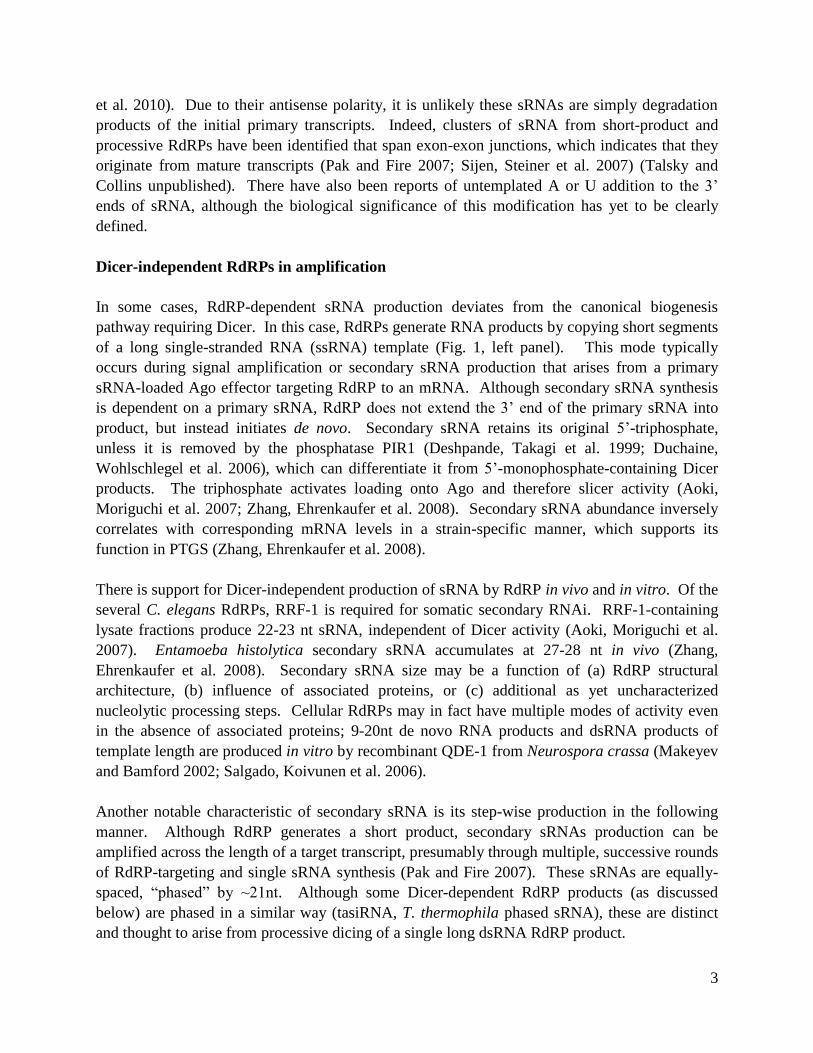
RNA-dependent RNA polymerases (RdRPs) induce sequence-specific gene silencing through the
production of double-stranded RNA (dsRNA) from a single-stranded RNA template. In
Tetrahymena thermophila, dsRNA products are synthesized by the only genome-encoded RdRP
(Rdr1) and then cleaved by an associated Dicer endonuclease (Dcr2) into 23-24 nt small RNAs
(sRNAs). These sRNAs accumulate constitutively, dependent on Rdr1 activity. They are
derived from endogenous transcripts and map in clusters to specific genomic loci. Rdr1 activity
is robust on all templates in vitro, yet the silencing process remains selective in the sequences it
targets in the cell. RNA targeting specificity is likely an absolute requirement for cell viability,
and, until now, the factors controlling it have not been clearly defined.
This dissertation details (1) the roles that Rdr1- interacting proteins play in the biogenesis of
multiple classes of 23-24 nt sRNA, (2) the in vitro template selectivity and initiation mechanisms
by distinct Rdr1 complexes (RDRCs) and (3) the identification of a new RNA silencing protein
(Rsp1) that, along with Rdr1, is required for bulk sRNA accumulation. Rsp1 shares several
protein partners with Rdr1, including the uridyltransferase Rdn1 and two factors (Rdf1 and Rdf2)
that control RNA target specificity in vivo through an unknown mechanism. Characterization of
Rdn1, Rdf1 and Rdf2 functions in vitro revealed that Rdf1 and Rdf2 are adaptors that activate
the polyuridylation of RNA by Rsp1-bound Rdn1. The data suggest that Rsp1 functions
upstream of Rdr1 in sRNA generation, as discussed in greater detail in Chapter 3. These
findings demonstrate that sRNA generation requires the coordination of a complex array of
protein machines. Future studies on how these proteins mediate RNA targeting in vivo will offer
exciting insights into the molecular mechanisms that are central to controlling the specificity of
RNA fates in the cell.


i
For anyone who faces challenges head on
and never gives up on lifelong goals.

ii

iii
TABLE OF CONTENTS
ABSTRACT ................................................................................................................................... 1
TABLE OF CONTENTS ............................................................................................................. iii
ACKNOWLEDGMENTS ............................................................................................................ v
INTRODUCTION ......................................................................................................................... 1
CHAPTER ONE: A single RNA-dependent RNA polymerase assembles with mutually exclusive
nucleotidyl transferase subunits to direct different pathways of small RNA biogenesis .............. 7
Abstract .............................................................................................................................. 7
Introduction ........................................................................................................................ 7
Results .............................................................................................................................. 10
Discussion ........................................................................................................................ 18
Materials and Methods ..................................................................................................... 20
Figures .............................................................................................................................. 22
CHAPTER TWO: Initiation by a eukaryotic RNA-dependent RNA polymerase requires looping
of the template end and is influenced by the template-tailing activity of an associated
uridyltransferase ........................................................................................................................... 31
Abstract ............................................................................................................................ 31
Introduction ...................................................................................................................... 31
Results............................................................................................................................... 33
Discussion ........................................................................................................................ 38
Materials and Methods ..................................................................................................... 40
Figures .............................................................................................................................. 42
CHAPTER THREE: Endogenous small RNA accumulation requires an RNA-dependent RNA
polymerase and the RNA silencing protein Rsp1 ........................................................................ 51
Abstract ............................................................................................................................ 51
Introduction ...................................................................................................................... 51
Results .............................................................................................................................. 53
Discussion ........................................................................................................................ 57
Materials and Methods ..................................................................................................... 60
Figures and Table ............................................................................................................. 63
FUTURE DIRECTIONS for studies on RNA silencing............................................................... 71
APPENDIX A: Rdf1 and Rdf2 serve distinct functions in sRNA biogenesis ............................. 72
APPENDIX B: Small RNA precursor structure may influence Rdr1 function and specificity ... 76
REFERENCES ............................................................................................................................ 80

iv

v
ACKNOWLEDGEMENTS
Thank you to all of my colleagues, friends and family members who have filled me with the
inspiration and determination necessary to complete this work of science and art.
I am forever grateful for the support and guidance of Kathleen Collins throughout the past four
plus years. Kathy introduced me to techniques related to biochemistry, RNA biology and one of
my best microbial friends, Tetrahymena thermophila. I could always depend on her for
countless suggestions for trouble-shooting and alternate methods. Kathy’s energy and focus are
impressive and contagious; we all notice and appreciate how much effort she puts into creating a
working environment that is highly intellectual and collaborative. Even more importantly, Kathy
encouraged my creativity and independence in designing and executing experiments. She gave
me room to develop my own ideas and the courage to share them with others.
My annual thesis guidance committee, consisting of Jeremy Thorner, Lin He and Ignacio Tinoco,
provided the highest level of mentorship and instruction I could have asked for. I am much
appreciative of their advice and thought-provoking questions, year after year. Jeremy Thorner is
also the one who introduced me to Berkeley when I interviewed with him as an MCB applicant.
Since then, he has pushed my intellectual and personal development as my mentor during my
first lab rotation, one of my first classes (MCB200) and throughout the succeeding years. Lin He
provided the perfect balance of breadth and depth into small RNA biology during her MCB 290
class. I also have much-appreciated her advice for improving my aptitude for scientific writing.
Kristin Scott supported me through my studies of neurodevelopment during my second rotation
in her lab, her MCB 260 class and in discussions of my outside proposal during my qualifying
exam. Others at Berkeley, including Don Rio, Caroline Kane and Jennifer Doudna, also played
important roles in my research pursuits and scientific growth.
For my colleagues in the Collins lab: my graduate experience would never have been the same
without all of you. Emily, Mary, Barbara, Kasper, Aaron, Bosun, Suzanne, Alec, Nicole,
George, Alex, Kyungah, Adam, Kwan, Debbie, Tim, Rama and Brandon are all are amazing
scientists and people. I thank you for being a resource for trouble-shooting, caring for my cells,
offering words of encouragement whenever I needed them and sharing your opinions and advice
on life decisions. I have much gratitude toward Suzanne Rebecca Lee (whose initials SRL will
forever be ingrained in my head), who began the RdRP expedition that I was proud to continue
throughout my graduate career.
To my friends and family: it makes me proud to know that you are so proud of me. Lance, you
served the honorable yet challenging role as my buffer between lab and home. Thank you with
all my heart for being there to listen when I needed it most. To my mom, dad, sister and the rest
of my family, thank you for your never-ending advice, encouragement and positivity. Thank you
Mai, Jess and my other amazing friends for keeping my life fun, active, balanced and satisfying.

vi

1
INTRODUCTION
COMPLEXITY OF SMALL RNA FUNCTION
Small RNA (sRNA) plays central roles in the regulation of gene expression in almost all
eukaryotes. Some sRNAs participate in regulation by RNA interference (RNAi) (Farazi, Juranek
et al. 2008; Carthew and Sontheimer 2009; Siomi and Siomi 2009; Ketting 2011). Traditionally,
RNAi refers to sequence-specific gene silencing mediated by sRNA (more specifically called
small interfering RNA, or siRNA) that accumulated after transgene overexpression in plants or
double-stranded RNA (dsRNA) injection into nematodes (Napoli, Lemieux et al. 1990; Fire, Xu
et al. 1998). With the rapid expansion of the RNA silencing field, it is now clear that sRNA also
regulates endogenous gene expression and plays an active role in normal cellular and organismal
biology.
Endogenous sRNA in mammals, plants, flies, nematodes and unicellular eukaryotes are produced
from genomic loci including expressed ORFs, pseudogenes, transposons, structured RNA
transcripts and overlapping convergent or antisense messages. Some of these sRNA families are
spatially or developmentally unique. For example, germline-specific Piwi-interacting RNA
(piRNA) suppresses mobile element expression in mammals and Drosophila melanogaster (Kim
review 2009). In Arabidopsis thaliana, trans-acting siRNA (tasiRNA) regulates mRNA
expression during development (Allen, Xie et al. 2005). Certain primary sRNA classes in
Caenorhabditis elegans only accumulate during embryogenesis while secondary sRNA
maintains silencing throughout adulthood (Vasale, Gu et al. 2010). In unicellular eukaryotes,
accumulation of sRNA can depend on developmental stage as well as strain (Lee and Collins
2006; Lepere, Nowacki et al. 2009).
Several genetically distinct small RNA types have been characterized in the ciliate Tetrahymena
thermophila. Ciliates contain a germline micronucleus and a transcriptionally active
macronucleus. During the sexual phase of its life cycle, two cells of different mating types
conjugate and exchange genetic material. The resulting progeny form a new macronucleus
derived from parental micronuclear genetic material. At this time, scan RNA (scnRNA, 27-30
nt) directs elimination of repetitive DNA elements and transposons in the developing
macronucleus (Mochizuki and Gorovsky 2004). A second class consists of tRNA halves (30-35
nt) that accumulate in response to stress such as starvation (Lee and Collins 2005). Only sRNA
(23-24 nt) accumulates during vegetative growth, starvation and conjugation (Lee and Collins
2006). This class may function in RNA surveillance by regulating accumulation of unnecessary
and potentially detrimental, aberrant macronuclear transcripts.
The one unifying theme between all 20-30 nt sRNAs is the involvement of Argonaute (Ago)
proteins (Farazi, Juranek et al. 2008; Ketting 2011). An sRNA is folded into an Ago protein and
targets it to RNA transcripts by base-pairing. Some Ago proteins have RNaseH (slicer) activity

2
that directs transcript cleavage (post-transcriptional gene silencing, PTGS) if its partner sRNA
shares 100% complementarity (Martinez and Tuschl 2004; Schwarz, Tomari et al. 2004; Faehnle
and Joshua-Tor 2007). Nuclear Ago RNPs function in transcriptional gene silencing (TGS)
through recruitment of chromatin modification machinery to a corresponding DNA locus
(Verdel, Jia et al. 2004). They can also perform a more microRNA- (miRNA) like function in
translational repression or enhancement of mRNA turnover (Tang 2005; Wu and Belasco 2008).
Additional less well-defined functions include influences on nascent transcript synthesis by RNA
Polymerase II (cotranscriptional gene silencing or CTGS).
PATHWAYS OF SMALL RNA BIOGENESIS
RNA silencing typically is triggered by long dsRNA that forms from structured or overlapping
transcripts or as products from an RNA-dependent RNA polymerase (RdRP). In fact, RdRPs
are indispensable for RNA silencing in many eukaryotes. Among RdRP-dependent pathways,
there are two modes of sRNA biogenesis (Fig 1). An RdRP can directly synthesize short RNA
products (Makeyev and Bamford 2002; Aoki, Moriguchi et al. 2007). Alternatively, processive
RdRP activity can be coupled to RNase III-related Dicer function (Tang, Reinhart et al. 2003;
Motamedi, Verdel et al. 2004; Lee and Collins 2007). Dicer produces sRNA duplexes
containing characteristic 2 nt 3’ overhangs. One strand is subsequently loaded onto a member of
the Argonaute family (Twi in T. thermophila), while the complementary strand is degraded.
RdRP-generated sRNAs shares some common characteristics. Although exceptions exist (Allen,
Xie et al. 2005), most sRNAs have an absolute or strong strand polarity bias and map antisense
along the length of predicted mRNAs (Lee and Collins 2006; Pak and Fire 2007; Sijen, Steiner et
al. 2007; Okamura and Lai 2008; Zhang, Ehrenkaufer et al. 2008; Couvillion, Lee et al. 2009;
Lepere, Nowacki et al. 2009; Gent, Lamm et al. 2010; Marker, Le Mouel et al. 2010; Vasale, Gu
3
et al. 2010). Due to their antisense polarity, it is unlikely these sRNAs are simply degradation
products of the initial primary transcripts. Indeed, clusters of sRNA from short-product and
processive RdRPs have been identified that span exon-exon junctions, which indicates that they
originate from mature transcripts (Pak and Fire 2007; Sijen, Steiner et al. 2007) (Talsky and
Collins unpublished). There have also been reports of untemplated A or U addition to the 3’
ends of sRNA, although the biological significance of this modification has yet to be clearly
defined.
Dicer-independent RdRPs in amplification
In some cases, RdRP-dependent sRNA production deviates from the canonical biogenesis
pathway requiring Dicer. In this case, RdRPs generate RNA products by copying short segments
of a long single-stranded RNA (ssRNA) template (Fig. 1, left panel). This mode typically
occurs during signal amplification or secondary sRNA production that arises from a primary
sRNA-loaded Ago effector targeting RdRP to an mRNA. Although secondary sRNA synthesis
is dependent on a primary sRNA, RdRP does not extend the 3’ end of the primary sRNA into
product, but instead initiates de novo. Secondary sRNA retains its original 5’-triphosphate,
unless it is removed by the phosphatase PIR1 (Deshpande, Takagi et al. 1999; Duchaine,
Wohlschlegel et al. 2006), which can differentiate it from 5’-monophosphate-containing Dicer
products. The triphosphate activates loading onto Ago and therefore slicer activity (Aoki,
Moriguchi et al. 2007; Zhang, Ehrenkaufer et al. 2008). Secondary sRNA abundance inversely
correlates with corresponding mRNA levels in a strain-specific manner, which supports its
function in PTGS (Zhang, Ehrenkaufer et al. 2008).
There is support for Dicer-independent production of sRNA by RdRP in vivo and in vitro. Of the
several C. elegans RdRPs, RRF-1 is required for somatic secondary RNAi. RRF-1-containing
lysate fractions produce 22-23 nt sRNA, independent of Dicer activity (Aoki, Moriguchi et al.
2007). Entamoeba histolytica secondary sRNA accumulates at 27-28 nt in vivo (Zhang,
Ehrenkaufer et al. 2008). Secondary sRNA size may be a function of (a) RdRP structural
architecture, (b) influence of associated proteins, or (c) additional as yet uncharacterized
nucleolytic processing steps. Cellular RdRPs may in fact have multiple modes of activity even
in the absence of associated proteins; 9-20nt de novo RNA products and dsRNA products of
template length are produced in vitro by recombinant QDE-1 from Neurospora crassa (Makeyev
and Bamford 2002; Salgado, Koivunen et al. 2006).
Another notable characteristic of secondary sRNA is its step-wise production in the following
manner. Although RdRP generates a short product, secondary sRNAs production can be
amplified across the length of a target transcript, presumably through multiple, successive rounds
of RdRP-targeting and single sRNA synthesis (Pak and Fire 2007). These sRNAs are equally-
spaced, “phased” by ~21nt. Although some Dicer-dependent RdRP products (as discussed
below) are phased in a similar way (tasiRNA, T. thermophila phased sRNA), these are distinct
and thought to arise from processive dicing of a single long dsRNA RdRP product.
4
Dicer-coupled RdRPs in initiation
Originally, it was believed that RdRPs function only in secondary amplification of silencing and
that primary sRNA only originates from dicing of dsRNA derived from RdRP-independent
mechanisms. Many RdRPs, however, cooperate with Dicer to initiate silencing. In contrast to
short RdRP products generated in a Dicer-independent fashion, RdRPs can produce long dsRNA
products that require Dicer processing into sRNA duplexes before Ago loading (Fig. 1, right
panel). In fact, many processive RdRPs directly interact with Dicer. An extreme case occurs in
Schizosaccharomyces pombe, whose genome encodes just one RdRP (Rdp1), one Dicer (Dcr1)
and one Ago (Ago1), which are all required for sRNA accumulation, co-transcriptional
heterochromatin formation through H3K9 methylation, and PTGS (Motamedi, Verdel et al.
2004; Iida, Nakayama et al. 2008). Dcr1 mediates the physical association between the Rdp1-
containing complex (RDRC) and the Ago1-containing RNA-induced transcriptional silencing
(RITS) complex, and is required for RITS recruitment to heterochromatic DNA loci (Motamedi,
Verdel et al. 2004). RdRP-Dicer physical interactions have also been described for C. elegans
RRF-3 and T. thermophila Rdr1 (Duchaine, Wohlschlegel et al. 2006; Lee and Collins 2007).
As with other processive (and non-processive) RdRPs, S. pombe Rdp1 does not require Dcr1 or
any other protein from the RDRC or RITS complexes for its in vitro RNA synthesis activity.
Moreover, the presence of these proteins does not seem to influence Rdp1 activity on synthetic
purified RNA templates (Motamedi, Verdel et al. 2004). Processive RdRP-catalyzed second
strand synthesis occurs across the length of a target RNA, as described for S. pombe Rdp1, A.
thaliana RDR6, wheat germ lysate RdRP activity, T. thermophila Rdr1, and N. crassa QDE-1
(Makeyev and Bamford 2002; Tang, Reinhart et al. 2003; Motamedi, Verdel et al. 2004; Lee and
Collins 2007; Curaba and Chen 2008). In fact, in a study where assays were run in parallel, S.
pombe Rdp1 and N. crassa QDE-1 produced comparable products to bacteriophage 6 RdRP
(Motamedi, Verdel et al. 2004).
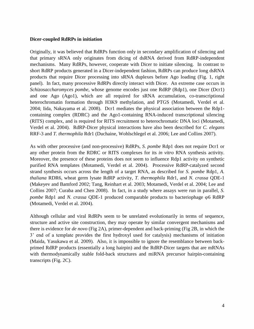
Although cellular and viral RdRPs seem to be unrelated evolutionarily in terms of sequence,
structure and active site construction, they may operate by similar convergent mechanisms and
there is evidence for de novo (Fig 2A), primer-dependent and back-priming (Fig 2B, in which the
3’ end of a template provides the first hydroxyl used for catalysis) mechanisms of initiation
(Maida, Yasukawa et al. 2009). Also, it is impossible to ignore the resemblance between back-
primed RdRP products (essentially a long hairpin) and the RdRP-Dicer targets that are mRNAs
with thermodynamically stable fold-back structures and miRNA precursor hairpin-containing
transcripts (Fig. 2C).
5
Dicer is not required for the RNA synthesis activity of RdRP in vitro, but the it can work
synergistically with RdRP both in vitro (Tang, Reinhart et al. 2003; Lee and Collins 2007) and in
vivo. Biological coupling of RdRP-Dicer activities extends across the phylogenic spectrum. In
organisms with multiple RdRPs and Dicers, a specific RdRP can provide the substrate for a
specific Dicer. For example, A. thaliana has six RdRPs, three of which have been characterized
in detail, and four Dicers (Ramachandran and Chen 2008). RDR2 and DCL3 produce 24 nt
sRNA required for heterochromatin formation and maintenance at genomic loci that contain
centromeres, mating type genes, transposons and retroelements (Wassenegger and Krczal 2006).
Without RDR2, 24 nt sRNA does not accumulate. In the absence of DCL3, though, RDR2
products can be processed by other Dicers to yield sRNAs of different sizes and of questionable
function (Kasschau, Fahlgren et al. 2007). RDR1 and RDR6 couple with DCR4 for PTGS to aid
in transgene silencing and antiviral defense. The biological coupling of A. thaliana RDR6 to
Dicer-like activity is also responsible for the successive production of phased, 21 nt tasiRNA
(Allen, Xie et al. 2005). In vivo, miRNA-bound Ago recruits RDR6 to a transcript and thereby
defines the tasiRNA phasing register. Second strand synthesis, coupled with processive dicing
produces perfectly phased tasiRNAs that regulate gene expression of transcription factors and
other proteins required for plant growth and development (Howell, Fahlgren et al. 2007).
Coordination also occurs during primary sRNA production in C. elegans: RRF-3 generates
primary sRNA in conjunction with DCR-1. Primary sRNA is distinct from RRF-1-generated
secondary sRNA in its accumulation level, 5’-end structure, and 26 nt size (Gent, Lamm et al.
2010; Vasale, Gu et al. 2010). In Paramecium tetraurelia, a subset of the three functional
RdRPs and the single canonical Dicer are required for generation of most, if not all, sRNA
derived endogenously, or induced by transgenes or dsRNA (Lepere, Nowacki et al. 2009;
Marker, Le Mouel et al. 2010).
STUDIES OF RDR1 IN TETRAHYMENA THERMOPHILA
In T. thermophila, biogenesis of 23-24 nt sRNA requires a single RdRP (Rdr1) and the most
canonical of its three genome-encoded Dicer-like proteins (Dcr2). T. thermophila mutants
lacking either Rdr1 or Dcr2 are inviable. Rdr1 and Dcr2 functionally and physically coordinate
6
dsRNA production and dicing in vitro and in the cell (Lee and Collins 2007). Sequencing of
sRNA purified from cells indicated they are derived from pseudogenes, structured RNA,
repetitive loci, and mRNA-encoding loci in T. thermophila (Couvillion, Lee et al. 2009). These
classes of sRNA associate with four of the five T. thermophila Ago proteins of the PIWI clade
(Twi2, 7, 8, 9) functioning during vegetative growth. Notably, accumulation of each sRNA class
has unique requirements for upstream (Rdr1 partners) and downstream (effector protein
association) factors. How this specificity is coordinated is an ongoing question in the field and
one focus of this dissertation.
Chapter One demonstrates that T. thermophila Rdr1 is found in distinct complexes that contain
four additional proteins. These different species seem to contribute to the complexity of sRNA
biogenesis. As judged by their knockout phenotypes, the Rdr1 interacting proteins control the
mRNA targets and sRNA accumulation profiles and function in distinct cellular processes.
Chapter Two explores the influence of Rdr1 protein partners and Rdr1 domain structure on RdR
activity in vitro. Finally, Chapter Three further explores the biological roles of sRNA-dependent
silencing machinery with the study of previously uncharacterized RNA silencing protein (Rsp1)
and its role in sRNA biogenesis.
Specificity of target selection and product fate is likely essential to survival, considering the
potential lethality of mis-silencing an essential transcript. This consideration may explain why
vertebrates may have lost their genome-encoded RdRP(s) (even though they encode functionally
active Dicers and Agos). Vertebrates rely on alternative mechanisms of dsRNA generation
(Maida, Yasukawa et al. 2009). By contrast, most other eukaryotic model organisms have
retained one (or multiple) RdRP(s). This conservation suggests that RdRPs must play critical
roles in physiological processes that give these invertebrate organisms an evolutionary
advantage. The multiple size classes of sRNA and the diverse genomic loci to which they map,
combined with the large number of sRNA biogenesis and effector proteins, suggests that T.
thermophila possesses similar complexity of RNA silencing pathways to those of other
eukaryotes. Characterization of the biochemical mechanisms of sRNA silencing in T.
thermophila will further our understanding of RdRP regulation and function in general.
7
CHAPTER ONE
A single RNA-dependent RNA polymerase assembles with mutually exclusive nucleotidyl
transferase subunits to direct different pathways of small RNA biogenesis
Based on S Lee, K Talsky & K Collins, RNA, 2009
These data are a combined effort between Kristin Talsky and Suzanne R. Lee, under the
direction of Kathleen Collins. SRL performed analysis of Rdn1 and Rdn2 (expression, gene
knockout and analysis and tagging). KT performed analysis of Rdf1 and Rdf2 (expression, gene
knockout and analysis). Both SRL and KT contributed to zz-Rdr1 purifications, activity assays
and small RNA accumulation analysis.
ABSTRACT
Members of the conserved family of eukaryotic RNA-dependent RNA polymerases (Rdrs)
synthesize double-stranded RNA (dsRNA) intermediates in diverse pathways of small RNA
(sRNA) biogenesis and RNA-mediated silencing. Rdr-dependent pathways of sRNA production
are poorly characterized relative to Rdr-independent pathways, and the Rdr enzymes themselves
are poorly characterized relative to their viral RNA-dependent RNA polymerase counterparts.
We previously described a physical and functional coupling of the Tetrahymena thermophila
Rdr, Rdr1, and a Dicer enzyme, Dcr2, in the production of ~24 nt sRNA in vitro. Here we
characterize the endogenous complexes that harbor Rdr1, termed RDRCs. Distinct RDRCs
assemble to contain Rdr1 and subsets of the total of four tightly Rdr1-associated proteins. Of
particular interest are two RDRC subunits, Rdn1 and Rdn2, which possess non-canonical
ribonucleotidyl transferase motifs. We show that the two Rdn proteins are uridine-specific
polymerases of separate RDRCs. Two additional RDRC subunits, Rdf1 and Rdf2, are present
only in RDRCs containing Rdn1. Rdr1 catalytic activity is retained in RDRCs purified from cell
extracts lacking any of the non-essential RDRC subunits (Rdn2, Rdf1, Rdf2) or if the RDRC
harbors a catalytically inactive Rdn. However, specific disruption of each RDRC imposes
distinct loss-of-function consequences at the cellular level and has a differential impact on the
accumulation of specific 23-24 nt sRNA sequences in vivo. The biochemical and biological
phenotypes of RDRC subunit disruption reveal a previously unanticipated complexity of Rdr-
dependent sRNA biogenesis in vivo.
INTRODUCTION
Endogenous eukaryotic RNA-templated RNA polymerases represent a new enzyme family with
an evolutionary origin distinct from that of other eukaryotic or viral polymerases (Iyer, Koonin et

8
al. 2003; Wassenegger and Krczal 2006). Interest in the Rdrs has grown with increasing
recognition of their roles in RNA interference (RNAi) and RNA-mediated silencing. RNAi and
related pathways exploit ~20-30 nt sRNAs as sequence-specific guides for regulation of gene
expression, heterochromatin assembly and defense against the disruptive impact of viruses and
mobile elements (Farazi, Juranek et al. 2008). Current evidence suggests that dsRNA products of
Rdr are processed by the cleavage activity of Dicer(s), and/or by helicase(s), and ultimately
assembled into Argonaute-family effector RNPs. Rdr-family polypeptides are encoded in the
genomes of a broad range of eukaryotes including amoebae, plants, fungi and nematodes (Cerutti
and Casas-Mollano 2006; Wassenegger and Krczal 2006). Despite the genetically critical roles
established for Rdrs in many organisms, much remains to be determined about their biochemical
activities and biological regulation.
Mechanisms that govern the in vivo specificity of single-stranded RNA (ssRNA) template
selection by an Rdr are largely unknown. One example of a template selection strategy was
revealed through studies of endogenous small-interfering RNA (siRNA) biogenesis in
Arabidopsis thaliana, in which transcripts targeted by specific microRNAs (miRNAs) were then
subject to endonucleolytic cleavage, dsRNA synthesis and subsequent processing by Dicer
(Allen, Xie et al. 2005; Yoshikawa, Peragine et al. 2005). In general, the biological specificity of
Rdr function is proposed to require interacting factors. Biochemical purification of the
Schizosaccharomyces pombe Rdr, Rdp1, revealed that it assembles as an RDRC with Hrr1, a
putative helicase, and Cid12, a protein with predicted non-canonical nucleotidyl transferase
motifs, both of which are required for Rdp1 function in heterochromatin silencing (Sugiyama,
Cam et al. 2005). For this S. pombe RDRC, individual subunit roles were not possible to discern
due cooperative subunit requirements for RDRC integrity (Motamedi et al. 2004). The
Caenorhabditis elegans Rdr-family protein RRF-1 co-purified the putative helicase DRH-1, and
the Rdr RRF-3 was copurified as one of several proteins associated with DCR-1 (Duchaine,
Wohlschlegel et al. 2006; Aoki, Moriguchi et al. 2007). In Neurospora crassa, perinuclear
localization and biological function of the Rdr SAD-1 depend on SAD-2 (Shiu, Zickler et al.
2006). These findings reveal coordination of Rdr function by other cellular factors, but the
specific biochemical properties and biological roles of Rdr-associated proteins have not been
well characterized.
The expressed macronuclear genome of the ciliated protozoan T. thermophila encodes a single
Rdr, Rdr1, which is genetically essential (Lee and Collins 2007). A putative role for Rdr1 in
sRNA biogenesis was inferred from the strand-asymmetric nature of an abundant class of
constitutively accumulated 23-24 nt sRNAs in vivo, which derive from the antisense strand of
predicted open reading frames lacking EST support (Lee and Collins 2006). The extreme bias in
strand origin foreshadowed the discovery of animal germline Piwi-associated RNAs (piRNAs),
which share this property (Seto, Kingston et al. 2007). T. thermophila possesses predicted genes
that encode up to 12 Argonaute family members, all of which cluster within the Piwi clade of
Argonautes (Cerutti and Casas-Mollano 2006; Seto, Kingston et al. 2007). The T. thermophila
Piwi protein Twi1 and one of three Dicer-like proteins, Dcl1, are induced only in a sexual cycle

9
of conjugation; they are required for biogenesis and stability of conjugation-specific 27-30 nt
sRNAs including the functionally defined scan RNAs that act as sequence-specific guides for
heterochromatin formation and macronuclear genome maturation (Mochizuki and Gorovsky
2004; Chalker 2008). The multiple size classes of T. thermophila sRNAs, along with the large
number of putative genes encoding sRNA biogenesis and effector machinery, suggest that T.
thermophila and other ciliates may possess a complexity of RNAi-related pathways comparable
to that of multicellular eukaryotes.
We previously showed that T. thermophila Rdr1 assembles with a set of associated proteins into
RDRC(s) that interact with the essential Dicer, Dcr2 (Lee and Collins 2007). Affinity
purification of endogenously expressed, epitope-tagged Rdr1 under gentle wash conditions
copurified Dcr2 and 3-4 other proteins; with more stringent washing, Dcr2 was released leaving
only Rdr1 and the tightly associated RDRC subunits. Biochemical assays of Rdr1 purified in
RDRC context, with or without associated Dcr2, revealed a functional as well as physical
coupling of T. thermophila RDRC and Dcr2 in the production of ~24 nt sRNA: Dcr2 cleavage of
only the RDRC product, not other dsRNA substrates, yielded a size of sRNA produced in vivo
(Lee and Collins 2006; Lee and Collins 2007). With or without associated Dcr2, RDRCs
harboring wild-type Rdr1 but not the active-site variant Rdr1-D1004A catalyzed long dsRNA
synthesis on a broad spectrum of ssRNA templates (Lee and Collins 2007). Almost the full
length of template was copied to produce dsRNA, as judged by treatment of 32
P-NTP
incorporation products with the single-strand specific Nuclease S1. In addition to Rdr1-
dependent synthesis of dsRNA, RDRC assays also generated radiolabeled ssRNA products that
were independent of catalysis by the Rdr1 active site.
Here we examine the contribution of T. thermophila Rdr1-associated proteins to dsRNA
synthesis in vitro and to sRNA biogenesis in vivo. We show that two RDRC subunits, Rdn1 and
Rdn2, are paralogs that possess the primary sequence motifs of non-canonical ribonucleotidyl
transferases, linking together RDRCs of ciliates and other organisms. We also demonstrate, for
the first time, the biochemical activity of these conserved RDRC subunits: both Rdn1 and Rdn2
catalyze non-templated uridine addition to RNA substrates in vitro, expanding the family of
known poly(U) polymerases. Although Rdn1 and Rdn2 have a similar specificity of biochemical
activity in vitro, the roles of the two proteins differ dramatically in vivo. We demonstrate that the
Rdn proteins have opposite developmental mRNA expression profiles, distinct gene knockout or
knockdown phenotypes, and mutually exclusive assembly with Rdr1 and the other RDRC
proteins Rdf1 and Rdf2. These findings demonstrate separable roles for Rdr1 in the content of
functionally specialized RDRCs, which are required to support distinct pathways of 23-24 nt
sRNA biogenesis in vivo.
10
RESULTS
Molecular characterization of four Rdr1-associated RDRC proteins
We previously characterized the biochemical activity of T. thermophila Rdr1 complexes isolated
by affinity purification of ZZ-Rdr1 (Rdr1 with a N-terminal tandem Protein A domain tag). N-
terminally tagged protein expressed from a transgene could functionally substitute for
endogenous untagged Rdr1, allowing disruption of the endogenous RDR1 locus, but C-terminal
tagging was not similarly successful in supporting viability (Lee and Collins 2007). SDS-PAGE
analysis of ZZ-Rdr1 purifications (Fig. 1A) suggests four associated polypeptides: a doublet of
~65 kDa proteins and a doublet of ~40 kDa proteins. Previously, mass spectrometry of ZZ-Rdr1
associated proteins identified, in addition to Rdr1 and Dcr2, predicted T. thermophila proteins
designated 6.m00629, 6.m00633, and 274.m00027 (Lee and Collins 2007). Subsequent ZZ-Rdr1
purifications and mass spectrometry identified a fourth protein, 274.m00028, represented by up
to 4 unique peptides when an isolated ~40 kDa gel slice from a ZZ-Rdr1 preparation was
analyzed.
Because gene predictions in T. thermophila are imprecise, we characterized the mRNA
transcripts expressed by these four predicted genes using RT-PCR. The largest open reading
frames of the experimentally defined mRNAs encode polypeptides with molecular weights
matching the RDRC polypeptides (two of ~65 kDa, two of ~40 kDa). BLAST and other analyses
of primary protein sequences revealed homology of both ~65 kDa proteins with the poly(A)
polymerase/2’-5’ oligo(A) synthetase family of non-canonical ribonucleotidyl transferases (Fig.
1B). This family includes proteins shown to have poly(A) and/or poly(U) polymerase activity
(Kwak and Wickens 2007; Martin and Keller 2007; Rissland and C.J. 2008). Following T.
thermophila nomenclature rules, the genes encoding these ~65 kDa Rdr1-associated proteins
were designated as the Rdr1-associated nucleotidyl transferases RDN1 and RDN2. The T.
thermophila Rdn1 and Rdn2 proteins are highly related, exhibiting 39% identity and 24%
additional similarity. Rdn1 and Rdn2 are more similar to each other than either is to other
putative nucleotidyl transferases encoded by T. thermophila macronuclear genome, including
proteins that we infer by sequence homology to represent the canonical poly(A) polymerase and
the non-canonical Trf4-family poly(A) polymerase involved in RNA turnover (Martin and Keller
2007).
In contrast to the Rdn proteins, the smaller Rdr1-associated proteins bear no structural motifs
that are readily discernible by either primary sequence analysis or tertiary structure threading
methods. Following T. thermophila nomenclature rules, the genes encoding the novel ~40 kDa
Rdr1-associated proteins were designated as the Rdr1-associated factors RDF1 and RDF2. While
no homologs of the Rdf proteins were found by BLAST of protein sequences deposited in
GenBank, comparison of the Rdf proteins to each other revealed 24% identity and 23%
additional similarity. The genes encoding the Rdf proteins are located in tandem in the genome,
with no intervening open reading frames, which suggests recent gene duplication. The genes

11
encoding the Rdn proteins are also located in close proximity in the genome, but they are
separated by ~12 kbp with three intervening predicted open reading frames.
We used Northern blot hybridization to examine mRNA expression for each of the RDRC
subunits in cells undergoing rapid growth and fission (vegetative growth) or cells starved for
nutrients and mixed with an alternate mating type to induce the sexual cycle of conjugation. All
of our Northern blot conclusions were recently supported and extended by whole-genome
microarray analysis of mRNA expression across highly sampled time-courses of T. thermophila
growth, starvation and conjugation (Miao, Xiong et al. 2009) and are therefore culled to show the
most relevant results. DCR2 and RDR1 are robustly expressed in vegetative growth (Fig. 1C,
left; (Lee and Collins 2006). RDN1 expression parallels that of RDR1 (Fig. 1C, left). Curiously,
RDN2 expression instead peaks in conjugation, when expression of RDN1 is relatively low (Fig.
1C, left; additional data not shown). This inverse relationship is consistent with the unequal
silver staining intensity of Rdn1 and Rdn2 in ZZ-Rdr1 purifications from growing or starving
cells, which suggests generally higher abundance of Rdn1 than Rdn2 in our RDRC preparations
(Fig. 1A; note that the indicated protein assignments are confirmed by additional mass
spectrometry and by protein tagging and genetic depletion studies described below).
Like RDN1 and RDN2, RDF1 and RDF2 show differential expression in vegetative growth
versus conjugation. RDF2 expression is high in vegetative growth and down-regulated by mid-
conjugation, while RDF1 expression is low in vegetative growth and increases in mid-
conjugation (Fig. 1C, right). Oddly, in silver-stained ZZ-Rdr1 purifications from growing or
starving cells, Rdf2 does not stain well and appears of equal or lesser abundance than Rdf1; we
suspect that this is due to the acidic isoelectric point of Rdf2, predicted to be 5.46. Together, our
observations indicate a discordance in the expression of RDN1 versus RDN2 and a discordance
in the expression of RDF1 versus RDF2. These results suggest that T. thermophila Rdr1-
associated proteins may form alternate RDRCs rather than the single RDRC of invariant
composition suggested for S. pombe (Sugiyama, Cam et al. 2005).
Distinct phenotypes of RDRC subunit depletion in vivo
We next tested whether genes encoding each of the newly identified RDRC subunits are essential
in T. thermophila, as is the case for RDR1 and DCR2 (Mochizuki and Gorovsky 2005; Lee and
Collins 2006; Lee and Collins 2007). We targeted the open reading frames of each of the Rdr1-
associated proteins for replacement with a gene cassette conferring resistance to neomycin.
Initial targeting replaces only a few of the 45 gene copies in the macronuclear genome, but
increased replacement by the neomycin-resistance cassette can be achieved by gradually
increasing the selective pressure. Non-essential genes can be eliminated entirely from the
macronucleus (KO strains), while essential genes reach a functionally limiting extent of gene
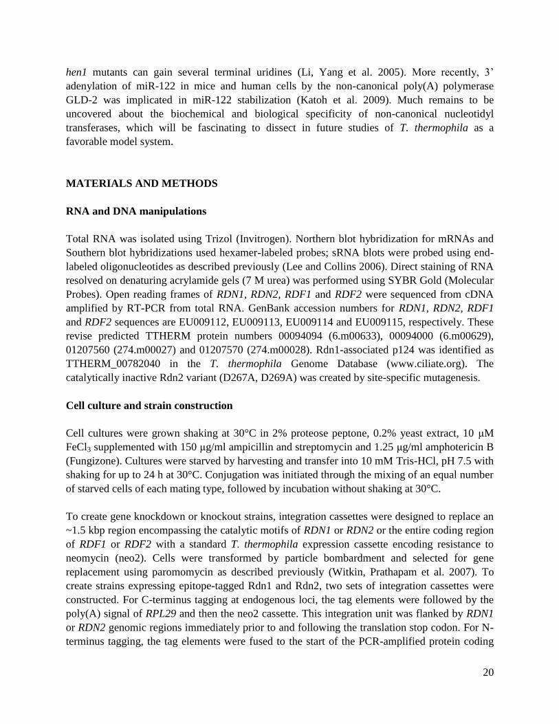
knockdown (KD strains). Genomic DNA was prepared from several independently selected
strains, which were subsequently released from selection to allow back-assortment of any
remaining copies of the endogenous locus. The state of each gene locus was assayed using

12
Southern blot hybridization. Probes were designed to distinguish wild-type chromosomes from
disrupted chromosomes by hybridization to differently sized DNA fragments in KO or KD
strains compared to wild-type (Fig. 2). Wild-type chromosomes were missing in strains where
RDN2, RDF1 or RDF2 was targeted, indicating that each of the genes had been replaced entirely
by the selectable marker. We conclude that RDN2, RDF1 and RDF2 are not essential genes. In
contrast, only incomplete loss of the RDN1 locus was attained. Thus, like RDR1 and DCR2,
RDN1 is essential in T. thermophila.
Up to 10% of cells in RDR1 KD strain cultures were enlarged, overly round and harboring an
often larger macronucleus than wild-type cells (Fig. 3A, right upper panels). This characteristic
“monster” phenotype has been frequently noted as a consequence of delayed or defective cell
division. Cells from RDN1 KD cultures also showed an increased frequency of monsters in
comparison to wild-type (Fig. 3A, left upper panels), even when wild-type cells were cultured at
a drug concentration that limited culture growth. Cells in RDF1 KO or RDF2 KO cultures
displayed phenotypes rarely observed in wild-type cell cultures but occasionally noted in RDR1
KD cultures as well: a minority of cells had more than a one macronucleus per cell and/or
incomplete DNA segregation between dividing cells (Fig. 3A, lower four panels). Curiously,
cells from RDN2 KO cultures showed no cellular phenotype during vegetative growth. To
confirm the generality of these phenotypes, we made each RDRC subunit gene knockdown or
knockout in different strain backgrounds. In strain CU522, a mutation in the beta-tubulin 1 gene
that confers taxol hypersensitivity (Gaertig et al. 1994) also sensitizes cells for division defects
(Smith, Yakisich et al. 2004). In the CU522 background even in the absence of taxol, the
phenotypes of RDN1 KD, RDF1 KO and RDF2 KO cells were similar to those described above
in SB210 background and were generally more penetrant; again, RDN2 KO cultures displayed no
cellular phenotype in vegetative growth.
SB210 and CU522 strains with macronuclear gene knockouts of the non-essential RDRC
subunits were mated to test for conjugation phenotypes. RDN2 KO or RDF1 KO cells paired and
appeared to complete meiosis with an efficiency and timing comparable to wild-type cells (Fig.
3B, step I), but few knockout pairs examined after these events appeared normal. Representative
images from the interval when wild-type cells have differentiating new macronuclei (Fig. 3B, 8-
12 h post-mixing) are shown in Fig. 3C. The number and arrangement of nuclei suggest that
knockout cells could not progress through zygotic mitoses and differentiation of two new
macronuclei (Fig. 3B, step III). Progression through conjugation was halted rather than delayed,
because paired KO cells examined at later time points (up to 19 h) still harbored a parental
macronucleus and several small nuclei rather than the two macronuclei plus one micronucleus
that result from successful conjugation (Fig. 3B, step V).
In contrast with RDN2 KO or RDF1 KO cells, RDF2 KO cells that paired did not exhibit a defect
in conjugation at the cellular level. The finding of conjugation defects resulting from loss of
Rdn2 or Rdf1 but not Rdf2 is consistent with the differential mRNA expression profiles of these
RDRC subunits (Fig. 1C). The use of macronuclear gene knockout strains for conjugation has

13
the potential to underestimate the importance of a gene product if it is needed late in conjugation,
because the zygotic nuclei may supply some mRNA prior to the completion of their
differentiation. Zygotic rescue is unlikely to account for the lack of conjugation phenotype for
RDF2 KO, however, because this locus is down-regulated in mRNA expression by mid-
conjugation (Fig. 1C). Overall, this phenotypic analysis of cells depleted or eliminated for
expression of individual RDRC subunits suggests different biological roles for each Rdn or Rdf
protein in vivo.
Mutually exclusive assembly of Rdn1 and Rdn2 into separate RDRCs
The differential mRNA expression profiles of RDRC subunits and their distinct genetic depletion
phenotypes suggested separation of function among RDRC assemblies. We focused subsequent
studies on the T. thermophila Rdn subunits, due in part to their disparate loss-of-function
phenotypes and in part to the presence of a potentially similar subunit with non-canonical
ribonucleotidyl transferase motifs in the S. pombe RDRC (Sugiyama, Cam et al. 2005). To
resolve RDRCs harboring Rdn1 versus Rdn2, we created two types of strains expressing tagged
Rdn proteins. At RDN1 and RND2 endogenous loci, we integrated a C-terminal tag with FLAG
epitopes, a TEV protease cleavage site and tandem Protein A domains (the FZZ tag) by selecting
for co-integration of a downstream neomycin resistance cassette (Fig. 4A, left). Because
complete replacement of the endogenous RDN1 locus with the tag cassette was achieved (data
not shown), we infer that Rdn function is not perturbed by tag fusion to the C-terminus. We also
expressed N-terminally tagged Rdn proteins with FLAG and ZZ modules placed in the opposite
orientation of the FZZ tag (the ZZF tag) by selecting for transgene replacement of the non-
essential, taxol-hypersensitive beta-tubulin encoded at BTU1 in strain CU522 (Fig. 4A, right).
Using strains that express the Rdn proteins tagged at their endogenous loci, optimal purification
of Rdn1-FZZ was accomplished using cell extracts from growing or starved cells whereas
optimal purification of Rdn2-FZZ was accomplished using extracts from conjugating cells (Fig.
4B, lanes 1-3), as predicted by RDN1 and RDN2 mRNA expression profiles. Using strains that
express tagged Rdn proteins from the BTU1 promoter of the transgene locus, both tagged
proteins could be readily purified from extracts of cells grown and starved in parallel (Fig. 4B,
lanes 4-5). Independent of tag location and whether the tagged protein was expressed from an
endogenous or transgene locus, Rdn1 copurified Rdr1 and both Rdf subunits (Fig. 4B, lanes 1
and 4). In contrast, tagged Rdn2 copurified only Rdr1 (Fig. 4B, lanes 3 and 5). Equal loadings of
tagged Rdn1 and Rdn2 confirmed this disparity in associated Rdf subunits (data not shown)
along with additional studies described below. Under the less stringent wash conditions used to
stabilize Dcr2 association with RDRC (Lee and Collins 2007), RDRCs with tagged Rdn1 or
Rdn2 each copurified Dcr2 (Fig. 4C; additional data not shown).
Tagged Rdn1 also copurified a protein of ~120 kDa, which was identified by mass spectrometry
as the predicted protein TTHERM_00782040. The putative ~124 kDa protein has no obvious
primary sequence motifs or homology with other proteins. No evidence of p124 was detected in

14
any purification of Rdn2 or in any previous affinity purification of tagged Rdr1 or Dcr2 (Lee and
Collins 2007). In summary (Fig. 4B, right), these findings suggest that distinct RDRCs harbor
Rdn1 or Rdn2 and that only RDRC harboring Rdn1 can recruit Rdf proteins; also, Rdn1
assembles a putative non-RDRC complex with p124.
We next examined the interdependence of RDRC subunit assembly. We created strains that
express ZZ-Rdr1 in the background of RDN2 KO, RDF2 KO or RDF1 KO. Cell extracts were
used for affinity purification of RDRC under the stringent wash conditions that release Dcr2
(Fig. 5A) or the gentle wash conditions that retain Dcr2-RDRC association (Fig. 5B).
Purifications of ZZ-Rdr1 in the absence of Rdn2, Rdf1 or Rdf2 confirmed a lack of association
of each genetically eliminated protein, but no additional subunits were depleted (Fig. 5A). Thus,
the loss of each non-essential RDRC subunit did not perturb the association of the remainder of
the subunits with Rdr1. Dcr2 association with RDRC was also unaffected by the absence of
Rdn2, Rdf2 or Rdf1 (Fig. 5B). These observations contrast with the interdependence of subunits
in the assembly of the S. pombe RDRC (Motamedi et al. 2004). Based on independent assembly
the two T. thermophila Rdf proteins with RDRC and their reciprocal mRNA expression profiles,
we suggest that the RDRC harboring Rdr1, Rdn1 and Rdf1 is distinct from the RDRC harboring
Rdr1, Rdn1 and Rdf2. However, the full complexity of Rdn1 association with the novel Rdf1,
Rdf2 and p124 polypeptides remains to be explored in future extensions of this work.
Nucleotidyl transferase activity of Rdn1 and Rdn2
To characterize the biochemical activities of Rdn1 and Rdn2 as potential nucleotidyl
transferases, we used RDRC complexes harboring the D1004A catalytic-dead variant of Rdr1
(Lee and Collins 2007). We performed separate reactions with each radiolabeled NTP and a 79
nt ssRNA template used in previous studies. Each radiolabeled NTP was supplemented with the
same NTP unlabeled or with the full set of unlabeled NTPs (Fig. 6A). Robust elongation of the
79 nt ssRNA was detected only in reactions with 32
P-UTP. Notably, product signal in reactions
with 32
P-UTP was not diminished by inclusion of other NTPs in the reaction (Fig. 6A, compare
lanes 3 and 6), suggesting that the activity is specific for incorporation of 32
P-UTP. Treatment of
the reaction products with ssRNA-specific nucleases entirely eliminated product signal (data not
shown; see (Lee and Collins 2007), confirming that the product did not represent residual
dsRNA synthesis activity by Rdr1 D1004A. Some ssRNA products were extended by one or a
few nucleotides, while others gained a longer polynucleotide tail. Because the specific activity of
product RNA varies with the number of nucleotides added, it is not possible to use radiolabel
intensity to infer which product is most abundant in vitro. However, we note that both short and
long products were generated under all in vitro reaction conditions tested. Similar results were
obtained in assays performed using input ssRNA templates of different lengths and sequence
compositions (data not shown).
We next compared the dependence of nucleotidyl transferase activity on RDRC composition
using a panel of complexes purified by tagged Rdr1, Rdn1 or Rdn2. ZZ-Rdr1 purification

15
enriched nucleotidyl transferase activity relative to mock purifications from control cells lacking
tagged protein (Fig. 6B, lanes 4-5). Importantly, ZZF-tagged Rdn1 and Rdn2 each enriched
nucleotidyl transferase activity in proportion to the amount of Rdn protein recovered by affinity
purification (Fig. 6B, lanes 1-2; additional data not shown). Rdn1-FZZ and Rdn2-FZZ tagged at
their endogenous loci also copurified nucleotidyl transferase activity in proportion to Rdn protein
(Fig. 6B, lanes 5 and 8). Because Rdn1 and Rdn2 do not co-purify each other (Fig. 4B), these
results suggest that each Rdn protein catalyzes ssRNA uridylation. Finally, we found that RDRC
purified by ZZ-Rdr1 from extract of any RDRC subunit knockout strain retained comparable
nucleotidyl transferase activity (Fig. 6B, lanes 9-12).
We attempted to create strains expressing tagged catalytic-dead (CD) versions of Rdn1 or Rdn2,
using the same transgene approach employed for expression of wild-type ZZF-Rdn1 and ZZF-
Rdn2 described above (Fig. 4A). Aspartic acids in the putative active site (Fig. 1B) were
substituted by alanines. Strains with complete replacement of BTU1 by the ZZF-Rdn2 CD
expression cassette were obtained, but expression of ZZF-Rdn1 CD was toxic enough to induce
loss of viability and prevent the establishment of strains fully replaced at the BTU1 locus by the
ZZF-Rdn1 CD cassette. Affinity purification of ZZF-Rdn2 CD failed to enrich for nucleotidyl
transferase activity (Fig. 6B, lane 3), despite enriching for the dsRNA synthesis and dicing
activities of Rdr1 and Dcr2 (see below). The disruption of RDRC nucleotidyl transferase activity
by substitution of two conserved aspartic acids in Rdn2 suggests that the T. thermophila Rdn
polypeptides rely on the same active site as other non-canonical poly(A) and poly(U)
polymerases to generate the nucleotidyl transferase activity detected in preparations of T.
thermophila RDRC. We observed no difference in nucleotidyl transferase activity associated
with wild-type versus catalytic-dead Rdr1, consistent with the requirement for the Rdn active
site. In contrast, studies of partially purified recombinant A. thaliana RDR6 suggest that this Rdr
by itself may act as a nucleotidyl transferase to extend ssRNA or ssDNA, with some preference
for use of UTP as the nucleotide substrate (Curaba and Chen 2008).
RDRCs share general properties of dsRNA synthesis and dicing in vitro
We next investigated whether Rdr1 catalytic activity was affected by RDRC composition. Rdr
reactions were performed using the same 79 nt ssRNA substrate tailed by Rdn proteins in studies
above, in reactions with all four unlabeled NTPs and 32
P-CTP. Reaction products were divided
for mock treatment or treatment with the single-strand specific Nuclease S1 prior to
electrophoresis (Fig. 7A). We found that this post-reaction processing resolves the nuclease-
resistant dsRNA portion of product from product region(s) with some ssRNA nature (Lee and
Collins 2007). As shown previously, ZZ-Rdr1 purification enriched for synthesis of products that
migrated larger than the input ssRNA template without Nuclease S1 treatment but resolved into a
series of products of slightly less than input template length with Nuclease S1 treatment (Fig.
7A, lane 5). Mock purifications from various cell extracts lacking a tagged RDRC subunit did
not enrich for this activity (Fig. 7A, lanes 4 and 7). RDRC complexes isolated by purification of
ZZF-Rdn1, ZZF-Rdn2 or ZZF-Rdn2 CD all harbored similar Rdr1 activity (Fig. 7A, lanes 1-3).
16
Thus, under the conditions used here, dsRNA synthesis is not influenced differentially by the two
Rdn proteins. Moreover, our results indicate that the transferase activity of an Rdn is not required
for dsRNA synthesis by Rdr1. RDRC complexes isolated by purification of Rdn1-FZZ or Rdn2-
FZZ, the tagged Rdn proteins expressed from endogenous loci, also did not display differential
dsRNA synthesis activity in vitro (Fig. 7A, lanes 6 and 8).
We also tested the activity of RDRCs isolated by purification of ZZ-Rdr1 from strains lacking
the non-essential RDRC subunits Rdn2, Rdf1 and Rdf2. Wild-type and gene-knockout strains all
yielded RDRCs with similar Rdr1 product synthesis activity (Fig. 7A, lanes 9-12). Together,
these Rdr1 activity assays demonstrate that synthesis of dsRNA is independent of the presence of
any individual RDRC subunit other than Rdr1, because normal Rdr1 activity was retained in
preparations of (1) tagged Rdn2, which does not co-purify Rdn1, Rdf1 and Rdf2; (2) tagged
Rdn1, which does not co-purify Rdn2; and (3) ZZ-Rdr1 from RDN2 KO, RDF1 KO or RDF2
KO cell extracts. Furthermore, Rdr1 activity in vitro does not require the catalytic activity of a
Rdn subunit: although ZZF-Rdn2 CD lacks nucleotidyl transferase activity (Fig. 6B, lane 3),
RDRC harboring ZZF-Rdn2 CD still carries out normal dsRNA synthesis (Fig. 7A, lane 3).
T. thermophila Rdr1 complexes containing Dcr2 are capable of coupled dsRNA synthesis and
dicing, such that input ssRNA generates ~24 nt sRNA products in short duplexes (Lee and
Collins 2007). We tested whether RDRC composition had an influence on Dcr2 activity in the
coupled reaction system in vitro. None of the changes in RDRC subunit composition affected
copurification of Dcr2 under gentle wash conditions (Fig. 4C and additional data not shown).
Likewise, none of the changes in RDRC subunit composition reduced the generation of ~24 nt
Dcr2 products from dsRNA synthesized by RDRC (Fig. 7B). Therefore, coupled dsRNA
synthesis and dicing in vitro is independent of the presence of any specific subunit other than
Rdr1 and Dcr2.
RDRC subunit requirements for 23-24 nt sRNA accumulation in vivo
Our previous finding of physical and functional coupling of Rdr1 and Dcr2 in generating ~24 nt
sRNA in vitro implicated these enzymes in the biogenesis of constitutively accumulated 23-24 nt
sRNAs (Lee and Collins 2007). Before investigating the role of Rdn and Rdf RDRC subunits in
23-24 nt sRNA biogenesis in vivo, we first wanted to verify dependence of the in vivo process on
Rdr1. Because RDR1 and DCR2 are both essential genes (Mochizuki and Gorovsky 2005; Lee
and Collins 2006; Lee and Collins 2007), it was not possible to use gene knockout strains to test
whether their loss of function also resulted in loss of 23-24 nt sRNA. Furthermore, although gene
knockdown strains often yield phenotypic insights, the genotypic variation in any growing cell
population will obscure molecular phenotypes by ‘averaging’ them across the culture. To escape
these limitations, we tested for potential reduction of 23-24 nt sRNA following short-term over-
expression of catalytic-dead Rdr1 D1004A. Because Rdr1-D1004A still assembles with all of the
RDRC-associated proteins including Dcr2 (Lee and Collins 2007), it could have a dominant-
17
negative impact by competing with wild-type Rdr1 for biological templates and by inhibiting
RDRC-associated activities that are coupled to dsRNA synthesis.
Expression of ZZ-Rdr1-D1004A was placed under the control of the cadmium-inducible MTT1
promoter integrated at BTU1. Selection for transgene integration was performed without
cadmium in the medium, allowing complete replacement of BTU1 with the transgene (data not
shown). After release from selection, cell cultures were expanded by vegetative growth. Protein
expression was induced by cadmium addition to cells either in vegetative growth or after transfer
of growing cells to starvation medium to halt cell growth. Cadmium addition induced similar
levels of ZZ-Rdr1-D1004A protein accumulation in growing and starving cells (data not shown).
Wild-type cells were cultured and induced with cadmium in parallel as a control.
At various time points within 24 h after cadmium addition, cells were harvested for total RNA
purification. Total RNA was normalized for recovery, size-enriched for sRNA and then
examined by denaturing gel electrophoresis and direct staining. Expression of catalytic-dead
Rdr1 in starved cells reduced the level of 23-24 nt sRNA (Fig. 8A, lanes 1-3). In continuously
growing cells, the impact of catalytic-dead Rdr1 expression was more dramatic: 23-24 nt sRNA
became almost undetectable after 16 hours (Fig. 8A, lanes 4-6). The greater impact of catalytic-
dead Rdr1 expression on 23-24 nt sRNA accumulation in growing cells could reflect greater
dilution of the sRNA present prior to cadmium addition or greater sRNA turnover. No cellular
phenotypes of catalytic-dead Rdr1 expression were detected in the 24 h interval of cell culture
employed for these studies.
We next investigated the accumulation of 23-24 nt sRNA in strains lacking Rdn2, Rdf1 or Rdf2.
Total RNA was size-enriched and used to visualize 23-24 nt sRNA by direct staining. In both
SB210 and CU522 backgrounds, RDF2 KO strains had extremely low levels of 23-24 nt sRNA
(Fig. 8B, top panel). This result was reproduced over many repetitions of sRNA purification. We
also examined the accumulation of 27-30 nt sRNA in conjugating RDN2 KO, RDF1 KO or
RDF2 KO cells sampled across the normal time-course of conjugation (data not shown). While
27-30 nt sRNA levels were largely unaffected, RDN2 KO cells exhibited a slight reduction in
sRNA that could reflect a direct contribution of Rdn2 to production of scan RNAs or more likely
an indirect impact of disrupted progression through conjugation (see Fig. 3C).
The genomic loci from which sequenced T. thermophila 23-24 nt sRNA originate harbor
predicted protein-coding genes antisense to the sRNA; these genes can be classified into several
homology groups or families (Lee and Collins 2006). In strains lacking Rdn2, Rdf1 or Rdf2, the
presence of known sRNA was probed by Northern blot hybridization with end-labeled
oligonucleotides. This approach revealed approximately wild-type levels of a specific sRNA,
sRNA2, in the RDF2 KO 23-24 nt sRNA population despite the much lower accumulation of 23-
24 nt sRNA overall (Fig. 8B, middle panel). Remarkably, sRNA2 was missing from the 23-24 nt
sRNA pool in the RDN2 KO strain, despite an abundance of 23-24 nt sRNA similar to wild-type.
Additional oligonucleotide probes complementary to known T. thermophila 23-24 nt sRNA were
18
used individually and as mixtures, with some sRNA found to be missing in the RDN2 KO strain
(i.e. sRNA2, and other sRNA from this sequence family) and others found to be missing in the
RDF2 KO strain (Fig. 8B, bottom panel).
Together these results suggest that accumulation of most 23-24 nt sRNA is dependent on Rdf2
but not Rdf1 or Rdn2, consistent with preferential expression of Rdf2 in growing cells. However,
because specific subsets of 23-24 nt sRNA require the presence of Rdn2, distinct forms of RDRC
have unique roles in sRNA biogenesis during growth. These findings indicate that
compositionally different RDRCs play functionally different roles in sRNA biogenesis in vivo.
DISCUSSION
Roles for Rdr polypeptides have been established at the levels of transcriptional and post-
transcriptional regulation (Wassenegger and Krczal 2006). Endogenous synthesis of dsRNA
complicates the necessary cellular repertoire of response to nucleic acids, because dsRNA is also
a hallmark of invasion by selfish foreign genomes. Much remains to be learned about how Rdr
activity is recruited to and/or restrained from acting on potential ssRNA targets in vivo.
The Rdr polypeptide itself has conserved N- and C-terminal extensions from the active site
motifs, so some functional specialization may be conferred by these accessory domains. In
addition, because Rdr proteins isolated from their endogenous sources form RDRCs, tightly
associated subunits are likely to be a general solution for increasing biological specificity. Here
we show that there is yet more gain in Rdr specificity by assembly of distinct RDRCs
responsible for separate sRNA biogenesis pathways. Mechanisms for RDRC subunit function in
the biogenesis of T. thermophila sRNAs remain to be addressed. The subunits may govern
specificity for recruitment to ssRNA templates, determine the synthesis of product structures
with endogenous rather than foreign dsRNA hallmarks, and/or direct the fate of product dsRNA
to siRNA generation or other currently unknown end-points.
In comparing the activities and in vivo functions of the T. thermophila RDRCs resolved here, it is
clear that some features are shared while others are distinct. By in vitro assays of dsRNA
synthesis, T. thermophila RDRCs differing in the presence of Rdn1 or Rdn2 have similar
activity. Likewise, both types of RDRC interact with Dcr2 and promote Dcr2 cleavage of RDRC
products to generate ~24 nt sRNA in vitro. In addition, both types of RDRC support equivalent
nucleotidyl transferase activity on purified ssRNA templates. Beyond these similarities in Rdr1,
Dcr2 and Rdn catalytic activities, differences are apparent. Curiously, only RDRC with Rdn1 can
assemble the Rdf1 and Rdf2 subunits. Furthermore, loss of Rdn2 precludes in vivo accumulation
of some sRNAs while loss of Rdf2 reduces accumulation of other sRNAs. Cellular phenotypes
also distinguish loss-of-function by the different RDRCs: loss of Rdf1 or Rdf2 induces DNA
segregation phenotypes, while loss of Rdn2 or Rdf1 halts conjugation. In addition to revealing a
division of labor among RDRCs sharing the same Rdr1 catalytic core, our results demonstrate
19
conclusively that T. thermophila Rdr1 and its associated proteins play important roles in
accumulation of 23-24 nt sRNA in vivo.
In the simplest model for the biogenesis of strand-asymmetric T. thermophila 23-24 nt sRNAs in
vivo, Rdr1 acts at the top of a pathway that selects RNA targets to yield sRNAs in a manner
specified by RDRC context. In other organisms, Rdr family members are proposed to act
downstream of initial sRNA generation. C. elegans RRF-1 acts downstream of primary siRNA to
generate secondary siRNA bearing a 5’-triphosphate (Aoki, Moriguchi et al. 2007; Pak and Fire
2007; Sijen, Steiner et al. 2007). The S. pombe RDRC functions in a positive feedback loop that
integrates transcription by DNA-dependent RNA polymerase, chromatin modification enzymes,
an Argonaute-containing RITS complex and Dicer (Bühler and Moazed 2007). A. thaliana
RDR6 generates endogenous siRNA from transcripts targeted by miRNA (Allen, Xie et al. 2005;
Yoshikawa, Peragine et al. 2005). In all these cases, whether dsRNA synthesis by the Rdr is
considered initiating or amplifying, RDRCs share the common need to recognize a non-mRNA
target transcript, such as one that may originate from a degenerate gene no longer encoding a
functional protein (as for T. thermophila Rdr1) or a nascent RNA (as for S. pombe Rdp1) or a
transcript otherwise compromised in its integrity (as for A. thaliana RDR6 or C. elegans RRF-1).
Our findings suggest that this specificity for transcripts potentially recognized as aberrant
mRNAs may be influenced by RDRC context.
Our finding that biochemically active poly(U) polymerases are subunits of T. thermophila
RDRCs is intriguing in light of the recent recognition that these non-canonical nucleotidyl
transferase proteins have wide conservation in diverse eukaryotes (Kwak and Wickens 2007;
Martin and Keller 2007; Rissland and C.J. 2008). Some members of the family have been
implicated to have function(s) in RNA silencing pathways in other organisms. Substitution of
putative active site residues of S. pombe Cid12 disrupts RNAi-dependent heterochromatin
formation and accumulation of centromeric siRNAs (Win, Stevenson et al. 2006). S. pombe
Cid14, the ortholog of Saccharomyces cerevisiae Trf4/5, also functions in heterochromatic gene
silencing (Bühler, Haas et al. 2007). For C. elegans RDE-3, substitutions predicted to inhibit
nucleotidyl transferase activity abrogate protein function in RNAi (Chen, Simard et al. 2005).
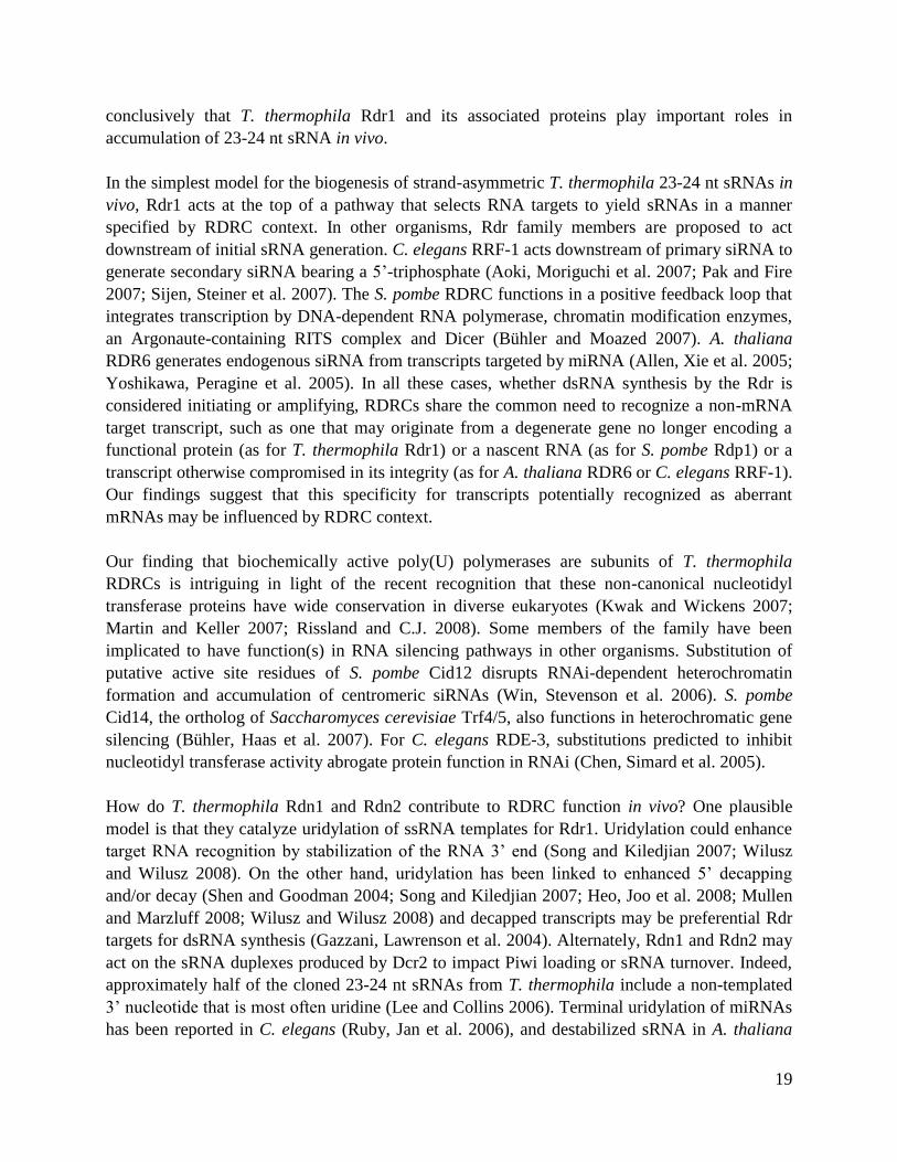
How do T. thermophila Rdn1 and Rdn2 contribute to RDRC function in vivo? One plausible
model is that they catalyze uridylation of ssRNA templates for Rdr1. Uridylation could enhance
target RNA recognition by stabilization of the RNA 3’ end (Song and Kiledjian 2007; Wilusz
and Wilusz 2008). On the other hand, uridylation has been linked to enhanced 5’ decapping
and/or decay (Shen and Goodman 2004; Song and Kiledjian 2007; Heo, Joo et al. 2008; Mullen
and Marzluff 2008; Wilusz and Wilusz 2008) and decapped transcripts may be preferential Rdr
targets for dsRNA synthesis (Gazzani, Lawrenson et al. 2004). Alternately, Rdn1 and Rdn2 may
act on the sRNA duplexes produced by Dcr2 to impact Piwi loading or sRNA turnover. Indeed,
approximately half of the cloned 23-24 nt sRNAs from T. thermophila include a non-templated
3’ nucleotide that is most often uridine (Lee and Collins 2006). Terminal uridylation of miRNAs
has been reported in C. elegans (Ruby, Jan et al. 2006), and destabilized sRNA in A. thaliana
20
hen1 mutants can gain several terminal uridines (Li, Yang et al. 2005). More recently, 3’
adenylation of miR-122 in mice and human cells by the non-canonical poly(A) polymerase
GLD-2 was implicated in miR-122 stabilization (Katoh et al. 2009). Much remains to be
uncovered about the biochemical and biological specificity of non-canonical nucleotidyl
transferases, which will be fascinating to dissect in future studies of T. thermophila as a
favorable model system.
MATERIALS AND METHODS
RNA and DNA manipulations
Total RNA was isolated using Trizol (Invitrogen). Northern blot hybridization for mRNAs and
Southern blot hybridizations used hexamer-labeled probes; sRNA blots were probed using end-
labeled oligonucleotides as described previously (Lee and Collins 2006). Direct staining of RNA
resolved on denaturing acrylamide gels (7 M urea) was performed using SYBR Gold (Molecular
Probes). Open reading frames of RDN1, RDN2, RDF1 and RDF2 were sequenced from cDNA
amplified by RT-PCR from total RNA. GenBank accession numbers for RDN1, RDN2, RDF1
and RDF2 sequences are EU009112, EU009113, EU009114 and EU009115, respectively. These
revise predicted TTHERM protein numbers 00094094 (6.m00633), 00094000 (6.m00629),
01207560 (274.m00027) and 01207570 (274.m00028). Rdn1-associated p124 was identified as
TTHERM_00782040 in the T. thermophila Genome Database (www.ciliate.org). The
catalytically inactive Rdn2 variant (D267A, D269A) was created by site-specific mutagenesis.
Cell culture and strain construction
Cell cultures were grown shaking at 30°C in 2% proteose peptone, 0.2% yeast extract, 10 μM
FeCl3 supplemented with 150 μg/ml ampicillin and streptomycin and 1.25 μg/ml amphotericin B
(Fungizone). Cultures were starved by harvesting and transfer into 10 mM Tris-HCl, pH 7.5 with
shaking for up to 24 h at 30°C. Conjugation was initiated through the mixing of an equal number
of starved cells of each mating type, followed by incubation without shaking at 30°C.
To create gene knockdown or knockout strains, integration cassettes were designed to replace an
~1.5 kbp region encompassing the catalytic motifs of RDN1 or RDN2 or the entire coding region
of RDF1 or RDF2 with a standard T. thermophila expression cassette encoding resistance to
neomycin (neo2). Cells were transformed by particle bombardment and selected for gene
replacement using paromomycin as described previously (Witkin, Prathapam et al. 2007). To
create strains expressing epitope-tagged Rdn1 and Rdn2, two sets of integration cassettes were
constructed. For C-terminus tagging at endogenous loci, the tag elements were followed by the
poly(A) signal of RPL29 and then the neo2 cassette. This integration unit was flanked by RDN1
or RDN2 genomic regions immediately prior to and following the translation stop codon. For N-
terminus tagging, the tag elements were fused to the start of the PCR-amplified protein coding

21
region, and its translation stop codon was followed by the BTU1 poly(A) signal and neo2
cassette; this integration unit was targeted for integration by flanking BTU1 genomic regions
upstream of the endogenous start codon and downstream of the poly(A) signal. Selection was
performed using paromomycin. Catalytic-dead Rdr1 D1004A was expressed from a transgene
integrated in substitution of the BTU1 locus under transcription control of a transplanted ~1 kbp
promoter region of MTT1 (Shang, Song et al. 2002).
Cell staining and microscopy
Cells were washed once with 10 mM Tris-HCl and fixed for 1 h at room temperature in 2%
paraformaldehyde prepared in PHEM buffer (60 mM PIPES, pH 6.9; 25 mM HEPES; 10 mM
EGTA; 2 mM MgCl2). Cells were then washed for five minutes with modified PBS, pH 7.2 (130
mM NaCl, 2 mM KCl, 8 mM Na2HPO4, 2 mM KH2PO4, 10 mM EGTA, 2 mM MgCl2) and
incubated in 0.1-1 microgram/ml DAPI in PBS for 10 min with end-over-end rotation. Following
three washes in PBS, cells were resuspended in PBS and mounted on a slide with 90% glycerol
containing anti-fade. Cells were imaged using the 40X objective of an Olympus BX61
fluorescent microscope. Images were captured using Metamorph software.
Affinity purification, mass spectrometry and activity assays
One-step affinity purifications of ZZ-tagged proteins using IgG agarose and mass spectrometry
were performed as described previously (Lee and Collins 2007). Silver staining was used to
detect proteins following SDS-PAGE. Rdr-mediated dsRNA synthesis, NTP transferase and
coupled Rdr/Dicer assays were performed as described previously (Lee and Collins 2007),
except that the unlabeled NTPs used were at 20 micromolar final concentration. Formamide
denaturing acrylamide gels containing 7 M urea and 45% formamide were used in the analysis of
Rdr and coupled Rdr/Dicer reaction products to eliminate dsRNA structure (Lee and Collins
2007).

22
FIGURE 1
FIGURE 1. Rdr1-associated RDRC subunits are two pairs of related proteins with differential
mRNA expression profiles. (A) Extracts from starved cells with no tagged protein (Mock) or
with ZZ-Rdr1 were used for affinity purification of RDRC. The four proteins recovered
specifically in association with Rdr1 are labeled in the enlargements, with assignments based on
mass spectrometry, protein tagging and genetic depletion assays. (B) An alignment of active site
residues in Rdn1, Rdn2 and other non-canonical ribonucleotidyl transferases is shown, with
consensus underneath. Identical and similar residues are boxed. Asterisks denote residues
mutated to alanine to render this class of enzyme catalytically inactive. Nucleotidyl transferase
and nucleotide recognition motifs are indicated as previously described (Martin and Keller
2007). Tt, Tetrahymena thermophila; Sp, Schizosaccharomyces pombe; Ce, Caenorhabditis
elegans; At, Arabidopsis thaliana; Xt, Xenopus tropicalis; Hs, Homo sapiens. (C) Northern blots
for mRNA expression were performed using total RNA isolated from cells in vegetative growth
(V), starvation (St) or conjugation (Conj). Time points after the initiation of conjugation are
noted in hours (h); where not noted, the 10 h time point of conjugation was used (see Fig. 3B for
conjugation stages).
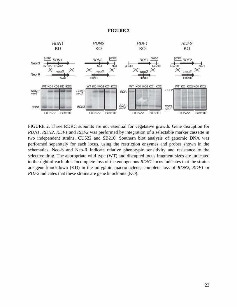
23
FIGURE 2
FIGURE 2. Three RDRC subunits are not essential for vegetative growth. Gene disruption for
RDN1, RDN2, RDF1 and RDF2 was performed by integration of a selectable marker cassette in
two independent strains, CU522 and SB210. Southern blot analysis of genomic DNA was
performed separately for each locus, using the restriction enzymes and probes shown in the
schematics. Neo-S and Neo-R indicate relative phenotypic sensitivity and resistance to the
selective drug. The appropriate wild-type (WT) and disrupted locus fragment sizes are indicated
to the right of each blot. Incomplete loss of the endogenous RDN1 locus indicates that the strains
are gene knockdown (KD) in the polyploid macronucleus; complete loss of RDN2, RDF1 or
RDF2 indicates that these strains are gene knockouts (KO).

24
FIGURE 3, part 1
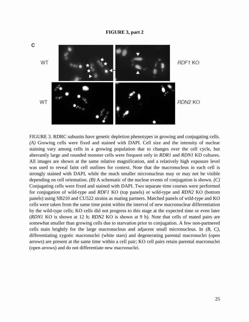
25
FIGURE 3, part 2
FIGURE 3. RDRC subunits have genetic depletion phenotypes in growing and conjugating cells.
(A) Growing cells were fixed and stained with DAPI. Cell size and the intensity of nuclear
staining vary among cells in a growing population due to changes over the cell cycle, but
aberrantly large and rounded monster cells were frequent only in RDR1 and RDN1 KD cultures.
All images are shown at the same relative magnification, and a relatively high exposure level
was used to reveal faint cell outlines for context. Note that the macronucleus in each cell is
strongly stained with DAPI, while the much smaller micronucleus may or may not be visible
depending on cell orientation. (B) A schematic of the nuclear events of conjugation is shown. (C)
Conjugating cells were fixed and stained with DAPI. Two separate time courses were performed
for conjugation of wild-type and RDF1 KO (top panels) or wild-type and RDN2 KO (bottom
panels) using SB210 and CU522 strains as mating partners. Matched panels of wild-type and KO
cells were taken from the same time point within the interval of new macronuclear differentiation
by the wild-type cells; KO cells did not progress to this stage at the expected time or even later
(RDN1 KO is shown at 12 h; RDN2 KO is shown at 9 h). Note that cells of mated pairs are
somewhat smaller than growing cells due to starvation prior to conjugation. A few non-partnered
cells stain brightly for the large macronucleus and adjacent small micronucleus. In (B, C),
differentiating zygotic macronuclei (white stars) and degenerating parental macronuclei (open
arrows) are present at the same time within a cell pair; KO cell pairs retain parental macronuclei
(open arrows) and do not differentiate new macronuclei.

26
FIGURE 4
FIGURE 4. Rdn1 and Rdn2 assemble as distinct RDRCs that share association with Dcr2. (A)
Strains expressing tagged Rdn1 or Rdn2 were created using two strategies: introduction of a C-
terminal tag cassette at the endogenous locus or integration of an N-terminally tagged protein
transgene at the non-essential BTU1 locus. Neo-S and Neo-R or Taxol-S and Taxol-R indicate
relative phenotypic sensitivity and resistance to the selective drug. (B) Protein complexes were
examined following purification of the indicated tagged proteins from extracts of starved cells
(lanes 1 and 4-5) or cells at the 9 h time point of conjugation (lanes 2-3). Parallel mock
purifications were performed using wild-type cell extracts, only one of which is shown but all of
which were similar in non-specific background. The composition of the multisubunit complexes
identified here is summarized on the right. Lighter text denotes the RDRC subunits that exhibit
increased expression during conjugation. (C) Dcr2 association with Rdr1 was examined
following purification of the indicated tagged proteins from extracts of starved cells using the
gentle washing conditions that preserve RDRC-Dcr2 interaction.

27
FIGURE 5
FIGURE 5. RDRC subunit interactions occur in the absence of Rdn2, Rdf1 or Rdf2. Protein
complexes were examined following purification of ZZ-Rdr1 from extracts of starved cells
lacking tagged protein (Mock) or the indicated gene knockout (KO) cells. Affinity purifications
employed either robust (A) or gentle (B) washing conditions.

28
FIGURE 6
FIGURE 6. Rdn1 and Rdn2 are uridine-specific nucleotidyl transferases. (A) Products of
nucleotidyl transferase assays using a 79 nt input ssRNA and purified RDRCs containing
catalytic-dead Rdr1-D1004A. (B) Uridylation of a 79 nt input ssRNA by specific RDRCs.
RDRCs were purified by the indicated tagged protein from extracts of cells that were starved (all
lanes except 7-8) or at the 9 h time point of conjugation (lanes 7-8). The intensity of reaction
product correlates with the level of Rdn present in the preparation (not shown). ZZ-Rdr1 was
also purified from extracts of starved wild-type or gene knockout (KO) cells as noted.

29
FIGURE 7
FIGURE 7. Synthesis and dicing of dsRNA do not require a specific RDRC composition in vitro.
(A) Products of dsRNA synthesis untreated or treated with Nuclease S1 prior to electrophoresis.
Assays were performed using 79 nt ssRNA template and radiolabeled CTP. The intensity of
reaction product correlates with the level of Rdr1 present in the preparation (not shown). (B)
Small RNA products of coupled dsRNA synthesis and dicing assays using the same 79 nt ssRNA
template as in (A) and RDRC complexes that were gently washed to preserve Dcr2 association.
The intensity of reaction product correlates with the level of Dcr2 protein present in the
preparation (not shown).

30
FIGURE 8
FIGURE 8. RDRC subunits differentially impact 23-24 nt sRNA accumulation in vivo. (A) Total
RNA isolated from starving (St) or growing (V) wild-type cells or cells expressing Rdr1-
D1004A was size-enriched for small RNA, resolved by denaturing gel electrophoresis and
stained directly. Starved or growing cell cultures were treated with 0.1 μg/ml CdCl2 or 1 μg/ml
CdCl2, respectively, for the times indicated to induce catalytic-dead Rdr1 over-expression. Wild-
type cells were similarly treated with CdCl2 as a control. (B) Aliquots of enriched sRNA from
growing wild-type or gene-knockout cells were used for direct staining (top panel) or Northern
blots. Representative sRNA expression results are shown for an individual sRNA (sRNA2) or 14
other sRNAs (sRNA mix) of known sequence used for Northern blot assays in previous work
(Lee and Collins 2006). A summary of sRNA phenotypes is also provided.
31
CHAPTER TWO
Initiation by a eukaryotic RNA-dependent RNA polymerase requires looping of the
template end and is influenced by the template-tailing activity of an associated
uridyltransferase
Based on K Talsky & K Collins, JBC, 2010
ABSTRACT
A conserved family of eukaryotic RNA-dependent RNA polymerases (RDRs) initiate or amplify
the production of small RNAs (sRNAs) to provide sequence specificity for gene regulation by
Argonaute/Piwi proteins. RDR-dependent silencing processes affect the genotype-to-phenotype
relationship in many eukaryotes, but the principles that underlie the specificity of RDR template
selection and product synthesis are largely unknown. Here I characterize the initiation specificity
of the Tetrahymena thermophila RDR, Rdr1, as a heterologously expressed single subunit and in
the context of its biologically assembled multisubunit complexes (RDRCs). Truncation analysis
of recombinant Rdr1 revealed domain requirements different from the only other similarly
characterized RDR, suggesting that there are subfamilies of RDR enzyme with distinct structural
requirements for activity. I demonstrate an apparently obligate Rdr1 mechanism of initiation in
which the template end is looped to provide the hydroxyl group priming the synthesis of double-
stranded RNA. RDRC subunits with poly(U) polymerase activity can act on the template end
prior to looping to increase the duplex length of product, thus impacting the sRNA sequences
generated by the RDRC-coupled Dicer. Overall, our findings give new perspective on
mechanisms of RDR initiation and demonstrate that non-RDR subunits of an RDRC can affect
the specificity of product synthesis.
INTRODUCTION
Argonaute/Piwi proteins loaded with small RNAs (sRNAs) direct diverse types of eukaryotic
gene regulation (Ghildiyal and Zamore 2009; Siomi and Siomi 2009). Most extensively
characterized are the post-transcriptional silencing roles of Argonaute-subfamily proteins bound
to sRNAs processed from annealed double-stranded (ds) RNAs or snap-back hairpin structures
(Carthew and Sontheimer 2009; Kim, Han et al. 2009). In addition to transcript base-pairing,
dsRNA precursors of sRNAs can be generated by the activity of an RNA-dependent RNA
polymerase. A family of eukaryotic RNA-dependent RNA polymerases (RDRs) is broadly
represented in protists, plants, fungi, and animals (Cerutti and Casas-Mollano 2006; Zong, Yao
et al. 2009). Notably, eukaryotic RDRs have an origin independent of their viral counterparts,
with a conserved active site and overall multidomain organization (Iyer, Koonin et al. 2003;

32
Salgado, Koivunen et al. 2006; Zong, Yao et al. 2009). Despite their apparently myriad essential
biological functions, RDRs remain largely uncharacterized in biochemical properties.
Only two RDRs have been assayed for catalytic activity following recombinant expression in a
heterologous system. Neurospora crassa QDE-1 produced in Saccharomyces cerevisiae
synthesized predominantly 9-21 nt short products with a 5’-triphosphate, as well as some longer
products (Makeyev and Bamford 2002). In comparison, Arabidopsis thaliana RDR6 produced in
Nicotiana benthamiana synthesized long products from near full-length copying of input
templates (Curaba and Chen 2008). RDR proteins from endogenous sources, which are purified
as multisubunit RDR complexes (RDRCs), have also been found to catalyze distinct types of
activity. The originally characterized RDR activity from tomato synthesized products with a 5’-
triphosphate (Schiebel, Haas et al. 1993), and triphosphate-capped 21-23 nt sRNA were
synthesized by the Caenorhabditis elegans RRF-1 RDRC (Aoki, Moriguchi et al. 2007). Wheat
germ lysate, the Schizosaccharomyces pombe Rdp1 RDRC, and the Tetrahymena thermophila
Rdr1 RDRCs instead synthesized long dsRNA products processed into sRNA duplexes by the
coupled action of a Dicer enzyme (Tang, Reinhart et al. 2003; Motamedi, Verdel et al. 2004; Lee
and Collins 2007).
It seems clear that at least two modes of dsRNA synthesis can be distinguished: constrained
synthesis of sRNA-length or shorter products versus processive synthesis from initiation to the
template 5’-end. Due to the non-parallel nature of previously employed assay conditions and
product analysis methods, it is difficult to draw more detailed parallels from the cross-
comparison of published studies. Importantly, in no case has the activity of a physiological
RDRC been compared to the activity of its RDR subunit alone. Therefore, it remains unclear
whether differences in the mode of product synthesis can arise from differences in the non-RDR
subunits of an RDRC.
We have used the ciliated protozoan T. thermophila as a model system for understanding the
biochemical and biological specificity of RDR function. The only T. thermophila RDR family
member, Rdr1, assembles several RDRCs (Lee and Collins 2007). Each RDRC harbors one of
two related nucleotidyl transferase subunits, Rdn1 or Rdn2, with poly(U) polymerase activity.
The Rdr1-Rdn1 complex additionally associates with one of two related novel subunits, Rdf1 or
Rdf2. T. thermophila RDRCs are stable to stringent wash conditions, but the use of gentle wash
conditions preserves RDRC interaction with Dcr2, the Dicer that produces 23-24 nt sRNAs in
vivo and in vitro (Lee and Collins 2006; Lee and Collins 2007). Rdr1, Rdn1, and Dcr2 are
essential, but the Rdn2, Rdf1, or Rdf2 subunit specific to a single RDRC can be depleted without
loss of strain growth or viability (Lee and Collins 2006; Lee and Collins 2007; Lee, Talsky et al.
2009). Cells lacking any one RDRC by RDN2, RDF1, or RDF2 gene-knockout have distinct
cellular and sRNA accumulation phenotypes, indicating that each RDRC has a non-redundant
function (Couvillion, Lee et al. 2009; Lee, Talsky et al. 2009).

33
Here I investigate the template requirements and product structures of recombinant T.
thermophila Rdr1 and purified T. thermophila RDRC complexes. I find that Rdr1 and its RDRCs
produce dsRNA products that do not result from de novo initiation. Instead, Rdr1 initiates by
looping the template 3’ end for intramolecular initiation of dsRNA synthesis. By comparing
Rdr1 and RDRCs purified using the same tag, I show that RDRC context can influence dsRNA
product structure. I demonstrate a novel role for RDRC-associated poly(U) polymerase activity,
which through uridylation of the template increases the length of duplex product synthesis. Our
findings suggest that template 3' tailing prior to template looping allows dsRNA synthesis and
Dicer cleavage of sRNA products from the original template 3' end. Overall, this work
characterizes a mode of RDR initiation distinct from the de novo initiation that produces
triphosphate-capped sRNAs for amplification of gene silencing. I propose that at least a subset of
RDR enzymes capable of long-product synthesis coupled to subsequent Dicer processing differ
in their initiation mechanism from the short-product RDRs that generate triphosphate-capped
sRNAs independent of Dicer. Our studies also provide the first evidence for an influence of
RDR-associated subunits on the specificity of dsRNA product synthesis.
RESULTS
Rdr1 domain requirements differ from previous findings for QDE-1
RDRs share regions of sequence conservation beyond the catalytic core (Zong, Yao et al. 2009).
The only published study of RDR domain requirements examined recombinant N. crassa QDE-
1, which is soluble and active without the RDR N-terminal extension (Makeyev and Bamford
2002). High-resolution structure determination for the remaining domains of QDE-1, designated
slab, catalytic, and head (Fig. 1A), revealed that the head domain mediates subunit dimerization
(Salgado, Koivunen et al. 2006). Because recombinant QDE-1 appears to have unique
biochemical features compared to subsequently characterized RDRCs, I analyzed the domain
requirements of T. thermophila Rdr1 for comparison. I used RRL to express a tagged version of
Rdr1, zzRdr1, known to be biologically functional (Lee and Collins 2007). Recombinant Rdr1
was recovered from RRL using IgG agarose to bind the N-terminal tag of protein A domains,
followed by elution with TEV protease. Full-length Rdr1 and several Rdr1 domain-truncation
variants were examined for dsRNA synthesis activity using the same conditions optimized for
the physiologically assembled T. thermophila RDRCs (Lee and Collins 2007; Lee, Talsky et al.
2009).
RNA products of Rdr1 were radiolabeled by incorporation of 32
P-CTP and resolved using highly
denaturing formamide-PAGE, which I have found to be essential for complete suppression of
product secondary structure formation (Lee and Collins 2007). For comparison to previous
studies of T. thermophila RDRC activity, I used the 79-nucleotide (nt) single-stranded RNA
template RNA1 derived from a genomic locus subject to Rdr1-mediated silencing in vivo (Lee
and Collins 2006; Couvillion, Lee et al. 2009). I compared the activity of wild-type (WT) Rdr1,
34
the catalytic-dead variant Rdr1-D1004A with an alanine substitution for aspartic acid in the
active site (Lee and Collins 2007), an internal deletion within the N-terminal extension, as well
as N- or C-terminal truncations designed based on primary sequence conservation with QDE-1 to
have end-points at putative subdomain boundaries (Fig. 1A). All of the proteins were expressed
at comparable levels (Fig. 1B, top panel). Full-length Rdr1 but not the catalytic-dead variant
catalyzed robust product synthesis (Fig. 1B, bottom panels). Curiously, the N-terminal truncation
of T. thermophila Rdr1 analogous to that used for recombinant expression of QDE-1 eliminated
enzyme activity, as did a smaller internal deletion of the Rdr1 N-terminal extension (Fig. 1B,
lanes 1-2). In contrast, C-terminal truncation reduced but did not eliminate activity (Fig. 1B, lane
5; in some experiments, weak activity was also detectable for the protein assayed in lane 6).
Recombinant QDE-1 forms a homodimer (Salgado, Koivunen et al. 2006). A similar interaction
seems unlikely for T. thermophila Rdr1 in RDRC context, because RDRCs with distinct subunit
composition are readily isolated from each other without the cross-purification that would be
expected from Rdr1 multimerization (Lee, Talsky et al. 2009). To test whether the isolated T.
thermophila Rdr1 subunit forms a multimer, I coexpressed differentially tagged versions of Rdr1
in RRL. In addition to zzRdr1 used above, I expressed Rdr1 with an N-terminal tag of three
tandem FLAG epitopes (F-Rdr1). Either tagged Rdr1 or the combination was expressed in RRL,
followed by binding to and elution from IgG agarose (Fig. 1C). Elution with TEV protease
removes the tag from zzRdr1, resulting in the isolation of untagged Rdr1 (Fig. 1C, lanes 1-2). A
low background binding of F-Rdr1 to IgG agarose was detectable (Fig. 1C, lane 3), but there was
no coenrichment of F-Rdr1 with zzRdr1. Furthermore, no enrichment of zzRdr1 by
copurification with F-Rdr1 was detected in the reciprocal purification on FLAG antibody resin
(data not shown). The Rdr1 isolated by zzRdr1 purification remained functional when
coexpressed with F-Rdr1 (Fig. 1C, bottom panel), as did F-Rdr1 when coexpressed with zzRdr1
(data not shown). These findings suggest that recombinant T. thermophila Rdr1 functions as a
subunit monomer. Overall, I conclude that there are likely to be subclasses of RDR with distinct
domain requirements for function.
Rdr1 and RDRCs can produce similar dsRNA products of less than full-template length
We next assayed product synthesis by recombinant Rdr1 compared to Rdr1 assembled in RDRC
context. I purified the pool of physiologically assembled RDRCs from extract of a T.
thermophila strain expressing zzRdr1, using stringent wash conditions to deplete the associated
Dcr2 dsRNA cleavage activity (Lee and Collins 2007). To our surprise, in assays using a variety
of template lengths and sequences including the standard template RNA1 (Fig. 2A), recombinant
Rdr1 and stringently washed RDRCs produced a similar profile of product synthesis. Notably,
despite the absence of RDRC subunits with poly(U) polymerase activity in the RRL-expressed
Rdr1 enzyme preparation, recombinant Rdr1 synthesized products that like RDRC products were
longer than the input 79-nt template length (Fig. 2B, lanes 1-2). If Rdr1 or RDRC products were
treated with S1 nuclease to remove any single-stranded regions, dsRNA product lengths were
resolved to be near full-template length and somewhat shorter (Fig. 2B, lanes 3-4). No bias
35
toward the synthesis of sRNA-sized products was observed, consistent with the Dcr2-
dependence of 23-24 nt sRNA production in vivo.
A likely cause of heterogeneity in dsRNA product lengths is the potential use of heterogeneous
sites within the template for dsRNA synthesis initiation. Extending the RNA1 template by
addition of a poly(A) tail (RNA1a, Fig. 2A) favored the synthesis of a dsRNA product with the
approximate length of RNA1, suggesting that the homopolymer A-tract did not support dsRNA
synthesis initiation (Fig. 2C, lanes 5-6). Insertion of a GC or C within the A-tract (RNA1aGCa or
RNA1aCa templates, Fig. 2A) provided an additional initiation site, resulting in the synthesis of
an extra product with the length expected for processive synthesis from the introduced initiation
site to the template 5’-end (Fig. 2B, lanes 7-10). Activity assays using numerous additional
templates did not suggest any specific sequence requirement for dsRNA synthesis initiation by
Rdr1 or RDRC, although some sequence preference was evident. For example, both Rdr1 and
RDRC greatly preferred a template with an introduced cytidine initiation site rather than a
uridine initiation site (Fig. 2B, lanes 9-12). Likewise, substitution of the 3 cytidine residues
within the 3’ 15 nt of the RNA1 template for guanosines (RNA1mut, Fig. 2A) decreased the
yield of the longest products and increased the yield of the slightly shorter ~64-65 nt products
(Fig. 2C). These findings suggest that T. thermophila Rdr1 can initiate dsRNA synthesis from
variable positions near the template 3’ end but then copies the remainder of the template
processively. Curiously, in contrast to the lack of requirement for any specific template
sequence, Rdr1 showed an absolute dependence on template 3’ end structure. If the 3’ terminal
template nucleotide was converted to a 3’ phosphate group, no activity was observed in reactions
with Rdr1 or RDRC (Fig. 2D).
Rdr1 can copy DNA if ribonucleotides are present at the template 3’ end
An endogenous tomato RDR activity, tagged N. crassa QDE-1 expressed in vivo, and
recombinant A. thaliana RDR6 each have been shown to synthesize RNA using DNA as
template (Schiebel, Haas et al. 1993; Curaba and Chen 2008; Lee, Chang et al. 2009). I therefore
tested T. thermophila Rdr1 and RDRC activity on DNA and RNA oligonucleotides of the same
32-nt sequence (Fig. 2A, RNA32). Rdr1 had robust activity on the synthetic RNA, yielding
heterogeneously sized dsRNA products shorter than the full-length template following treatment
with S1 nuclease (Fig. 3A, lane 6). No products were observed in Rdr1 reactions with the DNA
template, but chimeric templates with a DNA 5’-end and 2-6 residues of RNA at the 3’ end
supported the synthesis of RNA products resistant to treatment with S1 nuclease (Fig. 3A, lanes
1-5). Nearly identical results were observed for Rdr1 assayed in RDRC context (Fig. 3A, lanes 7-
12). Products from reactions with the template containing 26 nt of DNA were degraded by
RNase H, confirming the presence of product DNA-RNA hybrid, while RNA32 template
products were resistant to RNase H (Fig. 3B). I conclude that T. thermophila Rdr1 can act as a
DNA-dependent RNA polymerase in vitro, albeit with relatively low product yield compared to
its activity as an RNA-dependent RNA polymerase. However, even when copying DNA, Rdr1
has an absolute requirement for RNA structure at the template 3’ end. I note that in previous
36
studies it is possible that DNA templates were extended by ribonucleotide tailing prior to dsRNA
synthesis, in particular in the case of recombinant A. thaliana RDR6 which unlike T. thermophila
Rdr1 has an inherent nucleotidyl transferase activity (Curaba and Chen 2008; Lee, Talsky et al.
2009).
The duplex product contains an internal loop
Some of the single-stranded RNA content of RDRC products could derive from the poly(U)
polymerase activity of the Rdn subunits, but even recombinant Rdr1 lacking an associated
nucleotidyl transferase produced partially single-stranded products (Fig. 2B, lanes 1-4). The
migration of these partially single-stranded products was not affected by single-stranded RNA
exonuclease treatments (data not shown). Also, the specific activity of products from reactions
with radiolabeled CTP, UTP, or ATP was not greatly altered by digestion of the single-stranded
product regions with S1 nuclease, as would be expected if a nucleotide-selective transferase
activity was adding a 3’ tail to the dsRNA products (data not shown). I therefore tested a model
of product structure in which the template and product strands are joined by an internal loop of
single-stranded RNA. In Rdr1 products this loop would be derived from template residues only,
whereas the loop of RDRC products could include both template residues and nucleotides added
to the 3’ end of the template by Rdn activity (see below). A requirement for priming from a
“looped” rather than base-paired template 3’ end would explain why no product synthesis is
observed from an annealed primer (data not shown). Required use of the template 3’ end as
primer would preclude copying of templates with a blocked 3’ end, as I observed (Fig. 2D), and
would also explain why I have been unable to detect a product 5’-triphosphate group by direct or
indirect labeling approaches (data not shown).
To investigate template looping as a mechanism of initiation, I exploited the observation that
templates with a poly(A) 3’ region did not initiate dsRNA synthesis within the homopolymer A-
tract (Fig. 2B). I compared products of the 79-nt RNA1 and 104-nt RNA1a templates for their
susceptibility to digestion with RNase H in the presence of dT18 DNA oligonucleotide. For the
RNA1 template, Rdr1 products were insensitive to digestion with RNase H in the presence of
dA18 or dT18 DNA oligonucleotides; as a control, products were shifted to faster migration by
treatment with S1 nuclease (Fig. 4A, lanes 1-4). For the RNA1a template, Rdr1 products were
insensitive to digestion with RNase H in the presence of dA18 (Fig. 4A, lanes 5-6). On the other
hand, digestion with RNase H in the presence of dT18 produced discrete products not much larger
than products digested by S1 nuclease (Fig. 4A, lanes 7-8). Similar results were observed for
RDRC products, with slightly slower product mobility following RNase H treatment in the
presence of dT18 (Fig. 4A, lanes 9-12). These results support the template looping mechanism for
Rdr1 initiation of dsRNA synthesis (Fig. 4B). I note that the largest Rdr1 and RDRC products
were more efficiently digested by S1 nuclease than by RNase H and dT18 under our reaction
conditions (Fig. 4A, compare the longest products in lane 7 versus lane 8 or lane 11 versus lane
12). This could derive from the need for dT18 hybridization to compete with structure formation
in the looped poly(A) region of RNA1a products.

37
RDRC context and Rdr1-associated Rdn activity can influence template looping
RDRC activity assays described above used the cellular pool of Rdr1 complexes. The related
RDRC poly(U) polymerase subunits, Rdn1 and Rdn2, assemble with Rdr1 in a mutually
exclusive manner. Both enzymes can extend single-stranded RNA by mono-, oligo-, or poly-
uridine addition in vitro, but their in vivo depletion phenotypes are distinct: RDN1 knockdown or
expression of a catalytic-dead Rdn1 variant is lethal, whereas RDN2 knockout or expression of a
catalytic-dead Rdn2 variant (Rdn2cd) is not (Lee, Talsky et al. 2009). When assayed as
stringently washed complexes, RDRCs purified from cell extracts by tagged Rdn1, Rdn2, or
Rdn2cd had similar specificities of dsRNA product synthesis (Lee, Talsky et al. 2009).
Curiously, I found that gently washed RDRCs with associated Dcr2, designated RDRC*, show
differences in product synthesis dependent on the presence of a catalytically active Rdn.
RDRC purifications from extracts of cells expressing tagged Rdn1, Rdn2, or Rdn2cd (Lee,
Talsky et al. 2009) were conducted using gentle wash conditions to purify RDRC complexes
designated N1*, N2*, or N2cd* according to their Rdn subunit identity (Fig. 5A). These
complexes were assayed for dsRNA product synthesis using a 20-nt chimeric template with a
DNA 5’-end and RNA 3’ end (Aless, Fig. 2A), the short products of which are unsuitable for
Dcr2 processing. Because the Aless template lacks adenosines, UTP should not be required for
dsRNA synthesis. Indeed, N1*, N2*, and N2cd* RDRC reactions lacking UTP produced
predominant products longer than the template that were converted to dsRNA products of less
than template length by S1 nuclease treatment (Fig. 5B, lanes 1-6). Addition of UTP to the
reaction did not affect the profile of dsRNA products produced by the N2cd* RDRC, which
lacks an active poly(U) polymerase (Fig. 5B, lane 9). In contrast, in assays of the N1* and N2*
RDRCs harboring a catalytically active Rdn subunit, the presence of UTP allowed the synthesis
of longer dsRNA products (Fig. 5B, lanes 7-8). The poly(U) polymerase activity of N1* and N2*
but not N2cd* was confirmed in reactions with radiolabeled UTP, with other NTPs absent to
prevent dsRNA synthesis on the Aless template (Fig. 5C). Reactions using other templates also
showed a change in the product profile of dsRNA synthesis dependent on the presence of a
catalytically active Rdn: N1*, N2*, and the pooled RDRC* produced longer products in assays
with RNA1 (Fig. 5D) or RNA32 (Fig. 5E), while Rdr1 alone or N2cd* did not.
The Rdn-dependent increase in the length of template that is copied into dsRNA provides the
first biochemical demonstration of an RDRC subunit influence on dsRNA product synthesis by
an RDR. I suggest that Rdn-mediated template U-tailing provides an optimal structure for 3’
loop formation and/or priming of second-strand synthesis, favoring dsRNA synthesis across the
maximal extent of the original template (Fig. 5F). Only a limited length of template U-tailing is
likely to occur prior to second-strand synthesis, because the internal loop region of RDRC*
products was not digested by RNase H in the presence of dA18 (data not shown). Unfortunately,
our attempts to define loop sequences by cloning template-looped products were unsuccessful,
even for the relatively short products of the Aless template. Template use was not stimulated by

38
the pre-addition of various lengths of U-tail (data not shown), suggesting the need for
coordination of the Rdn and Rdr1 phases of template extension. Indeed, gently purified RDRCs
showed a greatly enhanced Rdn-dependence of dsRNA product synthesis, suggesting that an
RDRC* conformation correlated with Dcr2 association promotes productive coordination of Rdn
and Rdr1 activities.
Impact of Rdn activity on sRNA production by Dcr2
Because Rdn activity affects the profile of dsRNA product synthesis by Rdr1, it could alter the
sequence of sRNA products generated by Dcr2. I used the RNA1 template in assays of the
stringently washed N2 RDRC lacking associated Dcr2 or the gently washed N1*, N2*, and
N2cd* RDRCs with copurified Dcr2 (Fig. 5A). All of these Rdr1-containing RDRCs generated
products that without S1 nuclease treatment were longer than input template length (Fig. 6A). In
addition, as expected, the N1*, N2*, and N2cd* RDRCs generated 23-24 nt sRNA products from
dsRNA cleavage by Dcr2 (Fig. 6A, lanes 2-4). I note that not all RDRC* complexes retain Dcr2,
with generally higher Dcr2 stoichiometry in N1* compared to N2* purifications (Lee, Talsky et
al. 2009). As expected the N2 RDRC depleted of Dcr2 did not generate sRNA-sized products
(Fig. 6A, lane 1), and the N2cd* RDRC lacking nucleotidyl transferase activity did not generate
the background of long single-stranded products extending to the top of the gel (lane 4).
Parallel reactions were performed with unlabeled NTPs to evaluate sRNA sequences by blot
hybridization. End-labeled oligonucleotide probes complementary to the product strand were
used to detect putative sRNAs derived from RDRC synthesis across different regions of the
template (Fig. 6B). No sRNA sequences were detected in reactions with the stringently washed
N2 RDRC lacking Dcr2 (Fig. 6C, lane 1). Both N1* and N2* RDRC reactions generated sRNAs
complementary to the mid and 3’ regions of template, but sRNAs complementary to the template
3’ region were not detected in the N2cd* reaction (Fig. 6C, lanes 2-4). These results extend the
demonstration of Rdn-dependent copying of the template 3’ end into dsRNA. Without Rdn-
mediated U-tailing of the template, more of the template 3’ region must be incorporated into the
internal loop. Our attempts to determine whether RDRC-coupled Dcr2 cleavage occurred
preferentially on the loop side of a dsRNA product were not successful, due to the apparent lack
of partially Dcr2-cleaved dsRNA intermediates (data not shown). Dcr2 may be most active when
cotranscriptionally cleaving nascent dsRNA, prior to RDRC completion of dsRNA synthesis on
a long RNA template.
DISCUSSION
Eukaryotic RDR proteins play important roles in numerous cellular processes. Despite a clear
biological significance, little is known about the principles that govern RDR specificity for
template recognition or synthesis of a dsRNA product. Here I provide new insight into these

39
biochemical specificities. Also, for the first time I compare the properties of a recombinant Rdr1
subunit alone and its physiologically assembled RDRCs.
The N. crassa QDE-1 C-terminus mediates subunit dimerization, with an extensive interaction
surface proposed to promote its biological function (Salgado, Koivunen et al. 2006). Here, I did
not detect dimerization of recombinant T. thermophila Rdr1. T. thermophila Rdr1 also does not
appear to dimerize in RDRC context, because distinct RDRCs harboring the same Rdr1 subunit
purify separately from each other (Lee, Talsky et al. 2009). Also unlike recombinant QDE-1
(Makeyev and Bamford 2002), I find that recombinant T. thermophila Rdr1 requires its N-
terminal extension for catalytic activity. These differences between N. crassa QDE-1 and T.
thermophila Rdr1 suggest that there may be biochemically distinct requirements for activity
among subclasses of RDR enzyme. QDE-1 may be representative of an RDR class that
preferentially generates short products using de novo initiation, as clearly demonstrated for the
C. elegans RRF-1 RDRC (Aoki, Moriguchi et al. 2007). T. thermophila Rdr1 may be
representative of a distinct RDR class that initiates by template looping, with more processive
synthesis of dsRNA coupled to Dicer processing. A template-looping requirement for RDR
initiation has not been previously proposed, although products consistent with this mechanism
have been detected in previous studies. QDE-1 generates some products of approximately twice
the length of template when copying a subset of RNA templates (Makeyev and Bamford 2002),
and some S. pombe RDRC products are longer than template length as well (Motamedi, Verdel
et al. 2004; Sugiyama, Cam et al. 2005). Even using highly purified RDR or RDRC preparations,
trace contamination with a nuclease active on single-stranded RNA would be sufficient to
prevent detection of a covalent linkage between the template and product strands.
Our comparisons of T. thermophila Rdr1 and RDRCs uncovered evidence for an influence of
RDRC subunits in dsRNA synthesis. The Rdn subunits of T. thermophila RDRCs do not alter the
requirement for a template-looping mechanism of dsRNA synthesis, but they do modulate the
preferred site of dsRNA synthesis initiation. Likely through template 3’-tailing with a limited
number of uridines, Rdn catalytic activity allows a primary transcript to be more extensively
copied into dsRNA and more completely represented in the sRNA pool produced by Dcr2. In
vitro, RDRC purification conditions affected the coupling of Rdn and Rdr1 activities. Like the T.
thermophila RDRCs, the S. pombe RDRC contains a predicted nucleotidyl transferase subunit,
Cid12 (Motamedi, Verdel et al. 2004). I speculate that S. pombe Cid12 and other non-canonical
poly(A)-polymerase superfamily subunits of RDRCs serve functions similar to the T.
thermophila Rdn subunits, which may or may not be possible to detect in vitro depending on
purification and assay conditions.

40
MATERIALS AND METHODS
Rdr1 and RDRC expression and purification
Recombinant Rdr1 was expressed from a synthetic open reading frame (DNA2.0) in rabbit
reticulocyte lysate (RRL) by coupled transcription and translation. For assays requiring
analytical SDS-PAGE, recombinant protein was labeled with 35
S-methionine. Most experiments
used Rdr1 expressed with an N-terminal tag of tandem protein A domains (zz) joined to Rdr1 by
an intervening cleavage site for Tobacco Etch Virus (TEV) protease, identical to the tagged Rdr1
expressed in T. thermophila (Lee and Collins 2007; Lee, Talsky et al. 2009). For the Rdr1
dimerization assay, plasmids encoding Rdr1 tagged with zz and Rdr1 tagged with 3 tandem
repeats of the FLAG peptide (F) were expressed separately or in combination.
RRL-produced zzRdr1 was recovered from extract prior to activity assays by binding to IgG
agarose and elution with TEV protease, using conditions developed for RDRC purification from
T. thermophila extracts (Lee and Collins 2007). T2MG binding and wash buffer (20 mM Tris-
HCl at pH 7.5, 1 mM MgCl2, 10% glycerol) was supplemented with 50 mM NaCl for all
recombinant Rdr1 purifications. For dimerization assays, resin binding time was reduced from
the standard 1.5 hours to 30 minutes. T. thermophila RDRC complexes assembled in vivo on
zzRdr1 or a zzF-tagged Rdn subunit were purified by binding to IgG agarose and elution with
TEV protease as previously described (Lee and Collins 2007; Lee, Talsky et al. 2009). T2MG
with 50 mM NaCl was used for gentle washes, while T2MG with 200 mM NaCl was used for
high-stringency washes.
Template preparation and activity assays
With the exceptions noted, RNA templates for activity assays were transcribed from PCR
products using T7 RNA polymerase and purified by denaturing polyacrylamide gel
electrophoresis (PAGE) using gels of 9% acrylamide/bis-acrylamide (19:1), 7 M urea, 0.6X
TBE. RNA concentration was determined by spectrophotometry. RNA32, Aless, and DNA-
containing templates were synthesized chemically (IDT). RNA end-modification to produce a 3’
phosphate group was performed as described (Akbergenov, Si-Ammour et al. 2006). The purity,
integrity, and concentration of RNA template stocks were confirmed by denaturing PAGE with
SYBR Gold staining. Assays of dsRNA synthesis, dsRNA processing to sRNA, and poly(U)
polymerase activity were carried out largely as described (Lee and Collins 2007; Lee, Talsky et
al. 2009), using [α-32
P]-labeled CTP unless otherwise indicated. Reactions contained a final
concentration of 50 nM template.
Assay products were extracted with phenol/chloroform/isoamyl alcohol (25:24:1) and then
precipitated in ethanol with linear polyacrylamide carrier in the presence of 300 mM NaCl.
Products were resuspended in 2 μl of 10 mM Tris-HCl (pH 7.5) with 50 mM NaCl. S1 nuclease
treatment was performed for 10-20 minutes at 37°C in the manufacturer’s buffer (Fermentas).
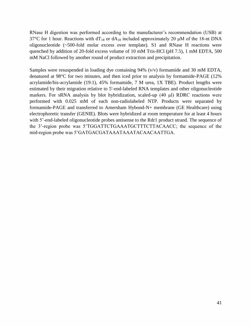
41
RNase H digestion was performed according to the manufacturer’s recommendation (USB) at
37°C for 1 hour. Reactions with dT18 or dA18 included approximately 20 μM of the 18-nt DNA
oligonucleotide (~500-fold molar excess over template). S1 and RNase H reactions were
quenched by addition of 20-fold excess volume of 10 mM Tris-HCl (pH 7.5), 1 mM EDTA, 500
mM NaCl followed by another round of product extraction and precipitation.
Samples were resuspended in loading dye containing 94% (v/v) formamide and 30 mM EDTA,
denatured at 98°C for two minutes, and then iced prior to analysis by formamide-PAGE (12%
acrylamide/bis-acrylamide (19:1), 45% formamide, 7 M urea, 1X TBE). Product lengths were
estimated by their migration relative to 5'-end-labeled RNA templates and other oligonucleotide
markers. For sRNA analysis by blot hybridization, scaled-up (40 μl) RDRC reactions were
performed with 0.025 mM of each non-radiolabeled NTP. Products were separated by
formamide-PAGE and transferred to Amersham Hybond-N+ membrane (GE Healthcare) using
electrophoretic transfer (GENIE). Blots were hybridized at room temperature for at least 4 hours
with 5’-end-labeled oligonucleotide probes antisense to the Rdr1 product strand. The sequence of
the 3’-region probe was 5’TGGATTCTGAAATGCTTTCTTACAACC; the sequence of the
mid-region probe was 5’GATGACGATAAATAAATACAACAATTGA.
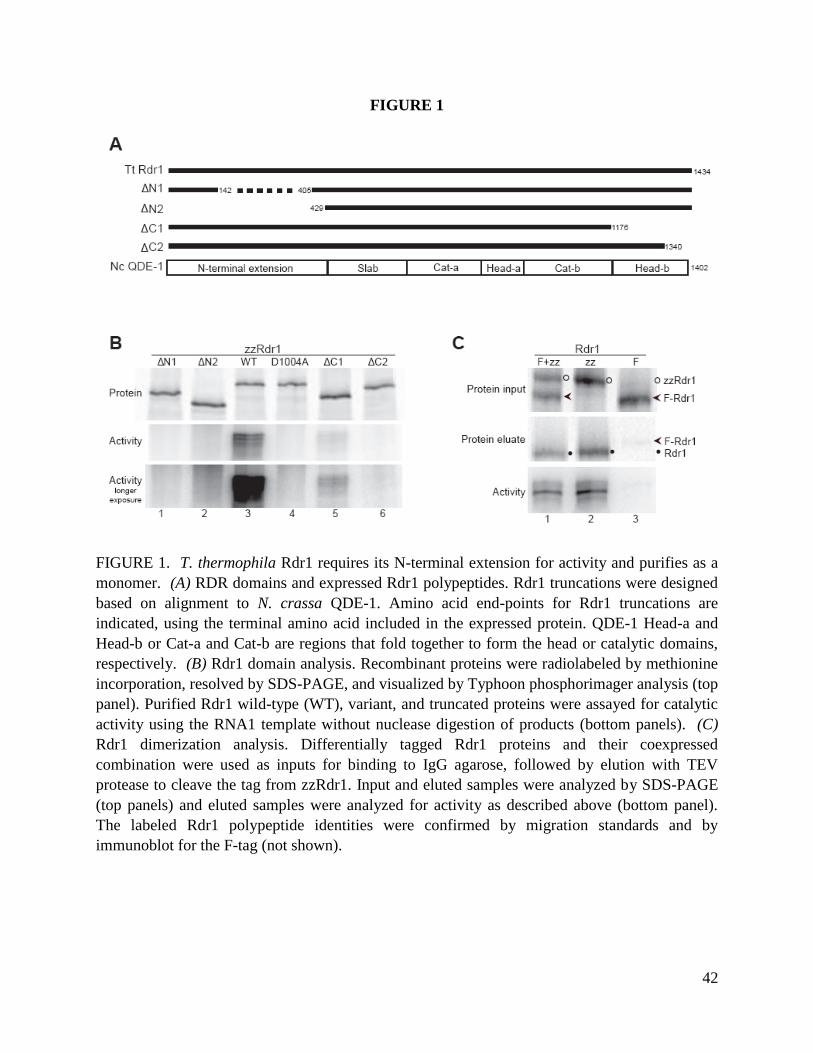
42
FIGURE 1
FIGURE 1. T. thermophila Rdr1 requires its N-terminal extension for activity and purifies as a
monomer. (A) RDR domains and expressed Rdr1 polypeptides. Rdr1 truncations were designed
based on alignment to N. crassa QDE-1. Amino acid end-points for Rdr1 truncations are
indicated, using the terminal amino acid included in the expressed protein. QDE-1 Head-a and
Head-b or Cat-a and Cat-b are regions that fold together to form the head or catalytic domains,
respectively. (B) Rdr1 domain analysis. Recombinant proteins were radiolabeled by methionine
incorporation, resolved by SDS-PAGE, and visualized by Typhoon phosphorimager analysis (top
panel). Purified Rdr1 wild-type (WT), variant, and truncated proteins were assayed for catalytic
activity using the RNA1 template without nuclease digestion of products (bottom panels). (C)
Rdr1 dimerization analysis. Differentially tagged Rdr1 proteins and their coexpressed
combination were used as inputs for binding to IgG agarose, followed by elution with TEV
protease to cleave the tag from zzRdr1. Input and eluted samples were analyzed by SDS-PAGE
(top panels) and eluted samples were analyzed for activity as described above (bottom panel).
The labeled Rdr1 polypeptide identities were confirmed by migration standards and by
immunoblot for the F-tag (not shown).

43
FIGURE 2
FIGURE 2. Rdr1 alone and in RDRC context have similar template specificities. (A) Template
sequences. Sequence substitutions made in the 3’ region of variants of RNA1 or RNA1a are
indicated with lower case letters and arrows. The region of the Aless template composed of DNA
is underlined. (B,C) Template dependence of dsRNA synthesis. Recombinant Rdr1 purified
following zzRdr1expression in RRL (Rdr1) and the mixed RDRC population purified from T.
thermophila expressing zzRdr1 (RDRC) were assayed for activity using the templates indicated.
Unless otherwise indicated (B, lanes 1-2), reaction products were digested with S1 nuclease to
remove the single-stranded region(s) of dsRNA products and the single-stranded products of
RDRC poly(U) polymerase activity prior to formamide-PAGE. End-labeled template RNAs and
smaller synthetic oligonucleotides were run in lanes not shown as markers, with the approximate

44
size of some of products estimated by relative migration. At right in B, a diagram indicates the
relative lengths of RNA1 template (solid line) and its 3' A-tract extension in RNA1a (black box
labeled +25nt) along with three representative product strands (dashed lines) that would remain
base-paired with portions of the template to be protected from digestion by S1 nuclease (dsRNA
products). Adding an initiation site within the A-tract allows the synthesis of an additional
product (lanes 7-10) of the expected length for complete template copying to its 5’-end. (D)
Influence of template 3’ end structure. Nuclease-treated reaction products of RNA1 with 3’-end
hydroxyl groups (RNA1) or a 3’ phosphate group (RNA1-P). Lanes are cropped from the same
exposure of the same gel.

45
FIGURE 3
FIGURE 3. Rdr1 can copy a DNA template if the 3’ end is RNA. (A) Rdr1 and RDRC assay
products from reactions containing 32-nt templates of entirely DNA (lanes 1 and 7), 5' DNA
(with the number of DNA nt in parenthesis) with 1, 2, 3 or 6 nt of RNA at the 3' end, or entirely
RNA (lanes 6 and 12) were treated with S1 nuclease before formamide-PAGE. (B) Rdr1 and
RDRC assay products from reactions with RNA32 and template (26)+6 (26 nt of DNA followed
by 6 nt of RNA at the 3' end) were treated with RNase H (H) or S1 nuclease (S1) or no nuclease
before formamide-PAGE.

46
FIGURE 4
FIGURE 4. Rdr1 links the template to product by looping the template 3’ end. (A) Rdr1 and
RDRC assay products from reactions containing RNA1 or RNA1a were reduced in length by S1
nuclease treatment. RNA1a assay products were also reduced in length by product treatment with
RNase H in the presence of dT18 but not dA18 oligonucleotide. Arrowheads mark the largest
product strands from the duplex region protected by S1 nuclease treatment, while filled circles
indicate slightly longer product strands likely to contain some single-stranded region not fully

47
removed by RNase H. (B) Model for Rdr1 and RDRC initiation by template looping. The 3’ end
of RNA1 and RNA1a templates (solid lines) is extended by product synthesis (dashed line).
Initiation does not occur within the 25 nt 3' A-tract of RNA1a, so this region of the template is
contained within the internal loop of the product and is therefore accessible for degradation by
either S1 nuclease or by RNase H in the presence of dT18.

48
FIGURE 5, part 1
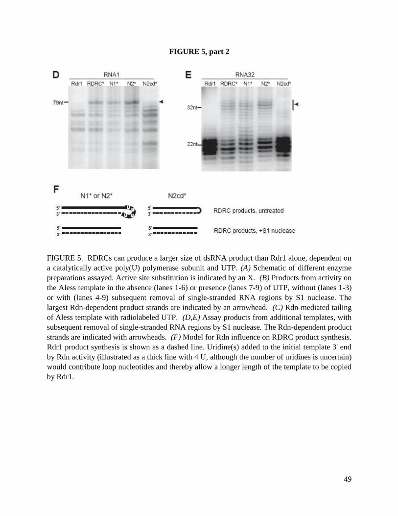
49
FIGURE 5, part 2
FIGURE 5. RDRCs can produce a larger size of dsRNA product than Rdr1 alone, dependent on
a catalytically active poly(U) polymerase subunit and UTP. (A) Schematic of different enzyme
preparations assayed. Active site substitution is indicated by an X. (B) Products from activity on
the Aless template in the absence (lanes 1-6) or presence (lanes 7-9) of UTP, without (lanes 1-3)
or with (lanes 4-9) subsequent removal of single-stranded RNA regions by S1 nuclease. The
largest Rdn-dependent product strands are indicated by an arrowhead. (C) Rdn-mediated tailing
of Aless template with radiolabeled UTP. (D,E) Assay products from additional templates, with
subsequent removal of single-stranded RNA regions by S1 nuclease. The Rdn-dependent product
strands are indicated with arrowheads. (F) Model for Rdn influence on RDRC product synthesis.
Rdr1 product synthesis is shown as a dashed line. Uridine(s) added to the initial template 3' end
by Rdn activity (illustrated as a thick line with 4 U, although the number of uridines is uncertain)
would contribute loop nucleotides and thereby allow a longer length of the template to be copied
by Rdr1.

50
FIGURE 6
FIGURE 6. Rdr1-associated poly(U) polymerase activity extends the length of template
represented in the Dicer cleavage products. (A) Assays of various RDRC preparations indicated
were performed using RNA1 template. Products were not treated with S1 nuclease prior to
formamide-PAGE. In addition to RDRC products, Dcr2 present in RDRC* purifications
generates 23-24 nt sRNAs. (B) Schematic of proposed Rdn influence on RDRC product
synthesis and subsequent cleavage by Dcr2. Rdr1 product synthesis is shown as a dashed line.
Oligonucleotide probes (Mid probe and 3' probe) were designed complementary to the indicated
regions of the product strand. (C) Assays were performed as in A except for the use of unlabeled
NTPs. Products of specific sequence were subsequently detected by blot hybridization with Mid
probe or 3' probe (top panels). The total amount of Dcr2 product was gauged by the parallel
reaction containing radiolabeled CTP (the boxed region of the gel in A; shown again as the
bottom panel in C).

51
CHAPTER THREE
Endogenous small RNA accumulation requires an RNA-dependent RNA polymerase and
the RNA silencing protein Rsp1
ABSTRACT
RNA silencing machinery directs transcript destruction and small RNA (sRNA) biogenesis in a
sequence-specific yet adaptable process, involving many biochemical steps that are conserved
among eukaryotes. Tetrahymena thermophila RNA-dependent RNA polymerase (Rdr1) and a
physically-associated Dicer nuclease (Dcr2) coordinate 23-24 nt sRNA biogenesis. As
demonstrated previously, sRNA sequence complexity requires the specialization of Rdr1 through
its interaction with two nucleotidyl transferases, Rdn1 and Rdn2, and two novel factors Rdf1 and
Rdf2. Rdn1 also interacts with a previously uncharacterized protein of 124 kDa (p124) in a
separate complex that lacks Rdr1 (Lee, Talsky et al. 2009). To further explore the protein
composition of p124 complexes, p124 and putative partners were epitope tagged and
immunopurified. Rdn1 and p124 assembled into two discrete complexes with either Rdf1 or
Rdf2. After knockout of the p124 gene locus, bulk 23-24 nt sRNA, as well as individual
sequences representing four sRNA families known to be Rdr1-dependent, failed to accumulate in
the knockout strain. Based on its newly defined role in sRNA generation, p124 is now referred
to as RNA silencing protein 1 (Rsp1). Considering Rsp1 knockout cells were viable for growth,
even though they lacked multiple sRNA classes, whereas Rdr1, Rdn1 and Dcr2 are essential for
growth, it is likely that Rdr1, Dcr2 and Rdn1 serve important functions aside from sRNA
production. The essential function(s) performed by RNA silencing machinery could include the
direct destruction of aberrant, potentially toxic RNAs. These findings provide new insights as to
the requirements and the purpose for Rdr1-dependent sRNA production.
INTRODUCTION
Small RNA (sRNA) biogenesis and function depends on an ever-expanding array of biochemical
steps, many of which require genetically-essential and highly-conserved protein machinery.
Initial targeting of an RNA into the silencing pathway often requires an RNA-dependent RNA
polymerase (RdRP) and Dicer (Dcr2) endonuclease, which catalyze double-stranded RNA
(dsRNA) synthesis and digestion into sRNA, respectively. The primary sRNA products of these
reactions function in downstream processes to provide sequence specificity to effector proteins
for gene regulation in trans, directing heterochromatin formation, secondary transcript
destruction and translational inhibition (Cerutti and Casas-Mollano 2006; Ghildiyal and Zamore
2009; Siomi and Siomi 2009; Ketting 2011).

52
In Tetrahymena thermophila, sRNA biogenesis is a step-wise process. T. thermophila’s only
genome-encoded RdRP (Rdr1) copies targeted RNA into dsRNA. The dsRNA is then cleaved
by RNaseIII-domain containing Dicer (Dcr2) into 23-24 nt sRNA (Lee and Collins 2007). Of
the several sRNA species that accumulate in T. thermophila, including starvation-induced tRNA
halves (Lee and Collins 2005) and conjugation-induced scan RNA (scnRNA) (Mochizuki and
Gorovsky 2004), 23-24 nt sRNA is the only known class of small RNAs that exists constitutively
(Lee and Collins 2006). Taken together with the fact that Rdr1 and Dcr2 are genetically
essential (Lee and Collins 2006; Lee and Collins 2007), Rdr1-directed RNA silencing likely
performs important functions throughout the T. thermophila lifecycle.
While the Rdr1 protein alone catalyzes dsRNA synthesis in vitro, many additional proteins are
required for 23-24 nt sRNA accumulation in vivo. In the cell, Rdr1 assembles several stable
complexes (RDRCs) through its association with two mutually exclusive uridyltransferases,
Rdn1 and Rdn2. Only Rdn1-containing RDRCs also contain factors Rdf1 and Rdf2.
Macronuclear knockout of each gene that encodes a nonessential RDRC component (Rdn2, Rdf1
and Rdf2) induces a distinct change in the profile of accumulated sRNA sequences (Lee, Chang
et al. 2009). Thus, RDRC components appear to specialize Rdr1 function in vivo, enabling Rdr1
to act on a diverse set of RNA targets overall.
Rdn1 is a catalytically active RDRC subunit that adds mono- and poly-uridine to a single-
stranded RNA template for Rdr1. At least in vitro, Rdn1 activity influences the size and
sequence content of Rdr1 and Dcr2 products (Talsky and Collins 2010). Like Rdr1 and Dcr2,
Rdn1 is essential for vegetative growth. Because Rdn1, but not Rdn2, is essential for growth,
and because Rdn1, but not Rdn2, also interacts with an uncharacterized protein of predicted
weight of 124 kDa (Rsp1), an Rsp1-Rdn1 complex may play a role in cellular processes other
than RDRC activity. Here, I further characterize Rsp1-interacting partners and Rsp1 function
using affinity purification, uridyltransferase activity assays, and gene knockout analysis. Rsp1
assembles stable complexes with Rdn1 and either Rdf1 or Rdf2 that do not include Rdr1 or Dcr2.
Curiously, Rdn1 activity was reduced when associated with Rsp1, as compared to when Rdn1 is
associated with Rdr1. In Rsp1 knockout (Rsp1ko) cells, accumulation of sRNAs that are
dependent on RDRC action was undetectable. However, in contrast to Rdr1 and despite the
severe sRNA accumulation phenotype, Rsp1 was not genetically essential for vegetative growth.
These results suggest that Rsp1 functions at a biochemical step distinct from Rdr1, yet in the
same pathways of sRNA biogenesis. Considering Rsp1ko cells were viable, bulk sRNA may be
dispensable for growth. These findings suggest that the important functions of essential RNA
silencing factors, such as Rdr1 and Dcr2, may extend well beyond their roles in sRNA
biogenesis for the sake of sRNA-effector complex function.

53
RESULTS
Rsp1 interacts with RDRC components but not with Rdr1.
We previously reported that epitope-tagged Rdn1 co-purifies a protein of 124 kDa (Rsp1), in
addition to the RDRC components Rdr1, Rdf1 and Rdf2 (Lee, Talsky et al. 2009). However,
neither tagged Rdr1 nor Rdn2 co-purify Rsp1 (Lee, Talsky et al. 2009) (Fig 1A, 1B). To confirm
that Rsp1 and Rdn1 interact in a complex separate from the RDRCs and to determine whether
Rsp1 associates with additional proteins, T. thermophila cell lines were generated to express
endogenous epitope-tagged Rsp1 with a carboxy-terminal triple-flag peptide followed by a
Tobacco Etch Virus (TEV) protease cleavage site and two tandem protein A domains (Rsp1-fzz).
Locus assortment went to completion (Fig S1A). Rsp1-fzz was immunoprecipitated on Protein
A agarose beads, stringently washed and then eluted by zz tag cleavage using TEV protease.
Consistent with previous findings, Rsp1-fzz co-purified endogenous Rdn1 (Fig 1B, lane 3).
Rsp1-fzz also co-purified Rdf1 and Rdf2. No additional stoichiometric protein partners were
detected. Thus, at least some Rsp1-Rdn1 complexes contain Rdf1 or Rdf2. Amino-terminally
tagged zz-Rdr1 (Lee and Collins 2007), zzf-Rdn1 and zzf-Rdn2 (Lee, Talsky et al. 2009) were
purified under identical conditions for comparison. Note that zz-Rdr1, zzf-Rdn1 and zzf-Rdn2
purifications contained a non-specific ~45 kDa protein also present in mock purifications from
wildtype cells (Fig 1A, lane 1). Rsp1-fzz did not co-purify endogenous Rdr1, which is consistent
with the absence of endogenous Rsp1 from zz-Rdr1 purifications. Rsp1-fzz co-purified Rdn1
from cells lacking the genomic region that contains both Rdf1 and Rdf2 (Rdf1,Rdf2ko) (Fig 1B,
lane 4), which suggests the Rsp1-Rdn1 association is direct.
To further assess the composition of Rdn1-containing complexes, Rdn1, Rdn2, Rdf1 and Rdf2
were also tagged at their endogenous loci. Integration constructs contained a carboxy-terminal
epitope tag with three copies of Flag, a TEV protease cleavage site and GFP (fg). Transgene-
containing chromosomes completely assorted for all but Rdn1-fg (Fig S1B-E). Rdf1-fg, Rdf2-fg
and Rdn2-fg were immunoprecipitated on Flag resin, stringently washed and eluted from beads
with Flag peptide. Both Rdf1-fg and Rdf2-fg co-purified Rdr1, Rdn1 and Rsp1 (Fig 1C, lanes
3,4), an expected result considering data from previous Rdn1 and Rdn2 purifications. Rdn2-fg,
like zzf-Rdn2, only co-purified Rdr1 (lane 2). Notably, endogenous Rdf1 and Rdf2 did not co-
purify with Rdf1-fg or Rdf2-fg, although they were present in zf-Rdn1 purifications (lane 5).
Therefore, Rdf1 and Rdf2 are mutually exclusive members that define two of the three RDRCs
and both Rsp1-containing complexes (RSPCs) (Fig 1 Key).
In T. thermophila, RDRCs retain co-purified Dcr2 only after gentle (low-salt) wash conditions
(Lee et al 2009), with the exception of Rdn1, which retains some Dcr2 even after stringent (high-
salt) wash conditions (Fig 1B lane 1). Rsp1-fzz purification with stringent washes did not co-
purify Dcr2 (Fig 1B lane 3, 4). To determine if an RSPC associates with Dcr2 under the same
conditions used to isolate RDRC-Dcr2 complexes, Rsp1-fzz was purified under gentle wash

54
conditions (Rsp1-fzz’). Rsp1-fzz’ did not co-purify Dcr2 (Fig 1B lane 5), which suggests RSPCs
do not associate stably with Dcr2.
Rdn1 activity depends on protein partner context
In the RDRC context, Rdn1 and Rdn2 have robust uridyltransferase activity, as measured by in
vitro activity assays on RDRCs purified from cells extracts (Lee, Talsky et al. 2009). In the
context of RSPCs, Rdn1 may display unique catalytic abilities. To compare the uridyltransferase
activity present in Rdn1-containing RDRCs and RSPCs, purified complexes were incubated with
79 nt, purified RNA (RNA1) (Talsky and Collins 2010) and 32
P UTP and analyzed by
formamide-urea acrylamide gel electrophoresis (formamide-PAGE), which reduces the impact of
RNA secondary structure on product migration. Previous activity assay analysis using this
method reported a mixture of non-processive end-labeled RNA products and processive poly(U)-
tailed products. RSPC-bound Rdn1 retained activity (Fig 2A). To compare Rdn1 activity in the
context of distinct RSPCs and RDRCs, purifications of Rsp1-fzz, zz-Rdr1(Rdn2ko), zzf-Rdn1,
Rdf1-fg and Rdf2-fg were normalized for Rdn1 content by silver staining after SDS-PAGE (Fig
2B, bottom panel) and assayed for RNA1-labeing activity with 32
P UTP (top panel). Rdn1
associated with Rsp1-fzz (Fig 2B, lane 1) displayed a weaker labeling activity than protein
complex mixtures containing at least one RDRC (lanes 2-5). The most active preparation was
from zz-Rdr1(Rdn2ko), which contains only Rdn1-bound RDRCs (Fig 2B, lane 2). To compare
the distribution of short-to-long products synthesized by RSPCs and RDRCs, immunopurified
zz-Rdr1(Rdn2ko) was serially diluted until its activity matched that of Rsp1-fzz. Rsp1-fzz and
diluted zz-Rdr1 complexes synthesized a comparable short:long product ratios, although product
synthesis was approximately 600 fold weaker in the Rsp1 context (Fig 2C). This difference
cannot be attributed to the polymerase activity of Rdr1, alone, because RDRCs purified by zz-
Rdr1 or by active site-mutant zz-Rdr1(D1004A) have the same uridyltransferase activity (Lee,
Talsky et al. 2009).
One possible function for Rsp1 in vivo is to regulate RNA silencing through the inhibition of
Rdn1 activity. To see if inhibition can occur in trans, RSPC and RDRC purifications were
mixed at a 1:1 ratio. The mixed complexes synthesized products comparable to RDRCs alone
(Fig 2C lanes 5, 6). Therefore, Rsp1 cannot inhibit the activity of Rdn1 already assembled in
separate complexes. This finding is consistent with the observation that purifications of zzf-
Rdn1, Rdf1-fg and Rdf2-fg, which contain both Rdr1 and Rsp1, all have robust uridyltransferase
activity (Fig 2B).
In previous analyses, Rdn1 activity was assayed using RDRCs purified via Rdr1 in an Rdf1 or
Rdf2 knockout background (Lee, Talsky et al. 2009). Rdf1 and Rdf2 did not contribute to Rdn1
activity in this context. Whether Rdf1 or Rdf2 may have an impact on Rdn1 activity in the Rsp1
context remains in question. To test this, Rsp1-fzz purifications from wildtype or Rdf1,Rdf2ko
cells were assayed for Rdn1 activity on RNA1. Rdn1 in the Rsp1-fzz (Rdf1,Rdf2ko) complex
was active, although possibly to a lesser extent than wildtype RSPCs (Fig 2D, lanes 3,4). Next,

55
RSPC activity was measured on two RNA templates of different size and sequence (RNA1 and
RNA2) (Fig 2E). When given either template, RSPCs that lack Rdf1 and Rdf2 synthesized less
of the high molecular weight product (Fig 2E, lanes 4, 5, 9, 10) compared to wildtype RSPCs
(lanes 2, 3, 7, 8) that were roughly normalized for Rdn1 content. Mono-uridylated product was
synthesized at more comparable amounts from wildtype and mutant RSPCs. These results are
consistent among assays using independent protein preparations (Fig 2D and 2E) and distinct
RNA templates (RNA1 and RNA2). Thus, the presence of Rdf1 or Rdf2 supports processive
product synthesis by Rdn1 in the RSPC context.
Cells that lack Rsp1 are viable but inefficiently complete conjugation.
To determine the genetic requirement for Rsp1 for culture growth, the Rsp1 locus was replaced
with the Bsr2 gene resistance cassette in the transcriptionally active T. thermophila
macronucleus. The replacement went to completion in both CU522 and SB210 strains (Fig 3A),
supporting a non-essential function for Rsp1 during vegetative growth. The Rsp1 knockout
(Rsp1ko) was performed in parallel to that of Rdn1 in CU522, which did not reach full
assortment (Fig 3B), consistent with its previously-identified essential requirement for growth.
Absence of Rsp1 mRNA was confirmed by RT-PCR from total RNA isolated from clonal cell
lines represented by CU522 clones 4 and 9 and SB210 clone 4 (data not shown). These strains
were used for subsequent analyses.
Rsp1 was not required for viability during vegetative growth. However, if Rsp1, or any Rsp1-
dependent sRNA, functions in maintaining chromosomal integrity, phenotypes from Rsp1ko
cells may only become apparent during the complex nuclear restructuring that occurs at
conjugation. Although Rsp1 is more highly expressed during vegetative growth than at other life
cycle stages (Miao, Xiong et al. 2009), it could also play a direct role during conjugation. To
address whether Rsp1ko cells exhibit defects during conjugation, SB210(Rsp1ko) clonal strain 4
and CU522(Rsp1ko) clonal strains 4 or 9 (Fig 3) were starved and mixed in equal volumes to
initiate conjugation. After nine hours, cells were fixed, DAPI-stained and assessed for their
ability to progress through conjugation. At this stage, wildtype cells developed the stereotyped
nuclear array that defines a conjugating pair at anlagen (Fig S2). However, the number of pairs
that reached anlagen was greatly reduced in Rsp1ko (Table 1). This conjugation phenotype was
more severe for CU522(Rsp1ko) 4 than 9 (1.2% and 23.3%, respectively, reached anlagen),
compared to wildtype cells (93.9% reached anlagen). Development of pre-anlagen Rsp1ko pairs
was variable. On average, CU522(Rsp1ko) 4 pairs resembled the wildtype 2-4 hour stage while
CU522(Rsp1ko) 9 pairs typically developed to the equivalent of wildtype 4-9 hour stage).
Consistent with this, after 18 hours of conjugation, when nuclear differentiation and pair
detachment was mostly complete in wildtype cells (>80%), Rsp1ko cells were greatly impaired
in their ability to produce progeny (separated, individual cells with two macronuclei). This
phenotype was, once again, more severe with clonal line 4 (<<1% cells contained two
macronuclei) compared to line 9 (~15%).

56
The conjugation phenotype exhibited by Rsp1ko cells was not a result from a general inability of
Rsp1ko cells to initiate conjugation; Rsp1ko pairs reached early (4 hours) stages of conjugation
at the same rate as wildtype pairs (data not shown). Therefore, in the absence of Rsp1, the
majority of paired cells cannot develop beyond early- or mid-conjugation. These findings
suggest Rsp1 function is important, either directly or indirectly, for the completion of
conjugation.
Accumulation of 23-24nt sRNA requires both Rsp1 and active Rdr1.
Deep sequencing of 23-24nt sRNA identified sequences that map to pseudogenes, structured
RNA, repetitive DNA sequences and mRNA-encoding loci (Couvillion, Lee et al. 2009). The
accumulation levels of distinct sRNA sub-populations change, depending on the presence of
Rdn2, Rdf1 or Rdf2, which are each unique to a single RDRC. These sRNA accumulation
profiles can be used to infer putative in vivo targets for each RDRC. Moreover, it is likely that
RDRC components provide specificity to Rdr1 such that each of the three RDRCs acts on
distinct RNA targets.
To address whether Rsp1 is required for sRNA accumulation, size-selected RNA isolated from
growing cells was analyzed by PAGE and stained with SYBR Gold (Fig 4A, Fig 4B bottom
panel). Individual sRNA sequences representing abundant sRNA sub-populations were then
detected by northern blot hybridization using oligo probes (Fig 4B). The same membrane was
probed for all sRNAs except for STR1, for which a separate blot containing the same RNA
preparation was probed. Each sRNA represents pseudogene loci (IIIB and IB), “phased” sRNA
clusters spaced at ~24 nt intervals (PH3), high-copy repeat loci (RPT1) and structured transcripts
(STR1). The sRNAs preferentially binds either Twi2 or Twi8, as noted (Fig 4B) (Couvillion,
Lee et al. 2009).
Consistent with previous findings (Couvillion, Lee et al. 2009; Lee, Talsky et al. 2009), Rdf1ko,
Rdf2ko and Rdn2ko cells each accumulated distinct sRNA profiles (Fig 4B). Moreover, cells
lacking the entire genomic locus containing Rdf1 and Rdf2 (Rdf1,Rdf2ko) and cells additionally
lacking the locus for Rdn2 (Rdf1,Rdf2,Rdn2ko) accumulated sRNAs consistent with each single
knockout. For example, IIIB and IB sRNAs did not accumulate in Rdn2ko or
Rdf1,Rdf2,Rdn2ko cells (Fig 4B, lanes 7, 8). PH3 was undetectable in all Rdf2ko strains (panel
3, lanes 5, 6, 8). RPT1 did not accumulate in Rdn2ko or Rdf1,Rdf2ko cells (panel 4, lanes). For
sRNAs whose accumulation increased (RPT1 in Rdf1ko, STR1 in Rdn2ko), it is unclear whether
this was a direct or indirect effect of the gene knockout.
Rdr1 is required for the biogenesis of 23-24nt sRNA, as bulk sRNA accumulation becomes
undetectable after overexpression of active site-mutant (D1004A) Rdr1 (Lee, Talsky et al. 2009).
Remarkably, accumulation of bulk and individual sRNAs was nearly identical between Rsp1ko
cells and cells overexpressing Rdr1(D1004A) (Fig 4A, lanes 1, 3; Fig 4B, lanes 1, 3). Even
STR1, which did not require any other RDRC component for accumulation, developed the same
57
mutant accumulation profile, marked by a wider size distribution, in both Rsp1ko and
Rdr1(D1004A) cells. These results support a related role for Rdr1 and Rsp1 in sRNA
biogenesis.
Rdf2ko, Rsp1ko and Rdr1(D1004A) cells lack detectable levels of the PH3 sRNA (Fig 4B).
Rdf2ko, but not wildtype, cells accumulate an ~800 nt RNA that contains sequence
corresponding to the sRNA cluster that contains the PH3 sRNA (phased-cluster Ph3)
(Couvillion, Lee et al. 2009). The simplest explanation is that the ~800 nt RNA is an sRNA
precursor and putative RDRC target. To better understand the structure of this putative RDRC
target, total RNA was reverse transcribed using sequence-specific oligonucleotides. The
resulting cDNA was amplified and sequenced, with results summarized (Fig 5A). The precursor
contained two regions that correspond to sRNA clusters (cluster 1 and 2), separated by an intron
(Fig 5A, top left). Cluster 1 overlapped with a predicted gene (TTHERM_00286880). Cluster 2
contained the PH3 sRNA sequence. The RNA was spliced, which juxtaposed sRNA clusters 1
and 2. The end of cluster 2 contained an in-frame stop codon followed by 20nt of sequence, a
163 nt stem-loop and at least 300 nt of 3’ sequence beyond the structured region. Ph3 transcript
was detected after reverse transcription with dT18 followed by PCR, which suggests the precursor
was polyadenylated (Fig 5A, bottom left). The Ph3 precursor may be further processed before it
can serve as a target for Rdr1 (Fig 5A, right).
Considering the production of PH3 sRNA requires Rdf2, and Rdf2 associates with Rdr1 and
Rsp1 in separate complexes, then processing of the Ph3 precursor could be performed either in
the RDRC or RSPC context. Thus, the Ph3 precursor may also accumulate in the absence of
Rsp1 or Rdr1 activity. To test this, total RNA was isolated from growing cells and analyzed by
mRNA northern blot hybridization with a probe (Fig 5A, top, grey line) to detect the cluster 2
region of the Ph3 precursor. As expected, the Ph3 precursor accumulated in Rdf2ko cells from
SB210 and CU522 strain backgrounds (Fig 5B, lanes 3, 6), but not in wildtype or Rdf1ko cells
(lane 1). Interestingly, the Ph3 precursor did not accumulate in Rsp1ko or Rdr1 (D1004A) cells
(Fig 5B, lanes 4, 7, 8). Therefore, although Rsp1 and Rdr1 both interact with Rdf2, they must
both act at separate steps from Rdf2. It is also possible that, in the absence of Rsp1 or Rdr1
function, the Ph3 precursor (which would not be processed into sRNA) may be shunted to a
separate degradation pathway. Models to account for the role for Rsp1 in sRNA production
include its putative functions either upstream (Fig 6A, B) or downstream (Fig 6C) of RDRC and
Dcr2 activities.
DISCUSSION
Rsp1 function in RNA silencing
Here I show that Rsp1 assembles two complexes with Rdn1 and either Rdf1 or Rdf2 that mediate
sRNA-mediated RNA silencing. Although Rsp1-bound Rdn1 synthesized only a fraction of

58
uridylated RNA product compared to Rdr1-bound Rdn1, all complexes generated the same
product profile of mono-uridylated and poly-uridylated RNA. However, when Rdf1 and Rdf2
were absent from RSPC purifications, the fraction of poly-uridylated product was reduced.
Thus, Rdf1 and Rdf2 activate Rdn1 in the RSPC context.
Rsp1 was required for the accumulation of bulk 23-24 nt sRNA in vivo. In fact, Rsp1ko and
Rdr1(D1004A) cells accumulated identical profiles of individual sRNA sequences from distinct
families of 23-24 nt sRNA. Thus, Rsp1, like Rdr1, serves a general role in RNA silencing.
Rsp1 may provide a range of functions in sRNA production or processing. RSPCs may bind
and/or process RNA targets, possibly in an Rdn1 activity-dependent manner (Fig 6, option A).
This could provide specificity to RNA target selection and even activate RNA for RDRC
processing. A second possibility is that Rsp1 mediates the assembly of Rdn1 with Rdf1 or Rdf2.
It may also promote RDRC formation (Fig 6, option B). Finally, it is also possible that Rsp1
regulates Rdn1 activity or specializes it for a RSPC-unique function in vivo.
Alternatively, Rsp1 could function downstream of RDRCs. It may process RDRC products,
modify sRNA or mediate sRNA loading onto Twi effectors (Fig 6, option C). In this context,
Rsp1 may be required for sRNA stability rather than biogenesis. Rsp1 does not seem to play a
direct role in Rdr1 or Dcr2-mediated steps because it did not influence their activity in vitro and
did not interact with them in vivo. Also, purified RSPC did not display any measurable RNA
synthesis or nuclease activity.
It is possible that Rsp1 plays an analogous role to known silencing-related protein machinery in
other organisms, although it does not contain significant domain or sequence similarities to
known proteins. Silencing factors in addition to RdRs, Dicers and poly(U) polymerases include
RNA binding proteins, helicases, methyltransferases and phosphatases. Of the latter two, Hen1-
directed sRNA 2’-O-methylation protects sRNAs from uridylation followed by degradation in A.
thaliana and D. melanogaster (Ramachandran and Chen 2008; Ameres, Horwich et al. 2010). T.
thermophila Hen1 is required for 2’-O-methylation of scnRNAs to provide stabilization and
allows them to accumulate throughout conjugation (Kurth and Mochizuki 2009). However, only
bulk Twi8-bound sRNA is known to be methylated. Twi2, which binds most abundant sRNA
families in T. thermophila, assembles 3’-unmodified sRNA (Couvillion, Lee et al. 2009). It is
possible that Twi2-bound sRNA is protected through another mechanism that may involve RSPC
function. Rsp1 could alternatively mediate sRNA modifications such as removal of 5’-
triphosphates that remain after de novo RNA synthesis by Rdr1 (Deshpande, Takagi et al. 1999;
Duchaine, Wohlschlegel et al. 2006).
Essential Roles of sRNA silencing machinery
Although Rsp1 is required for bulk sRNA accumulation, it is not growth-essential (Fig 3).
Consistent with this, Twi2 binds bulk sRNAs during growth but is also not essential (Couvillion,

59
Lee et al. 2009). Thus, the essential functions of Rdr1, Dcr2 and Rdn1 may not be in sRNA
production but instead in their catalytic functions related to RNA degradation. In the absence of
Rdr1, Dcr2 or Rdn1, aberrant transcripts and/or processing intermediates may accumulate in a
manner similar to Ph3 precursor accumulation in Rdf2ko cells. In some cases, these
intermediates may be toxic.
There is the potential for RNA toxicity at several steps of sRNA biogenesis, during which Rdn1,
Rdr1 and Dcr2 act sequentially. RNA templates are first elongated at their 3’ end through Rdn1-
or Rdn2-mediated uridylation, and then subsequently extended by Rdr1 to generate dsRNA
products, each with an oligo(U)-loop between the original template and the newly synthesized
strand (Talsky and Collins 2010). Then, Dcr2 digests the RDRC products into 23-24 nt duplexed
sRNA (Lee and Collins 2007). Growth-essential Rdn1 assembles with Rdr1 and Rdf2 to form
the predominantly expressed RDRC during growth. Considering Rdn1 defines the first known
RNA processing step during silencing, cells without Rdn1 may be unable to target RNA for
silencing and therefore would accumulate aberrant RNA molecules that clog general RNA
processing and turnover machinery and impede on normal cellular processes. In the absence of
Rdr1, Rdn1 may be free to mislabel RNA that is normally not destined for silencing.
Accumulating 3’-uridylated RNAs could overwhelm machinery for RNA surveillance or other
pathways of degradation. Finally, in the absence of Dcr2, Rdr1 products in the form of dsRNA
would remain unprocessed. These intermediates could aberrantly activate antiviral and other
dsRNA defense pathways, as reported for hairpin RNA-induced upregulation of Dicer and
Argonaute family genes in T. thermophila (Howard-Till and Yao 2006).
Silencing machinery in conjugation
While Rsp1 is not required for vegetative growth, it is essential for the completion of
conjugation. Cells that lack Rdn2 or Rdf1 also have conjugation defects (Lee et al 2009). It is
possible that these factors and/or their sRNA products may play a direct role during conjugation
processes. Alternatively, they may more generally maintain nuclear integrity. In the latter case,
nuclear defects that arise during growth may inhibit the ability of Rsp1ko cells to restructure new
nuclei during conjugation. In fact, depending on which nuclear defects accumulate in vegetative
growth, different cell lines may demonstrate varying phenotypic severity. This is consistent with
the phenotypic variation between CU522 Rsp1ko strains #4 and #9 during conjugation.
Cellular control of Rdr1 specificity
RDRC components provide RNA target specificity that is linked to downstream sRNA assembly
onto Twi effectors. As a consequence, Rdn2, Rdf1 and Rdf2 knockouts have unique sRNA
accumulation profiles. Moreover, each of the four vegetatively-expressed Twi proteins that bind
23-24 nt sRNA (Twi2, Twi7, Twi8, and Twi9) stably associates with only sub-populations of
sRNAs in vivo (Couvillion, Lee et al. 2009). Thus, each component unique to an RDRC may
specialize its association with only a subset of RNA targets and load its sRNA products

60
specifically onto one or a subset of Twis. Considering Rsp1 shares protein partners with Rdr1
and, like Rdr1, is also specialized into distinct complexes by the alternative presence of Rdf1 and
Rdf2. Thus, Rdf1 and Rdf2 in the RSPC context may mediate RNA targeting to appropriate
RDRCs. Alternatively, RSPCs may transfer RDRC specialization downstream; they could
mediate appropriate sRNA transfer between RDRC-Dcr2 complexes and Twi proteins.
I favor the upstream positioning of Rsp1 in sRNA silencing for several reasons. First of all, if
RSPCs were to function downstream of RDRC-Dcr2 complexes, one would expect to detect an
interaction between at least a subset of Rsp1 molecules and either Dcr2 or any of the Twi family
proteins. Instead, Rsp1 interacts with Rdn1, which, as previously reported, functions upstream
of Rdr1 (Talsky and Collins 2010). Secondly, Rsp1 does not influence RDRC and Dcr2
activities in vitro. It is unlikely that RSPCs are dsRNA or sRNA modifications machines,
considering they do not have an effect on product length or structure. Instead, these findings are
consistent with a role for RSPCs in promoting upstream events such as RNA selectivity or
RDRC assembly in vivo. Finally, Rsp1 was required for the accumulation of STR1 sRNA of the
appropriate length. Considering the sRNA products synthesized by Dcr2 are usually 23-24 nt, it
is possible that, in the absence of Rsp1, processing of the STR1 precursor is performed by an
imperfectly assembled RDRC-Dcr2, or that STR1 precursor was mis-targeted to an alternate
enzyme altogether.
Neither the upstream or downstream model accounts for the finding that Rdn2-dependent IIIB
and IB sRNAs require Rsp1 for accumulation. Curiously, no stable physical interaction was
detected between Rsp1 and Rdn2. It is possible that the Rsp1-Rdn2 relationship may be indirect
or that their coordination was not detectable under the experimental conditions used here.
Nevertheless, these data provide further support for the involvement of Rsp1 in the generation of
all 23-24 nt sRNA.
These studies of Rsp1 partners and function reveal yet another level of pathway complexity in
endogenous sRNA-mediated silencing. Rsp1 and Rdr1 have remarkable similarities, as outlined
above, which suggest the two proteins function in coordination despite a lack of stable physical
interaction. Details as to the biochemical functions of Rsp1, its relationship to Rdr1 and its place
in sRNA biogenesis will expand the current models of RNA silencing specificity.
MATERIALS AND METHODS
Cell lines and culture conditions
Strains for gene knockouts or expression of epitope-tagged proteins were made in CU522 or
SB210 background using integration cassettes targeted to the appropriate endogenous locus.
Knockout cassettes replaced the endogenous open reading frame with the Bsr2 cassette (MTT1
promoter, blasticidin-resistance gene (Bsr2) and 3’UTR from BTU2). Constructs for protein
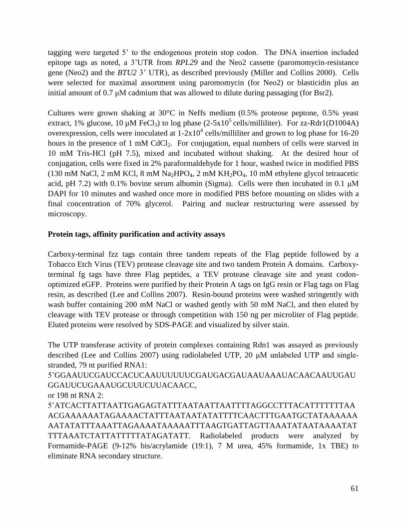
61
tagging were targeted 5’ to the endogenous protein stop codon. The DNA insertion included
epitope tags as noted, a 3’UTR from RPL29 and the Neo2 cassette (paromomycin-resistance
gene (Neo2) and the BTU2 3’ UTR), as described previously (Miller and Collins 2000). Cells
were selected for maximal assortment using paromomycin (for Neo2) or blasticidin plus an
initial amount of 0.7 μM cadmium that was allowed to dilute during passaging (for Bsr2).
Cultures were grown shaking at 30°C in Neffs medium (0.5% proteose peptone, 0.5% yeast
extract, 1% glucose, 10 µM FeCl3) to log phase (2-5x10
5 cells/milliliter). For zz-Rdr1(D1004A)
overexpression, cells were inoculated at 1-2x10⁴ cells/milliliter and grown to log phase for 16-20
hours in the presence of 1 mM CdCl2. For conjugation, equal numbers of cells were starved in
10 mM Tris-HCl (pH 7.5), mixed and incubated without shaking. At the desired hour of
conjugation, cells were fixed in 2% paraformaldehyde for 1 hour, washed twice in modified PBS
(130 mM NaCl, 2 mM KCl, 8 mM Na2HPO4, 2 mM KH2PO4, 10 mM ethylene glycol tetraacetic
acid, pH 7.2) with 0.1% bovine serum albumin (Sigma). Cells were then incubated in 0.1 μM
DAPI for 10 minutes and washed once more in modified PBS before mounting on slides with a
final concentration of 70% glycerol. Pairing and nuclear restructuring were assessed by
microscopy.
Protein tags, affinity purification and activity assays
Carboxy-terminal fzz tags contain three tandem repeats of the Flag peptide followed by a
Tobacco Etch Virus (TEV) protease cleavage site and two tandem Protein A domains. Carboxy-
terminal fg tags have three Flag peptides, a TEV protease cleavage site and yeast codon-
optimized eGFP. Proteins were purified by their Protein A tags on IgG resin or Flag tags on Flag
resin, as described (Lee and Collins 2007). Resin-bound proteins were washed stringently with
wash buffer containing 200 mM NaCl or washed gently with 50 mM NaCl, and then eluted by
cleavage with TEV protease or through competition with 150 ng per microliter of Flag peptide.
Eluted proteins were resolved by SDS-PAGE and visualized by silver stain.
The UTP transferase activity of protein complexes containing Rdn1 was assayed as previously
described (Lee and Collins 2007) using radiolabeled UTP, 20 μM unlabeled UTP and single-
stranded, 79 nt purified RNA1:
5’GGAAUUCGAUCCACUCAAUUUUUUCGAUGACGAUAAUAAAUACAACAAUUGAU
GGAUUCUGAAAUGCUUUCUUACAACC,
or 198 nt RNA 2:
5’ATCACTTATTAATTGAGAGTATTTAATAATTAATTTTAGGCCTTTACATTTTTTTAA
ACGAAAAAATAGAAAACTATTTAATAATATATTTTCAACTTTGAATGCTATAAAAAA
AATATATTTAAATTAGAAAATAAAAATTTAAGTGATTAGTTAAATATAATAAAATAT
TTTAAATCTATTATTTTTATAGATATT. Radiolabeled products were analyzed by
Formamide-PAGE (9-12% bis/acrylamide (19:1), 7 M urea, 45% formamide, 1x TBE) to
eliminate RNA secondary structure.
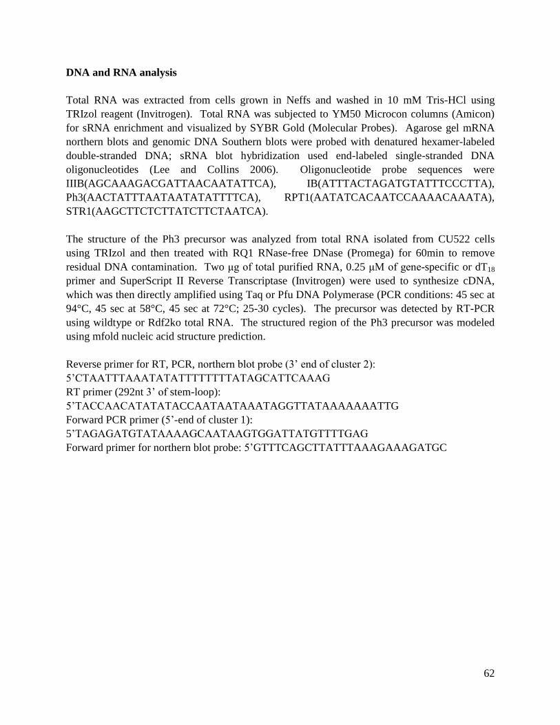
62
DNA and RNA analysis
Total RNA was extracted from cells grown in Neffs and washed in 10 mM Tris-HCl using
TRIzol reagent (Invitrogen). Total RNA was subjected to YM50 Microcon columns (Amicon)
for sRNA enrichment and visualized by SYBR Gold (Molecular Probes). Agarose gel mRNA
northern blots and genomic DNA Southern blots were probed with denatured hexamer-labeled
double-stranded DNA; sRNA blot hybridization used end-labeled single-stranded DNA
oligonucleotides (Lee and Collins 2006). Oligonucleotide probe sequences were
IIIB(AGCAAAGACGATTAACAATATTCA), IB(ATTTACTAGATGTATTTCCCTTA),
Ph3(AACTATTTAATAATATATTTTCA), RPT1(AATATCACAATCCAAAACAAATA),
STR1(AAGCTTCTCTTATCTTCTAATCA).
The structure of the Ph3 precursor was analyzed from total RNA isolated from CU522 cells
using TRIzol and then treated with RQ1 RNase-free DNase (Promega) for 60min to remove
residual DNA contamination. Two μg of total purified RNA, 0.25 μM of gene-specific or dT18
primer and SuperScript II Reverse Transcriptase (Invitrogen) were used to synthesize cDNA,
which was then directly amplified using Taq or Pfu DNA Polymerase (PCR conditions: 45 sec at
94°C, 45 sec at 58°C, 45 sec at 72°C; 25-30 cycles). The precursor was detected by RT-PCR
using wildtype or Rdf2ko total RNA. The structured region of the Ph3 precursor was modeled
using mfold nucleic acid structure prediction.
Reverse primer for RT, PCR, northern blot probe (3’ end of cluster 2):
5’CTAATTTAAATATATTTTTTTTATAGCATTCAAAG
RT primer (292nt 3’ of stem-loop):
5’TACCAACATATATACCAATAATAAATAGGTTATAAAAAAATTG
Forward PCR primer (5’-end of cluster 1):
5’TAGAGATGTATAAAAGCAATAAGTGGATTATGTTTTGAG
Forward primer for northern blot probe: 5’GTTTCAGCTTATTTAAAGAAAGATGC

63
FIGURE 1
FIGURE 1. Rsp1 assembles two complexes with Rdn1 that contain either Rdf1 or Rdf2. Silver-
stained SDS-PAGE gels reflect purified protein complexes from extracts of cells expressing
epitope-tagged proteins or wildtype cells (mock). Protein complexes containing Rsp1 (RSPCs)
and Rdr1 (RDRCs) are diagramed below each lane and summarized in the Figure Key. On gels,
protein complex components are indicated as Rdn1 (open circle), Rdn2 (closed circle) and Rdf1
or Rdf2 (*). Proteins were washed using stringent conditions except for gently-washed Rsp1-
fzz’. (A, B) Proteins were purified by their tandem protein A domains (zz) on agarose IgG resin
and eluted by TEV protease-directed cleavage at site adjacent to the zz tag. For example, zz-
Rdr1 purifications after TEV elution contain untagged Rdr1 and its interacting proteins (which
comprise all three RDRCs, as diagramed below lane). TEV elution of zzf-Rdn1 or Rsp1-fzz
purifications removes the zz tag, leaving f-Rdn1 or Rsp1-f and each of their respective protein
complexes. (C) Proteins were purified by their 3x Flag (f) affinity tags and eluted from beads by
competition with Flag peptide. Note: Flag purifications consistently co-purified more non-
specific proteins.
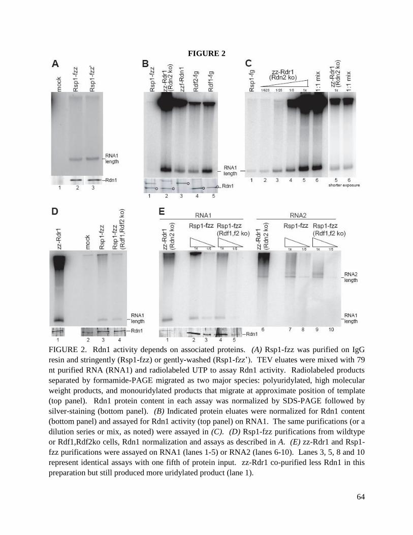
64
FIGURE 2
FIGURE 2. Rdn1 activity depends on associated proteins. (A) Rsp1-fzz was purified on IgG
resin and stringently (Rsp1-fzz) or gently-washed (Rsp1-fzz’). TEV eluates were mixed with 79
nt purified RNA (RNA1) and radiolabeled UTP to assay Rdn1 activity. Radiolabeled products
separated by formamide-PAGE migrated as two major species: polyuridylated, high molecular
weight products, and monouridylated products that migrate at approximate position of template
(top panel). Rdn1 protein content in each assay was normalized by SDS-PAGE followed by
silver-staining (bottom panel). (B) Indicated protein eluates were normalized for Rdn1 content
(bottom panel) and assayed for Rdn1 activity (top panel) on RNA1. The same purifications (or a
dilution series or mix, as noted) were assayed in (C). (D) Rsp1-fzz purifications from wildtype
or Rdf1,Rdf2ko cells, Rdn1 normalization and assays as described in A. (E) zz-Rdr1 and Rsp1-
fzz purifications were assayed on RNA1 (lanes 1-5) or RNA2 (lanes 6-10). Lanes 3, 5, 8 and 10
represent identical assays with one fifth of protein input. zz-Rdr1 co-purified less Rdn1 in this
preparation but still produced more uridylated product (lane 1).
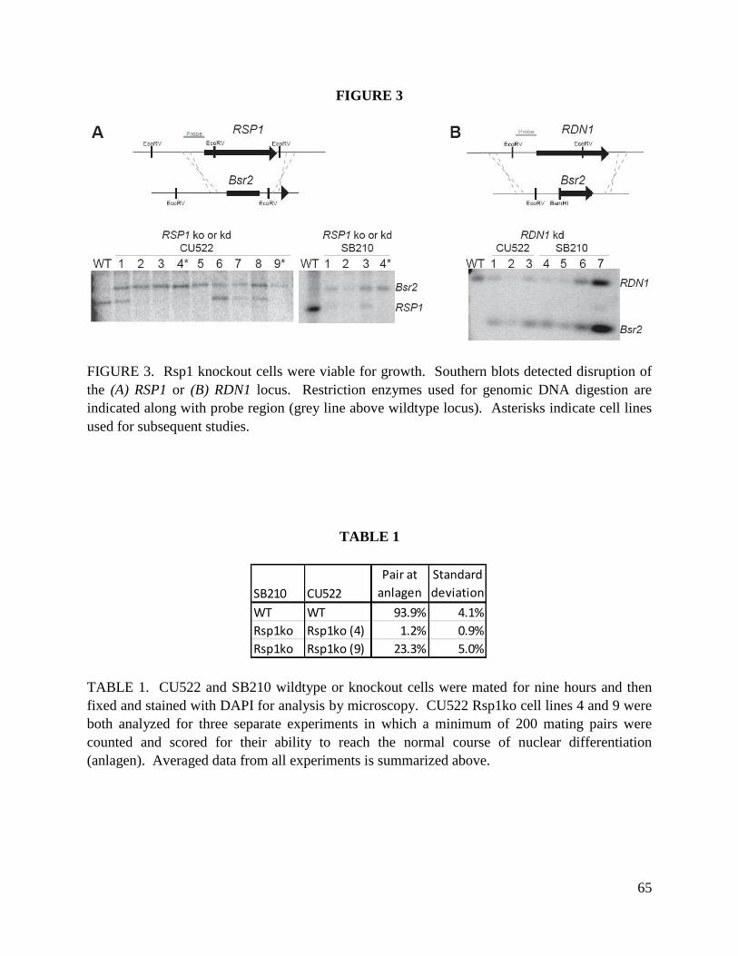
65
FIGURE 3
FIGURE 3. Rsp1 knockout cells were viable for growth. Southern blots detected disruption of
the (A) RSP1 or (B) RDN1 locus. Restriction enzymes used for genomic DNA digestion are
indicated along with probe region (grey line above wildtype locus). Asterisks indicate cell lines
used for subsequent studies.
TABLE 1
TABLE 1. CU522 and SB210 wildtype or knockout cells were mated for nine hours and then
fixed and stained with DAPI for analysis by microscopy. CU522 Rsp1ko cell lines 4 and 9 were
both analyzed for three separate experiments in which a minimum of 200 mating pairs were
counted and scored for their ability to reach the normal course of nuclear differentiation
(anlagen). Averaged data from all experiments is summarized above.
SB210 CU522
Pair at
anlagen
Standard
deviation
WT WT 93.9% 4.1%
Rsp1ko Rsp1ko (4) 1.2% 0.9%
Rsp1ko Rsp1ko (9) 23.3% 5.0%
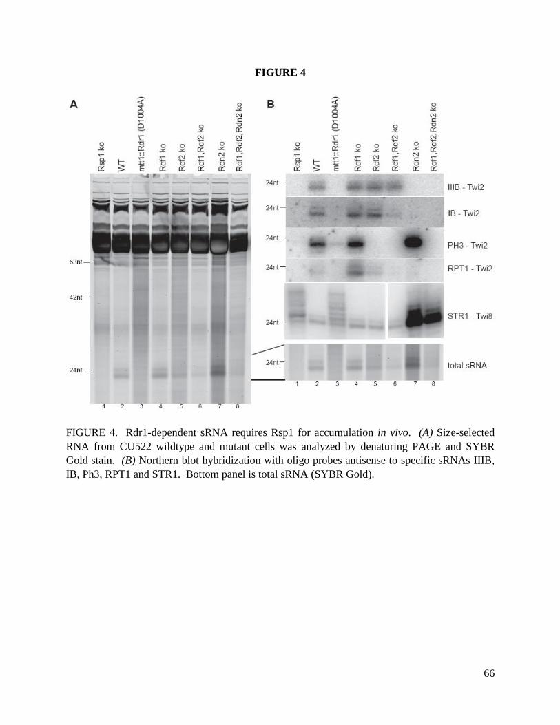
66
FIGURE 4
FIGURE 4. Rdr1-dependent sRNA requires Rsp1 for accumulation in vivo. (A) Size-selected
RNA from CU522 wildtype and mutant cells was analyzed by denaturing PAGE and SYBR
Gold stain. (B) Northern blot hybridization with oligo probes antisense to specific sRNAs IIIB,
IB, Ph3, RPT1 and STR1. Bottom panel is total sRNA (SYBR Gold).

67
FIGURE 5
FIGURE 5. A putative Ph3 sRNA precursor accumulates only in Rdf2ko cells, not Rsp1ko. (A)
An RNA Polymerase transcript (solid black line) contains two exons to which sRNAs overlap
(grey boxes: cluster 1 and cluster 2) that are separated by one confirmed intron (dashed line).
Ph3 sRNA is a member of cluster 2. The 3’ end of exon 2 contains an in-frame translation stop
codon. The 3’UTR contains a 163 nt stem-loop that is 20 nt after the stop codon. A potential
mRNA was detected from total RNA by RT-PCR; at least some RNA Pol transcript fraction was
polyadenylated and the intron removed (bottom left panel). Further processing of the mRNA,
such as removal of the 3’ stem-loop, may occur before Rdr1 targeting (right panel,
experimentally unconfirmed steps in grey). (B) Detection of the Ph3 precursor by mRNA
northern blot using a cluster 2-region-directed hexamer-primed probe (thin labeled line in A, top
panel). An rRNA doublet from the ethidium bromide-stained agarose gel before transfer to
northern blot membrane is shown as a loading control.
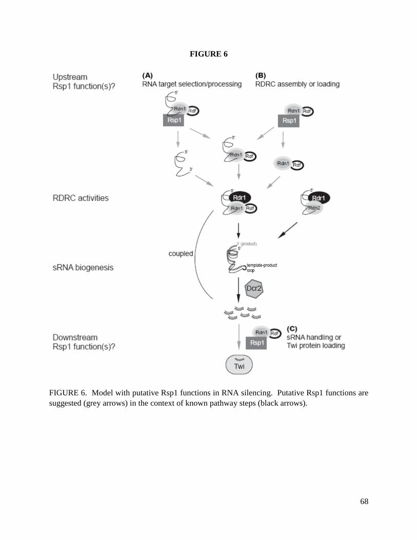
68
FIGURE 6
FIGURE 6. Model with putative Rsp1 functions in RNA silencing. Putative Rsp1 functions are
suggested (grey arrows) in the context of known pathway steps (black arrows).
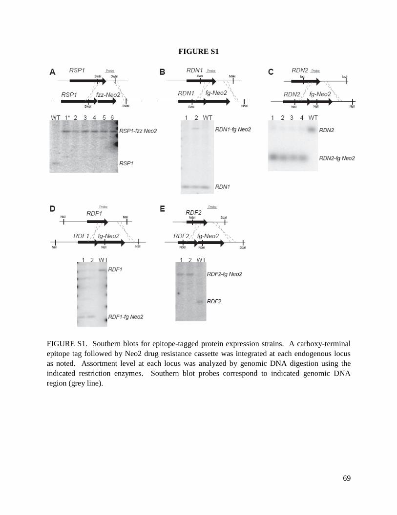
69
FIGURE S1
FIGURE S1. Southern blots for epitope-tagged protein expression strains. A carboxy-terminal
epitope tag followed by Neo2 drug resistance cassette was integrated at each endogenous locus
as noted. Assortment level at each locus was analyzed by genomic DNA digestion using the
indicated restriction enzymes. Southern blot probes correspond to indicated genomic DNA
region (grey line).

70
FIGURE S2
FIGURE S2. Rsp1 has a genetic depletion phenotype in conjugating cells. Cells were fixed after
nine hours of conjugation and stained with DAPI. All images are shown at the same
magnification and high exposure level to allow for visualization of each cell’s outline. Each
conjugating pair (* *) represents the most abundant state of nuclear differentiation seen in
wildtype (top panel) or SB210 Rsp1ko mated to either CU522(Rsp1ko) clonal strain 4 (middle)
or CU522(Rsp1ko) clonal strain 9 (bottom). In each wildtype cell, the degenerating parental
macronucleus stains more brightly than the two newly-developing zygotic macronuclei. The
micronuclei are visible between each developing zygotic macronucleus. Most Rsp1ko pairs did
not develop to anlagen but instead resembled early (2-4 hours, middle panel) or mid (4-9 hours)
of conjugation. Also shown is one unpaired cell (middle panel, arrow). The white scale bar
(upper right) represents 10 μm.
71
FUTURE DIRECTIONS FOR STUDIES OF RNA SILENCING
1. Why does Rdn1 in the RSPC context synthesize fewer products? Is this due to changes in
binding or activity? Compare RNA binding by RSPCs and RDRCs. The sRNA precursors
outlined in Appendix B would be especially interesting to test, considering the known RDRC
specificity in vivo.
2. Do Rdf1 and Rdf2 have binding preferences for RNA sequence or structure? For example,
does the PH3 stemloop mediate binding by complexes that contain Rdf2?
3. Rsp1 shares protein partners with Rdr1. One potential function of Rsp1 is to assemble Rdn1,
Rdf1 and Rdf2 with Rdr1 in vivo. To test this, generate cell lines with tagged RDRC
components in Rsp1ko cells and determine if RDRCs can still assemble.
4. What targets RNA to appropriate RDRCs in vivo? GFP was targeted for silencing using the
Ph3 stem-loop sequence (unpublished data, Kyungah Hong and K. Collins). Determine if the
transgene can still be targeted in the Rdf2ko background. It would be interesting to test the
ability of mutagenized precursors, alternative stem-loop-like structures and alternative stem-
loop placement within the transgene to target a transcript for silencing.

72
APPENDIX A
Rdf1 and Rdf2 serve distinct functions in sRNA biogenesis
SUMMARY AND RESULTS
Rdf1 and Rdf2 are mutually exclusive in their assembly into two of the three RDRCs and the two
RSPCs. Due to their similarities in protein partners, I thought it possible that each may perform
the same or similar functions, but at different times of the T. thermophila life cycle. Different
developmental regulation of Rdf1 and Rdf2 could account for the distinct sRNA accumulation
profiles between Rdf1ko and Rdf2ko cells, as described in preceding chapters.
Rdf2 is expressed constitutively while Rdf1 expression is low except during mid and late-stage
conjugation (Lee, Talsky et al. 2009). Considering this, Rdf2 occupies the most abundant RDRC
during vegetative growth. Its two RDRC partners, Rdr1 and Rdn1, are both essential for growth,
but Rdf2 is not (Lee, Talsky et al. 2009). In the absence of Rdf2, low levels of Rdf1 may
substitute for any essential function Rdf2 may perform. To test this, I attempted to replace the
entire DNA locus that contains neighboring ORFs for Rdf1 and Rdf2 with a drug resistance
casette. Assortment went to completion (Fig 1A). Thus, Rdf1 and Rdf2 do not serve redundant
functions that are essential for growth. In fact, cells accommodate the triple knockout of all three
non-essential RDRC components Rdf1, Rdf2 and Rdn2 (Fig 1B).
Accumulation of bulk sRNA depends on Rdr1 and Rsp1 function (Chapter 3). Individual sRNA
sequences that represent a phased sRNA family (Ph3), a high-copy repeat locus (RPT1) and
pseudogene-derived loci (IB and IIIB) also require a subset of RDRC components (Chapter 3,
Fig 4). For example, Ph3 does not accumulate in any strain that lacks Rdf2, which includes the
single, double or triple knockouts (Fig 2A, middle panel, lanes 11, 13, 14). RPT1 does not
accumulate in cells lacking both Rdf1 and Rdf2 (bottom panel, lanes 11, 14). Both sRNAs are
absent from Rsp1ko and Rdr1(D1004A)-overexpression strains (lanes 1,2) but accumulate in
wildtype and Rdf1ko (3-5, 12).
To assess whether Rdf1 is capable of substituting for Rdf2, I attempted to rescue the Rdf2ko
sRNA accumulation phenotype with Rdf1 or Rdf2 expression during growth. I integrated zzf-
Rdf1 and zzf-Rdf2 under the MTT1 promoter at the BTU1 locus in CU522 (Rdf1,Rdf2ko) cells
using Neo2 selection. Assortment went to completion. In the absence of CdCl2, expression
under the MTT1 promoter was low but detectable. Transgene and wildtype cells were grown
without CdCl2 or with 3 or 16 hours of induction using 1 μM CdCl2. Total RNA was isolated
from growing cells, size-selected and analyzed using denaturing PAGE stained by SYBR Gold to
detect bulk sRNA (Fig 2B, top panel). Individual sRNAs were detected by northern blot using
end-labeled oligonucleotides. Each sRNA accumulated in wildtype cells grown in all three
conditions (Fig 2B, lanes 3-5). CU522(Rdf1,Rdf2ko) cells did not accumulate Ph3 or RPT1,

73
which, for these purposes, is considered baseline (lane 13). Expression of Rdf2-fzz in the
double-knockout cells rescued the accumulation of Ph3 and RPT1 after 0 or 3 hours of protein
induction with CdCl2 (lanes 9, 10). However, Rdf1-fzz was unable to rescue accumulation of
either sRNA after 0, 3 or 16 hours of protein induction (lanes 6-8). In an independent
experiment, Rdf1-fzz or Rdf2-fzz expression was induced for three hours such that each protein
accumulated to comparable levels (Fig 2B, bottom panel). In these cells, IB accumulated in
Rdf2-fzz and not in Rdf1-fzz. A control sRNA, IIIB, which does not depend on either Rdf1 or
Rdf2, accumulated in both transgene strains (Fig 2B, middle panels).
Expression of Rdf2-fzz in CU522(Rdf1,Rdf2ko) cells rescued the accumulation of Rdf2-
dependent sRNAs. However, Rdf1-fzz expression under the same conditions did not rescue
sRNA accumulation. Thus, Rdf1 and Rdf2 do not serve redundant functions in sRNA
biogenesis, at least during vegetative growth.
MATERIALS AND METHODS
Please see the materials and methods section of Chapter 3.

74
FIGURE 1
FIGURE 1. Southern blots. (A) Generation of Rdf1,Rdf2 double knockout by replacement of the
entire locus containing both Rdf1 and Rdf2 ORFs with a Neo2 drug resistance cassette in
wildtype CU522 cells. (B) Triple knockout by replacement of Rdf1,Rdf2 locus with Bsr2 in
CU522(Rdn2ko) cells, which were first selected for Rdn2ko using Neo2 drug resistance cassette.
Assortment of both knockout strains went to completion.
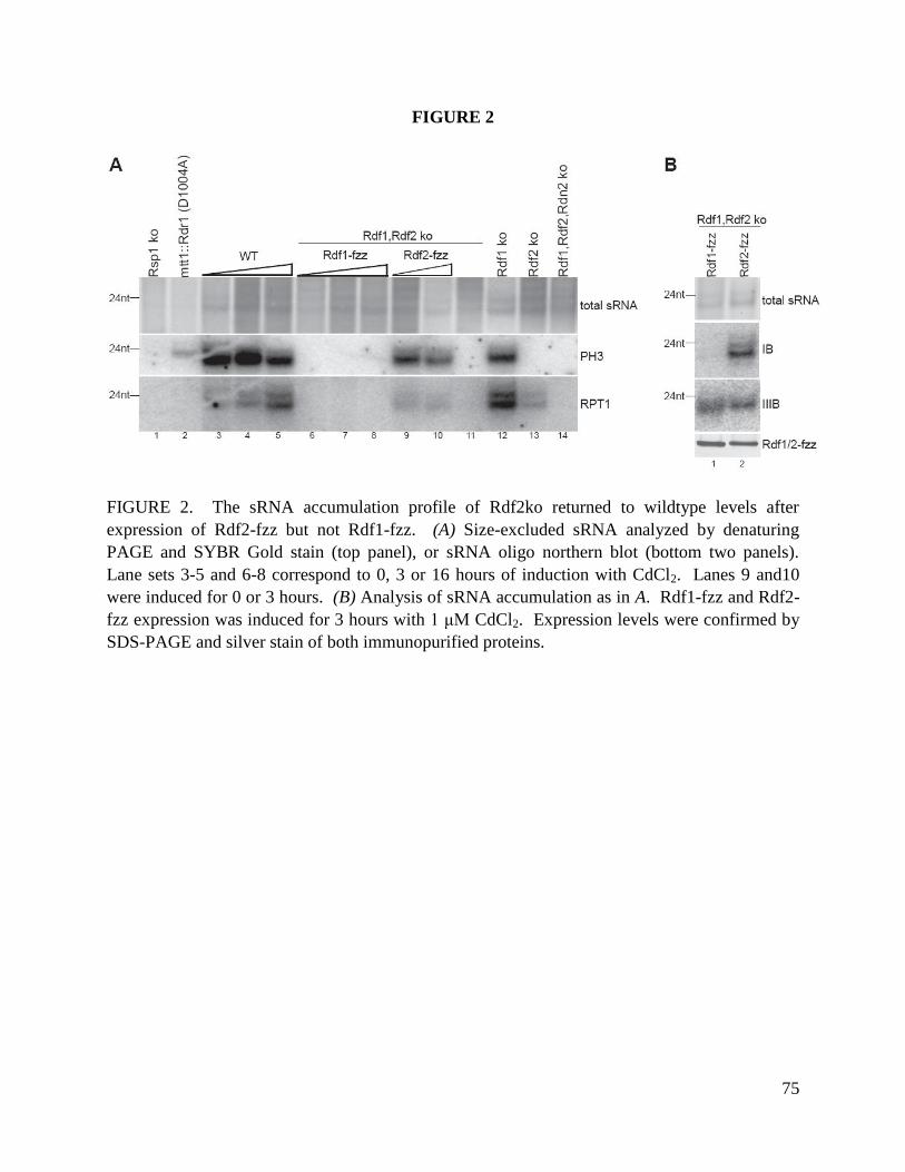
75
FIGURE 2
FIGURE 2. The sRNA accumulation profile of Rdf2ko returned to wildtype levels after
expression of Rdf2-fzz but not Rdf1-fzz. (A) Size-excluded sRNA analyzed by denaturing
PAGE and SYBR Gold stain (top panel), or sRNA oligo northern blot (bottom two panels).
Lane sets 3-5 and 6-8 correspond to 0, 3 or 16 hours of induction with CdCl2. Lanes 9 and10
were induced for 0 or 3 hours. (B) Analysis of sRNA accumulation as in A. Rdf1-fzz and Rdf2-
fzz expression was induced for 3 hours with 1 μM CdCl2. Expression levels were confirmed by
SDS-PAGE and silver stain of both immunopurified proteins.

76
APPENDIX B
Small RNA precursor structure may influence Rdr1 function and specificity
SUMMARY AND RESULTS
“Phased” clusters of sRNA mark one of the more predominant families of sRNA that accumulate
in T. thermophila (Couvillion, Lee et al. 2009). These sRNAs map to the genome with absolute
strand bias, in regularly-spaced intervals, mostly 24 nt apart. The sRNAs from one phased
cluster, Ph3, fail to accumulate in Rdf2ko cells, which is correlated with the accumulation of an
~800 nt RNA (Couvillion, Lee et al. 2009). This precursor contains two exons followed by a
3’UTR containing a 163 nt stem-loop structure (Chapter 3, Fig 5). To further define what
features distinguish putative RDRC target-RNAs, I identified the structural and transcript-
processing characteristics for the precursors of four abundantly-accumulating sRNA clusters.
The effect of precursor structure on RDRC activity was then assessed.
Included in this analysis are two phased clusters of sRNA (Ph2 and Ph3) and two pseudogene-
derived sRNAs (IIIB and IIA). Pseudogene-derived sRNAs are a second abundant family of
sRNAs that accumulate in T. thermophila. These sRNAs map antisense to predicted genes
containing an A-rich tract in the 3’ UTR (Couvillion et al 2009). Total RNA was reverse
transcribed and amplified (RT-PCR) and results summarized (Fig 1A). Ph2 and Ph3 precursors
(solid black lines) each contained two sRNA clusters (light grey boxes) separated by an intron
(dotted lines) and extended at the 3’ UTR by a stem-loop structure. Both 5’ regions abruptly
ended at the 5’-end of cluster 1. Precursors for IIIB and IIA both overlapped with predicted
genes (noted with TTHERM numbers). A single IIIB precursor contained three such predicted
genes, each with an ~80 nt A-rich tract (black box). Between each of the three pseudogenes
were regions in which sRNA overlap was greatly reduced (white triangles). Precursor IIA
mapped to a single gene prediction. Both 5’-ends of IIIB and IIA were extended at least 200bp
beyond the predicted gene start codons. All four sRNA precursors contained an extended 3’
UTR, as indicated. All four also could be detected by RT-PCR using oligo dT18, which suggests
they were polyadenylated and at least partially processed. It is impossible to ignore the
possibility that some of the newly identified characteristics shared by Ph2 and Ph3 (processed
intron, 3’ stem-loop) as well as IIIB and IIA (A-rich tracts, possibly polycistronic) may
contribute to RNA targeting to appropriate RDRCs.
There is a conserved stem-loop structure in the 3’ UTR of both Ph2 and Ph3 precursors. The
stem-loop may mediate RNA targeting to the Rdf2-containing RDRC and could also potentially
influence Rdr1 activity. To test this, the activity of Rdr1, in the context of distinct RDRCs, was
assayed on three variations of in vitro-transcribed and purified Ph3 precursor RNA sequence (Fig
1B, top diagram). All three templates contained a single-stranded leader sequence 5’ to the
stemloop sequence. Template 1 included the sequence for the first half of the stem-loop (198 nt),

77
template 2 contained the full stem-loop with a blunt end (281 nt) and template 3 extended past
the stem-loop with single-stranded sequence (351 nt). Rdr1 was expressed recombinantly using
rabbit reticulocyte lysate (Talsky and Collins 2010) and compared to RDRCs isolated by
immunopurification of zz-Rdr1 from Rdf1ko, Rdf2ko or Rdn2ko cells (Lee, Talsky et al. 2009).
RDRCs were purified under gentle wash conditions, leaving Dcr2 present. Assays were
performed in the presence of radiolabeled CTP. Rdr1 products were treated with S1 single-
stranded nuclease, leaving only dsRNA regions that correspond to second strand synthesis.
Recombinant Rdr1 (rRdr1) and Rdr1 from each RDRC mix synthesized dsRNA products from
templates 1 (Fig 1B, top panel, lanes 1-4), template 2 (lanes 5-8) and template 3 (lanes 9-12).
For each reaction except those containing recombinant Rdr1, sRNA also accumulated from
Dcr2-mediated cleavage of Rdr1 products (Fig 1B, bottom panel). These findings do not support
the involvement of Rdf2 in sRNA biogenesis from phased-cluster precursors in vitro, as product
synthesis by Rdr1 and Dcr2 did not change in the presence or absence of Rdf2.
Rdr1 is a processive polymerase that synthesizes dsRNA products that are approximately
template-length (Talsky and Collins 2010). Consistent with this, Rdr1 products from templates 1
and 3 migrated at template-length (Fig 1B, lanes 1-4 and 9-12, open circles). In contrast, only
minor products from template 2 were near template-length (lanes 5-8, open circle), while most
products were shorter. Considering the entire sequence for template 2 was contained within
template 3, and that Rdr1 does not require a precise initiation sequence (Talsky and Collins
2010), it is likely that the presence of the blunt-ended stem-loop inhibited dsRNA synthesis from
the 3’ end of template 2. Instead, initiation by Rdr1 probably occurred within the open loop in
the middle of the stem-loop, which would result in products approximately the length of template
1 (asterisk). Some products also likely arose from initiation 5’ to the stem-loop (arrowheads).
Precursors of phased and pseudogene-derived sRNAs contain sequence and structure that is
conserved among sRNA families. Ph2 and Ph3 precursors each contain a 3’ ~160 nt stem-loop
which suggests, despite the lack of evidence in vitro, that stem-loops may help target RNA to its
appropriate Rdf2-containing RDRC in vivo. When given Ph3 precursor sequence as template,
Rdr1 was unable to initiate within a 3’, blunt-ended stem-loop in vitro. This suggests that RNA
structures influence initiation of second-strand synthesis (and, thus, the sequences included in the
sRNA population) during RNA silencing within the cell.
MATERIALS AND METHODS
See Chapter 3 for details on precursor analysis and Chapter 2 or (Talsky and Collins 2010) for
details on activity assays. Assay templates:
template 1 (198 nt):
5’ATCACTTATTAATTGAGAGTATTTAATAATTAATTTTAGGCCTTTACATTTTTTTAA
ACGAAAAAATAGAAAACTATTTAATAATATATTTTCAACTTTGAATGCTATAAAAAA

78
AATATATTTAAATTAGAAAATAAAAATTTAAGTGATTAGTTAAATATAATAAAATAT
TTTAAATCTATTATTTTTATAGATATT
template 2 (281 nt):
5’ATCACTTATTAATTGAGAGTATTTAATAATTAATTTTAGGCCTTTACATTTTTTTAA
ACGAAAAAATAGAAAACTATTTAATAATATATTTTCAACTTTGAATGCTATAAAAAA
AATATATTTAAATTAGAAAATAAAAATTTAAGTGATTAGTTAAATATAATAAAATAT
TTTAAATCTATTATTTTTATAGATATTTTTTTCTAATAAAAATAATATTTTTATCATTA
TTTTATTGTATTTAAAAATATCATTCAAATATTTATTGTTTAATTTAAGTA
template 3 (351 nt):
5’ATCACTTATTAATTGAGAGTATTTAATAATTAATTTTAGGCCTTTACATTTTTTTAA
ACGAAAAAATAGAAAACTATTTAATAATATATTTTCAACTTTGAATGCTATAAAAAA
AATATATTTAAATTAGAAAATAAAAATTTAAGTGATTAGTTAAATATAATAAAATAT
TTTAAATCTATTATTTTTATAGATATTTTTTTCTAATAAAAATAATATTTTTATCATTA
TTTTATTGTATTTAAAAATATCATTCAAATATTTATTGTTTAATTTAAGTAAAATTTA
TGTTAGTATTATCTTTAAATTTTATATTAAAACAATTCAATTTATTTTTTTTTCTAAAC
AATT

79
FIGURE 1
FIGURE 1. (A) Structural analysis of sRNA precursors. (B) Recombinant Rdr1 or
endogenously-assembled and purified RDRCs (Figure Key) were assayed for activity on three in
vitro-transcribed and purified RNA templates derived from the Ph3 precursor sequence (top).
Rdr1 products were detected by incorporation of radiolabeled CTP, treated with S1 single-
stranded nuclease, and resolved by formamide-PAGE (middle). Template-length dsRNA
products (open circle), and shorter products (asterisk and arrowheads) are noted. sRNA products
also accumulated in RDRC assays that contained co-purifying Dcr2 (bottom).

80
REFERENCES
Akbergenov, R., A. Si-Ammour, et al. (2006). "Molecular characterization of geminivirus-derived small RNAs in different plant species." Nucleic Acids Res. 34: 462-471.
Allen, E., Z. Xie, et al. (2005). "microRNA-directed phasing during trans-acting siRNA biogenesis in plants." Cell 121(2): 207-221.
Ameres, S. L., M. D. Horwich, et al. (2010). "Target RNA-directed trimming and tailing of small silencing RNAs." Science 328(5985): 1534-1539.
Aoki, K., H. Moriguchi, et al. (2007). "In vitro analyses of the production and activity of secondary small interfering RNAs in C. elegans." EMBO J. 26: 5007-5019.
Aoki, K., H. Moriguchi, et al. (2007). "In vitro analyses of the production and activity of secondary small interfering RNAs in C. elegans." EMBO J 26(24): 5007-5019.
Bühler, M., W. Haas, et al. (2007). "RNAi-dependent and -independent RNA turnover mechanisms contribute to heterochromatic gene silencing." Cell 18: 707-721.
Bühler, M. and D. Moazed (2007). "Transcription and RNAi in heterochromatic gene silencing." Nat. Struct. Mol. Biol. 14: 1041-1048.
Carthew, R. W. and E. J. Sontheimer (2009). "Origins and Mechanisms of miRNAs and siRNAs." Cell 136(4): 642-655.
Carthew, R. W. and E. J. Sontheimer (2009). "Origins and mechanisms of miRNAs and siRNAs." Cell 136: 642-655.
Cerutti, H. and J. A. Casas-Mollano (2006). "On the origin and functions of RNA-mediated silencing: from protists to man." Curr. Genet. 50: 81-99.
Chalker, D. L. (2008). "Dynamic nuclear reorganization during genome remodeling of Tetrahymena." Biochim. Biophys. Acta. 1783: 2130-2136.
Chen, C. C., M. J. Simard, et al. (2005). "A member of the polymerase beta nucleotidyltransferase superfamily is required for RNA interference in C. elegans." Curr. Biol. 15: 378-383.
Couvillion, M. T., S. R. Lee, et al. (2009). "Sequence, biogenesis, and function of diverse small RNA classes bound to the Piwi-family proteins of Tetrahymena thermophila." Genes Dev. 23: 2016-2032.
Couvillion, M. T., S. R. Lee, et al. (2009). "Sequence, biogenesis, and function of diverse small RNA classes bound to the Piwi family proteins of Tetrahymena thermophila." Genes Dev 23(17): 2016-2032.
Curaba, J. and X. Chen (2008). "Biochemical activities of Arabidopsis RNA-dependent RNA polymerase 6." J Biol Chem 283(6): 3059-3066.
Curaba, J. and X. Chen (2008). "Biochemical activities of Arabidopsis RNA-dependent RNA polymerase 6." J. Biol. Chem. 283: 3059-3066.
Deshpande, T., T. Takagi, et al. (1999). "Human PIR1 of the protein-tyrosine phosphatase superfamily has RNA 5'-triphosphatase and diphosphatase activities." J Biol Chem 274(23): 16590-16594.
Duchaine, T. F., J. A. Wohlschlegel, et al. (2006). "Functional proteomics reveals the biochemical niche of C. elegans DCR-1 in multiple small-RNA-mediated pathways." Cell 124(2): 343-354.
Duchaine, T. F., J. A. Wohlschlegel, et al. (2006). "Functional proteomics reveals the biochemical niche of C. elegans DCR-1 in multiple small-RNA-mediated pathways." Cell 124: 343-354.
Faehnle, C. R. and L. Joshua-Tor (2007). "Argonautes confront new small RNAs." Curr Opin Chem Biol 11(5): 569-577.
Farazi, T. A., S. A. Juranek, et al. (2008). "The growing catalog of small RNAs and their association with distinct Argonaute/Piwi family members." Development 135(7): 1201-1214.
81
Farazi, T. A., S. A. Juranek, et al. (2008). "The growing catalog of small RNAs and their association with distinct Argonaute/Piwi family members." Development 135: 1201-1214.
Fire, A., S. Xu, et al. (1998). "Potent and specific genetic interference by double-stranded RNA in Caenorhabditis elegans." Nature 391(6669): 806-811.
Gazzani, S., T. Lawrenson, et al. (2004). "A link between mRNA turnover and RNA interference in Arabidopsis." Science 306: 1046-1048.
Gent, J. I., A. T. Lamm, et al. (2010). "Distinct phases of siRNA synthesis in an endogenous RNAi pathway in C. elegans soma." Mol Cell 37(5): 679-689.
Ghildiyal, M. and P. D. Zamore (2009). "Small silencing RNAs: an expanding universe." Nat Rev Genet 10(2): 94-108.
Ghildiyal, M. and P. D. Zamore (2009). "Small silencing RNAs: an expanding universe." Nat. Rev. Genet. 10: 94-108.
Heo, I., C. Joo, et al. (2008). "Lin28 mediates the terminal uridylation of let-7 precursor MicroRNA." Mol. Cell 32: 276-284.
Howard-Till, R. A. and M. C. Yao (2006). "Induction of gene silencing by hairpin RNA expression in Tetrahymena thermophila reveals a second small RNA pathway." Mol Cell Biol 26(23): 8731-8742.
Howell, M. D., N. Fahlgren, et al. (2007). "Genome-wide analysis of the RNA-DEPENDENT RNA POLYMERASE6/DICER-LIKE4 pathway in Arabidopsis reveals dependency on miRNA- and tasiRNA-directed targeting." Plant Cell 19(3): 926-942.
Iida, T., J. Nakayama, et al. (2008). "siRNA-mediated heterochromatin establishment requires HP1 and is associated with antisense transcription." Mol Cell 31(2): 178-189.
Iyer, L. M., E. V. Koonin, et al. (2003). "Evolutionary connection between the catalytic subunits of DNA-dependent RNA polymerases and eukaryotic RNA-dependent RNA polymerases and the origin of RNA polymerases." BMC Struct. Biol. 28: 1-23.
Kasschau, K. D., N. Fahlgren, et al. (2007). "Genome-wide profiling and analysis of Arabidopsis siRNAs." PLoS Biol 5(3): e57.
Ketting, R. F. (2011). "The many faces of RNAi." Dev Cell 20(2): 148-161. Kim, V. N., J. Han, et al. (2009). "Biogenesis of small RNAs in animals." Nat. Rev. Mol. Cell Biol. 10: 126-
139. Kurth, H. M. and K. Mochizuki (2009). "2'-O-methylation stabilizes Piwi-associated small RNAs and
ensures DNA elimination in Tetrahymena." RNA 15(4): 675-685. Kwak, J. E. and M. Wickens (2007). "A family of poly(U) polymerases." RNA 13: 860-867. Lee, H. C., S. S. Chang, et al. (2009). "qiRNA is a new type of small interfering RNA induced by DNA
damage." Nature 459(7244): 274-277. Lee, H. C., S. S. Chang, et al. (2009). "qiRNA is a new type of small interfering RNA induced by DNA
damage." Nature 459: 274-277. Lee, S. R. and K. Collins (2005). "Starvation-induced cleavage of the tRNA anticodon loop in Tetrahymena
thermophila." J Biol Chem 280(52): 42744-42749. Lee, S. R. and K. Collins (2006). "Two classes of endogenous small RNAs in Tetrahymena thermophila."
Genes Dev 20(1): 28-33. Lee, S. R. and K. Collins (2006). "Two classes of endogenous small RNAs in Tetrahymena thermophila."
Genes Dev. 20: 28-33. Lee, S. R. and K. Collins (2007). "Physical and functional coupling of RNA-dependent RNA polymerase
and Dicer in the biogenesis of endogenous siRNAs." Nat Struct Mol Biol 14(7): 604-610. Lee, S. R. and K. Collins (2007). "Physical and functional coupling of RNA-dependent RNA polymerase
and Dicer in the biogenesis of endogenous siRNAs." Nat. Struct. Mol. Biol. 14: 604-610.

82
Lee, S. R., K. B. Talsky, et al. (2009). "A single RNA-dependent RNA polymerase assembles with mutually exclusive nucleotidyl transferase subunits to direct different pathways of small RNA biogenesis." RNA 15(7): 1363-1374.
Lee, S. R., K. B. Talsky, et al. (2009). "A single RNA-dependent RNA polymerase assembles with mutually exclusive nucleotidyl transferase subunits to direct different pathways of small RNA biogenesis." RNA 15: 1363-1374.
Lepere, G., M. Nowacki, et al. (2009). "Silencing-associated and meiosis-specific small RNA pathways in Paramecium tetraurelia." Nucleic Acids Res 37(3): 903-915.
Li, J., Z. Yang, et al. (2005). "Methylation protects miRNAs and siRNAs from a 3'-end uridylation activity in Arabidopsis." Curr. Biol. 15(16): 1501-1507.
Maida, Y., M. Yasukawa, et al. (2009). "An RNA-dependent RNA polymerase formed by TERT and the RMRP RNA." Nature 461(7261): 230-235.
Makeyev, E. V. and D. H. Bamford (2002). "Cellular RNA-dependent RNA polymerase involved in posttranscriptional gene silencing has two distinct activity modes." Mol Cell 10(6): 1417-1427.
Makeyev, E. V. and D. H. Bamford (2002). "Cellular RNA-dependent RNA polymerase involved in posttranscriptional gene silencing has two distinct activity modes." Mol. Cell 10: 1417-1427.
Marker, S., A. Le Mouel, et al. (2010). "Distinct RNA-dependent RNA polymerases are required for RNAi triggered by double-stranded RNA versus truncated transgenes in Paramecium tetraurelia." Nucleic Acids Res 38(12): 4092-4107.
Martin, G. and W. Keller (2007). "RNA-specific ribonucleotidyl transferases." RNA 13: 1834-1849. Martinez, J. and T. Tuschl (2004). "RISC is a 5' phosphomonoester-producing RNA endonuclease." Genes
Dev 18(9): 975-980. Miao, W., J. Xiong, et al. (2009). "Microarray analyses of gene expression during the Tetrahymena
thermophila life cycle." PLoS ONE 4: e4429. Miao, W., J. Xiong, et al. (2009). "Microarray analyses of gene expression during the Tetrahymena
thermophila life cycle." PLoS One 4(2): e4429. Miller, M. C. and K. Collins (2000). "The Tetrahymena p80/p95 complex is required for proper telomere
length maintenance and micronuclear genome stability." Mol Cell 6(4): 827-837. Mochizuki, K. and M. A. Gorovsky (2004). "Small RNAs in genome rearrangement in Tetrahymena." Curr
Opin Genet Dev 14(2): 181-187. Mochizuki, K. and M. A. Gorovsky (2004). "Small RNAs in genome rearrangement in Tetrahymena." Curr.
Opin. Genet. Dev. 14: 181-187. Mochizuki, K. and M. A. Gorovsky (2005). "A Dicer-like protein in Tetrahymena has distinct functions in
genome rearrangement, chromosome segregation, and meiotic prophase." Genes Dev. 19: 77-89.
Motamedi, M. R., A. Verdel, et al. (2004). "Two RNAi complexes, RITS and RDRC, physically interact and localize to noncoding centromeric RNAs." Cell 119: 789-802.
Motamedi, M. R., A. Verdel, et al. (2004). "Two RNAi complexes, RITS and RDRC, physically interact and localize to noncoding centromeric RNAs." Cell 119(6): 789-802.
Mullen, T. E. and W. F. Marzluff (2008). "Degradation of histone mRNA requires oligouridylation followed by decapping and simultaneous degradation of the mRNA both 5' to 3' and 3' to 5'." Genes Dev. 22: 50-65.
Napoli, C., C. Lemieux, et al. (1990). "Introduction of a Chimeric Chalcone Synthase Gene into Petunia Results in Reversible Co-Suppression of Homologous Genes in trans." Plant Cell 2(4): 279-289.
Okamura, K. and E. C. Lai (2008). "Endogenous small interfering RNAs in animals." Nat Rev Mol Cell Biol 9(9): 673-678.
Pak, J. and A. Fire (2007). "Distinct populations of primary and secondary effectors during RNAi in C. elegans." Science 315: 241-244.
83
Pak, J. and A. Fire (2007). "Distinct populations of primary and secondary effectors during RNAi in C. elegans." Science 315(5809): 241-244.
Ramachandran, V. and X. Chen (2008). "Small RNA metabolism in Arabidopsis." Trends Plant Sci 13(7): 368-374.
Rissland, O. S. and N. C.J. (2008). "The Cid1 poly(U) polymerase." Biochim. Biophys. Acta. 1779: 286-294. Ruby, J. G., C. Jan, et al. (2006). "Large-scale sequencing reveals 21U-RNAs and additional microRNAs
and endogenous siRNAs in C. elegans." Cell 127: 1193-1207. Salgado, P. S., M. R. Koivunen, et al. (2006). "The structure of an RNAi polymerase links RNA silencing
and transcription." PLoS Biol 4(12): e434. Salgado, P. S., M. R. Koivunen, et al. (2006). "The structure of an RNAi polymerase links RNA silencing
and transcription." PLoS Biol. 4: e434. Schiebel, W., B. Haas, et al. (1993). "RNA-directed RNA polymerase from tomato leaves. II. Catalytic in
vitro properties." J Biol Chem. 268: 11858-11867. Schwarz, D. S., Y. Tomari, et al. (2004). "The RNA-induced silencing complex is a Mg2+-dependent
endonuclease." Curr Biol 14(9): 787-791. Seto, A. G., R. E. Kingston, et al. (2007). "The coming of age for Piwi proteins." Mol. Cell 26: 603-609. Shang, Y., X. Song, et al. (2002). "A robust inducible-repressible promoter greatly facilitates gene
knockouts, conditional expression, and overexpression of homologous and heterologous genes in Tetrahymena thermophila." Proc. Natl. Acad. Sci. USA 99: 3734-3739.
Shen, B. and H. M. Goodman (2004). "Uridine addition after microRNA-directed cleavage." Science 306: 997.
Shiu, P. K., D. Zickler, et al. (2006). "SAD-2 is required for meiotic silencing by unpaired DNA and perinuclear localization of SAD-1 RNA-directed RNA polymerase." Proc. Natl. Acad. Sci. USA 103: 2243-2248.
Sijen, T., F. A. Steiner, et al. (2007). "Secondary siRNAs result from unprimed RNA synthesis and form a distinct class." Science 315: 244-247.
Sijen, T., F. A. Steiner, et al. (2007). "Secondary siRNAs result from unprimed RNA synthesis and form a distinct class." Science 315(5809): 244-247.
Siomi, H. and M. C. Siomi (2009). "On the road to reading the RNA-interference code." Nature 457: 396-404.
Siomi, H. and M. C. Siomi (2009). "On the road to reading the RNA-interference code." Nature 457(7228): 396-404.
Smith, J. J., J. S. Yakisich, et al. (2004). "A beta-tubulin mutation selectively uncouples nuclear division and cytokinesis in Tetrahymena thermophila." Eukaryot. Cell. 3: 1217-1226.
Song, M. G. and M. Kiledjian (2007). "3' Terminal oligo U-tract-mediated stimulation of decapping." RNA 13: 2356-2365.
Sugiyama, T., H. Cam, et al. (2005). "RNA-dependent RNA polymerase is an essential component of a self-enforcing loop coupling heterochromatin assembly to siRNA production." Proc. Natl. Acad. Sci. USA 102: 152-157.
Talsky, K. B. and K. Collins (2010). "Initiation by a eukaryotic RNA-dependent RNA polymerase requires looping of the template end and is influenced by the template-tailing activity of an associated uridyltransferase." J Biol Chem 285(36): 27614-27623.
Tang, G. (2005). "siRNA and miRNA: an insight into RISCs." Trends Biochem Sci 30(2): 106-114. Tang, G., B. J. Reinhart, et al. (2003). "A biochemical framework for RNA silencing in plants." Genes Dev
17: 49-63. Tang, G., B. J. Reinhart, et al. (2003). "A biochemical framework for RNA silencing in plants." Genes Dev
17(1): 49-63.

84
Vasale, J. J., W. Gu, et al. (2010). "Sequential rounds of RNA-dependent RNA transcription drive endogenous small-RNA biogenesis in the ERGO-1/Argonaute pathway." Proc Natl Acad Sci U S A 107(8): 3582-3587.
Verdel, A., S. Jia, et al. (2004). "RNAi-mediated targeting of heterochromatin by the RITS complex." Science 303(5658): 672-676.
Wassenegger, M. and G. Krczal (2006). "Nomenclature and functions of RNA-directed RNA polymerases." Trends Plant Sci 11(3): 142-151.
Wassenegger, M. and G. Krczal (2006). "Nomenclature and functions of RNA-directed RNA polymerases." Trends Plant Sci. 11: 142-151.
Wilusz, C. J. and J. Wilusz (2008). "New ways to meet your (3') end oligouridylation as a step on the path to destruction." Genes Dev. 22: 1-7.
Win, T. Z., A. L. Stevenson, et al. (2006). "Fission yeast Cid12 has dual functions in chromosome segregation and checkpoint control." Mol. Cell. Biol. 26: 4435-4447.
Witkin, K. L., R. Prathapam, et al. (2007). "Positive and negative regulation of Tetrahymena telomerase holoenzyme." Mol. Cell. Biol. 27(6): 2074-2083.
Wu, L. and J. G. Belasco (2008). "Let me count the ways: mechanisms of gene regulation by miRNAs and siRNAs." Mol Cell 29(1): 1-7.
Yoshikawa, M., A. Peragine, et al. (2005). "A pathway for the biogenesis of trans-acting siRNAs in Arabidopsis." Genes Dev. 19: 2164-2175.
Zhang, H., G. M. Ehrenkaufer, et al. (2008). "Small RNAs with 5'-polyphosphate termini associate with a Piwi-related protein and regulate gene expression in the single-celled eukaryote Entamoeba histolytica." PLoS Pathog 4(11): e1000219.
Zong, J., X. Yao, et al. (2009). "Evolution of the RNA-dependent RNA polymerase (RdRP) genes: duplications and possible losses before and after the divergence of major eukaryotic groups." Gene 447: 29-39.