Generation of recombinant MVA-norovirus: a comparison ...
12
METHODOLOGY Open Access Generation of recombinant MVA-norovirus: a comparison study of bacterial artificial chromosome- and marker-based systems Franziska Kugler 1 , Ingo Drexler 2 , Ulrike Protzer 1 , Dieter Hoffmann 1* and Hassan Moeini 1* Abstract Background: Recombinant Modified Vaccinia Virus Ankara has been employed as a safe and potent viral vector vaccine against infectious diseases and cancer. We generated recMVAs encoding norovirus GII.4 genotype capsid protein by using a marker-based approach and a BAC-based system. In the marker-based approach, the capsid gene together with a reporter gene was introduced into the MVA genome in DF-1 cells. Several rounds of plaque purification were carried out to get rid of the WT-MVA. In the BAC-based approach, recMVA-BAC was produced by en passant recombineering in E. coli. Subsequently, the recMVAs were rescued in DF-1 cells using a helper rabbit fibroma virus. The BAC backbone and the helper virus were eliminated by passaging in DF-1 cells. Biochemical characteristics of the recMVAs were studied. Results: We found the purification of the rare spontaneous recombinants time-consuming in the marker-based system. In contrast, the BAC-based system rapidly inserted the gene of interest in E. coli by en passant recombineering before virion production in DF-1 cells. The elimination of the reporter gene was found to be faster and more efficient in the BAC-based approach. With Western blotting and electron microscopy, we could prove successful capsid protein expression and proper virus-assembly, respectively. The MVA-BAC produced higher recombinant virus titers and infected DF-1 cells more efficiently. Conclusions: Comparing both methods, we conclude that, in contrast to the tedious and time-consuming traditional method, the MVA-BAC system allows us to quickly generate high titer recMVAs. Keywords: Bacterial artificial chromosome, Recombinant MVA, Self-excising, Norovirus Background Norovirus is highly infectious and can be serious to indi- viduals with underlying conditions, the elderly, young chil- dren and immunocompromised patients. GII.4 genotype evolves faster than other genotypes and global pandemic outbreaks often correlate to the emergence of new GII.4 variants [1–3]. Major capsid protein (VP1) is responsible for virus attachment to the histo-blood group antigen [4]. The capsid protein is divided into shell (S) domain and a protruding (P) domain linked together by a hinge of eight amino acids [4, 5]. The aim of this study was to generate a recMVA expressing norovirus (NoV) capsid protein of GII.4 genotype. We plan to apply the constructs as vaccine candidates against this most prevalent gastro- enteritis virus. Modified Vaccinia Ankara (MVA) is a highly attenu- ated strain of vaccinia virus derived by extensive serial passages in chicken embryo fibroblasts (CEF) [6]. MVA can be produced in large scale in chicken cell lines under bio safety level 1. Temporal high expression of MVA-delivered antigens can modulate antigen-specific immune responses, making it a suitable vaccine carrier [6–9]. We followed a traditional marker-based method (as reviewed by Drexler et al. [7]) and a BAC-based approach as described by Dai [10] and Cottingham et al. [11] for generation of recMVA-NoV constructs. Both techniques produced recombinant MVA delivering the desired antigen. In the marker-based approach, a shuttle vector harbor- ing a fluorescence reporter gene, the gene of interest © The Author(s). 2019 Open Access This article is distributed under the terms of the Creative Commons Attribution 4.0 International License (http://creativecommons.org/licenses/by/4.0/), which permits unrestricted use, distribution, and reproduction in any medium, provided you give appropriate credit to the original author(s) and the source, provide a link to the Creative Commons license, and indicate if changes were made. The Creative Commons Public Domain Dedication waiver (http://creativecommons.org/publicdomain/zero/1.0/) applies to the data made available in this article, unless otherwise stated. * Correspondence: [email protected]; [email protected] 1 Institute of Virology, Faculty of Medicine, Technische Universität München, Munich, Germany Full list of author information is available at the end of the article Kugler et al. Virology Journal (2019) 16:100 https://doi.org/10.1186/s12985-019-1212-y
Transcript of Generation of recombinant MVA-norovirus: a comparison ...

METHODOLOGY Open Access
Generation of recombinant MVA-norovirus:a comparison study of bacterial artificialchromosome- and marker-based systemsFranziska Kugler1, Ingo Drexler2, Ulrike Protzer1, Dieter Hoffmann1* and Hassan Moeini1*
Abstract
Background: Recombinant Modified Vaccinia Virus Ankara has been employed as a safe and potent viral vectorvaccine against infectious diseases and cancer. We generated recMVAs encoding norovirus GII.4 genotype capsidprotein by using a marker-based approach and a BAC-based system. In the marker-based approach, the capsidgene together with a reporter gene was introduced into the MVA genome in DF-1 cells. Several rounds of plaquepurification were carried out to get rid of the WT-MVA. In the BAC-based approach, recMVA-BAC was produced byen passant recombineering in E. coli. Subsequently, the recMVAs were rescued in DF-1 cells using a helper rabbitfibroma virus. The BAC backbone and the helper virus were eliminated by passaging in DF-1 cells. Biochemicalcharacteristics of the recMVAs were studied.
Results: We found the purification of the rare spontaneous recombinants time-consuming in the marker-basedsystem. In contrast, the BAC-based system rapidly inserted the gene of interest in E. coli by en passantrecombineering before virion production in DF-1 cells. The elimination of the reporter gene was found to be fasterand more efficient in the BAC-based approach. With Western blotting and electron microscopy, we could provesuccessful capsid protein expression and proper virus-assembly, respectively. The MVA-BAC produced higherrecombinant virus titers and infected DF-1 cells more efficiently.
Conclusions: Comparing both methods, we conclude that, in contrast to the tedious and time-consumingtraditional method, the MVA-BAC system allows us to quickly generate high titer recMVAs.
Keywords: Bacterial artificial chromosome, Recombinant MVA, Self-excising, Norovirus
BackgroundNorovirus is highly infectious and can be serious to indi-viduals with underlying conditions, the elderly, young chil-dren and immunocompromised patients. GII.4 genotypeevolves faster than other genotypes and global pandemicoutbreaks often correlate to the emergence of new GII.4variants [1–3]. Major capsid protein (VP1) is responsiblefor virus attachment to the histo-blood group antigen [4].The capsid protein is divided into shell (S) domain and aprotruding (P) domain linked together by a hinge of eightamino acids [4, 5]. The aim of this study was to generate arecMVA expressing norovirus (NoV) capsid protein ofGII.4 genotype. We plan to apply the constructs as
vaccine candidates against this most prevalent gastro-enteritis virus.Modified Vaccinia Ankara (MVA) is a highly attenu-
ated strain of vaccinia virus derived by extensive serialpassages in chicken embryo fibroblasts (CEF) [6]. MVAcan be produced in large scale in chicken cell linesunder bio safety level 1. Temporal high expression ofMVA-delivered antigens can modulate antigen-specificimmune responses, making it a suitable vaccine carrier[6–9]. We followed a traditional marker-based method(as reviewed by Drexler et al. [7]) and a BAC-basedapproach as described by Dai [10] and Cottingham et al.[11] for generation of recMVA-NoV constructs. Bothtechniques produced recombinant MVA delivering thedesired antigen.In the marker-based approach, a shuttle vector harbor-
ing a fluorescence reporter gene, the gene of interest
© The Author(s). 2019 Open Access This article is distributed under the terms of the Creative Commons Attribution 4.0International License (http://creativecommons.org/licenses/by/4.0/), which permits unrestricted use, distribution, andreproduction in any medium, provided you give appropriate credit to the original author(s) and the source, provide a link tothe Creative Commons license, and indicate if changes were made. The Creative Commons Public Domain Dedication waiver(http://creativecommons.org/publicdomain/zero/1.0/) applies to the data made available in this article, unless otherwise stated.
* Correspondence: [email protected]; [email protected] of Virology, Faculty of Medicine, Technische Universität München,Munich, GermanyFull list of author information is available at the end of the article
Kugler et al. Virology Journal (2019) 16:100 https://doi.org/10.1186/s12985-019-1212-y
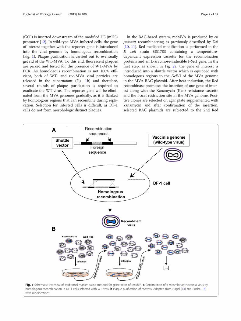
(GOI) is inserted downstream of the modified H5 (mH5)promoter [12]. In wild-type MVA-infected cells, the geneof interest together with the reporter gene is introducedinto the viral genome by homologous recombination(Fig. 1). Plaque purification is carried out to eventuallyget rid of the WT-MVA. To this end, fluorescent plaquesare picked and tested for the presence of WT-MVA byPCR. As homologous recombination is not 100% effi-cient, both of WT- and rec-MVA viral particles arereleased in the supernatant (Fig. 1b) and therefore,several rounds of plaque purification is required toeradicate the WT virus. The reporter gene will be elimi-nated from the MVA genomes gradually, as it is flankedby homologous regions that can recombine during repli-cation. Selection for infected cells is difficult, as DF-1cells do not form morphologic distinct plaques.
In the BAC-based system, recMVA is produced by enpassant recombineering as previously described by Dai[10, 11]. Red-mediated modification is performed in theE. coli strain GS1783 containing a temperature-dependent expression cassette for the recombinationproteins and an L-arabinose-inducible I-SecI gene. In thefirst step, as shown in Fig. 2a, the gene of interest isintroduced into a shuttle vector which is equipped withhomologous regions to the DelVI of the MVA genomein the MVA-BAC plasmid. After heat induction, the Redrecombinase promotes the insertion of our gene of inter-est along with the Kanamycin (Kan) resistance cassetteand the I-SceI restriction site in the MVA genome. Posi-tive clones are selected on agar plate supplemented withkanamycin and after confirmation of the insertion,selected BAC plasmids are subjected to the 2nd Red
Fig. 1 Schematic overview of traditional marker-based method for generation of recMVA. a Construction of a recombinant vaccinia virus byhomologous recombination in DF-1 cells infected with WT MVA. b Plaque purification of recMVA. Adapted from Nagel [13] and Rocha [14]with modifications
Kugler et al. Virology Journal (2019) 16:100 Page 2 of 12
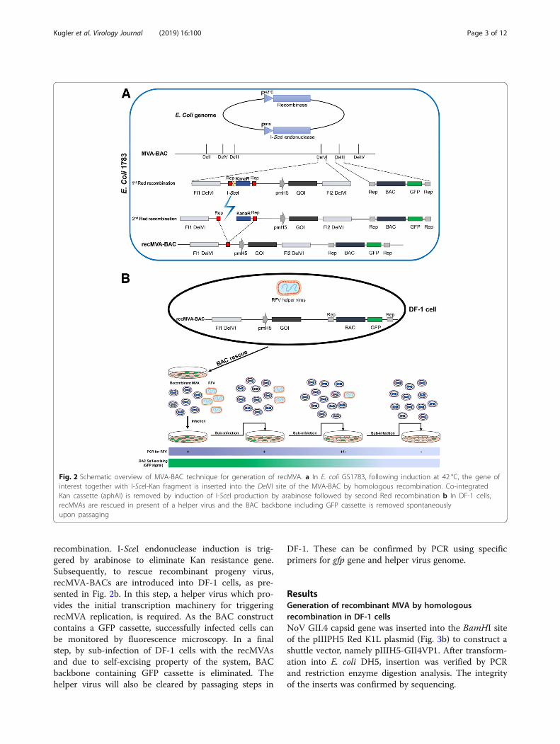
recombination. I-SceI endonuclease induction is trig-gered by arabinose to eliminate Kan resistance gene.Subsequently, to rescue recombinant progeny virus,recMVA-BACs are introduced into DF-1 cells, as pre-sented in Fig. 2b. In this step, a helper virus which pro-vides the initial transcription machinery for triggeringrecMVA replication, is required. As the BAC constructcontains a GFP cassette, successfully infected cells canbe monitored by fluorescence microscopy. In a finalstep, by sub-infection of DF-1 cells with the recMVAsand due to self-excising property of the system, BACbackbone containing GFP cassette is eliminated. Thehelper virus will also be cleared by passaging steps in
DF-1. These can be confirmed by PCR using specificprimers for gfp gene and helper virus genome.
ResultsGeneration of recombinant MVA by homologousrecombination in DF-1 cellsNoV GII.4 capsid gene was inserted into the BamHI siteof the pIIIPH5 Red K1L plasmid (Fig. 3b) to construct ashuttle vector, namely pIIIH5-GII4VP1. After transform-ation into E. coli DH5, insertion was verified by PCRand restriction enzyme digestion analysis. The integrityof the inserts was confirmed by sequencing.
Fig. 2 Schematic overview of MVA-BAC technique for generation of recMVA. a In E. coli GS1783, following induction at 42 °C, the gene ofinterest together with I-SceI-Kan fragment is inserted into the DelVI site of the MVA-BAC by homologous recombination. Co-integratedKan cassette (aphAI) is removed by induction of I-SceI production by arabinose followed by second Red recombination b In DF-1 cells,recMVAs are rescued in present of a helper virus and the BAC backbone including GFP cassette is removed spontaneouslyupon passaging
Kugler et al. Virology Journal (2019) 16:100 Page 3 of 12

The shuttle vector was introduced into DF-1 cellsinfected with the WT-MVA. Forty-eight hours post-transfection, the cells were checked for expression ofmCherry. As shown in Fig. 4a, in the 1st passage, wecould detect red plaques producing recMVA. Insertionof NoV capsid gene was confirmed by PCR (Fig. 4b). Ineach step, the presence of the WT MVA was checked byPCR. After several rounds of plaque purification, WT-MVA DNA became undetectable for the recMVA-GII.4(Fig. 4c).
Generation of recombinant MVA-BACRecombinant shuttle vectors carrying NoV capsid genewere generated by insertion of the GII.4 VP1 gene intothe BglII and XbaI sites of the pEPMVAdVI PH5 plas-mid downstream of the PmH5 promoter (Fig. 5b). Aftertransformation into E. coli competent cells, correctclones were identified by RE digestion and PCR usinggene-specific and DelVI primers. Potential correct cloneswere checked for integrity of the inserts by sequencing.To generate recombinant MVA-BAC, the transgene cas-sette along with the Kan cassette, I-SecI sequence andDelVI flanking sequences were amplified from the shut-tle vectors and introduced into the GS1783 E. coli com-petent cells carrying the MVA/BAC. As shown inFig. 5c, PCR amplification confirmed the insertion of theI-SecI-Kan-pH 5/NoVGII4 fragment into the MVA
genome in the BAC plasmid. The KanS-cassette wasremoved by arabinose induction of endonuclease, I-SceIfollowed by induction of the second Red recombinationat 42 °C in the GS1783 E. coli cells. Correct clones wereconfirmed by PCR using specific DelVI primers (Fig. 5d).Restriction fragment analysis using BamHI and EcoRIalso confirmed correct insertion of the VP1 capsid gene(Data not shown).The MVA-BAC-GII4VP1 plasmid was transfected
into DF-1 cells after infection of the cells with thehelper virus RFV. After 2–3 days of incubation at37 °C, the cells were harvested and the recombinantviruses were used to infect new DF-1 cells. PCR ana-lysis revealed successful integration of the capsidgenes (Fig. 6a). Clearance of the helper virus fromthe recMVA-producing cells was confirmed, after fourrounds of passaging (Fig. 6b). Due to self-excisingproperty of the MVA-BAC, the BAC-GFP cassettewas expected to be eliminated from the MVA genomeby an inverse-oriented sequence replication in theDelIII of the genome [15]. As shown in Fig. 6d, afterfive rounds of recMVA passaging we observed asignificant reduction in GFP population indicating theloss of BAC cassette from the DelIII locus of therecMVA genome. Successful removal of BAC-GFPmarker was assured by PCR using specific primers forthe gfp gene, as shown in Fig. 6c.
Fig. 3 a Schematic map of shuttle vector pIIIH5 Red K1L. b Targeting expression cassette into MVA DelIII region by homologous recombinationof adjacent flank sequences FI1 DelIII and FI2 DelIII
Kugler et al. Virology Journal (2019) 16:100 Page 4 of 12

Biochemical characterization of the recMVAsBinding assay clearly showed binding of the recMVAson the cell surface, as shown in Fig. 7a. PropagatedrecMVAs were purified from cell suspension and afternegative staining were visualized by EM where envel-oped, brick-shaped MVA virions with a size of approxi-mately 300 × 250 nm [16] were detected (Fig. 7b).Successful expression of NoV capsid protein in therecMVA-producing DF-1 cells was confirmed by West-ern blotting, where we could clearly detect a 58 kDa VP1band for both constructs (Fig. 7c). A 32 kDa proteinband, the P domain size, was also detected. According toa previous study [17], soluble VP1 is susceptible to invivo proteolytic trypsin cleavage at aa residue 227, leav-ing a 32 kDa protein. The cleavage site is thought to beburied in assembled virions, while it becomes exposedwhen the particles assemble to multimers or unfold. The
absence of the S domain is thought to be due to intercel-lular degradation [15, 18]. The bands we assume as Pparticles or multimers were not present in the unin-fected control. Recombinant GST-tagged GII.4 VP1 pro-tein expressed in E. coli and non-infected cells wereused as positive and negative controls, respectively.Plaque-forming units (PFU) were determined for thevirus stocks in DF-1 cells infected in duplicates with therespective virus in 10-fold serial dilutions. Virus titers of4.1 × 108- and 1.7 × 10⁹ PFU/ml were measured for themarker-based- and the BAC-based systems, respectively.
DiscussionThe highly attenuated vaccinia virus, MVA, has beenestablished as a safe and potent viral vector for thedevelopment of recombinant vaccines [7, 19–21]. MVA-based vaccines have been shown to induce a robust T
Fig. 4 Generation of recMVA-NoV using marker-based approach. a After transfection of the pIIIH5-GII4VP1 into MVA-infected DF-1 cells, mCherry-expressing cells producing recMVAs were picked and plaque purification was carried out. After some rounds of plague picking, a significantincrease in the signal was observed. Scale: 100 μm. b Confirmation of gene insertion into the MVA genome by PCR. After extraction of viral DNAfrom infected DF-1 cells, insertion of the NoV capsid gene was checked by amplifying the DelIII cassette with gene-specific primers. An amplifiedfragment of 1.6 Kb was expected for the GII.4. DF-1 cells infected with WT-MVA were used as negative control for PCR. c Confirmation of WT-MVA clearance. The presence of the WT-MVA was checked by amplifying DelIII cassette where a PCR product of 446 bp was expected
Kugler et al. Virology Journal (2019) 16:100 Page 5 of 12

cell response to the transgene [16, 22, 23]. A short burstof antigen expression following immunization makes italso suited for B cell boosting [24]. In the present study,we generated recMVA vectors encoding norovirus GII.4capsid protein to be used as B- and T- cell booster in a
prime-boost vaccination regime. In this work, beside theavailable marker-based approach, a BAC-based systemwas applied for generation of recombinant MVA vectors.The marker-based approach relies upon homologousrecombination with a shuttle vector in virus-infected
Fig. 5 Generation of recMVA-NoV BAC plasmid. a Schematic map of the shuttle vector pEP-MVAdVI-PH5. b Linear map of insertion region in therecombinant pEPMVAdVI-PH5VP1. The recombinant plasmid was constructed by insertion of the NoV GII.4 capsid gene VP1 into the shuttlevector downstream of the viral promoter PmH5. c Confirmation of first Red recombination. After first recombination in E. coli 1783, insertion ofthe I-SecI-Kan-pH 5/VP1 fragment into MVA-BAC genome was confirmed by PCR (expected size: 3.1 kb). d Confirmation of resolution of co-integrated Kan cassette by PCR using DelVI-specific primers (expected size: 2.1 kb)
Fig. 6 Rescue of recMVA-NoV from recBAC plasmid in DF-1 cells. a Confirmation of gene insertion into the MVA genome by PCR. Insertion of thecapsid gene was checked by amplifying the DelVI cassette using DelVI primers for the WT (expected size: 498 bp) and gene-specific primers forthe recMVA-BAC-GII.4 (expected size: 1.6 kb). Uninfected DF-1 cells were used as negative control. b Confirmation of RFV clearance by PCR usingRFV-specific primers with an expected size of 265 bp. RFV-related band was detected in the first rescue sample; it was not detectable from the4th passage. Purified RFV was used as positive control. c and d BAC self-excision: After transfection of the shuttle vector into RFV-infected DF-1cells, the BAC backbone and the GFP cassette were spontaneously lost by passaging: (C) Confirmation of removal of BAC backbone by PCR usingspecific primers for gfp gene. (D) significant reduction in GFP population indicating the loss of BAC cassette from the recMVA genome.Scale: 100 μm
Kugler et al. Virology Journal (2019) 16:100 Page 6 of 12

cells. We used pIIIH5 Red K1L as shuttle vector wherethe NoV GII.4 capsid gene was inserted downstream ofthe modified H5 promoter PmH5 which contains bothnative early and late vaccinia promoter regions [25].PmH5 is a strong vaccinia virus promoter that overex-presses the respective antigens in a high dose, during theearly and late phase of virus infection [12]. It has beenshown that antigen driven by PmH5 elicited dramaticallya higher immunogenicity than those by other vacciniavirus promoters [26, 27]. In addition, mH5 promoter im-proves stability of insert genes during extended passageswhen compared with some other strong promoters suchas pSyn I or II [12]. The transgene was designed so thatit can recombine into the DelIII site of the MVA genomeby homologous flanks adjacent to the gene of interest.Forty-eight hours after introducing the shuttle vectorsinto DF-1 cells, recMVA-producer cells were recognizedby mCherry expression. In early passages, plaque pickingwas tedious, as only a few cells were infected and a highnumber of plaques had to be isolated for the secondround of purification. After several rounds of plaque
purification from infected cells with highly diluted virus-titers we managed to eliminate wild type MVA. Thereporter gene mCherry is removed by spontaneous hom-ologous recombination of two flanking sequences.Therefore, after some rounds of plaque purification weexpected a decrease in fluorescence signal with anincrease in the recMVA yield. Eliminating the reportergene is particularly important when the potential vaccineis required for clinical applications. The production ofthis markerless construct brought up another drawbackas MVA does not form morphologically distinct plaquesin DF-1 cells. Hence, picking of mCherry-negative pla-ques issued another challenge. We could not eliminatethe mCherry reporter marker after 10 rounds of passaging(data not shown). Even though the marker-based gener-ation of MVA recombinants is robust, purification of therare spontaneous recombinants is time-consuming andcumbersome.In the BAC-based system, a self-excising variant of
MVA-BAC was applied in which the BAC cassette har-boring the selection marker is spontaneously removed
Fig. 7 Biochemical characterization of recMVAs. a Binding assay: Control cells are depicted on the left side. Virus binding to DF-1 cells wasvisualized by staining with anti-Vaccinia virus antibody. Top: recMVAs derived by the marker-based method; Bottom: BAC-recMVAs. b Electronmicroscopy with negative staining. Scale: 300 nm. c Expression of NoV VP1 in DF-1 cells infected with recMVAs. Immunoblotting was carried outusing human serum from GII-infected patients as primary- and anti-human IgG as secondary antibody. VP1 protein (58 kDa) was detected in bothrecMVAs. Non-infected- and WT-MVA-infected cells were used as negative controls. Recombinantly expressed GST-tagged GII.4VP1 was used aspositive control. d Schematic workflow for recMVA generation using traditional approach (left) and MVA-BAC system (right)
Kugler et al. Virology Journal (2019) 16:100 Page 7 of 12

from derived recombinant viruses. The major advantagecompared to the marker-based attempt is the utilizationof E. coli as a production platform. In this way, positiveclones harboring the expression cassette can easily beidentified by their Kanamycin resistance. In contrast, theonly way to determine positive clones in the marker-based system is mCherry expression in infected DF-1cells. Hence, the MVA-BAC system guarantees the suc-cessful incorporation of the gene of interest at a stagemuch ahead of the actual production of virions in DF-1cells. GFP-expression facilitated evaluation of correcttransfection of DF-1 cells significantly and, importantly,the cassette was lost after few rounds of sub-passaging.Compared to the marker-based approach, the elimin-ation of the marker gene scar was found to be vastlysuperior in the BAC-system. In contrast to the marker-based methods, where several rounds of plaque purifica-tion are required for elimination of the WT virus fromthe culture, in MVA-BAC approach RFV, providinginitial transcriptional products for virus replication, waseliminated very fast, after two rounds of passaging. RFVinfection in DF-1 cells is abortive as the virus is unableto replicate in these cells.After successful production of recMVAs, stable inte-
gration of the capsid gene into the genome was con-firmed. Successful expression of the capsid protein wasthen confirmed by visible VP1 band in Western blot forthe recombinants. With electron microscopy, we couldsee the expected round 300 × 250 nm viral particles [16].We also visualized recMVAs by a binding assay. Attach-ment of the recombinant MVA particles on the DF-1cell surface was clearly visible; even though our subject-ive impression suggested a higher binding rate for therecMVA-BACs.Not only high virus titer was measured with recMVA-
BACs, but also infection of new DF-1 cells was moreefficient than with the recMVAs obtained by themarker-based method. We therefore hypothesize thatthe MVA-BAC platform provides more functional viralparticles. This might be due to the location of transgeneinsertion. The marker-based approach uses DelIII,whereas MVA-BAC aims for DelVI. DelIII has occurredin earlier passaging stages during MVA generationwhereas DelVI has occurred 190 passages later. Interest-ingly, these deletion sites are in close proximity withinthe MVA genome. It still is not known what effects thedeletion sites have on attenuation [28, 29]. However,these factors may contribute to the anticipated differentprotein expression levels as well as infection- and ampli-fication properties of the virus.
ConclusionsIn this study, we successfully constructed recombinantMVA expressing norovirus capsid protein of GII.4
genotype by two different approaches. We conclude thatthe MVA-BAC system quickly produces high amountsof functional recombinant viral particles. The generationof the expression cassette, manipulate the MVA genomeand removal of the selective marker resistance gene isdone in E. coli, which simplifies handling and generationof recombinant MVAs.Although MVA is known to be a potent boosting
vector, it has limitations for priming immunostimulatoryresponses [20]. MVA can induce CD8+ and accompany-ing CD4+ T cell response and a short burst of antigenexpression also boosts B cells [16, 19, 21]. Therefore wesuggest using recMVAs as booster for B- and T- cellresponses in combination with protein-, DNA- and/ormRNA vaccines.
MethodsPlasmids, bacterial strains, cells and virusesDF-1 chicken fibroblast cells were used for MVA replica-tion. Wild-type MVA [30] was obtained from the Instituteof Infectious Diseases and Zoonosis, University of MunichLMU. MVA-BAC plasmid and helper Rabbit FibromaVirus (RFV) was obtained from the Institute of Virology,Universitätklinikum Düsseldorf. E. coli GS1783 was usedfor generation of recMVA-BAC plasmid. Cloning plasmidpEX-K4-NoVGII.4 was used as a template for amplifica-tion of the NoV capsid gene. The pIIIPH5-RedK1L plas-mid (Fig. 3a) was used as a shuttle vector for generation ofrecMVA by the marker-based approach. It contains anexpression unit under control of a PmH5 promoter [12].Homologous recombination with the non-essential DelIIIof MVA is directed by two stretches of genomic MVAsequences flanking the foreign gene and the mCherrycassette. The pEP-MVAdVI-PH5 plasmid containing twohomologous sequences to the DelVI of the MVA genomewas used as a shuttle vector for generation of recMVA-BAC. It contains MVA DNA homologous flankingsequences (FL1/2) adjusted to the MVA deletion VI(DelVI) region. The plasmid encodes for the ampicillinresistance gene as well as for the kanamycin resistancegene aphAI. There is an 18-bp recognition site of theendonuclease I-SceI upstream the Kan cassette. I-SceIgenerates a double-strand break at the recognition sitewithout cleavage of human, bacterial or viral DNA [31].Adjacent to the I-SceI sequence and the Kan cassette,the 51 bp homologous sequences DX´ and DX´´ ensurethe elimination of the marker gene by homologousrecombination.
Generation of recMVA in MVA-infected DF-1 cellsConstruction of MVA shuttle vectorThe full length of NoV GII.4 capsid gene was amplifiedfrom the pEX-K4-GII.4 plasmid using a pair of
Kugler et al. Virology Journal (2019) 16:100 Page 8 of 12

specific primers listed in Table 1, and inserted intothe BamHI site of the shuttle vector pIIIH5-RedK1Ldownstream of the PmH5 promoter using In-Fusioncloning kit (Takara Bio, USA). After transformationinto Top10 E. coli competent cells, positive cloneswere screened on LB-Amp (100 μg/ml) plates. Re-combinant plasmids were checked for gene insertionby restriction enzyme (RE) digestion. Integrity andcorrect orientation of the insert were verified bydouble-stranded sequencing. The constructs werepurified in large-scale using the Plasmid Plus MidiKit (Qiagen, Germany).
Generation of recMVA by homologous recombinationAt confluency of approximately 90%, DF-1 cells wereinfected with WT-MVA at MOI 0.05 for 1.5 h at37 °C. The cells were then transfected with thepIIIH5-RedK1L-GII.4VP1 plasmid using Lipofecta-min® 2000 transfection reagent (Thermo Scientific,USA) as described by Moeini et al. [32]. Forty-eight-hour post-transfection, the cells were harvested byscraping and centrifugation at 4000 rpm, 4 °C for 10min. The cells were resuspended in 1 ml media andviral particles were released from the cells by 3times freeze-thawing. Cell debris was pelleted at 14,000 rpm for 10 min, and 200 μl of the virus-contain-ing supernatant was used to infect new DF-1 cells in6-well plates in a serial dilution (10− 1–10− 6). Twodays post-infection, the cells were checked underfluorescence microscope for the expression of the re-porter gene, mCherry. Red plaques were picked andused to infect new DF-1 cells for further purificationof the mCherry-expressing plaques. Plaque pickingwas carried out for several rounds to get rid of theWT-MVA. In each step of plaque purification, thecells were checked for the presence of the WT-MVAby PCR using DelIII-specific primers listed inTable 1.
Generation of recMVA by BAC-based systemConstruction of MVA shuttle vectorNorovirus GII.4 capsid gene was amplified from thepEX-K4GII.4 plasmid using a pair of specific primerslisted in Table 1 and inserted into the XbaI and BglIIsites of the shuttle vector pEP-MVAdVI-pH 5 using In-Fusion cloning kit. After transformation into E. coli com-petent cells, positive clones were screened on LB-Ampagar; plasmids were extracted and subsequently gene inser-tion was confirmed by PCR and RE digestion. Finally, theintegrity of the gene insert was confirmed by sequencing.
Insertion of the VP1 gene into the MVA-BAC genomeE. coli GS1783 carrying MVA-BAC (GS1783/MVA) wasgrown overnight at 32 °C in LB broth supplemented with30 μg/ml chloramphenicol (Cam); electrocompetent cellswere prepared as previously described [33]. The GII4 VP1gene along with the KanS fragment (including Kan cas-sette and I-SceI restriction site) was amplified from thepEP-MVAdVI-pH 5-GII4VP1 plasmid using MVA DelVIprimers listed in Table 1. The PCR products were treatedwith DpnI, purified and introduced into the electrocompe-tent E. coli GS1783/MVA cells for the first Red recombin-ation. Briefly, in a pre-chilled electroporation cuvette, 100ng of the PCR products was added to 50 μl of the electro-competent cells and electro-transformation was carriedout at 15 kV/cm, 25 μF, 200Ω using the Gene Pulser XcellElectroporation device (Bio-Rad, USA). The cells wereimmediately mixed with 1ml LB broth followed by 1 hincubation at 32 °C. Positive clones were screened on LB/cam agar after 24 h incubation at 32 °C. RecombinantMVA-BAC plasmids were extracted using the GeneJetPlasmid Miniprep Kit (Thermo Scientific, USA) and geneinsertion was confirmed by PCR using the DelVI primers.To remove the co-integrated Kan cassette from the
inserts, in 1ml LB-Cam broth medium, E. coli cells carryingrecombinant MVA-BAC were inoculated at 32 °C and 220rpm for 2 h until the culture became cloudy. Thereafter, to
Table 1 List of primers used in this study
Primer Sequence 5′→ 3´ Description
Kan control F: CGTACTCCTGATGATGCATGR: ATTCGTGATTGCGCCTGAGC
For control PCR/sequencing afterfirst recombination into MVA-BAC
MVA DelIII F: GATGAGTGTAGATGCTGTTATTTTGR: GCAGCTAAAAGAATAATGGAATTG
To check the presence of WT andgene insertion after plaque picking
MVA DelVI F: CTCCGCATCTAGTTGATATTCCAACCTCTTR: CCTGGACATTTAGTTTGAGTGTTCCTGAAT
For first homologous recombinationinto the MVA-BAC by En-passant
pEPH5-GII4 F: CATAAATAAGGTTGACTCTAGAGCCACCATGAAGATGGCCTCR: ACGTAGAGCTCTTAAGGAATTCTTATACGGCTCGTCTTCTACCT
For cloning of NoV GII.4 intopEP-MVAdVI-PH5 plasmid
pIIIH5-GII.4 F: TAAGGTTGACTCTAGAGCTAGCGCCACCATGAAGATGGCCTCR: CGGCCGCGTTTAAACCTCGAGTTATACGGCTCGTCTTCTACCT
For cloning of NoV GII.4 intothe pIIIH5 shuttle vector
RFV F: AAAGATGCGTACATTGGACCCR: GTTCGAGACTAGAAAAGCGCC
To check the presence of helper virus RFVin the transfect cells with recMVA-BAC
Kugler et al. Virology Journal (2019) 16:100 Page 9 of 12

induce the production of I-SceI, 1ml pre-warmed LB-Cammedium + 2% arabinose was added to the culture followedby 1 h incubation at 32 °C and 220 rpm. The cells were thenheat-shocked at 42 °C in a shaker water bath for 30min toinduce Red recombinase. After 2–3 h incubation at 32 °C ina shaker incubator, the cells were screened on a LB-Camagar plate supplemented with 1% arabinose. After 2 days ofincubation at 32 °C, positive clones were tested for theresolution of the co-integrated cassette by PCR.
MVA BAC-rescue in DF-1 cellsIn 6-well plates, at confluency of 80–90%, DF-1 cellswere transfected with the recMVA- BAC GII4VP1 plas-mid using Lipofectamin® 2000 transfection reagent. Forreactivation of MVA, after 3 h incubation at 37 °C, thecells were infected with the helper virus RFV at MOI 1with gentle rotation [34]. One hour later, the wells werefilled up with RPMI medium supplemented with 10%FCS. After 24–48 h incubation at 37 °C, the cells werechecked for the expression of GFP with the FluorescenceMicroscope CKK41 (Olympus, Germany).After successful rescue of viral progeny from the BAC,
to remove the helper virus, the cells were harvested byscraping and spun at 4000 rpm and 4 °C for 10 min. Thecell pellet was resuspended in 1 ml DMEM GlutaMAXX(Gibco/Thermo Scientific, USA) and viral particles werereleased from the cells by 3 times freeze-thawing. Celldebris was precipitated by 10 min centrifugation at 14,000 rpm. Two-hundred μl of the virus-containing super-natant in serial dilutions (10− 1–10− 6) was used to infectnew DF-1 cells at confluency of 70–80%, in a 6-wellplate. Three days later, cells from the most infected wellswere harvested and blind passages were repeated untilRFV was eradicated. This was tested by PCR using theRFV-specific primers listed in Table 1. Passages werecontinued to get rid of the plasmid backbone and GFPcassette from the viral genome. Finally, in positiverecMVA-producing cells, the integrity of the gene insertswas checked by sequencing of the DelVI PCR product.
Virus stock preparation by ultracentrifugationDF-1 cells were seeded in T-75 flasks and then atconfluency of 80–90% were infected with the recMVAviruses. Two days post-infection, the cells were har-vested by scrapping; washed with 1 × PBS and thenresuspended in 10 mM Tris pH 9. After 3 times freeze-thawing, the samples were sonicated 3 times on ice, eachfor 30 s, at 100% power. The cell debris was sedimentedat 4000×g, 4 °C for 5 min. In UltraClear tubes (BechmanCoulter, Germany), the supernatant containing viral par-ticles was added to 36% sucrose and viral particles werepelleted with ultracentrifugation at 13,500 rpm and 4 °Cfor 1.5 h. Virus pellets were air-dried and resuspended in200 μl 10 mM Tris pH 9 for further analysis.
Electron microscopyNegative staining was carried out to visualize recMVAparticles by electron microscopy (EM). For this, 5 μl ofthe purified virus suspension was put on a S162 flyingcopper grid. After 5 min, the copper grid waswashed with 5 μl water and then were stained with0.5% uranyl acetate in water for 20 s. The fluid film wasremoved with a paper and the samples were then analyzedby EM.
Immunofluorescence staining for binding assayIn 12-well plates, UV-sterilized cover slips were coatedwith 1:20 diluted L-lysine in PBS at 37 °C for 1 h. Afterwashing with 1 × PBS, DF-1 cells were seeded on thecoated cover slips. At confluence of 70–80%, the culturemedium was removed; the cells were washed twice withice-cold PBS and then incubated with virus supernatantat 4 °C for 2–3 h. The cells were then washed (3x) care-fully with PBS; fixed with 4% PFA for 20 min and thenincubated for 30 min at 37 °C with anti-Vaccinia virusprimary antibody (Acris Antibodies, Germany) diluted inPBS + 10% serum. Slides are washed twice, each for 5min, with 1 × PBS before adding goat Alexa Fluor 594-conjugated anti-rabbit IgG (H + L) secondary antibodyin PBS + 10% serum. After 30 min incubation at 37 °C ina dark chamber, the cover slips were washed (× 2, eachfor 5 min) and then transferred upside down to a glassslide with DAPI mounting solution. The next day, thecells were analyzed with the Confocal MicroscopeFluoview FV10 (Olympus, Germany).
Virus titration - plaque forming unitsVirus titer was measured for three virus preparations.Briefly, ten-fold serial dilutions (10− 4–10− 9) of virussuspension in 2% FCS medium were plated out in dupli-cates on confluent DF-1 monolayers in 6-well plates.After 2 h infection at 37 °C, the inoculum was removedand the cells were washed with 1 × PBS followed by 2days incubation at 37 °C in 2% FCS medium. After that,the medium was removed and the cells were fixed witha 1:1 mixture of ice-cold (− 20 °C) acetone:methanol for5 min at RT. The cells were then blocked for 1 h at RTwith PBS + 3% FCS and were incubated with rabbit anti-Vaccinia virus primary antibody (Acris Antibodies,Germany), diluted 1:2000 in PBS + 3% FCS, for 1 h atRT. After washing (× 3) with PBS + 3% FCS, the cellswere incubated with peroxidase-conjugated anti-rabbitIgG (1:5000 in PBS + 3% FCS) for 1 h at RT with gentlerocking. The cells were washed (× 3) and then incubatedwith the True-Blue Peroxidase Substrate for about 10min. Stained plaques were counted and virus titer wascalculated in PFU/ml by multiplying the mean plaquenumber (big, clearly blue plaques, not including satelliteplaques) by the magnitude of dilution.
Kugler et al. Virology Journal (2019) 16:100 Page 10 of 12
AbbreviationsBAC: Bacterial Artificial Chromosome; CEF: Chicken Embryo Fibroblast;Del: Deletion; GFP: Green Fluorescence Protein; Kan: Kanamycin;MVA: Modified Vaccinia Virus Ankara; NoV: Norovirus; PCR: Polymerase ChainReaction; PFU: Plaque Forming Unite; recMVA: recombinant MVA; RFP: RabbitFibroma Virus; WT: Wild Type
AcknowledgementsWe thank Prof. Gerd Sutter and Michael Lehmann for providing wild-typeMVA. We also thank Martin Kächele for helping to produce andbiochemically characterize recombinant MVAs.
Authors’ contributionsFK carried out the experiment. ID and UP helped supervise the project andcontributed to the final version of the manuscript. HM and DH conceivedand planned the experiments and supervised the project. All authors readand approved the final manuscript.
FundingThe DH received funding from the German Center for Infection Research(http://www.dzif.de/en/); grant number TTU06.804. ID received funding fromthe German research Foundation (http://www.dfg.de/en/); grant numberGRK1449. The funders had no role in study design, data collection andanalysis, or preparation of the manuscript.
Availability of data and materialsAll data are fully available without restriction.
Ethics approval and consent to participateNot applicable
Consent for publicationNot applicable
Competing interestsThe authors declare that they have no competing interests.
Author details1Institute of Virology, Faculty of Medicine, Technische Universität München,Munich, Germany. 2Institute for Virology, Universitätklinikum Düsseldorf,Heinrich Heine Universität, Düsseldorf, Germany.
Received: 5 April 2019 Accepted: 5 August 2019
References1. Hasing ME, Lee BE, Preiksaitis JK, Tellier R, Honish L, et al. Emergence of a
new norovirus GII. 4 variant and changes in the historical biennial pattern ofnorovirus outbreak activity in Alberta, Canada, from 2008 to 2013. J ClinMicrobiol. 2013;51:2204–11.
2. Zakikhany K, Allen DJ, Brown D, Iturriza-Gómara M. Molecular evolution ofGII-4 norovirus strains. PLoS One. 2012;7:e41625.
3. Boon D, Mahar JE, Abente EJ, Kirkwood CD, Purcell RH, et al.Comparative evolution of GII. 3 and GII. 4 norovirus over a 31-yearperiod. J Virol. 2011;85:8656–66.
4. Thorne LG, Goodfellow IG. Norovirus gene expression and replication. J GenVirol. 2014;95:278–91.
5. Tan M, Jiang X. Norovirus P particle: a subviral nanoparticle for vaccinedevelopment against norovirus, rotavirus and influenza virus.Nanomedicine. 2012;7:889–97.
6. Sutter G, Staib C. Vaccinia vectors as candidate vaccines: the developmentof modified vaccinia virus Ankara for antigen delivery. Current Drug Targets-Infectious Disorders. 2003;3:263–71.
7. Drexler I, Staib C, Sutter G. Modified vaccinia virus Ankara as antigendelivery system: how can we best use its potential? Curr OpinBiotechnol. 2004;15:506–12.
8. Kreijtz J, Suezer Y, de Mutsert G, van den Brand J, van Amerongen G, et al.Preclinical evaluation of a modified vaccinia virus Ankara (MVA)-basedvaccine against influenza a/H5N1 viruses. Vaccine. 2009;27:6296–9.
9. Leung-Theung-Long S, Gouanvic M, Coupet C-A, Ray A, Tupin E, et al. Anovel MVA-based multiphasic vaccine for prevention or treatment of
tuberculosis induces broad and multifunctional cell-mediated immunity inmice and primates. PLoS One. 2015;10:e0143552.
10. Dai L. Generation, Characterization and Application of a Novel BAC Systemfor MVA Mutagenesis to Investigate the Function of Vaccinia Virus ImmuneModulatory Gene N1L. Munich: Technische Universität München; 2014.
11. Cottingham MG, Gilbert SC. Rapid generation of markerless recombinantMVA vaccines by en passant recombineering of a self-excising bacterialartificial chromosome. J Virol Methods. 2010;168:233–6.
12. Wang Z, Martinez J, Zhou W, La Rosa C, Srivastava T, et al. Modified H5promoter improves stability of insert genes while maintainingimmunogenicity during extended passage of genetically engineered MVAvaccines. Vaccine. 2010;28:1547–57.
13. Nagel C-H, Pohlmann A, Sodeik B. Construction and characterization ofbacterial artificial chromosomes (BACs) containing herpes simplex virus full-length genomes. Methods Mol Biol. 2014;1144:43-62.
14. Rocha CD, Caetano BC, Machado AV, Bruña-Romero O. Recombinant virusesas tools to induce protective cellular immunity against infectious diseases.Int Microbiol. 2004;7:83–94.
15. Bertolotti-Ciarlet A, White LJ, Chen R, Prasad BV, Estes MK. Structural requirementsfor the assembly of Norwalk virus-like particles. J Virol. 2002;76:4044–55.
16. Draper SJ, Heeney JL. Viruses as vaccine vectors for infectious diseases andcancer. Nat Rev Microbiol. 2010;8:62.
17. Hardy ME, White LJ, Ball JM, Estes MK. Specific proteolytic cleavage ofrecombinant Norwalk virus capsid protein. J Virol. 1995;69:1693–8.
18. Ausar SF, Foubert TR, Hudson MH, Vedvick TS, Middaugh CR.Conformational stability and disassembly of Norwalk virus-like particleseffect of pH and temperature. J Biol Chem. 2006;281:19478–88.
19. Gudmundsdotter L, Nilsson C, Brave A, Hejdeman B, Earl P, et al.Recombinant modified vaccinia Ankara (MVA) effectively boosts DNA-primed HIV-specific immune responses in humans despite pre-existingvaccinia immunity. Vaccine. 2009;27:4468–74.
20. Cooney E, Collier A, Greenberg P, Coombs R, Zarling J, et al. Safety of andimmunological response to a recombinant vaccinia virus vaccine expressingHIV envelope glycoprotein. Lancet. 1991;337:567–72.
21. Ura T, Okuda K, Shimada M. Developments in viral vector-based vaccines.Vaccines. 2014;2:624–41.
22. Gómez CE, Perdiguero B, Cepeda MV, Mingorance L, García-Arriaza J, et al.High, broad, polyfunctional and durable T cell immune responses inducedin mice by a novel hepatitis C virus (HCV) vaccine candidate based on MVAexpressing the near full-length HCV genome (MVA-HCV). J Virol. 2013;87(13):7282-300.
23. Mooij P, Balla-Jhagjhoorsingh SS, Koopman G, Beenhakker N, Van Haaften P,et al. Differential CD4+ versus CD8+ T-cell responses elicited by differentpoxvirus-based human immunodeficiency virus type 1 vaccine candidatesprovide comparable efficacies in primates. J Virol. 2008;82:2975–88.
24. Geiben-Lynn R, Greenland JR, Frimpong-Boateng K, Letvin NL. Kinetics ofrecombinant adenovirus type 5, vaccinia virus, modified vaccinia Ankaravirus, and DNA antigen expression in vivo and the induction of memory T-lymphocyte responses. Clin Vaccine Immunol. 2008;15:691–6.
25. Wyatt LS, Shors ST, Murphy BR, Moss B. Development of a replication-deficient recombinant vaccinia virus vaccine effective against parainfluenzavirus 3 infection in an animal model. Vaccine. 1996;14:1451–8.
26. Baur K, Brinkmann K, Schweneker M, Pätzold J, Meisinger-Henschel C, et al.Immediate-early expression of a recombinant antigen by modified vacciniavirus Ankara breaks the immunodominance of strong vector-specific B8Rantigen in acute and memory CD8 T-cell responses. J Virol. 2010;84:8743–52.
27. Orubu T, Alharbi NK, Lambe T, Gilbert SC, Cottingham MG. Expressionand cellular immunogenicity of a transgenic antigen driven byendogenous poxviral early promoters at their authentic loci in MVA.PLoS One. 2012;7:e40167.
28. Meisinger-Henschel C, Schmidt M, Lukassen S, Linke B, Krause L, et al.Genomic sequence of chorioallantois vaccinia virus Ankara, the ancestor ofmodified vaccinia virus Ankara. J Gen Virol. 2007;88:3249–59.
29. Meyer H, Sutter G, Mayr A. Mapping of deletions in the genome of thehighly attenuated vaccinia virus MVA and their influence on virulence. JGen Virol. 1991;72:1031–8.
30. Kremer M, Volz A, Kreijtz JH, Fux R, Lehmann MH, Sutter G. Easy andefficient protocols for working with recombinant vaccinia virus MVA.Methods Mol Biol. 2012;890:59-92.
31. Jamsai D, Orford M, Nefedov M, Fucharoen S, Williamson R, et al.Targeted modification of a human β-globin locus BAC clone using
Kugler et al. Virology Journal (2019) 16:100 Page 11 of 12

GET recombination and an I-SceI counterselection cassette. Genomics.2003;82:68–77.
32. Moeini H, Omar AR, Rahim RA, Yusoff K. Development of a DNA vaccineagainst chicken anemia virus by using a bicistronic vector expressing VP1 andVP2 proteins of CAV. Comp Immunol Microbiol Infect Dis. 2011;34:227–36.
33. Tischer BK, Smith GA, Osterrieder N. En passant mutagenesis: a two stepmarkerless red recombination system. Methods Mol Biol. 2010;634:421-30.
34. Yao X-D, Evans DH. High-frequency genetic recombination and reactivationof orthopoxviruses from DNA fragments transfected into leporipoxvirus-infected cells. J Virol. 2003;77:7281–90.
Publisher’s NoteSpringer Nature remains neutral with regard to jurisdictional claims inpublished maps and institutional affiliations.
Kugler et al. Virology Journal (2019) 16:100 Page 12 of 12