General Surgery PEDS ABDOMEN - Schulich School … abdo... · 2017-07-30 · PEDS ABDOMEN General...
116
PEDS ABDOMEN General Surgery Seminar Martina Mudri Dr. Andreana Bütter September 7, 2016 General Surgery
-
Upload
vuongduong -
Category
Documents
-
view
216 -
download
0
Transcript of General Surgery PEDS ABDOMEN - Schulich School … abdo... · 2017-07-30 · PEDS ABDOMEN General...

PEDS ABDOMEN General Surgery Seminar
Martina Mudri
Dr. Andreana Bütter
September 7, 2016
General Surgery
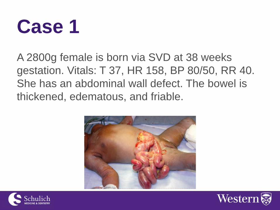
Case 1
A 2800g female is born via SVD at 38 weeks
gestation. Vitals: T 37, HR 158, BP 80/50, RR 40.
She has an abdominal wall defect. The bowel is
thickened, edematous, and friable.

Case 1
What kind of defect is this?
Gastroschisis
What are the associated anomalies?
None.
10% of babies have bowel abnormalities such as
atresia and stenosis. These are related to the
bowel trauma and position in utero.

Case 1
What are the initial steps in managing this patient
after delivery?
ABCs
Insert NG/OG
Cover bowels with saline soaked gauze and plastic
Administer IV fluids, Abx
Maintain normothermia

Case 1
What are the surgical management options?
1.Primary closure in OR. Risk of abdominal
compartment syndrome.
2.Umbilical cord flap. Temporary coverage.
3.Placement of silastic silo. This will prevent
heat/water losses and keep bowel in sterile
environment. Silo tightened over time to slowly
reduce bowel.

Case 1
When would you proceed directly to the OR?
- Closing gastroschisis: defect closes around
viscera causing ischemia and infarction

Case 1
You have repaired the defect primarily and the
baby was started on TPN. 3 weeks later, the NG is
still draining 60 cc bile per day. The baby has not
passed meconium. What do you do next?
A. Observation without intervention
B. Upper GI series
C. Contrast enema
D. Gastric emptying study
E. Initiate oral feeds

Case 1
You have repaired the defect primarily and the
baby was started on TPN. 3 weeks later, the NG is
still draining 60 cc bile per day. The baby has not
passed meconium. What do you do next?
A. Observation without investigation
A period of ileus is expected and does not justify
investigation. Mean time from operation to initiation
of oral feeds in gastroschisis is 3-6 weeks.

Case 1
What is the most likely long-term complication of
gastroschisis?
Short gut syndrome
Adhesive small bowel obstruction

Gastroschisis • Definition: full thickness, paraumbilical
abdominal wall defect (usually < 4 cm)
associated with evisceration of bowel
• No peritoneal sac covering bowel; bowel in
direct contact with amniotic fluid which leads
to serositis
• Liver is rarely involved

Gastroschisis • Incidence: 1 in 3,000 – 8,000 live births
• Etiology: occlusion of right omphalomesenteric
artery during embryogenesis results in
disruption of umbilical ring and bowel
herniation
• Possible factors: premature infants, low birth
weight, young mothers (< 20 years old),
ASA/ibuprofen/vasoconstrictive agents during
1st trimester, EtOH, smoking, recreational
drugs
\
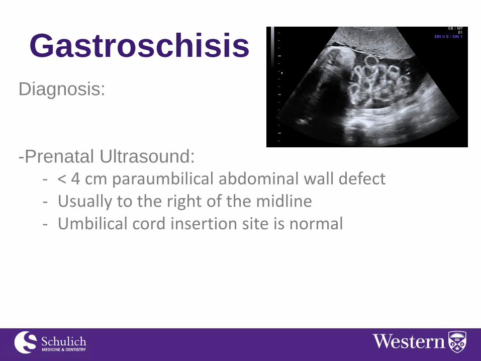
Gastroschisis Diagnosis:
-Prenatal Ultrasound: - < 4 cm paraumbilical abdominal wall defect - Usually to the right of the midline - Umbilical cord insertion site is normal

Gastroschisis
Management: Delivery Room
- Wrap bowel in sterile saline dressings covered
with plastic wrap
- Insert OG to decompress stomach
- IV insertion – IVF and broad spectrum Abx
- Keep neonate in thermoneutral environment
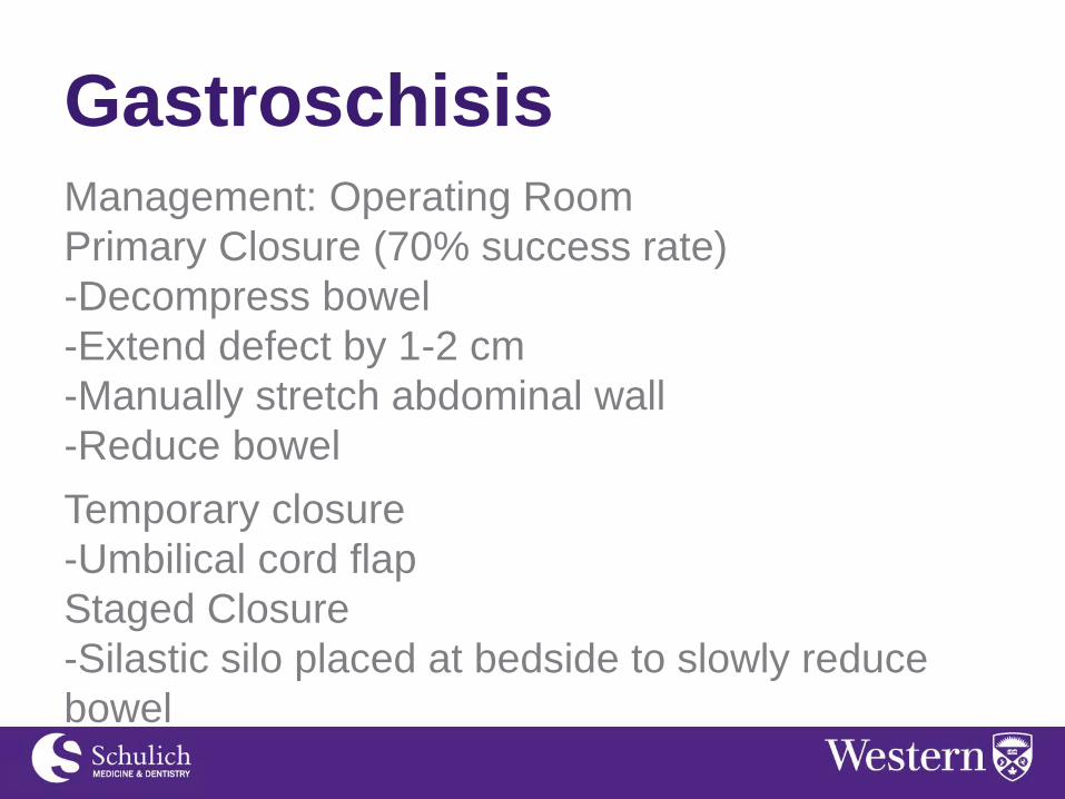
Gastroschisis Management: Operating Room
Primary Closure (70% success rate)
-Decompress bowel
-Extend defect by 1-2 cm
-Manually stretch abdominal wall
-Reduce bowel
Temporary closure
-Umbilical cord flap
Staged Closure
-Silastic silo placed at bedside to slowly reduce
bowel

Gastroschisis

Gastroschisis
If associated with atresia:
1.Primary repair at time of
abdominal wall closure –
OR–
2.Intestinal diversion
(ostomy or long intestinal
tube) followed by delayed
repair

Gastroschisis
Prognosis:
-Survival rate >90%
-Favorable prognosis because typically not
associated with other congenital anomalies
-Prolonged postoperative ileus
-25% complex cases associated with higher risk of
in hospital mortality, short bowel syndrome, bowel
obstruction, NEC, and TPN/tube feedings on
discharge
Case 2
Male born at 34 wk 3 days gestation via C/S
secondary to maternal and fetal distress. Baby
was hypotonic. Apgars: 1 (1 min), 6 (5 min), 7 (10
min). Required PPV and was transferred to NICU.
Consulted for abdominal wall defect.

Case 2
Prenatal hx:
- Born to 25 year old female
- Antenatal U/S showed a midline herniation
with the sac containing bowel loops
- Fetal echo normal
- Spontaneous rupture of membranes at 28 wks
– mother briefly admitted and treated with
steroids and Abx

Case 2 Physical exam –
Large defect (~10cm) with intact amnion
containing bowels and liver
What is the diagnosis?
Giant omphalocele

Case 2
Next steps:
-Stabilize (ABCs)
-Cover with gauze/plastic
-NG, IV Abx
-Rule out associated congenital anomalies - Echo, CXR, genetic testing
-Small omphalocele repair
-Giant omphalocele delayed repair - Topical sclerosing agent

3 weeks 4 weeks
5 weeks
6 weeks 2 months 3 months

4 months 9 months
2.5 years

Case 2
Brought to the operating room at age 3 for
laparotomy and primary repair of omphalocele.
Discharged POD 7

Omphalocele
• Definition: abdominal wall defect of varying
size with herniated viscera contained in a sac
• Incidence: 1 in 6,000 – 10,000 live births

Omphalocele Development of abdominal wall and GI tract
depends on growth and fusion of cephalic, caudal,
and lateral embryologic folds.
Omphalocele occurs when there is failure of
migration and fusion of these folds.
Can be associated with other midline defects.
-Pentalogy of Cantrell - Failure of cephalic fold: epigastric omphalocele,
anterior diaphragmatic defect, sternal cleft, pericadial and cardiac defects

Omphalocele Environmental and social factors play less of a role
compared to gastroschisis
Karyotype abnormalities present in 30%
-Trisomy 13, 18, 21
50% associated with malformations
-Cardiac*, MSK, GI, GU
Beckwith-Wiedemann syndrome
-Omphalocele, macroglossia, hyperinsulinism

Omphalocele Defect: small (2-5 cm) to large (>8 cm)
Liver containing or Non liver containing
“Giant omphalocele” – contains 75% of liver
Sac: amniotic membrane, mesenchymal tissue
(i.e. Wharton jelly), and peritoneum
Bowel: healthy (not exposed to amniotic fluid);
malrotation is usually present

Omphalocele Diagnosis:
Prenatal U/S
(2nd Trimester)
Physical Exam

Omphalocele Management:
-C-section if giant omphalocele with herniation of
liver
-External warmer/incubator
-NG/OG tube
-Cover with saline soaked gauze and plastic wrap
-IVF + IV Abx
-Pre-op work-up: - CXR, echo, renal U/S, bloodwork

Omphalocele Management: Surgical
-Reduce herniated viscera
-Primary fascial closure (60-70%)
-Resect sac
-May consider staged closure (silo)
-Limiting factor is intra-abdominal pressure - Avoid abdominal compartment syndrome - Intra-operative intragastric/intravesical pressure,
end tidal CO2, CVP, regional oximetry

Omphalocele Management: Giant Omphalocele (10%)
-Primary closure not possible due to poorly
developed abdominal wall
-Promote epithelialization of sac with secondary
closure of ventral hernia at later date - Topical agents used include: mercurochrome, silver
nitrate, silver sulfadiazine

Omphalocele
Post-op:
- Prolonged ileus is not common
- Increased frequency of gastroesophageal
reflux
- Excellent prognosis if not associated with
severe malformations - Increased mortality in infants with chromosomal
syndromes and cardiac defects
Gastroesophageal reflux
(GER) Epidemiology:
Symptoms begin within 6 weeks of life
80% become symptom free by age 1
20% continue to be symptomatic
4% of these patients develop esophageal
strictures

GER
Mechanism: inappropriate LES relaxation
Clinical manifestations:
-Regurgitation
-Coughing, wheezing, stridor secondary to
aspiration
-Refusal to eat (secondary to esophagitis) - May lead to failure to thrive and malnourishment
-Sandifer’s syndrome - Spastic posturing of head, neck, upper trunk associated
with eating; disappears during sleep

GER
Diagnosis:
- Clinical
- Scintigraphy – technetium (gastric emptying)
- 24 hr pH monitoring - Indications: respiratory symptoms, intractable crying,
reactive airways, recurrent pneumonia
- Endoscopy and biopsy - Indications: suspicion of esophagitis, dysphagia
- Manometry

GER
Treatment:
Positioning – prone with head elevated
Thicken foods
Medications
- Antacids
- H2 receptor antagonists: ranitidine
- Prokinetic agents: cisapride (increase LES,
improve esophageal peristalsis and gastric
emptying)
- Proton pump inhibitor: omeprazole

GER
Surgery:
- Intractable esophagitis or emesis that does
not respond to PPIs
- Principles - Lengthening intra-abdominal esophagus - Accentuation of the angle of His - Increase in pressure barrier at the GE junction - Approximation of the crura
GER
- Nissen Fundoplication**
- Boix-Ochoa Technique
- Thal procedure
- Toupet procedure

Umbilical Hernia
Most common abdominal wall defect
Incidence:
10 x more common in African Americans
25-50% vs 5-10%
Increased risk in premature infants

Umbilical Hernia
Umbilical ring closes by contracture after cord
ligated and umbilical vessels thrombose
Failure of recti to approximate and failure of round
ligament to attach to both superior and inferior
margins of the umbilical ring predispose fetus to
developing umbilical hernia
Umbilical Hernia
Diagnosis:
- Physical exam – usually first noted after
separation of umbilical cord remnant from
umbilicus
- Usually asymptomatic and reducible

Umbilical Hernia
Management:
-Majority spontaneously close
-If hernia persists by age 4-5 then should be
repaired
-Defect > 1.5-2 cm less likely to close and repair
may be considered earlier (age 2-3)
-10% persist into adulthood
-Risk of incarceration or strangulation is rare but
does increase in adults
Epigastric Hernia
Result from defects in linea alba
Incidence: 5%
Usually very small (0.5 – 1 cm palpable mass)
Do not resolve spontaneously – surgical repair
required

Epigastric Hernia
Diastasis recti – rectus abdominis not fully
developed

Case 3
13 day old boy presents with 1 day history of
bilious emesis x 2 and BRBPR x 1
PMHx:
Born at 37 wks 3 days
Mother on methadone
NICU stay (no intubation) for a few days
Case 3
Differential diagnosis:
Next steps?

Case 3
O/E: T 37, HR 132, RR 54, BP 116/70
Abdomen distended, firm
Bulge in right groin
DRE - melena

Case 3

Case 3
Initial cap gas 3 hours later

Case 3
What is your management plan?
Resuscitate
NG tube
OR

Case 3
- Laparotomy (omega incision)
- Dilated proximal small bowel with 5-8 cm
incarcerated in right inguinal canal (10-15 cm
from ileocecal valve) – reduced with mild
traction, appeared ischemic
- Colon normal; no malrotation
- Right inguinal hernia repair (groin incision)
- Ischemic bowel resected, side to side stapled
anastomosis

Case 3
On POD 8, you note that the baby’s abdomen is
distended and discolored.

Case 3
What next?
Exploratory laparotomy
Findings:
-3 tiny holes (1 mesenteric, 2 antimesenteric at
staple line) closed primarily
-No intra abdominal contamination
-Next?
-Diverting loop ileostomy

Case 3
Patient was discharged 29 days after initial
presentation (POD 21 from diverting ileostomy)
Admitted briefly for rehydration after increased
ostomy outputs
Plan for closure of ileostomy in September

Inguinal Hernia
Most common elective pediatric general surgical
procedure
Types:
-Congenital Indirect (99%)
-Direct (0.5%)
-Femoral (<0.5%)

Inguinal Hernia
Incidence: 1-3% of all children; 3-5% of premature
infants
Increased incidence in those with connective
tissue disorders (i.e. Ehlers-Danlos Syndrome or
Marfan Syndrome)
8:1 male predominance
Right sided (56%) more common than left (27%)

Inguinal Hernia Indirect Inguinal Hernia
-Abnormal patent continuation of peritoneum
(processus vaginalis) through internal inguinal ring
-Hernia sac lateral to inferior epigastric vessels,
anterior/medial to spermatic cord structures which
are retroperitoneal
-Hernia sac descends along spermatic cord within
cremasteric fascia
-Sac can reside within inguinal canal or descend
through external inguinal ring into scrotum

Inguinal Hernia
Direct Inguinal Hernia
- Originates medial to deep inferior epigastric
vessels
- External to cremasteric fascia
- Hernia sac protrudes directly through posterior
wall of inguinal canal
- Can descend through external inguinal ring
into scrotum

Inguinal Hernia
Signs and symptoms of incarcerated hernia:
- Non-reducible mass
- Inconsolable infant
- Feeding intolerance
- Pain
- Abdominal distention, vomiting, obstipation
- Edematous groin – reactive hydrocele

Inguinal Hernia
Operative Considerations
-Elective repair to prevent incarceration - 70% of infants who require operative reduction of
incarcerated inguinal hernia are <11 months old
-May attempt reduction of incarcerated hernia - If remains unreduced for 1-2 hours then urgent
operative reduction and repair
-Recurrence is low (0-1%)
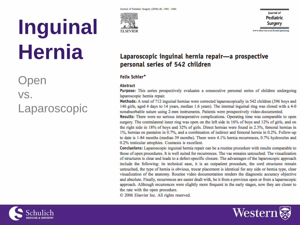
Inguinal
Hernia
Open
vs.
Laparoscopic

Inguinal Hernia
- Operative exploration of asymptomatic contralateral groin controversial
- 60-70% patent processus vaginalis at age < 2
- However, risk of contralateral hernia following open unilateral repair is 6% - Reserve for patients with associated disorders, high suspicion of bilateral
clinical hernia, underlying risk to anesthesia, etc.

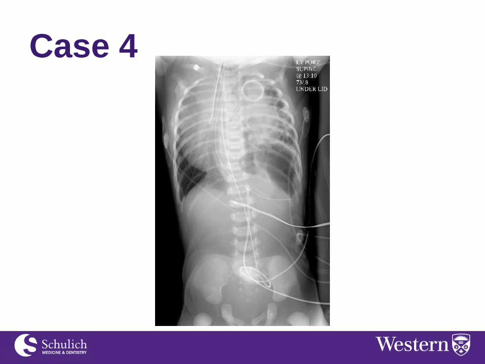
Case 4 Male born at 39 wk 6 days gestation via C/S due
to failure to progress and fetal distress. The
pregnancy was normal other than polyhydramnios
on U/S.
The baby is blue, HR 53. Apgar at 1 minute is 2.
The abdomen is scaphoid. Baby is resuscitated
with bag mask ventilation and nasal intubation.
CXR shows the following:
Case 4

Case 4
What is the diagnosis?
Congenital diaphragmatic hernia
Case 4
Initial steps in management:
Intubation
NG or OG
IVF
NICU
Rule out associated anomalies
-Echo: most common cardiac association is left
heart hypoplasia

Case 4
When would you proceed with surgical repair and
what would you do in the operating room?

Case 4 At DOL 3 – laparotomy and repair of left CDH
1. Left subcostal incision
2. Small bowel, cecum, ascending, transverse,
descending colon, left kidney and spleen in
chest cavity; liver intra abdominal
3. Viscera reduced
4. Large defect spanning entire width of posterior
diaphragm; diaphragm folded with adhesions
5. Posterior diaphragm released – primary repair
6. Viscera inspected – normal

Case 4

Congenital Diaphragmatic
Hernia (CDH) Definition: opening in diaphragm allowing viscera to
herniate into chest
Incidence: 1 in 5000 births
Etiology: unknown
Left side predominance: 80% vs 20%
Location: posterolateral (90%) vs anteriomedial (10%)

CDH
Embryology:
-Septum transversum fuses dorsally with
mesoderm of mediastinum
-Pleuroperitoneal canals connect pleural space
and peritoneal cavity
-Closure of pleuroperitoneal canal fetal
diaphragm (8th wk gestation)
-Defective formation of pleuroperitoneal membrane
or post-hepatic mesenchymal plate results in CDH

CDH

CDH
Lung Development: 4 stages
1.Pseudoglandular (5-17 wks) – major bronchi,
terminal bronchi formed
2.Canalicular (16-25 wks) - respiratory
bronchioles, alveolar ducts, pulmonary vessels
3.Terminal sac (24 wks-birth)
4.Alveolar (late fetal life – childhood)

CDH • Visceral herniation during pseudoglandular
stage Ipsilateral pulmonary hypoplasia
– Decrease in pulmonary mass and weight
– Reduction in bronchiole divisions
– Reduction in number of alveoli and respiratory bronchioles
– Hypolastic pulmonary vascular tree with abnormal muscularization of pulmonary arterioles
– Surfactant deficiency

CDH

CDH
Associated anomalies (50%):
- Cardiac (60%): VSD, ASD, heart hypoplasia
- Neural tube defects
- Pulmonary sequestration
- Renal and genital anomalies
- Trisomies 13, 18, 21
- Midline defects – omphalocele, cleft palate,
EA

CDH
Diagnosis:
-Antenatal ultrasound - Can be identified as early as 25 wk gestation - Polyhydramnios (80%) - Bowel/liver/spleen in chest
-On exam: respiratory distress, decreased air
entry, bowel sounds in chest, scaphoid abdomen
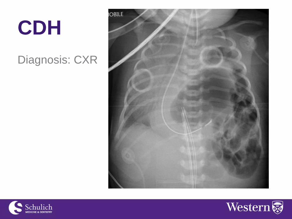
CDH
Diagnosis: CXR

CDH
Management:
-ABCs - Endotracheal intubation, neuromuscular blockade,
PPV - If conventional mechanical ventilation fails, consider:
HFV, HFOV, nitric oxide administration, ECMO
-Insert NG
-Maintain normothermia
-Echo rule out cardiac anomalies
-Repair when stable

CDH
Surgical considerations:
-Transabdominal approach preferred with
subcostal incision
-Bowel, liver, spleen carefully reduced from
thoracic cavity
-Excise hernia sac if present (20%)
-Close small defects primarily with 2-0 or 3-0
permanent sutures
-If large defect, use mesh - Higher risk of recurrence

CDH
Post-Op Care:
-Chest tube to water seal to avoid
overexpansion/pneumothorax of contralateral lung
-Continue mechanical ventilation - Initial decrease in chest compliance - Surgical stress can precipitate pulmonary
vasoconstriction pulmonary hypertension

CDH
Outcomes:
60-80% survival
- Highly variable
- Depends on associated anomalies
- Depends on severity of lung hypoplasia

CDH
Indications for fetal surgery:
-Severe pulmonary hypoplasia
-0% survival with optimal postnatal care
-Predictors of high mortality on U/S - Early cardiac ventricular disproportion - Reduced lung area to head circumference ratio - Hepatic herniation into chest - Polyhydramnios diagnosed before 25 weeks gestation - Left ventricular hypoplasia

CDH
Fetal tracheal occlusion:
- Causes lung growth and gradual reduction of
herniated viscera
- Mediated by mechanical and hormonal growth
stimuli triggered by trapped fetal lung fluid
(unable to exit into amniotic cavity)
- Goal is for baby to be born without lung
hypoplasia or associated pulmonary
hypertension
- Minimally invasive procedure; experimental

Case 5
7 week old girl born at term. Hospitalized a few
days after birth due to hyperbilirubinemia. Her
pediatrician is concerned as she still appears
visibly jaundiced.
She is thriving, feeding well, and gaining weight
appropriately. She passed meconium after delivery
and now passes 2-3 yellow stools per day.

Case 5
O/E: Jaundiced. Alert, VSS. Abdomen slightly
distended. Liver palpable 4 cm below costal
margin.
Bloodwork is normal other than elevated total
(250) and direct (15) bilirubin

Case 5
What is the differential diagnosis?
Congenital TORCH infections
Neonatal giant cell hepatitis
Alpha 1 antitrypsin deficiency
Alagille syndrome
Biliary atresia
Jaundice that persists longer than 4 weeks and
jaundice due to conjugated hyperbilirubinemia is
pathologic


Case 5
Which investigations would you order?
Bloodwork
-Liver enzymes, bilirubin, serum alpha 1
antitrypsin, hepatitis work up
U/S
Nuclear scintigraphy (i.e. HIDA scan)***
-Uptake by liver without excretion for 48 hours is
highly suspicious for biliary atresia

Case 5
Tests performed are inconclusive. Baby is now 8
weeks and jaundice continues. You decide to
operate on the baby.
How will you proceed?

Case 5
Intraoperative
cholangiogram:

Case 5
Next step?
Kasai portoenterostomy
- Excision of biliary tree
- Anastomosis of jejunum to portal plate
Liver biopsy – determine prognosis

Biliary Atresia Definition: progressive obliteration of normally
developed extrahepatic biliary tract Biliary hypoplasia - diminutive but patent biliary tree secondary
to neonatal hepatitis or alpha 1 antitrypsin deficiency
Incidence: 1 in 10, 000 births
Mechanism unknown
Most common cause of chronic cholestasis in
pediatric population

Biliary Atresia
Subtypes:
A.Complete obliteration of biliary ducts
B.Obliteration of proximal ducts with patent distal
ducts
C.Patent proximal ducts with distal fibrosis

Biliary Atresia
Presentation and Diagnosis:
- Progressive neonatal jaundice during first few
weeks of life
- Dark urine
- Acholic stools
- Late findings: failure to thrive, feeding
intolerance, portal hypertension, fat soluble
vitamin deficiency

Biliary Atresia Investigations:
-Conjugated hyperbilirubinemia
-Serum alpha 1 antitrypsin
-Radioisotope scanning (technetium 99m) - Biliary atresia will have prompt uptake and no excretion
into gut because of obliterated extrahepatic bile ducts
-Cholangiography - Ruled out If bile ducts patent from liver to duodenum
-U/S - Diminutive or absent gallbladder without associated
intrahepatic duct dilatation

Biliary Atresia
Management:
Surgery
-Early biliary drainage within 2-4 months of age
may be associated with reversal of liver injury and
increase long term survival
Medical therapy – post-op management of chronic
liver disease

Biliary Atresia

Biliary Atresia
Portoenterostomy:
- Developed in Japan by Morio
Kasai (1950s)
- Biliary drainage to arrest or
reverse parenchymal liver
injury
- Probability of bile flow depends
on age at time of operation - 65-75% chance of bile flow if <2
months of age

Biliary Atresia Post-operative cholangitis (40-50%)
-Fever, leukocytosis, decreased bile flow
-Medical management: IVF, antibiotics
1/3 – do not require transplant
1/3 – liver failure after 5 years, requiring transplant
1/3 – liver failure post-op, requiring transplant
5 year survival rate ~50%
Biliary Atresia
Liver Transplantation:
Indications: - Progressive hepatic failure despite portoenterostomy - Growth retardation - Complications of portal hypertension
5 year survival 75-95%
Consider effects of lifelong immunosuppression - Risk of infection and treatment related malignancies

Case 6
3 year old female referred to outpatient clinic for
”cystic lesion” on U/S of RUQ. History of
abdominal pain since 3 months of age. Treated for
constipation with daily PEG.

Case 6
Physical exam:
VSS
Abdomen non distended, soft, non tender
No palpable masses
No jaundice

Case 6 U/S:
4.1x 2.4 cm
cyst originating
from biliary tree
Involves extrahepatic ducts – tapers proximally
and distally
Normal intrahepatic ducts
Bloodwork normal

Case 6
What is the diagnosis?
How would you manage this patient?

Case 6
Operation: Da Vinci assisted choledochal cyst
resection, cholecystectomy, Roux en Y
hepaticojejunostomy
Discharged POD 3
Choledochal Cysts
Definition: cystic dilatations of the bile ducts
Incidence: 1 in 100, 000
Possible mechanisms:
-Abnormal recanalization of primitive bile duct
cords
-Inflammation caused by reflux of pancreatic
secretions into CBD
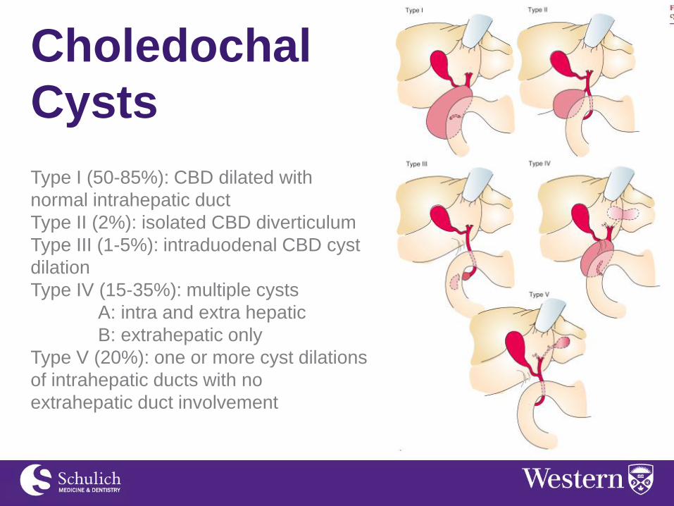
Type I (50-85%): CBD dilated with
normal intrahepatic duct
Type II (2%): isolated CBD diverticulum
Type III (1-5%): intraduodenal CBD cyst
dilation
Type IV (15-35%): multiple cysts
A: intra and extra hepatic
B: extrahepatic only
Type V (20%): one or more cyst dilations
of intrahepatic ducts with no
extrahepatic duct involvement
Choledochal
Cysts

Choledochal Cysts
Choledochal Cysts
Presentation and Diagnosis:
Jaundice
Abdominal pain
Abdominal mass
Imaging:
U/S
Radioscintigraphy (technetium 99m)
MRCP or ERCP

Choledochal Cysts
Treatment:
- Excision of cyst with direct anastomosis of
proximal normal bile duct to Roux en Y loop of
jejunum
- External drainage is temporizing measure
reserved for emergency decompression
- Cyst enterostomy should not be done - High rate of stricture - Possibility of biliary malignancy

Choledochal Cysts Treatment:
Type I
-Surgical exploration and cholangiography
-Cholecystectomy
-Primary excision with Roux en Y
Type II
-Resection and reanastomosis of CBD
Type III
-Transduodenal approach – excision of cyst and
sphincteroplasty
Type IV and V intrahepatic cysts
-Roux en Y

Choledochal Cysts
Complications:
- Cholangitis
- Stricture formation
- Choledocholithiasis
- Cholangiocarcinoma - Incidence is 2.5-5% if incomplete excision of
choledochal cyst - Survival time is <1 year after cancer detected


References
1) Mulholland, M et al. Greenfield’s Surgery: Scientific Principles and
Practice 5th edition. 2011.’
2) Ashcraft et al. Pediatric Surgery 3rd edition. 2000.
3) Moss, L. R. et al. Case Studies in Pediatric Surgery. 2000.
4) UpToDate