General Issues: Meeting to Discuss the Evaluation of ... · Endovascular Medical Devices Intended...
50
CO-1 General Issues: Meeting to Discuss the Evaluation of Safety and Effectiveness of Endovascular Medical Devices Intended to Treat Intracranial Aneurysms Neurological Devices Panel March 1, 2018
Transcript of General Issues: Meeting to Discuss the Evaluation of ... · Endovascular Medical Devices Intended...

CO-1
General Issues: Meeting to Discuss the Evaluation of Safety and Effectiveness of Endovascular Medical Devices Intended to Treat Intracranial Aneurysms
Neurological Devices Panel
March 1, 2018

CO-2
Introduction
Jacques Dion, MD
Vice President Scientific Affairs
MicroVention
CO-3
Unified Industry Presentation

CO-4
Target aneurysm treatment populations and challenges associated
with natural history data
▪ All aneurysms, including small aneurysms, present risks to
patients and should be considered for treatment
How to use current safety and effectiveness data to evaluate new
device technology
Recommendations and post-marketing studies
Presentation Focus to Advance Aneurysm Treatment and Patient Care
CO-5
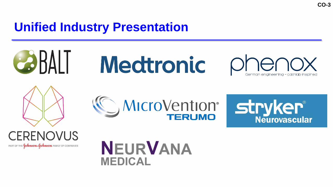
Agenda
Aneurysm Disease
Background
Jacques Dion, MDVice President Scientific Affairs
MicroVention
Current Clinical Trial Data
to Support Safety and
Effectiveness
Stacey PughVice President and General Manager
Medtronic Neurovascular
Recommendations and
Conclusion
John Allison, RACVice President, Regulatory and Clinical Affairs
Stryker Neurovascular

CO-6
Aneurysm Disease Background
Jacques Dion, MD
Vice President Scientific Affairs
MicroVention

CO-7
2–5% of adults have an IA1
Screening for IAs not standard practice
Majority of IAs asymptomatic and undiagnosed prior to rupture
Ruptures typically occur suddenly and often lead to cerebral
bleeding or subarachnoid hemorrhage (SAH)
SAH is a devastating disease2
▪ ~45% of events are fatal
▪ ~50% of survivors experience significant disability
Significant Consequences of Intracranial Aneurysms (IAs)
1) Thompson, 2015; 2) Lantigua, 2015

CO-8
Aneurysm rupture attributed to many factors
▪ Size, morphology, location, prior history of SAH
Consistent trends in literature demonstrate increased risk
▪ Larger vs. smaller
▪ Posterior circulation vs. anterior circulation
Severity and consequences associated with rupture
independent of size and location
Difficult to Predict Risk of Rupture
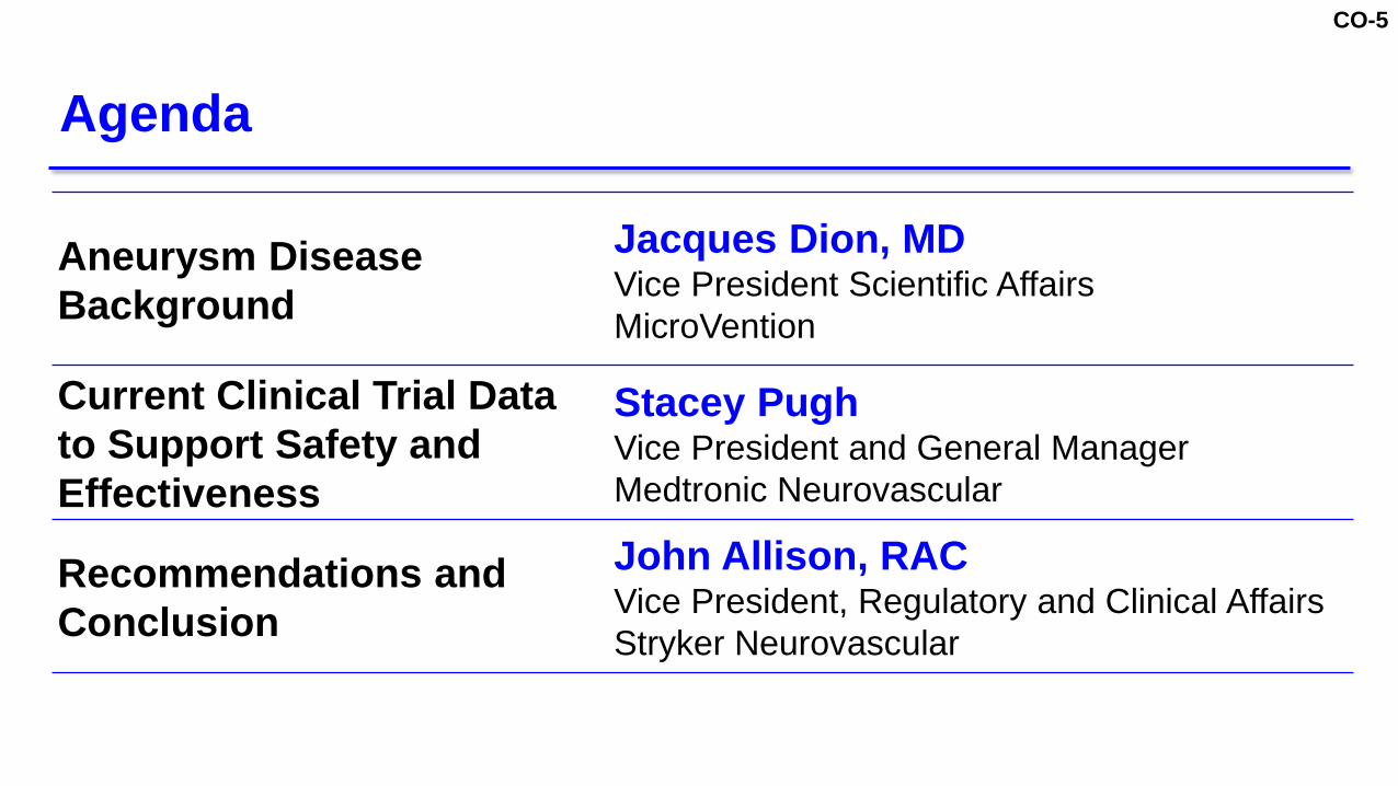
CO-9
International Study Unruptured Intracranial Aneurysms (ISUIA)
▪ Initial report published 1998
▪ Post-hoc re-analysis of data 2003
Two natural history of aneurysm studies in large cohorts in
Finland and Japan
Inconsistency in studies creates uncertainty regarding
prevalence
Reliable Conclusions Challenging to Draw from Natural History Studies
International Study of Unruptured Intracranial Aneurysms Investigators, 1998 and 2003
The UCAS Japan Investigators, 2012; JUVELA, 2000

CO-10
Large, retrospective and prospective, cohort study
▪ 60 centers in USA, Canada and Europe
Patients evaluated in 3 non-randomized cohorts
▪ Observation, surgical, and endovascular treatment
2 groups broadly defined for observation
▪ Group 1 without history of SAH
▪ Group 2 with history of SAH
Patients followed annually for 4 years with standardized questionnaire
1998 retrospective analysis in 1449 patients
▪ Group 1 aneurysm < 10 mm had rupture rate of < 0.05%
ISUIA Study Design

CO-11
ISUIA (2003) Post-Hoc Re-Analysis Suggest No Risk to Patients with Aneurysms < 7 mm
5-Year Cumulative Risk, %
< 7 mm
7–12 mm 13–24 mm ≥ 25 mm
No SAH
Separate
Aneurysm
SAH
Separate
Aneurysm
Cavernous Carotid Artery (N=210) 0 0 0 3.0 6.4
AC/MC/IC (N=1037) 0 1.5 2.6 14.5 40
Post-PCom (N=445) 2.5 3.4 14.5 18.4 50
AC=Anterior communicating or anterior cerebral artery; IC=internal carotid artery (not cavernous carotid artery); MC=middle cerebral artery;
Post-PCom=vertebrobasilar, posterior cerebral arterial system, or posterior communicating artery
CO-12
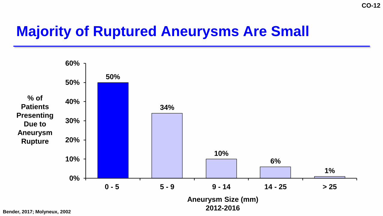
Majority of Ruptured Aneurysms Are Small
50%
34%
10%6%
1%0%
10%
20%
30%
40%
50%
60%
0 - 5 5 - 9 9 - 14 14 - 25 > 25
Aneurysm Size (mm)
2012-2016
% of
Patients
Presenting
Due to
Aneurysm
Rupture
Bender, 2017; Molyneux, 2002

CO-13
Post-hoc reconstructions of artificial subgroups
Methodological factors impacting low rate of reported rupture
▪ Selection bias
▪ Arbitrary assignment of PCom aneurysms to posterior
circulation
▪ High crossover rate from observation to treatment group
▪ Undefined observational periods with no predefined
hypotheses, sample size, subgroup definitions
▪ Aneurysms < 2 mm excluded
ISUIA Study Limitations
Raymond, 2008

CO-14
Primary goal to prevent rupture and related morbidity and
mortality
Secondary goals
▪ Symptom relief due to mass effect
▪ Prevent further growth
▪ Prevent thrombus formation
Goals of Treatment

CO-15
Current options are surgical or endovascular
▪ Surgical clipping associated with high occlusion success, but safety varies
according to location
1.7 – 2.6% mortality rate1,2
5 – 10.9% permanent morbidity rate1,2
ISUIA: 2.3 / 12.1%3
Surgical risk related to location2:
▪ Small (< 10 mm): 4%
▪ Large (10 – 24 mm): 12.1%
▪ Giant (> 25 mm): 26.5%
▪ Anterior vs. posterior: RR = 4.1
Surgical Clipping High Occlusion Success but Limited to Certain Anatomical Locations
1) Kotowski; 2) Raaymakers, 1998; 3) ISUIA, 2003

CO-16
Progression of endovascular treatment
▪ Coiling
▪ Stent-assisted coiling
▪ Balloon assisted coiling
▪ Flow diversion
Innovative and refined endovascular treatments reduce
complications and improve outcomes
Endovascular Treatment Options Evolving
Etminan, 2016

CO-17
What patient characteristics justify foregoing treatment for an
aneurysm that would otherwise be considered for treatment?
FDA Question 3

CO-18
Life expectancy
Family history of aneurysmal SAH
Co-morbidities (poorly controlled HTN, PKD, smoking)
Aneurysmal growth on sequential imaging
Aneurysm location
Risk of treatment
Patient choice
Factors to Consider for Aneurysm Treatment

CO-19
Who and when to treat
Risks of surgical and endovascular treatments well-described
Inconsistent literature reports make interpretation of natural
history difficult1
All Patients Need Treatment Options
1) ISUIA

CO-20
Current Clinical Trial Data to Support Safety and Effectiveness
Stacey Pugh
Vice President and General Manager
Medtronic Neurovascular

CO-21
Ongoing IDE Trials for Aneurysm Treatment
WEB Intra-saccular FD
Barrel VRD
Liberty Stent-assisted Coiling
ATLAS Stent-assisted Coiling
LVIS Stent-assisted Coiling
FRED FDS
Premier Pipeline FDS
SCENT Surpass FDS
2012 2013 2014 2015 2016 2017 2018
Q1 Q2 Q3 Q4 Q1 Q2 Q3 Q4 Q1 Q2 Q3 Q4 Q1 Q2 Q3 Q4 Q1 Q2 Q3 Q4 Q1 Q2 Q3 Q4 Q1 Q2 Q3 Q4
n=180 12-month follow-up
12-month follow-up
12-month follow-up
12-month follow-up
12-month follow-up
12-month follow-up
12-month follow-up
Enrollment Follow-up
n=141
n=145
n=153
n=180
n=120
n=138
n=150 12-month follow-up
> 10 mm, WN ICA
< 12 mm, WN ICA/Vert
> 10 mm WN ICA
WN ICA, rupt/unrupt
WN ICA, rupt/unrupt
WN ICA, rupt/unrupt
WNBA in MCA/Basilar
WNBA in Basilar, MCA/ACOM, ICA

CO-22
Operating Characteristics Common Features
Prospective, multi-center, single-arm, PG driven studies
12-month primary safety endpoints
12-month primary effectiveness endpoints
Formal hypothesis and predetermined statistical analysis plan
Core Lab adjudications of imaging endpoints
Independent DSMB and CEC review
Similar Characteristics Across All 8 Studies
✓
✓
✓
✓
✓
✓

CO-23
Industry Perspective on FDA Questions

CO-24
Can the mRS at 1 year also be a potential primary safety
outcome measure for all endovascular device trials?
FDA Question 2

CO-25
Challenging in evaluation of ruptured aneurysm treatment due to
significant disabilities present at or near time of treatment
▪ Pre-rupture: not reflective of disability from rupture
▪ Post-treatment: could mask procedure related harm
Non-specific to cause of functional dependency
Changes in mRS scores could be due to factors other than
aneurysm treatment
Period of observation for ischemic stroke is 3 months, not 12
months as in aneurysm therapy
mRS Suitable for Ischemic Stroke but Challenging for Aneurysm Therapy

CO-26
4a: Do you consider the Raymond Classification Scale to be the
standard to assess effectiveness for ALL endovascular
intracranial aneurysm treatment devices?
4b: If the Raymond Classification scale is used, is Raymond II (or
higher) classification a satisfactory outcome for aneurysm patients
with unruptured aneurysms? And is Raymond II (or higher)
classification a satisfactory outcome for aneurysm patients with
ruptured aneurysms?
FDA Question 4

CO-27
Raymond-Roy Classification System Most Established and Reasonable Method to Assess Aneurysm Occlusion
Raymond
Classification Definition Example
Class IComplete occlusion of
aneurysm including neck
Class II
Persistence of original arterial
wall defect without opacification
of aneurysmal sac
Class III Opacification of aneurysmal sac

CO-28
Do aneurysm occlusion assessment recommendations using
Raymond differ for endosaccular devices?
FDA Question 6

CO-29
Evaluation of Occlusion via Raymond-Roy in Aneurysm Treatment Trials
Intra-Luminal Intra-Saccular
Flow Diversion Coiling
Stent-Assisted Coiling or
Balloon Assisted Coiling
Intra-Saccular
Flow Disruption
Raymond I
Raymond II
(Stable)
Raymond II
(Not Stable)
Raymond III
✓ ✓ ✓
✓✓✓×
×
×
×
×
×
×
×
×
✓
Mazur, et al. J NeuroIntervent Surg. 2016

CO-30
Evaluation of Stable Raymond II for Intra-Saccular Technologies
Coiling
Stent-Assisted
Coiling or
Balloon Assisted
Coiling
Intra-
Saccular
Flow
Disruption
Raymond I ✓ ✓ ✓
Raymond II
(Stable) ✓ ✓ ✓
Raymond II
(Not Stable) × × ×
Raymond III × × ×
What is stable Raymond II?
▪ Defined by serial observations
via MRA/DSA required to
establish “stability”
▪ ≥ 6 months apart from first
assessment
▪ Assessments must demonstrate
equal or better occlusion of the
neck remnant

CO-31
Evaluation of Stable Raymond II for Intra-Saccular Technologies
Coiling
Stent-Assisted
Coiling or
Balloon Assisted
Coiling
Intra-
Saccular
Flow
Disruption
Raymond I ✓ ✓ ✓
Raymond II
(Stable) ✓ ✓ ✓
Raymond II
(Not Stable) × × ×
Raymond III × × ×
▪ Raymond II stable outcomes ONLY acceptable for intra-saccular technology evaluation
▪ Evaluation must be adjudicated by independent core lab
▪ Primary effectiveness analysis at 1 year for Raymond II could not occur until 2 stable assessments
▪ Raymond II occlussions must be followed for 2 years post-efficacy assessment for recurrence or growth

CO-32
Does a worsening in the Raymond scale at follow-up imaging
warrant retreatment and should FDA consider a worsening of
the Raymond scale during 1 year follow-up to represent a
failure of treatment?
FDA Question 8

CO-33
7: What length of follow-up is recommended to assess effectiveness for
endovascular aneurysm treatment devices?
10: What is a sufficient long term follow-up period for a post-approval study
where the majority of patients have the following outcomes for ruptured or
unruptured aneurysms?
FDA Questions 7 and 10

CO-34
Recommendations for Duration of Follow-Up by Raymond-Roy Status
If Novel Technology
Raymond III
Raymond II
(not stable)
Raymond II
(stable)
Raymond I
Year 1 Year 2 Year 3
Failure - Consider Retreatment
Failure - Consider Retreatment
Primary Effectiveness Assessment Post-Market
CO-35

CO-36
We consider digital subtraction angiography (DSA) to be the gold
standard to assess aneurysm occlusion at follow-up. Can
magnetic resonance angiography (MRA) or computed
tomography angiography (CTA) serve as a surrogate follow-up
examination and when should this take place?
FDA Question 9

CO-37
DSA gold standard to assess aneurysm occlusion
▪ Invasive and not without risks
MRA offers advantages compared to DSA1
▪ May be appropriate alternative to DSA for some treatment
technologies
▪ MRA positive correlation to DSA with assessing occlusion2,3
Non-invasive MRA eliminates risk of cerebral thromboembolism and
ionizing radiation2
AHA Guidelines state MRA is reasonable alternative to DSA for
follow-up for treated aneurysms1
Alternative Imaging Assessments
1) Thompson. Stroke, 2015; 2) S.R. Boddu et al. 2014; 3) M.J. van Amerongen et al. 2014

CO-38
Studies can be assessed for effectiveness via Raymond-Roy
scale of aneurysm occlusion
Provided clarity regarding nuances of this scale as it relates to
technology and acceptable outcome
▪ Recommendations for subject follow-up and reporting
Articulate specific challenges for requirement for aneurysm
study follow-up imaging
IDE Studies Conducted Allow Meaningful Analysis of Safety and Effectiveness
CO-39
Recommendations for Current and Future Studies for Aneurysm Treatment and Conclusion
John Allison, RAC
Vice President, Regulatory and Clinical Affairs
Stryker Neurovascular

CO-40
Ongoing Multiple Single-Arm IDE Studies
WEB Intra-saccular FD
Barrel VRD
Liberty Stent-assisted Coiling
ATLAS Stent-assisted Coiling
LVIS Stent-assisted Coiling
FRED FDS
Premier Pipeline FDS
SCENT Surpass FDS
2012 2013 2014 2015 2016 2017 2018
Q1 Q2 Q3 Q4 Q1 Q2 Q3 Q4 Q1 Q2 Q3 Q4 Q1 Q2 Q3 Q4 Q1 Q2 Q3 Q4 Q1 Q2 Q3 Q4 Q1 Q2 Q3 Q4
n=180 12-month follow-up
12-month follow-up
12-month follow-up
12-month follow-up
12-month follow-up
12-month follow-up
12-month follow-up
Enrollment Follow-up
n=141
n=145
n=153
n=180
n=120
n=138
n=150 12-month follow-up
> 10 mm, WN ICA
< 12 mm, WN ICA/Vert
> 10 mm WN ICA
WN ICA, rupt/unrupt
WN ICA, rupt/unrupt
WN ICA, rupt/unrupt
WNBA in MCA/Basilar
WNBA in Basilar, MCA/ACOM, ICA

CO-41
Most practical and pragmatic approach to understanding
success and failure of innovative devices
Well-designed, multi-center, and core lab adjudicated
▪ Builds evidence in area of high unmet medical need
Generates sufficient evidence for PG assessment in high
heterogeneous, low volume population
Serves as future standard for well-defined OPC models
Current Single-Arm Studies with PGs Generate Sufficient Evidence for Approvals

CO-42
Appointment of independent 3rd party to oversee OPC creation
▪ Participation from industry partners, medical societies and FDA
Published data from current IDE studies to validate OPC(s) per
aneurysm type and influence evidence-based guidance
Pooling patient level data to better answer questions on subgroups
Enable FDA to include OPC(s) in future guidance document
Unified Industry Proposal to Generate OPCs

CO-43
Trials with PGsUtilize 3rd Party
to Develop OPC
Stakeholder Acceptance of
OPC
PMS/RWE and New
Technology Data
Timeline for Generation of OPC
2018 2019+ Periodic
Updates2012-2018

CO-44
Implementation of OPC
Appoint Multidisciplinary
Team
Define Research Question
Develop SAPIntegrate Study
Data
Generate OPCs and Subgroup
Data
CO-45
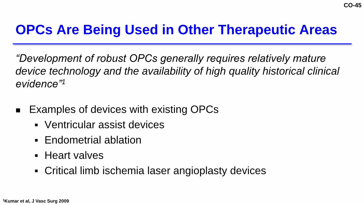
“Development of robust OPCs generally requires relatively mature
device technology and the availability of high quality historical clinical
evidence”1
Examples of devices with existing OPCs
▪ Ventricular assist devices
▪ Endometrial ablation
▪ Heart valves
▪ Critical limb ischemia laser angioplasty devices
OPCs Are Being Used in Other Therapeutic Areas
1Kumar et al, J Vasc Surg 2009
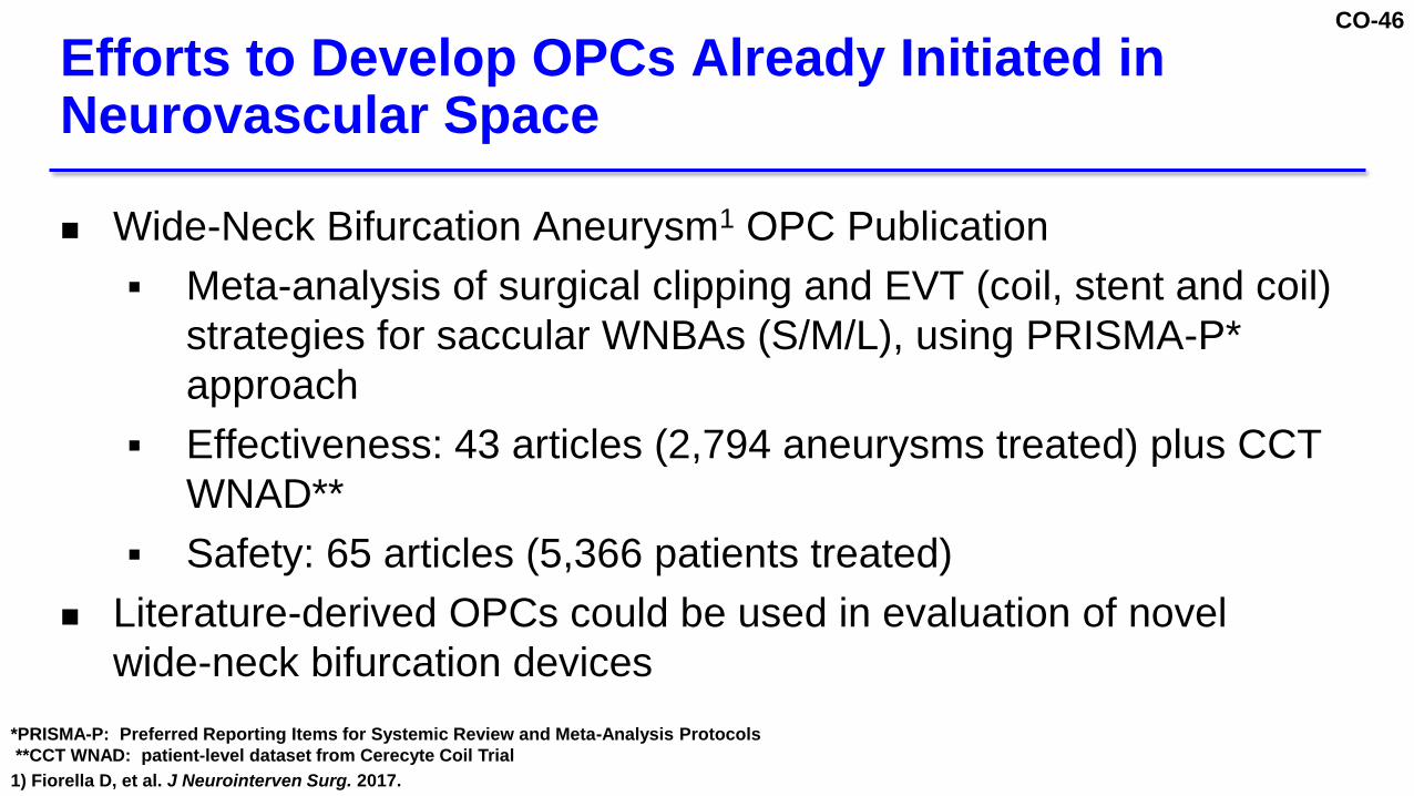
CO-46
Wide-Neck Bifurcation Aneurysm1 OPC Publication
▪ Meta-analysis of surgical clipping and EVT (coil, stent and coil)
strategies for saccular WNBAs (S/M/L), using PRISMA-P*
approach
▪ Effectiveness: 43 articles (2,794 aneurysms treated) plus CCT
WNAD**
▪ Safety: 65 articles (5,366 patients treated)
Literature-derived OPCs could be used in evaluation of novel
wide-neck bifurcation devices
Efforts to Develop OPCs Already Initiated in Neurovascular Space
*PRISMA-P: Preferred Reporting Items for Systemic Review and Meta-Analysis Protocols
**CCT WNAD: patient-level dataset from Cerecyte Coil Trial
1) Fiorella D, et al. J Neurointerven Surg. 2017.
CO-47
Aneurysms at risk of rupture regardless of size warrant
consideration for treatment
Provided industry perspective and practical solutions
Current single-arm PG studies can provide reasonable
assurance of safety and effectiveness
Numerous IDE studies near completion and evidence maturing
to derive OPC model
OPCs can establish clinical trial design standards
Conclusion

CO-48
General Issues: Meeting to Discuss the Evaluation of Safety and Effectiveness of Endovascular Medical Devices Intended to Treat Intracranial Aneurysms
Neurological Devices Panel
March 1, 2018

CO-49
Q&A Slides Shown

CO-50