GENERAL DENTISTRY
12
QUINTESSENCE INTERNATIONAL | volume 51 • number 9 • October 2020 763 Krastl et al Initial management of dental trauma: musts, shoulds, and cans Gabriel Krastl, Prof Dr med dent/Andreas Filippi, Prof Dr med dent/Roland Weiger, Prof Dr med dent When dental trauma occurs, initial management on the day of injury has a determining influence on healing and thus on the prognosis of the affected teeth. Improper, delayed, and/or in - consistent treatment often has far-reaching consequences that cannot be reversed later, even with great effort, especially in children and adolescents. In most cases, it is unrealistic or impossible for the patient to get to a specialized dental trauma facility in time. Therefore, it is every dental practitioner’s duty to provide adequate initial diagnosis and treatment of dental trauma at their dental practice, even if they do not have rou - tine experience in this area. This article serves as a guide to the initial management of dental trauma. It utilizes a three-tiered approach to illustrate which initial management measures are absolutely essential (MUSTS), which should ideally be per - formed (SHOULDS), and which are not top priorities but can be performed (CANS) if the necessary time, training and expe - rience, and equipment and facilities are available. For further treatment, dental practitioners should realistically assess the limits of their ability to treat complex dental trauma cases and, if necessary, they should refer the patient to a specialist or specialized treatment center. (Quintessence Int 2020;51: 763–774; doi: 10.3290/j.qi.a45103 ; modified from a previously published article (in German) Quintessenz 2019;70(9):990–1002) Key words: dental trauma, emergency treatment, initial management The prognosis of traumatic dental injuries depends on both the severity of the injuries and on the treatment received. In addi- tion to first responses at the scene of the accident, the initial treatment measures initiated by the dental practitioner who first manages the case have a particularly decisive influence on the subsequent healing process. 1 Therefore, clinicians must be able to make quick and competent decisions regarding a wide range of patterns of dental trauma during the initial manage- ment phase. These initial decisions form the basis for later treat- ment steps. The dental practitioner must select treatments so as to avoid negative effects on jaw growth on the one hand, while taking the high life expectancy of these mostly young patients into account on the other . 2 This article focuses exclusively on the requirements for the initial management of traumatic dental injuries, to be per- formed at initial presentation on the day of injury. Since dental trauma patients usually arrive at the dental office unannounced and the number of appointments kept open for emergency cases is generally limited, the practitioner must decide what treatment is absolutely necessary and what can be omitted without compromising the prognosis of the injured structures. A three-tiered approach is utilized to illustrate which measures are absolutely essential (MUSTS), which should ideally be per- formed (SHOULDS), and which are not top priorities but can be performed (CANS) if the necessary time, training and experi- ence, and equipment and facilities are available (Table 1). Initial diagnosis of dental trauma During the initial diagnosis of dental trauma (Table 2), it is of utmost importance to exclude traumatic brain injury. Fractures of the alveolar process, mandible, and midface as well as other possibly more serious non-odontogenic injuries must also be excluded. The patient’s tetanus immunization status must be GENERAL DENTI S TRY
Transcript of GENERAL DENTISTRY
QUINTESSENCE INTERNATIONAL | volume 51 • number 9 • October 2020 763
Krastl et al
Initial management of dental trauma: musts, shoulds, and cansGabriel Krastl, Prof Dr med dent/Andreas Filippi, Prof Dr med dent/Roland Weiger, Prof Dr med dent
When dental trauma occurs, initial management on the day of
injury has a determining influence on healing and thus on the
prognosis of the affected teeth. Improper, delayed, and/or in-
consistent treatment often has far-reaching consequences
that cannot be reversed later, even with great effort, especially
in children and adolescents. In most cases, it is unrealistic or
impossible for the patient to get to a specialized dental trauma
facility in time. Therefore, it is every dental practitioner’s duty
to provide adequate initial diagnosis and treatment of dental
trauma at their dental practice, even if they do not have rou-
tine experience in this area. This article serves as a guide to the
initial management of dental trauma. It utilizes a three-tiered
approach to illustrate which initial management measures are
absolutely essential (MUSTS), which should ideally be per-
formed (SHOULDS), and which are not top priorities but can
be performed (CANS) if the necessary time, training and expe-
rience, and equipment and facilities are available. For further
treatment, dental practitioners should realistically assess the
limits of their ability to treat complex dental trauma cases and,
if necessary, they should refer the patient to a specialist
or specialized treatment center. (Quintessence Int 2020;51:
763–774; doi: 10.3290/j.qi.a45103; modified from a previously
published article (in German) Quintessenz 2019;70(9):990–1002)
Key words: dental trauma, emergency treatment, initial management
The prognosis of traumatic dental injuries depends on both the
severity of the injuries and on the treatment received. In addi-
tion to first responses at the scene of the accident, the initial
treatment measures initiated by the dental practitioner who
first manages the case have a particularly decisive influence on
the subsequent healing process.1 Therefore, clinicians must be
able to make quick and competent decisions regarding a wide
range of patterns of dental trauma during the initial manage-
ment phase. These initial decisions form the basis for later treat-
ment steps. The dental practitioner must select treatments so
as to avoid negative effects on jaw growth on the one hand,
while taking the high life expectancy of these mostly young
patients into account on the other.2
This article focuses exclusively on the requirements for the
initial management of traumatic dental injuries, to be per-
formed at initial presentation on the day of injury. Since dental
trauma patients usually arrive at the dental office unannounced
and the number of appointments kept open for emergency
cases is generally limited, the practitioner must decide what
treatment is absolutely necessary and what can be omitted
without compromising the prognosis of the injured structures.
A three-tiered approach is utilized to illustrate which measures
are absolutely essential (MUSTS), which should ideally be per-
formed (SHOULDS), and which are not top priorities but can be
performed (CANS) if the necessary time, training and experi-
ence, and equipment and facilities are available (Table 1).
Initial diagnosis of dental trauma
During the initial diagnosis of dental trauma (Table 2), it is of
utmost importance to exclude traumatic brain injury. Fractures
of the alveolar process, mandible, and midface as well as other
possibly more serious non-odontogenic injuries must also be
excluded. The patient’s tetanus immunization status must be
GENERAL DENTISTRY
QUINTESSENCE INTERNATIONAL | volume 51 • number 9 • October 2020764
GENERAL DENTISTRY
checked. For forensic reasons, it must be documented that an
evaluation of these important general medical aspects took
place. Before performing the actual dental examination, the
dental practitioner must ensure that any avulsed teeth brought
to the dental office are placed in a tooth rescue box immediately.
Traumatic dentoalveolar injuries can potentially involve five
types of tissues: hard dental tissues, pulp, periodontal tissues,
alveolar bone, and oral mucosa/gingiva. Thorough individual
diagnosis of all tissues concerned is required for an overall
assessment of the extent of injury and adequate treatment.3,4
The clinical examination includes examination of the teeth
for abnormal mobility, displacement, assessment of soft tissue
injuries, as well as sensitivity percussion tests. Although sensi-
tivity tests performed immediately after dental trauma can pro-
vide information regarding the severity of pulp injury, this has
no bearing on treatment choices on the day of injury.
A radiologic examination of the potentially involved teeth
is performed after the clinical examination. Two-dimensional
radiographs are sufficient in most cases. If there is tooth dis-
placement, there is a risk that traumatic alveolar enlargement
might be mistaken for an apical lesion.5 Cone-beam computed
tomography (CBCT) can provide valuable additional informa-
tion for later treatment decision-making in complex cases
(visualize the fracture lines of subgingival tooth fractures,
etc),6,7 but this usually has no influence on initial trauma man-
agement on the day of injury. Combined injuries frequently
occur so it is important to ensure not to overlook any less obvi-
ous injuries such as concomitant minor luxations of fractured
teeth or of adjacent or opposing teeth. Clear and complete
recording and documentation of the examination findings is
crucial for establishing the correct diagnosis and deciding
which treatment steps are necessary.8
Initial management of crown fracture
without pulp involvement
Measures related to the initial management of crown fractures
without pulp exposure (Table 3) consist of preventing infection
of the pulp via exposed dentinal tubules (Fig 1). A hard-setting
calcium hydroxide cement provides acceptable short-term
dentin wound coverage (for a few days), but dentin sealing
with a dental adhesive and flowable composite is more reli-
able.9,10 If tooth fragments are available and not dehydrated,
they can be adhesively reattached. Dehydrated fragments
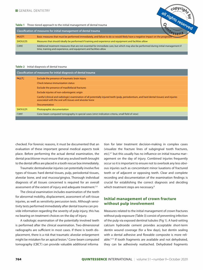
Table 1 Three-tiered approach to the initial management of dental trauma
Classification of measures for initial management of dental trauma
MUSTS Basic measures that must be performed immediately, and failure to do so would likely have a negative impact on the prognosis
SHOULDS Measures that should ideally be performed if training and experience and equipment and facilities allow
CANS Additional treatment measures that are not essential for immediate care, but which may also be performed during initial management if time, training and experience, and equipment and facilities allow
Table 2 Initial diagnosis of dental trauma
Classification of measures for initial diagnosis of dental trauma
Exclude the presence of traumatic brain injury
Check tetanus immunization status
Exclude the presence of maxillofacial fractures
Exclude injuries of non-odontogenic origin
Careful (clinical and radiologic) examination of all potentially injured teeth (pulp, periodontium, and hard dental tissues) and injuriesassociated with the oral soft tissues and alveolar bone
Documentation
SHOULDS Photographic documentation
CANS Cone-beam computed tomography in special cases (strict indication criteria, small field of view)

QUINTESSENCE INTERNATIONAL | volume 51 • number 9 • October 2020 765
Krastl et al
should be placed in water and allowed to rehydrate overnight
before performing reattachment on the next day. Interim rehy-
dration of the fragment improves both shade adaptation and
bond strength to the tooth.9 If a fragment is to be reattached
on the following day, an adhesive sealing with composite
should not be used to cover the dentin during the initial treat-
ment period because adhesive sealings are difficult to remove
completely and can thus impair the accuracy of fragment re-attachment. Hard-setting calcium hydroxide materials are eas-
ier to remove and should be used instead.10
Initial management of crown fracture with
pulp involvement
Initial treatment of crown fractured teeth with pulp exposure
(Table 4 and Fig 2) should aim at maintaining pulp vitality, inde-
pendent of patient age. To reach this goal, partial pulpotomy is
a highly predictable treatment approach.11,12 Unlike direct cap-
ping, partial pulpotomy enables the clinician to successfully
manage even those cases with broad or longer-term exposure
within the first few days after trauma. Partial pulpotomy can
but must not necessarily be performed in the scope of initial
trauma management. It can also be carried out as a secondary
treatment within a few days after pulp coverage, provided that
the exposed area (pulp and dentin) has been adequately sealed
during primary treatment.13
Initial management of crown-root fracture
Restoration of teeth with crown-root fractures (Table 5 and Fig 3)
is often a challenge. Deep subgingival fracture lines greatly
reduce the long-term survival of these teeth.9,14 Notwithstand-
ing, initial management of teeth with crown-root fractures is
comparable with the initial treatment of crown-fractured teeth.
However, the mobile coronal fragment, which is often still
attached to the gingiva, must be removed for fracture line
assessment. This usually provokes bleeding, further complicat-
ing primary treatment which usually consists of sealing the
exposed pulp-dentin complex.
Adhesive fixation of the mobile but still attached coronal
fragment in the generally accessible labial region is a simple
and time-saving alternative primary treatment modality. This
treatment is regarded as a compromise due to the unreliable
seal against bacterial ingress, but it eliminates the symptoms
originating from the mobile fragment in the majority of cases.
Due to the extremely limited durability of the fragment fixa-
tion, the patient should receive further treatment soon (ideally,
on the following day).10
Table 3 Initial management of crown fracture without pulp involvement
Classification of measures for crown fracture without pulp involvement
Fig 1 Enamel or enamel-dentin fracture without pulp exposure.
MUSTS Cover the exposed dentin with a calcium hydroxide cement
Tooth fragments, if available, should be stored in water
Refer for further treatment, which should be carried out soon (ideally, on the next day)
SHOULDS Seal the exposed dentin with a dental adhesive and flowable composite*
Refer for further treatment, which should be carried out soon (within 2 weeks)
CANS Composite buildup as definitive restoration
Immediate reattachment of available fragments if not dehydrated
*If a fragment is to be reattached, initial treatment should be with a calcium hydroxide cement instead of a bonded material.
1
QUINTESSENCE INTERNATIONAL | volume 51 • number 9 • October 2020766
GENERAL DENTISTRY
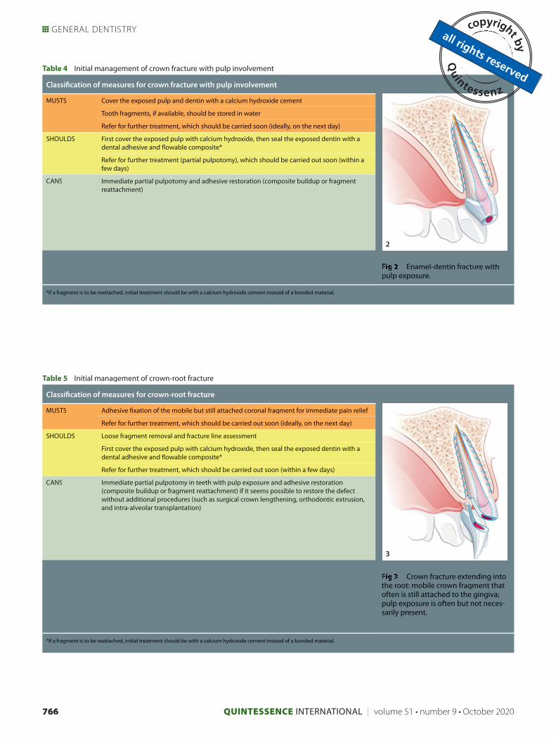
Table 4 Initial management of crown fracture with pulp involvement
Classification of measures for crown fracture with pulp involvement
Enamel-dentin fracture withpulp exposure.
MUSTS Cover the exposed pulp and dentin with a calcium hydroxide cement
Tooth fragments, if available, should be stored in water
Refer for further treatment, which should be carried soon (ideally, on the next day)
SHOULDS First cover the exposed pulp with calcium hydroxide, then seal the exposed dentin with a dental adhesive and flowable composite*
Refer for further treatment (partial pulpotomy), which should be carried out soon (within a few days)
CANS Immediate partial pulpotomy and adhesive restoration (composite buildup or fragment reattachment)
*If a fragment is to be reattached, initial treatment should be with a calcium hydroxide cement instead of a bonded material.
Table 5 Initial management of crown-root fracture
Classification of measures for crown-root fracture
Crown fracture extending into the root: mobile crown fragment that often is still attached to the gingiva;pulp exposure is often but not neces-sarily present.
MUSTS Adhesive fixation of the mobile but still attached coronal fragment for immediate pain relief
Refer for further treatment, which should be carried out soon (ideally, on the next day)
SHOULDS Loose fragment removal and fracture line assessment
First cover the exposed pulp with calcium hydroxide, then seal the exposed dentin with a dental adhesive and flowable composite*
Refer for further treatment, which should be carried out soon (within a few days)
CANS Immediate partial pulpotomy in teeth with pulp exposure and adhesive restoration (composite buildup or fragment reattachment) if it seems possible to restore the defect without additional procedures (such as surgical crown lengthening, orthodontic extrusion,and intra-alveolar transplantation)
*If a fragment is to be reattached, initial treatment should be with a calcium hydroxide cement instead of a bonded material.
2
3
QUINTESSENCE INTERNATIONAL | volume 51 • number 9 • October 2020 767
Krastl et al
Table 7 Initial management of concussion
Classification of measures for concussion
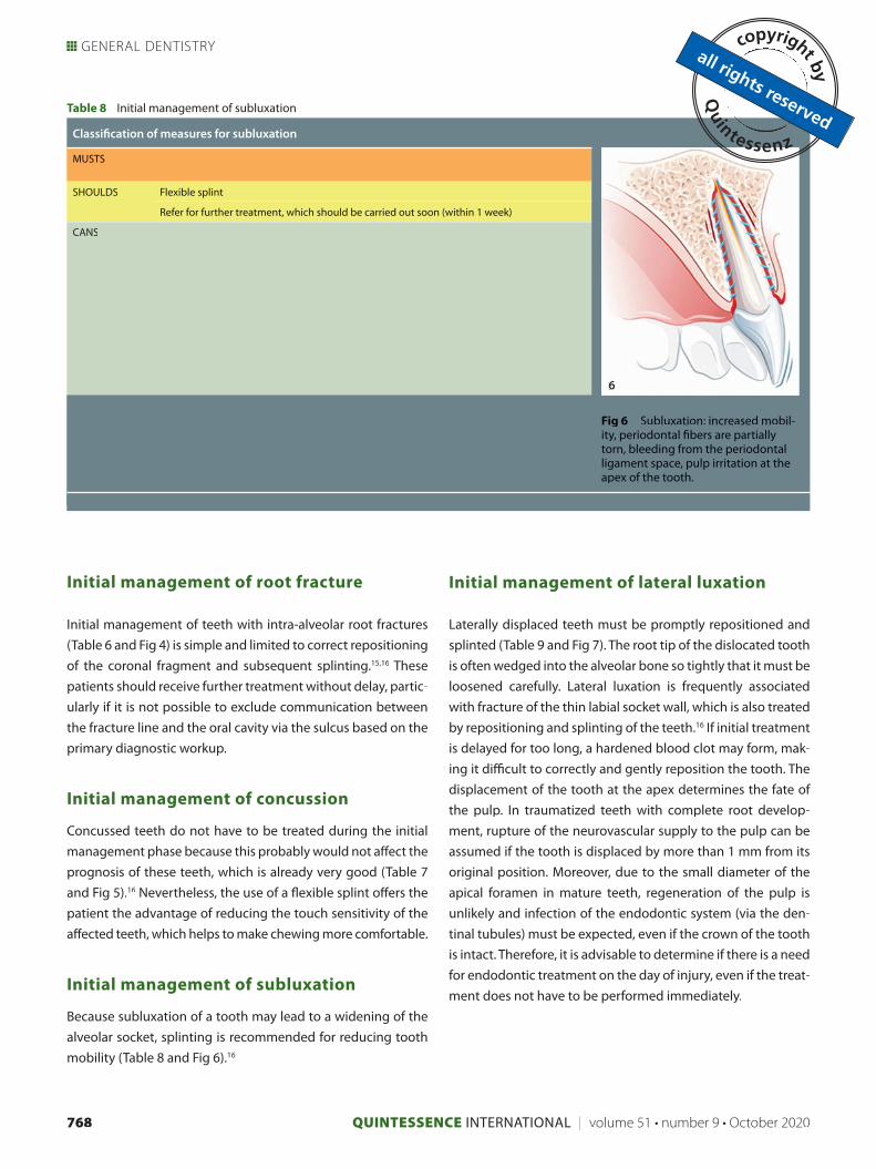
Fig 5 Concussion: touch sensitivity of the affected tooth, no signs of in-creased mobility, no signs of luxation, edema and bleeding in the periodon-tium and apex.
MUSTS
SHOULDS
CANS Flexible splint
Refer for further treatment, which should be carried out soon (within a week)
Table 6 Initial management of crown-root fracture
Classification of measures for crown-root fracture
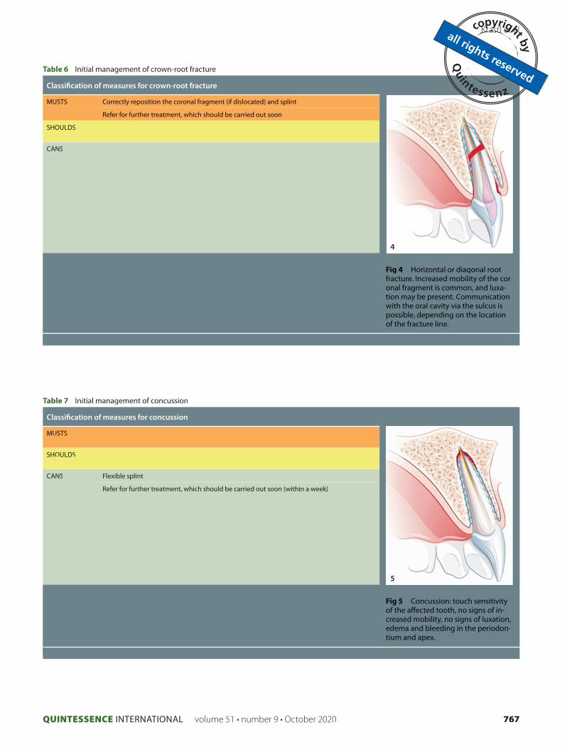
Fig 4 Horizontal or diagonal root fracture. Increased mobility of the cor-onal fragment is common, and luxa-tion may be present. Communicationwith the oral cavity via the sulcus is possible, depending on the location of the fracture line.
MUSTS Correctly reposition the coronal fragment (if dislocated) and splint
Refer for further treatment, which should be carried out soon
SHOULDS
CANS
4
5
QUINTESSENCE INTERNATIONAL | volume 51 • number 9 • October 2020768
GENERAL DENTISTRY
Initial management of root fracture
Initial management of teeth with intra-alveolar root fractures
(Table 6 and Fig 4) is simple and limited to correct repositioning
of the coronal fragment and subsequent splinting.15,16 These
patients should receive further treatment without delay, partic-
ularly if it is not possible to exclude communication between
the fracture line and the oral cavity via the sulcus based on the
primary diagnostic workup.
Initial management of concussion
Concussed teeth do not have to be treated during the initial
management phase because this probably would not affect the
prognosis of these teeth, which is already very good (Table 7
and Fig 5).16 Nevertheless, the use of a flexible splint offers the
patient the advantage of reducing the touch sensitivity of the
affected teeth, which helps to make chewing more comfortable.
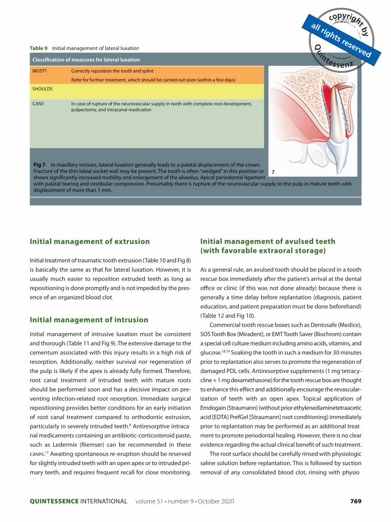
Initial management of subluxation
Because subluxation of a tooth may lead to a widening of the
alveolar socket, splinting is recommended for reducing tooth
mobility (Table 8 and Fig 6).16
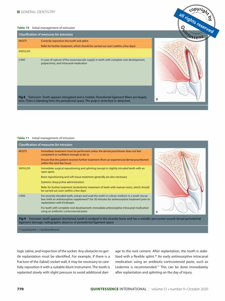
Initial management of lateral luxation
Laterally displaced teeth must be promptly repositioned and
splinted (Table 9 and Fig 7). The root tip of the dislocated tooth
is often wedged into the alveolar bone so tightly that it must be
loosened carefully. Lateral luxation is frequently associated
with fracture of the thin labial socket wall, which is also treated
by repositioning and splinting of the teeth.16 If initial treatment
is delayed for too long, a hardened blood clot may form, mak-
ing it difficult to correctly and gently reposition the tooth. The
displacement of the tooth at the apex determines the fate of
the pulp. In traumatized teeth with complete root develop-
ment, rupture of the neurovascular supply to the pulp can be
assumed if the tooth is displaced by more than 1 mm from its
original position. Moreover, due to the small diameter of the
apical foramen in mature teeth, regeneration of the pulp is
unlikely and infection of the endodontic system (via the den-
tinal tubules) must be expected, even if the crown of the tooth
is intact. Therefore, it is advisable to determine if there is a need
for endodontic treatment on the day of injury, even if the treat-
ment does not have to be performed immediately.
Table 8 Initial management of subluxation
Classification of measures for subluxation
Fig 6 Subluxation: increased mobil-ity, periodontal fibers are partiallytorn, bleeding from the periodontalligament space, pulp irritation at theapex of the tooth.
MUSTS
SHOULDS Flexible splint
Refer for further treatment, which should be carried out soon (within 1 week)
CANS
6
QUINTESSENCE INTERNATIONAL | volume 51 • number 9 • October 2020 769
Krastl et al
Initial management of extrusion
Initial treatment of traumatic tooth extrusion (Table 10 and Fig 8)
is basically the same as that for lateral luxation. However, it is
usually much easier to reposition extruded teeth as long as
repositioning is done promptly and is not impeded by the pres-
ence of an organized blood clot.
Initial management of intrusion
Initial management of intrusive luxation must be consistent
and thorough (Table 11 and Fig 9). The extensive damage to the
cementum associated with this injury results in a high risk of
resorption. Additionally, neither survival nor regeneration of
the pulp is likely if the apex is already fully formed. Therefore,
root canal treatment of intruded teeth with mature roots
should be performed soon and has a decisive impact on pre-
venting infection-related root resorption. Immediate surgical
repositioning provides better conditions for an early initiation
of root canal treatment compared to orthodontic extrusion,
particularly in severely intruded teeth.8 Antiresorptive intraca-
nal medicaments containing an antibiotic-corticosteroid paste,
such as Ledermix (Riemser) can be recommended in these
cases.17 Awaiting spontaneous re-eruption should be reserved
for slightly intruded teeth with an open apex or to intruded pri-
mary teeth, and requires frequent recall for close monitoring.
Initial management of avulsed teeth
(with favorable extraoral storage)
As a general rule, an avulsed tooth should be placed in a tooth
rescue box immediately after the patient’s arrival at the dental
office or clinic (if this was not done already) because there is
generally a time delay before replantation (diagnosis, patient
education, and patient preparation must be done beforehand)
(Table 12 and Fig 10).
Commercial tooth rescue boxes such as Dentosafe (Medice),
SOS Tooth Box (Miradent), or EMT Tooth Saver (Biochrom) contain
a special cell culture medium including amino acids, vitamins, and
glucose.18,19 Soaking the tooth in such a medium for 30 minutes
prior to replantation also serves to promote the regeneration of
damaged PDL cells. Antiresorptive supplements (1 mg tetracy-
cline + 1 mg dexamethasone) for the tooth rescue box are thought
to enhance this effect and additionally encourage the revascular-
ization of teeth with an open apex. Topical application of
Emdogain (Straumann) (without prior ethylenediaminetetraacetic
acid [EDTA] PrefGel [Straumann] root conditioning) immediately
prior to replantation may be performed as an additional treat-
ment to promote periodontal healing. However, there is no clear
evidence regarding the actual clinical benefit of such treatment.
The root surface should be carefully rinsed with physiologic
saline solution before replantation. This is followed by suction
removal of any consolidated blood clot, rinsing with physio-
Table 9 Initial management of lateral luxation
Classification of measures for lateral luxation
MUSTS Correctly reposition the tooth and splint
Refer for further treatment, which should be carried out soon (within a few days)
SHOULDS
CANS In case of rupture of the neurovascular supply in teeth with complete root development, pulpectomy, and intracanal medication
Fig 7 In maxillary incisors, lateral luxation generally leads to a palatal displacement of the crown. Fracture of the thin labial socket wall may be present. The tooth is often “wedged” in this position or
tshows significantly increased mobility and enlargement of the alveolus. Apical periodontal ligament uwith palatal tearing and vestibular compression. Presumably there is rupture of the neurovascular supply to the pulp in mature teeth with
displacement of more than 1 mm.
7
QUINTESSENCE INTERNATIONAL | volume 51 • number 9 • October 2020770
GENERAL DENTISTRY
logic saline, and inspection of the socket. Any obstacles to gen-
tle replantation must be identified. For example, if there is a
fracture of the (labial) socket wall, it may be necessary to care-
fully reposition it with a suitable blunt instrument. The tooth is
replanted slowly with slight pressure to avoid additional dam-
age to the root cement. After replantation, the tooth is stabi-
lized with a flexible splint.20 An early antiresorptive intracanal
medication using an antibiotic-corticosteroid paste, such as
Ledermix is recommended.17 This can be done immediately
after replantation and splinting on the day of injury.
Table 11 Initial management of intrusion
Classification of measures for intrusion
MUSTS Immediate treatment must be performed unless the dental practitioner does not feel competent or confident enough to do so
Ensure that the patient receives further treatment (from an experienced dental practitioner) within the next few hours
SHOULDS Immediate surgical repositioning and splinting (except in slightly intruded teeth with an open apex)
Bone repositioning and soft tissue treatment generally are also necessary
Systemic doxycycline administration
Refer for further treatment (endodontic treatment of teeth with mature roots), which should be carried out soon (within a few days)
CANS For severely intruded teeth, extract and soak the tooth in culture medium in a tooth rescue box (with an antiresorptive supplement*) for 30 minutes for antiresorptive treatment prior to replantation with Emdogain
For teeth with complete root development: immediate antiresorptive intracanal medicationusing an antibiotic-corticosteroid paste
Fig 9 Intrusion: tooth appears shortened; tooth is wedged in the alveolar bone and has a metallic percussion sound; broad periodontal ligament damage; radiographic absence of periodontal ligament space.
*1 mg tetracycline + 1 mg dexamethasone.
Table 10 Initial management of extrusion
Classification of measures for extrusion
MUSTS Correctly reposition the tooth and splint
Refer for further treatment, which should be carried out soon (within a few days)
SHOULDS
CANS In case of rupture of the neurovascular supply in teeth with complete root development, pulpectomy, and intracanal medication
Fig 8 Extrusion. Tooth appears elongated and is mobile. Periodontal ligament fibers are largelytorn. There is bleeding from the periodontal space. The pulp is stretched or detached. 8
9
QUINTESSENCE INTERNATIONAL | volume 51 • number 9 • October 2020 771
Krastl et al
Initial management of avulsed teeth
(after unfavorable extraoral storage)
If extraoral storage conditions were so adverse that periodontal
healing is unlikely and ankylosis must be expected, the necrotic
periodontal ligament tissue should be mechanically removed
from the root surface and the tooth should be stored in a fluoride
solution before replantation (Table 13 and Fig 11). This is thought
to slow down the process of root resorption.20 Because improper
storage eliminates the chances of periodontal healing, the use of
Emdogain cannot be expected to have a positive effect on heal-
ing in this case either. However, Emdogain may be able to prevent
the occurrence of invasive cervical resorption as an additional
complication in teeth with ankylosis and osseous replacement.
In these cases, the required endodontic treatment, includ-
ing root canal filling, can be performed extraorally. Unlike teeth
with vital periodontal tissues, there is no concern that extraoral
manipulation will cause further damage to these teeth. Replan-
tation of improperly stored teeth with incomplete root devel-
opment leads to ankylosis, which results in localized disruption
of jaw growth and infraposition of the tooth. Even so, replanta-
tion and tooth preservation are still recommended for esthetic,
functional, and psychologic reasons until the most suitable
long-term multidisciplinary treatment approach is defined.17,20,21
Initial management of trauma to primary
teeth
From a biologic point of view, the principles for initial manage-
ment of dental trauma to primary teeth are the same as those
for permanent teeth, except in the case of avulsion (where
replantation is not indicated).
However, the individual treatment and stress tolerance of
the injured child determines and often limits the possibilities
for consistent initial management of dental trauma. For this
reason and to prevent (further) damage to the permanent
tooth germ, primary teeth with deep fractures, severe mobility,
or displacement are usually extracted.22 Waiting for sponta-
neous re-eruption of intruded primary teeth is justified if the
accident resulted in labial displacement relative to the perma-
nent tooth germ and therefore is unlikely to have an effect on
it. However, if intrusion does have an effect on the tooth germ,
extraction is indicated as a damage control measure.23,24
Minimum equipment for initial treatment
of dental trauma
Every dental office should have on hand the minimum equip-
ment required for initial management of dental trauma (Table 14).
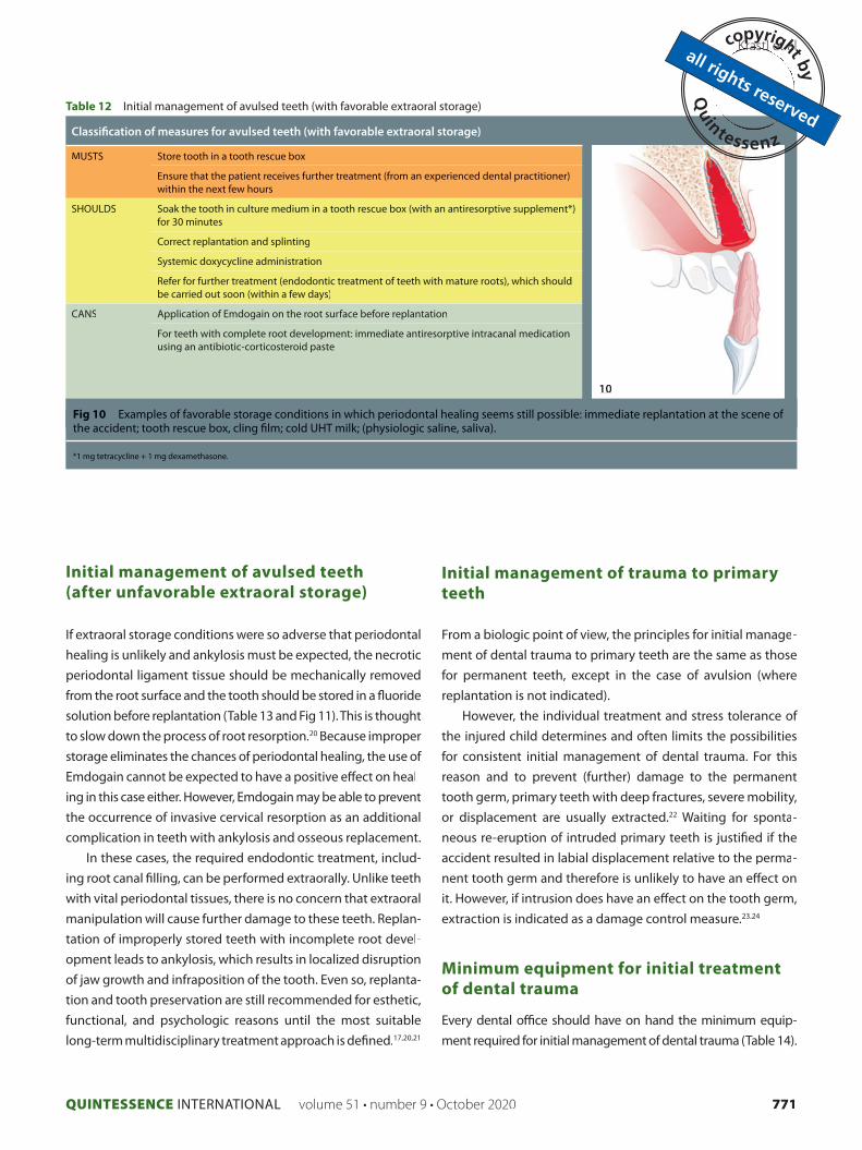
Table 12 Initial management of avulsed teeth (with favorable extraoral storage)
Classification of measures for avulsed teeth (with favorable extraoral storage)
MUSTS Store tooth in a tooth rescue box
Ensure that the patient receives further treatment (from an experienced dental practitioner) within the next few hours
SHOULDS Soak the tooth in culture medium in a tooth rescue box (with an antiresorptive supplement*) for 30 minutes
Correct replantation and splinting
Systemic doxycycline administration
Refer for further treatment (endodontic treatment of teeth with mature roots), which should be carried out soon (within a few days)
CANS Application of Emdogain on the root surface before replantation
For teeth with complete root development: immediate antiresorptive intracanal medicationusing an antibiotic-corticosteroid paste
Fig 10 Examples of favorable storage conditions in which periodontal healing seems still possible: immediate replantation at the scene of the accident; tooth rescue box, cling film; cold UHT milk; (physiologic saline, saliva).
*1 mg tetracycline + 1 mg dexamethasone.
10
QUINTESSENCE INTERNATIONAL | volume 51 • number 9 • October 2020772
GENERAL DENTISTRY
Only a few materials are required, and most of them are already
part of the basic equipment. The most crucial element is a
tooth rescue box containing a cell culture storage medium,
which should be kept in a place where it can be accessed
quickly in case of an emergency.
Systemic doxycycline administration
Systemic doxycycline administration is recommended for the
treatment of severe luxation injuries to permanent teeth (espe-
cially avulsion and intrusion). Due to the antiresorptive proper-
ties of tetracycline derivates, the likelihood of periodontal heal-
ing is expected to increase,20 although clinical evidence is
inconclusive.25 Since tetracyclines are incorporated into tissues
that are calcifying at the time of their administration, the risk of
tooth discoloration should be considered.26 After the age of
8 years calcifi cation of the crowns of permanent teeth is com-
pleted, with the exception of the third molar, and clinically rel-
evant discoloration resulting from a short-term tetracycline
administration is unlikely.
Systemic doxycycline is administered for 7 days starting
on the day of replantation. The dosage is 100 mg per day for
adults and adolescents with a body weight of over 50 kg, and
2 mg/kg per day for children 8 years of age and older who
weigh less than 50 kg; all patients receive a double dose on
the fi rst day.
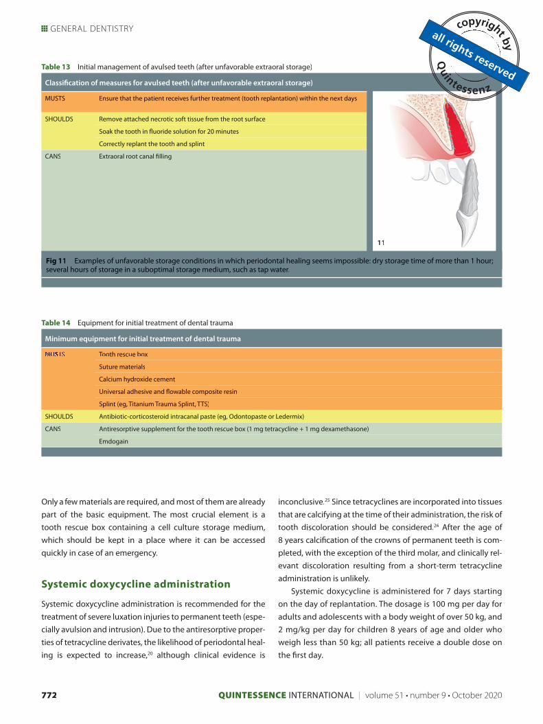
Table 13 Initial management of avulsed teeth (after unfavorable extraoral storage)
Classifi cation of measures for avulsed teeth (after unfavorable extraoral storage)
MUSTS Ensure that the patient receives further treatment (tooth replantation) within the next days
SHOULDS Remove attached necrotic soft tissue from the root surface
Soak the tooth in fl uoride solution for 20 minutes
Correctly replant the tooth and splint
CANS Extraoral root canal fi lling
Fig 11 Examples of unfavorable storage conditions in which periodontal healing seems impossible: dry storage time of more than 1 hour; several hours of storage in a suboptimal storage medium, such as tap water.
Table 14 Equipment for initial treatment of dental trauma
Minimum equipment for initial treatment of dental trauma
Tooth rescue box
Suture materials
Calcium hydroxide cement
Universal adhesive and fl owable composite resin
Splint (eg, Titanium Trauma Splint, TTS)
SHOULDS Antibiotic-corticosteroid intracanal paste (eg, Odontopaste or Ledermix)
CANS Antiresorptive supplement for the tooth rescue box (1 mg tetracycline + 1 mg dexamethasone)
Emdogain
11
QUINTESSENCE INTERNATIONAL | volume 51 • number 9 • October 2020 773
Krastl et al
Further treatment
Even if initial management of dental trauma was ideal, subop-
timal or delayed further treatment can compromise the later
outcome of treatment. In particular, prompt root canal treat-
ment is crucial for teeth with severe luxation injuries, to prevent
infection-related root resorption. In order to be able to make
correct decisions regarding endodontic treatment, the dental
practitioner providing further treatment must rely on the initial
diagnostic findings (particularly regarding the extent of dislo-
cation) documented in the patient records. Therefore, the refer-
ring practitioner must make the utmost effort to ensure that
the patient receives adequate further treatment and that the
patient and/or the clinician providing further treatment
receives all important information of relevance to treatment.
References
1. Andreasen JO, Andreasen FM, Anders-son L. Textbook and Color Atlas of TraumaticInjuries to the Teeth. Oxford: Wiley Blackwell, 2018.
2. Krastl G, Filippi A, Weiger R. Therapie vonZahnunfällen bei Kindern und Jugendlichen: eine Übersicht. Wissen Kompakt 2008;2: 31–43.
3. Filippi A, Tschan J, Pohl Y, Berthold H,Ebeleseder K. A retrospective classification of tooth injuries using a new scoring system.Clin Oral Investig 2000;4:173–175.
4. Ebeleseder K. A suggestion of a new classification system of traumatic dental in-juries. Endod Dent Traumatol 1994;10:39.
5. Weiger R, Krastl G. Endodontische Spätfolgen nach Zahntrauma: Update 2019.Die Quintessenz 2019;70:1012–1019.
6. Dula K, Bornstein MM, Buser D, et al. SADMFR guidelines for the use of cone-beam computed tomography/ digital volume tomography. Swiss Dent J2014;124:1169–1183.
7. Patel S, Brown J, Semper M, Abella F, Mannocci F. European Society of Endodon-tology Position Statement: Cone Beam Computed Tomography. Int Endod J 2019;52: 1675–1678.
8. Weiger R, Krastl G, Filippi A, Lienert N. AcciDent 3.0 (App for iOS and Android),2019.
9. Krastl G, Filippi A, Zitzmann NU, Walter C, Weiger R. Current aspects of restoring traumatically fractured teeth. Eur J Esthet Dent 2011;6:124–141.
10. Krastl G, Amato J. Management of crown fractures and crown-root fractures. In: Neuhaus KW, Lussi A (eds). Management of Dental Emergencies in Children and Adoles-cents. Oxford: Wiley Blackwell, 2019:79–90.
11. Krastl G, Weiger R. Vital pulp therapyafter trauma. ENDO (London, Engl)2014;8:293–300.
12. Dammaschke T, Galler KM, Krastl G.Current recommendations for vital pulptreatment. Dtsch Zahnärztl Z Int 2019;1: 43–52.
13. Wang G, Wang C, Qin M. Pulp progno-sis following conservative pulp treatment inteeth with complicated crown fractures: a retrospective study. Dent Traumatol 2017;33:255–260.
14. Krug R, Krastl G. Therapieoptionennach Kronen-Wurzel-Fraktur Die Quintessenz2019;70:1032–1039.
15. Cvek M, Tsilingaridis G, Andreasen JO.Survival of 534 incisors after intra-alveolar root fracture in patients aged 7–17 years.Dent Traumatol 2008;24:379–387.
16. Diangelis AJ, Andreasen JO, EbelesederKA, et al. International Association of Dental Traumatology guidelines for the managementof traumatic dental injuries: 1. Fractures and luxations of permanent teeth. Dent Trauma-tol 2012;28:2–12.
17. Trope M. Avulsion of permanent teeth: theory to practice. Dent Traumatol2011;27:281–294.
18. Malhotra N. Current developments ininterim transport (storage) media in den-tistry: an update. Br Dent J 2011;211:29–33.
19. Lee W, Stover S, Rasoulianboroujeni M,et al. The efficacy of commercial tooth stor-age media for maintaining the viability of human periodontal ligament fibroblasts. Int Endod J 2018;51:58–68.
20. Andersson L, Andreasen JO, Day P, et al.International Association of Dental Trauma-tology guidelines for the management of traumatic dental injuries: 2. Avulsion of per-manent teeth. Dent Traumatol 2012;28:88–96.
21. Lauridsen E, Andreasen JO, Bouaziz O, Andersson L. Risk of ankylosis of 400 avulsed and replanted human teeth in relation tolength of dry storage: A re-evaluation of a long-term clinical study. Dent Traumatol2020;36:108–116.
22. Filippi A, Krastl G. Traumatologie imMilch- und Wechselgebiss. Quintessenz 2007;58:739–752.
23. Malmgren B, Andreasen JO, Flores MT, et al. International Association of Dental Traumatology guidelines for the manage-ment of traumatic dental injuries: 3. Injuriesin the primary dentition. Dent Traumatol2012;28:174–182.
24. Krastl G, Weiger R. Milchzahntrauma. Die Quintessenz 2009;60:531–539.
25. Hinckfuss SE, Messer LB. An evi-dence-based assessment of the clinicalguidelines for replanted avulsed teeth. PartII: prescription of systemic antibiotics. DentTraumatol 2009;25:158–164.
26. Sanchez A, Roger R, Sheridan P. Tetra-cycline and other tetracycline-derivative staining of the teeth and oral cavity. Int JDermatol 2004;43:709–715.
QUINTESSENCE INTERNATIONAL | volume 51 • number 9 • October 2020774
GENERAL DENTISTRY
Gabriel Krastl Gabriel Krastl Professor and Head, Department of ConservativeDentistry and Periodontology, Center of Dental Traumatology, University Hospital of Würzburg, Germany
Andreas Filippi Professor and Head, Department of Oral Sur-gery, Center of Dental Traumatology, University Center for Dental Medicine, University of Basel, Basel, Switzerland
Roland Weiger Professor and Head, Department of Periodontol-ogy, Endodontology and Cariology, Center of Dental Traumatolo-gy, University Center for Dental Medicine, University of Basel, Basel, Switzerland
Correspondence: Prof Dr Gabriel Krastl, Department of Conservative Dentistry and Periodontology, Center of Dental Traumatology,University Hospital of Würzburg, Pleicherwall 2 D-97070 Würzburg, Germany. Email: [email protected]