Gastric cancer: Metabolic and metabolomics perspectives ...
13
INTERNATIONAL JOURNAL OF ONCOLOGY 51: 5-17, 2017 Abstract. Gastric cancer is one of the most malignant tumors worldwide and remains a major health threat in Asia-Pacific regions, while its pathological mechanism is generally unknown. Recent research has advanced the understanding of the relationship between metabolic repro- gramming and carcinogenesis. In particular, metabolic regulation and cancer research are being further brought into sharp focus with the emergence of metabolomics. Not only can metabolomics provide global information on metabolic profiles of specific tumors, but it can also act as a promising tool to discover biomarkers regarding diagnosis, metastatic surveillance and chemotherapeutic sensitivity prediction. Meanwhile, metabolism-based anticancer therapies will be further discovered. Up to now, accumulative studies have highlighted the application of metabolomics in gastric cancer research regarding different aspects; therefore we summarized the current available results of how metabolic changes are linked to gastric carcinogenesis, and how metabolomics holds promise for the diagnosis, metastatic surveillance, treatment and prognosis prediction of gastric cancer. Contents 1. Introduction 2. Metabolic alteration in gastric cancer 3. Metabolomics in diagnosis, treatment and prognostic prediction of gastric cancer 4. Current perspectives and future directions 5. Conclusions 1. Introduction Gastric cancer remains third in ranking in cancer death worldwide, although its overall incidence is declining in recent years (1). In the past decades, studies aimed at Helicobacter pylori infection (2,3), hereditary susceptibility (4) and envi- ronmental factors (5) have made a great breakthrough in investigating its precise pathogenesis. Recently, application of various ‘-omics’ technologies opened a new field to investigate the mechanisms behind this disease. With the emergency of metabolomics, major progress has been made in the understanding of the relationship between metabolic regulation and cancer. Warburg, in fact, showed a characteristic metabolic pattern of tumors in the 1920s, that is, tumor cells consume a large amount of glucose for glycolysis even under the condition of sufficient oxygen (Warburg effect) (6). Extensive research also indicates that metabolic reprogramming is one of the hallmarks of cancer (7), and intricately linked to oncogenesis (8-10) and cancer immune escape (11-13). On the other hand, study methods combined conventional oncology research and metabolomics are more likely to provide deeper insights in this field. The procedure of these methods is illustrated in Fig. 1, and more detailed information can be found in literature (14-16). Several excellent reviews have been published on metabo- lomics application in different diseases (17-19) especially cancer research (20-23). Hence, this report presents fresh and profound insights into metabolic changes in gastric cancer and possible mechanism behind these alterations is further discussed. Then, we focus on some studies including our data targeted on biomarkers involving diagnosis, metastasis and prognosis, and treatment in this disease. Finally, future direc- tions are presented. 2. Metabolic alteration in gastric cancer Up to now, several studies aimed at identifiable metabolic changes in macroenvironment-blood (24-29) (Table I) and urine (30-34) (Table II) or microenvironment-carcinoma tissues (35-41) (Table III) and gastric juice (42-44) (Table IV) have been done to map globally metabolic profiles and interpret its possible mechanism in the process of gastric carcinogenesis. Typical changes in metabolites of this disease are illustrated in Fig. 2. Gastric cancer: Metabolic and metabolomics perspectives (Review) SHIYU XIAO and LIYA ZHOU Department of Gastroenterology, Peking University Third Hospital, Haidian, Beijing 100191, P.R. China Received March 17, 2017; Accepted May 2, 2017 DOI: 10.3892/ijo.2017.4000 Correspondence to: Professor Liya Zhou, Department of Gastro- enterology, Peking University Third Hospital, 49 North Huayuan Road, Haidian, Beijing 100191, P.R. China E-mail: [email protected] Key words: gastric cancer, metabolomics, metabolic reprogramming, diagnosis, metastatic prediction, treatment
Transcript of Gastric cancer: Metabolic and metabolomics perspectives ...

INTERNATIONAL JOURNAL OF ONCOLOGY 51: 5-17, 2017
Abstract. Gastric cancer is one of the most malignant tumors worldwide and remains a major health threat in Asia-Pacific regions, while its pathological mechanism is generally unknown. Recent research has advanced the understanding of the relationship between metabolic repro-gramming and carcinogenesis. In particular, metabolic regulation and cancer research are being further brought into sharp focus with the emergence of metabolomics. Not only can metabolomics provide global information on metabolic profiles of specific tumors, but it can also act as a promising tool to discover biomarkers regarding diagnosis, metastatic surveillance and chemotherapeutic sensitivity prediction. Meanwhile, metabolism-based anticancer therapies will be further discovered. Up to now, accumulative studies have highlighted the application of metabolomics in gastric cancer research regarding different aspects; therefore we summarized the current available results of how metabolic changes are linked to gastric carcinogenesis, and how metabolomics holds promise for the diagnosis, metastatic surveillance, treatment and prognosis prediction of gastric cancer.
Contents
1. Introduction2. Metabolic alteration in gastric cancer3. Metabolomics in diagnosis, treatment and prognostic prediction of gastric cancer4. Current perspectives and future directions5. Conclusions
1. Introduction
Gastric cancer remains third in ranking in cancer death worldwide, although its overall incidence is declining in recent years (1). In the past decades, studies aimed at Helicobacter pylori infection (2,3), hereditary susceptibility (4) and envi-ronmental factors (5) have made a great breakthrough in investigating its precise pathogenesis. Recently, application of various ‘-omics’ technologies opened a new field to investigate the mechanisms behind this disease.
With the emergency of metabolomics, major progress has been made in the understanding of the relationship between metabolic regulation and cancer. Warburg, in fact, showed a characteristic metabolic pattern of tumors in the 1920s, that is, tumor cells consume a large amount of glucose for glycolysis even under the condition of sufficient oxygen (Warburg effect) (6). Extensive research also indicates that metabolic reprogramming is one of the hallmarks of cancer (7), and intricately linked to oncogenesis (8-10) and cancer immune escape (11-13). On the other hand, study methods combined conventional oncology research and metabolomics are more likely to provide deeper insights in this field. The procedure of these methods is illustrated in Fig. 1, and more detailed information can be found in literature (14-16).
Several excellent reviews have been published on metabo-lomics application in different diseases (17-19) especially cancer research (20-23). Hence, this report presents fresh and profound insights into metabolic changes in gastric cancer and possible mechanism behind these alterations is further discussed. Then, we focus on some studies including our data targeted on biomarkers involving diagnosis, metastasis and prognosis, and treatment in this disease. Finally, future direc-tions are presented.
2. Metabolic alteration in gastric cancer
Up to now, several studies aimed at identifiable metabolic changes in macroenvironment-blood (24-29) (Table I) and urine (30-34) (Table II) or microenvironment-carcinoma tissues (35-41) (Table III) and gastric juice (42-44) (Table IV) have been done to map globally metabolic profiles and interpret its possible mechanism in the process of gastric carcinogenesis. Typical changes in metabolites of this disease are illustrated in Fig. 2.
Gastric cancer: Metabolic and metabolomics perspectives (Review)
SHIYU XIAO and LIYA ZHOU
Department of Gastroenterology, Peking University Third Hospital, Haidian, Beijing 100191, P.R. China
Received March 17, 2017; Accepted May 2, 2017
DOI: 10.3892/ijo.2017.4000
Correspondence to: Professor Liya Zhou, Department of Gastro-enterology, Peking University Third Hospital, 49 North Huayuan Road, Haidian, Beijing 100191, P.R. ChinaE-mail: [email protected]
Key words: gastric cancer, metabolomics, metabolic reprogramming, diagnosis, metastatic prediction, treatment

XIAO and ZHOU: GASTRIC CANCER AND METABOLOMICS6
Glucose metabolism. Cumulative evidence demonstrates that concentration of lactic acid shows a consistent increase in urine (30) or tissue (31,35,36,38) samples of gastric cancer groups, but glucose is considerably depleted compared with those healthy counterparts or non-malignant patients (like chronic superficial gastritis and chronic atrophic gastritis without intestinal metaplasia) (35,36). The high lactate level might be attributed to the special metabolism of most cancer cells, known as ‘Warburg effect’ we mentioned above (6). Scarce glucose might result from the overexpression of glucose transporters (42) and type II hexokinase (43), which are both confirmed in gastric cancer tissues. Higher fructose-6-phosphokinase (6-FPK) activity can also result in low glucose in gastric cancer tissues (44), as it regulates the output of glucose to glycolysis pathway. The glycolytic switch has been identified to be associated with oncogenic transformation and molecular signal transduction, such as hypoxia-inducible factor pathway, insulin signaling pathway and PI3K-Akt-mTOR pathway (45). Furthermore, over-
expression of pyruvate kinase and lactate dehydrogenase is positively associated with tumor proliferation and poor prognosis, downregulation of them in vitro experiment can impair tumor invasion (38,46-49). On the other hand, such special microenvironment might be the requirement of rapid propagation of tumor cells. To our understanding, it has been reported that accumulated lactic acid moderates the activity of proteases that decompose extracellular matrix, which can produce some peptides and amino acids that are consumable for energy generation (44). Acidosis microenvironment is also ascribed to the formation of cancer blood vessels, meeting the plentiful supply of nutrients and leading to tumor invasion and metastasis (50). Moreover, tumor-derived lactate shows strongly negative effects on cytotoxic T-cell/NK cell function (11,51) and blocks differentiation of monocytes to dendritic cells (52), finally leading to tumor immune escape. However, such outcome demands further verification in gastric cancer.
Considering tricarboxylic acid cycle (TCA) intermediates, an increase of five metabolites (α-ketoglutaric acid, malic acid,
Figure 1. Metabolism and metabolomics in cancer research. Concerning tumor cell lines cultured in vitro, either conventional cell biology research or isotopic tracer experiment is available. Tumor cytobiological methods (cell morphology, cell proliferation assay and cell invasion assay) can be utilized to assess cytobiological behaviors under specific nutrient-stressed or stress-free condition, and further investigations targeted at precise mechanism and significance can be confirmed via molecular biology techniques. With regard to isotopic tracer experiment, the flow of nutrients and metabolites can be identified with isotopic tracer, then the significance of specific nutrients or metabolites and its potential divergent fates toward meeting the demands of either energetic utilization or synthesizing macromolecules in cancer cells can be identified (14). Metabolites that are different between tumor groups and control groups are able to be detected through metabolomics analysis (such as 1H-NMR, 1hydrogen-nuclear magnetic resonance; LC-MS, liquid chromatography-mass spectrometry; GC-MS, gas chromatography-mass spectrometry) and data analysis, metabolic biomarkers or metabolic pathway that is specific to certain cancers were discovered to benefit cancer research (15,16). Of note, combination of genomics, transcriptomics and proteomics plus metabolomics can further give us comprehensive understanding of cancers toward systematic biology.
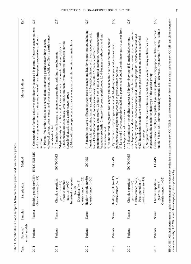
INTERNATIONAL JOURNAL OF ONCOLOGY 51: 5-17, 2017 7Ta
ble
I. M
etab
olite
s in
bloo
d sa
mpl
es b
etw
een
canc
er g
roup
s and
non
-can
cer g
roup
s.
Year
Pa
tient
s/
Sam
ples
Sa
mpl
e si
ze
Met
hod
Maj
or fi
ndin
gs
Ref
.
anim
al m
odel
s
2011
Pa
tient
s Pl
asm
a H
ealth
y pe
ople
(n=9
85)
HPL
C-E
SI-M
S i)
Conc
entra
tion
of a
min
o ac
ids w
as si
gnifi
cant
ly d
ecre
ased
in p
lasm
a of
gas
tric
canc
er p
atie
nts
(24)
G
astri
c ca
ncer
(n=1
99)
an
d th
is c
hang
e oc
curs
in e
arly
stag
e re
gard
less
of t
he su
bseq
uent
pro
gres
sion
and
poo
r
nutri
tion
ii)
Pla
sma-
free
am
ino
acid
s hav
e sh
ared
alte
ratio
n am
ong
gast
ric c
ance
r, lu
ng c
ance
r,
colo
rect
al c
ance
r, br
east
can
cer a
nd p
rost
ate
canc
er, b
ut sp
ecifi
c pr
ofile
s in
gast
ric c
ance
r
wer
e al
so d
etec
ted
2011
Pa
tient
s Pl
asm
a C
hron
ic su
perfi
cial
G
C-T
OFM
S i)
15 m
etab
olite
s (in
crea
se, g
luta
mat
e, a
spar
agin
e, o
rnith
ine,
pyr
oglu
tam
ate,
(2
5)
gast
ritis
(n=1
9)
2-
hydr
oxyb
utyr
ate,
aze
laic
aci
d, 1
1-ei
cose
noic
aci
d, 1
-mon
ohex
adec
anoy
lgly
cero
l
Chr
onic
atro
phic
-γ-to
coph
erol
, ura
te; d
ecre
ase:
cre
atin
ine,
thre
onat
e) w
ere
diffe
rent
bet
wee
n ch
roni
c
gast
ritis
(n=1
0)
su
perfi
cial
gas
tritis
and
gas
tric
canc
er g
roup
In
test
inal
met
apla
sia
ii)
Met
abol
ic p
heno
type
of g
astri
c ca
ncer
was
gre
atly
sim
ilar t
o in
test
inal
met
apla
sia
(n
=10)
D
yspl
asia
(n=1
5)
Gas
tric
canc
er (n
=22)
2012
Pa
tient
s Se
rum
H
ealth
y pe
ople
(n=3
0)
GC
-MS
i) 18
met
abol
ites w
ere
diffe
rent
bet
wee
n ga
stric
can
cer a
nd h
ealth
y co
ntro
l gro
up, i
nclu
ding
(2
6)
Gas
tric
canc
er (n
=30)
fum
aric
aci
d, g
luta
min
e, v
alin
e, sa
rcos
ine,
9,1
2-oc
tade
cadi
enoi
c ac
id, 9
-oct
adec
enoi
c ac
id,
tra
ns-1
3-oc
tade
ceno
ic a
cid,
non
ahex
acon
tano
ic, c
hole
sta-
3,5-
dien
e, c
hole
ster
ol
pent
afluo
ropi
onat
e, c
hole
ster
ol, C
hole
st-5
-en-
3-ol
, 2-O
-mes
yl a
rabi
nose
, hex
adec
anen
itrile
,
benz
enea
ceto
nitri
le, 2
-am
ino-
4-hy
drox
y-pt
erid
inon
e, 1
,2,4
-ben
zene
trica
rbox
ylic
aci
d an
d
hexa
nedi
oic
acid
ii)
Val
ine
show
ed th
e gr
eate
st fo
ld c
hang
e an
d he
xane
dioi
c ac
id w
as th
e m
ost d
eple
ted
2012
Pa
tient
s Se
rum
H
ealth
y pe
ople
(n=1
2)
GC
-MS
i) Py
ruvi
c ac
id, 3
-hyd
roxy
prop
ioni
c ac
id, 3
-hyd
roxy
isob
utyr
ic a
cid,
oct
anoi
c ac
id,
(27)
G
astri
c ca
ncer
(n=1
1)
ph
osph
oric
aci
d si
gnifi
cant
ly c
hang
ed in
gas
tric
canc
er p
atie
nts
ii)
Lev
els o
f 3-h
ydro
xypr
opio
nic
acid
and
pyr
uvic
aci
d co
uld
disc
rimin
ate
gast
ric c
ance
r fro
m
esop
hage
al o
r col
orec
tal c
ance
r20
12
Patie
nts
Plas
ma
Chr
onic
supe
rfici
al
GC
-TO
FMS
i) 15
dis
crim
inat
ory
met
abol
ites (
incr
ease
, β-h
ydro
xybu
tyra
te, β
-D-m
ethy
lglu
copy
rano
- (2
8)
gast
ritis
(n=2
0)
si
de, h
epta
noic
aci
d; d
ecre
ase,
succ
inat
e, m
alat
e, fu
mar
ate,
citr
ate,
serin
e, g
lyci
ne, c
yste
ine,
Gas
tric
canc
er (n
=17)
and
S-m
ethy
l-cys
tein
e, d
ocos
ahex
aeno
ic a
cid,
inos
itol-p
hosp
hate
, oct
adec
enoi
c ac
id, a
nd
Post
-ope
ratio
n of
9-(Z
)-he
xade
ceno
ic a
cid)
wer
e id
entifi
ed b
etw
een
gast
ric c
ance
r and
chr
onic
supe
rfici
al
gast
ric c
ance
r (n=
15)
ga
strit
is g
roup
ii)
Sur
gica
l rem
oval
of t
he c
ance
r tis
sues
cou
ld c
hang
e le
vels
of m
any
met
abol
ites t
hat
ch
arac
teriz
ed th
e m
etab
olic
phe
noty
pe o
f the
can
cer g
roup
2016
Pa
tient
s Se
rum
C
hron
ic su
perfi
cial
LC
-MS
Gas
tric
canc
er d
ispl
ays u
preg
ulat
ed k
ynur
enin
e pa
thw
ay o
f try
ptop
han
met
abol
ism
: inc
reas
e,
(29)
ga
strit
is (n
=17)
indo
le-3
-lact
ic a
cid,
ant
hran
ilic
acid
, kyn
uren
ic a
cid;
dec
reas
e, k
ynur
enin
e, 3
-indo
xyl-s
ulfa
te
Gas
tric
canc
er (n
=32)
HPL
C-E
SI-M
S, h
igh
perf
orm
ance
liqu
id c
hrom
atog
raph
y-el
ectro
spra
y io
niza
tion-
mas
s sp
ectro
met
ry; G
C-T
OM
S, g
as c
hrom
atog
raph
y-tim
e-of
-flig
ht m
ass
spec
trom
etry
; GC
-MS,
gas
chr
omat
ogra
phy-
mas
s spe
ctro
met
ry; L
C-M
S, li
quid
chr
omat
ogra
phy-
mas
s spe
ctro
met
ry.

XIAO and ZHOU: GASTRIC CANCER AND METABOLOMICS8
Tabl
e II
. Met
abol
ites i
n ur
ine
sam
ples
bet
wee
n ca
ncer
gro
ups a
nd n
on-c
ance
r gro
ups.
Year
Pa
tient
s/
Sam
ple
size
M
etho
d
Maj
or fi
ndin
gs
Ref
.
anim
al m
odel
s
2011
SC
ID m
ice
(mal
e)
Gas
tric
canc
er (n
=16)
G
C-M
S i)
10 m
etab
olite
s wer
e di
ffere
nt b
etw
een
canc
er g
roup
(met
asta
sis a
nd n
on-m
etas
tasi
s) a
nd c
ontro
l gro
up:
(30)
SC
G-7
901
(met
asta
sis g
roup
(=8
and
lact
ic a
cid,
mal
ic a
cid,
citr
ic a
cid,
gly
cero
l, he
xade
cano
ic a
cid,
pyr
imid
ine,
uric
aci
d, b
utan
oic
acid
,
cell
line
non-
met
asta
sis (
=8)
pr
opan
oic,
but
aned
ioic
aci
d
C
ontro
l gro
up (n
=8)
ii)
7 m
etab
olite
s wer
e ch
arac
teris
tic b
etw
een
met
asta
sis a
nd n
on-m
etas
tasi
s gro
ups:
ala
nine
, L-p
rolin
e,
glyc
erol
, but
anoi
c ac
id, b
utan
edio
ic a
cid,
L-th
reon
ic a
cid,
myo
-inos
itol.
iii) C
hang
es in
lact
ic a
cid
and
buta
noic
aci
d sh
owed
dia
gnos
tic v
alue
2014
Pa
tient
s Tr
aini
ng se
t/val
idat
ion
set
1 H-N
MR
i)
Alte
red
met
abol
ites i
n ur
ine
sam
ples
of g
astri
c ca
ncer
are
mai
nly
rela
ted
to a
min
o ac
ids a
nd
(31)
Gas
tric
canc
er (n
=50/
23)
lip
id m
etab
olis
m; l
evel
s of 4
-hyd
roxy
phen
ylac
etat
e, a
lam
ine,
phe
nyla
cety
lgly
cine
, man
mito
l, gl
ycol
ate
and
Hea
lthy
peop
le (n
=50/
31)
ar
gini
ne a
re re
late
d to
T st
age
of g
astri
c ca
ncer
ii) H
ypox
anth
ine
is a
ble
to p
redi
ct a
reco
very
tren
d in
pos
tope
rativ
e gr
oups
of g
astri
c ca
ncer
2015
Pa
tient
s G
astri
c ca
ncer
(n=1
3)
LC-M
S 16
met
abol
ites w
ere
diffe
rent
ly e
xpre
ssed
bet
wee
n ca
ncer
and
hea
lthy
grou
p: su
ccin
ic a
cid,
mal
ic a
cid,
(3
2)
H
ealth
y pe
ople
(n=9
)
alan
ine,
gly
cine
, L-p
rolin
e, h
exad
ecan
oic
acid
, pyr
imid
ine,
uric
aci
d, g
lyco
chol
ic a
cid,
hip
pura
te, u
rea,
4-de
oxyt
hreo
nic
acid
, phe
nyla
cety
lgly
cine
, tau
rine,
2-o
xogl
utar
ate
2016
Pa
tient
s G
astri
c ca
ncer
(n=4
3)
1 H-N
MR
i)
Leve
ls o
f suc
rose
, dim
ethy
lam
ine,
1-m
ethy
lnic
otin
amid
e, 2
-fur
oylg
ylyc
ine,
N-a
cety
l-ser
oton
in, t
rans
- (3
3)
H
ealth
y pe
ople
(n=4
0)
ac
onita
te, f
orm
ate
and
sero
toni
n gr
eatly
alte
red
in u
rine
sam
ples
of g
astri
c ca
ncer
Bar
rett'
s eso
phag
us
ii)
Ala
nine
, 2-h
ydro
xyis
obut
yrat
e an
d 3-
indo
xyls
ulfa
te p
rodu
ced
a di
scrim
inat
ory
mod
el re
gard
ing
(n=4
0)
di
scrim
inat
ing
canc
er a
nd c
ontro
l gro
up
2016
Pa
tient
s G
astri
c ca
ncer
(n=1
99)
GC
-MS
i) 17
met
abol
ites a
re la
rgel
y di
ffere
nt b
etw
een
canc
er a
nd c
ontro
l gro
ups i
n tra
inin
g se
t: gl
ycin
e, v
alin
e,
(34)
Hea
lthy
peop
le (n
=87)
isol
euci
ne, s
erin
e, th
reon
ine,
pro
line,
met
hion
ine,
tyro
sine
, try
ptop
han,
eth
yl 2
-met
hyla
ceto
acet
ate,
levu
linic
aci
d, p
-cre
sol,
benz
ylm
alon
ic a
cid,
4-h
ydro
xybe
nzoi
c ac
id, h
ippu
ric a
cid,
ben
zil,
alam
ine
ii) 1
4 of
them
show
dia
gnos
tic v
alue
that
is b
ette
r tha
n cl
assi
c bl
ood
biom
arke
rs o
n va
lidat
ion
set,
mos
t
of
whi
ch w
ere
rela
ted
to a
min
o ac
id m
etab
olis
m
iii
) Pro
line,
p-c
reso
l and
4-h
ydro
xybe
nzoi
c ac
id p
rodu
ce o
utco
me-
pred
ictio
n va
lue
by su
rviv
al a
naly
sis
SCID
, sev
ere
com
bine
d im
mun
e de
ficie
ncy;
1 H-N
MR
, 1 hydr
ogen
-nuc
lear
mag
netic
reso
nanc
e; L
C-M
S, li
quid
chr
omat
ogra
phy-
mas
s spe
ctro
met
ry.

INTERNATIONAL JOURNAL OF ONCOLOGY 51: 5-17, 2017 9Ta
ble
III.
Met
abol
ites i
n tis
sue
sam
ples
bet
wee
n ca
ncer
gro
ups a
nd n
on-c
ance
r gro
ups.
Year
Pa
tient
s/
Sam
ple
size
M
etho
d
Maj
or fi
ndin
gs
Ref
.
anim
al m
odel
s
2009
Pa
tient
s 12
pai
rs o
f mat
ched
tum
or
CE-
TOFM
S i)
Extre
mel
y lo
w g
luco
se, h
igh
lact
ate
and
glyc
olyt
ic in
term
edia
te c
once
ntra
tions
wer
e fo
und
in b
oth
(35)
and
norm
al g
astri
c tis
sues
colo
n an
d st
omac
h tu
mor
tiss
ues
ii) S
igni
fican
t acc
umul
atio
n of
all
amin
o ac
ids e
xcep
t glu
tam
ine
in th
e tu
mor
tiss
ues
20
10
Mal
e SC
ID m
ice
Can
cer g
roup
G
C-M
S i)
29 m
etab
olite
s wer
e di
ffere
ntly
exp
ress
ed b
etw
een
met
asta
sis a
nd n
on-m
etas
tasi
s gro
up: g
luco
se,
(36)
SC
G-7
901
cell
line
(Non
-met
asta
sis 8
succ
inat
e, m
alic
acid
, lac
tate
, ala
nine
, gly
cine
, val
ine,
leuc
ine,
dim
ethy
lgly
cine
, iso
leuc
ine,
M
etas
tasi
s 8)
pr
opan
amid
e, b
utan
edio
ic, p
rolin
e, m
ethi
onni
ne, s
erin
e, th
reon
ine,
asp
arag
ine,
glu
tam
ine,
pho
spho
serin
e,
C
ontro
l gro
up 6
glut
amat
e, ly
sine
, arg
inin
e, d
ocos
anoi
c, o
ctad
ecan
oic,
pyr
imid
ine,
hyp
oxan
thin
e, in
osito
l,
pr
opan
edio
c, p
yrro
lidin
e
ii)
Ser
ine
and
prol
ine
met
abol
ism
s wer
e hi
ghlig
hted
in m
etas
tatic
gro
up
20
10
Patie
nts
18 p
airs
of m
atch
ed tu
mor
G
C-M
S i)
L-gl
utam
ine,
pho
spho
serin
e, L
-val
ine,
L-is
oleu
cine
, ser
ine,
hep
tane
dioi
c ac
id, p
ropa
noic
aci
d,
(37)
and
norm
al g
astri
c tis
sues
phen
anth
reno
l, bu
tane
triol
, ace
tam
id, b
uten
oic
acid
, oxa
zole
thio
ne, n
apht
hale
ne, L
-altr
ose,
L-m
anno
fura
nose
, gal
acto
fura
nosi
de, m
yo-in
osito
l, D
-rib
ofur
anos
e w
ere
dete
cted
diff
eren
tly
be
twee
n th
e m
alig
nant
tiss
ues a
nd th
e ad
jace
nt n
on-m
alig
nant
tiss
ues o
f gas
tric
muc
osa
ii) 5
of t
hem
wer
e de
tect
ed d
iffer
ently
bet
wee
n th
e no
n-in
vasi
ve tu
mor
s and
the
inva
sive
tum
ors:
high
er le
vels
of L
-cys
tein
e, L
-tyro
sine
, hyp
oxan
thin
e an
d lo
wer
leve
ls o
f phe
nant
hren
ol, b
utan
oic
acid
in th
e in
vasi
ve g
roup
2010
Pa
tient
s 65
pai
rs o
f mat
ched
gas
tric
GC
-TO
FMS
i) D
ysre
gula
tion
of p
yruv
ic a
cid
efflu
x w
as a
n im
porta
nt g
luco
se m
etab
olic
sign
atur
e in
the
(38)
card
iac
canc
er a
nd a
djac
ent
de
velo
pmen
t of g
astri
c ca
rdia
c ca
ncer
norm
al ti
ssue
s
ii) T
rans
ition
from
gly
coly
sis t
o th
e K
rebs
cyc
le h
ad a
n in
hibi
tory
effe
ct o
n G
CC
pro
gres
sion
, whi
ch
coul
d be
serv
ed a
s a p
oten
tial t
hera
peut
ic ta
rget
for g
astri
c ca
rdia
c ca
ncer
2011
Pa
tient
s 30
pai
rs o
f mat
ched
tum
or
GC
-MS
i) 15
diff
eren
tial m
etab
olite
s wer
e id
entifi
ed: α
-ket
oglu
taric
aci
d, fu
mar
ic a
cid,
val
ric a
cid,
(3
9)
an
d no
rmal
gas
tric
tissu
es
9-
hexa
dece
nnoi
c ac
id, 3
-hyd
roxy
buta
noic
aci
d, h
exad
ecan
oic
acid
, oct
adec
anoi
c ac
id, c
is-v
acce
nic
acid
,
ar
achi
doni
c ac
id, 1
-phe
nant
hren
e-ca
rbox
ylic
aci
d, 9
-oct
adec
enam
ide,
squa
lene
, xyl
onic
aci
d,
be
nzen
epro
pano
ic a
cid
ii) C
urre
nt m
odel
s cou
ld n
ot d
iscr
imin
ate
norm
al m
ucos
a an
d di
ffere
nt p
atho
logi
cal s
tage
s of G
C
tis
sues
bas
ed o
n th
eir i
dent
ified
met
abol
ic p
rofil
es
2012
Pa
tient
s G
astri
c ca
ncer
(n=1
7)
GC
-TO
FMS
Dis
crim
inat
ing
met
abol
ites a
ssoc
iate
d w
ith g
luco
se, a
min
o ac
ids,
lipid
and
nuc
leot
ide
met
abol
ism
wer
e (2
8)
C
hron
ic su
perfi
cial
gas
tritis
dete
cted
bet
wee
n tw
o gr
oups
: inc
reas
e, c
itrat
e, m
alat
e, fu
mar
ate,
succ
inat
e), c
yste
ine,
(n
=20)
2-am
inoa
dipa
te, 9
-(Z)
-hex
adec
enoi
c ac
id, d
ocos
ahex
aeno
ic a
cid,
β-h
ydro
xybu
tyra
te, u
raci
l,
m
onom
ethy
lpho
spha
te; d
ecre
ase,
glu
cose
, mal
tose
, rib
ose,
β-D
-met
hylg
luco
pyra
nosi
de, f
ruct
ose-
6-
(31)
phos
phat
e, in
osito
l and
ribi
tol,
glyc
eric
aci
d-2,
3-di
phos
phat
e, n
ones
terifi
ed c
hole
ster
ol, u
ridin
e 20
14
Patie
nts
30 p
airs
of m
atch
ed tu
mor
H
R-M
AS-
Li
pid
met
abol
ites w
ere
sign
ifica
ntly
low
er, w
hile
som
e am
ino
acid
s (su
ch a
s iso
leuc
ine,
glu
tam
ate,
and
norm
al g
astri
c tis
sues
N
MR
le
ucin
e, v
alin
e, a
lani
ne, l
ysin
e an
d ph
enyl
alan
ine)
, tau
rine
and
lact
ate
wer
e si
gnifi
cant
ly h
ighe
r in
tum
or ti
ssue
s
CE-
TOFM
S, c
apill
ary
elec
troph
ores
is ti
me-
of-fl
ight
mas
s spe
ctro
met
ry; H
R-M
AS
NM
R, h
igh-
reso
lutio
n m
agic
ang
le sp
inni
ng n
ucle
ar m
agne
tic re
sona
nce.

XIAO and ZHOU: GASTRIC CANCER AND METABOLOMICS10
fumarate, succinate, citric acid) is noticed regardless of blood (26,28), urine (30,32) or tissue (28,35,36,39) samples in gastric cancer. There are some possible reasons that can explain this phenomenon. One account is that cancer cells still use a small portion of glucose for oxidative phosphorylation. Secondly, cancer cells might also utilize fumarate respiration to generate energy under special conditions of glucose deprivation and severe hypoxia in microenvironment (53), and succinate is one of the byproducts in this process except for originating from TCA. Hence, it provides a likely explanation for the accumula-tion of fumarate and succinate. Another reason is that some amino acids, such as glutamine, threonine, phenylalanine, tyrosine or proline, can be converted into these intermediates involving in TCA (Fig. 2). Additionally, elevated levels of citric acid can be used in the de novo fatty acid synthesis, but it is noted that citrate can also induce apoptosis in two gastric cancer cell lines in vitro experiment (54,55).
Amino acid metabolism. Availability of amino acids is pivotal for cellular protein biosynthesis and cytoskeleton formation, while it has been pointed out that amino acids especially those linking to TCA (Fig. 2) are an alternative energy source of cancer cell proliferation (56). By employing metabolomics technologies, levels of various amino acids (including serine, valine, phenylalanine, tryptophan, glycine, and proline) and their primary derivatives (such as kynurenine, kynurenic acid, anthranilic acid and nicotinic acid) are significantly higher in tissue specimens (31,36,37) and gastric content (29,40,41), but decreased concentration in some of them is observed in blood (24). The overexpression of L-type amino acid transporter 1 (LAT1) might be proposed to explain this dissimilarity (57). Free amino acids are greatly assimilated to cancer tissues via LAT1 from bloodstream, resulting in the low accumulation of amino acid in contrast to normal counterparts. Malnourishment may also be a contributing factor to these reduced levels of plasma amino acids. Apart from these, degradation of extracellular matrix mediated by the overexpressed matrix metalloproteinases (MMPs) and activated autophagic degradation of intracellular proteins are considered as the potential source of accumulative amino acids in tumor tissues (58-60).
Elevated amino acids in microenvironment are contrib-uting factors in carcinogenesis. Most strikingly, it is indicated that many cancer cell lines cannot survive in the absence of glutamine (61), because it is required for anabolic growth of mammalian cells through its ability to control the master regu-lator of protein translation mTORC1 (62). Reprogramming of glutamine metabolism further contributes to the proliferative and metabolic responses regulated by oncogenic transcrip-tion factor c-MYC (63). In addition, it is also the nitrogen donor for several key metabolic enzymes and for the de novo synthesis of both purines and pyrimidines (Fig. 2). Serine also participates in the de novo synthesis of nucleotides by serving one carbon unit. Functional genomics further indi-cates that serine biosynthesis pathway is significant for breast cancer event, which can be attributable to the overexpression of phosphoglycerate dehydrogenase (PHGDH) that controls the flow of intermediates originated from glycolysis (64). Inhibition of PHGDH in cells can result in lower serine and decrease cellular proliferation in vitro. However, this remains
Tabl
e IV
. Met
abol
ites i
n ga
stric
juic
e be
twee
n ca
ncer
gro
ups a
nd n
on-c
ance
r gro
ups.
Year
Pa
tient
s/
Sam
ple
size
M
etho
d
Maj
or fi
ndin
gs
Ref
.
anim
al m
odel
s
2011
Pa
tient
s Be
nign
gas
tric
dise
ases
(n=6
8)
HPL
C
i) A
rom
atic
am
ino
acid
s in
gastr
ic ju
ice
can
be u
sed
as d
iagn
ostic
bio
mar
kers
to sc
reen
gas
tric
mal
igna
ncie
s, (4
0)
G
astri
c m
alig
nanc
ies (
n=33
) LC
-MS
area
s und
er re
ceiv
er o
pera
ting
char
acte
ristic
cur
ves f
or ty
rosi
ne, p
heny
lala
nine
and
trypt
opha
n w
ere
0.83
8, 0
.856
and
0.8
16, r
espe
ctiv
ely
ii) T
he se
nsiti
vity
and
spec
ifici
ty o
f gas
tric
mal
igna
ncy
dete
ctio
n w
ith p
heny
lala
nine
reac
hed
87.9
% a
nd
79
.4%
20
12
Patie
nts
Non
-neo
plas
tic g
astri
c H
PLC
Le
vels
of t
yros
ine,
phe
nyla
lani
ne a
nd tr
ypto
phan
in g
astri
c ju
ice
incr
ease
d in
the
early
pha
se o
f gas
tric
(41)
dise
ase
(n=7
0)
ca
rcin
ogen
esis
, whi
ch c
ould
func
tion
as a
bio
mar
ker t
o sc
reen
this
dis
ease
at e
arly
stag
e in
the
gene
ral
Early
gas
tric
canc
er (n
=49)
popu
latio
n
A
dvan
ced
gast
ric c
ance
r
(n
=66)
20
16
Patie
nts
Chr
onic
supe
rfici
al g
astri
tis
LC-M
S U
preg
ulat
ed k
ynur
enin
e pa
thw
ay o
f try
ptop
han
met
abol
ism
: lev
els o
f try
ptop
han,
ant
hran
ilic
acid
, (2
9)
(n
=17)
nico
tinic
aci
d, k
ynur
enic
aci
d, k
ynur
enin
e an
d in
dole
-3-la
ctic
aci
d (P
>0.0
5) w
ere
incr
ease
d
G
astri
c ca
ncer
(n=3
2)
HPL
C, h
igh
perf
orm
ance
liqu
id c
hrom
atog
raph
y.

INTERNATIONAL JOURNAL OF ONCOLOGY 51: 5-17, 2017 11
unclear in gastric cancer. Tryptophan and its downstream metabolites (mainly including kynurenine, kynurenic acid, anthranilic acid, nicotinic acid) via kynurenine pathway are related to the pathogenesis and prognosis of various malig-nancies including gastric cancer (65,66). Kynurenine pathway catalyzed by indoleamine-2, 3-dioxygenase (IDO) plays a key role in adapting the tumor microenvironment to favor cancer progression because higher IDO expression is associated with an increase in immunosuppressive T-regulatory cell activity (67), and its immunosuppressive role inhibits T-cell mediated cytotoxicity and cell proliferation of gastric cell lines in vitro (68). Additionally, 3-hydroxyanthanilie acid (downstream metabolites in kynurenine pathway) also has suppressive effects on inflammation and immune response (69). Glycine used in living organism as building blocks of purines is strongly correlated with the rapid proliferation rates, and then antagonizing glycine uptake and its mitochondrial biosynthesis preferentially impair rapidly proliferating cells (70). The indi-rect anti-angiogenic impact of glycine is also identified in vitro (71,72) and in vivo (73,74), possibly because it might inhibit
the proliferation of vascular endothelial cells, finally leading to angiogenesis (74). Elevated proline in tumor tissues might begin with the activation of MMPs and degradation of micro-environmentally extracellular matrix (ECM), subsequently the degradation of collagen catalyzed by proline dehydrogenase (PRODH) that can be regulated under conditions of nutrient stress linked to mTOR signaling system (75). Other elevated amino acids, such as tyrosine, valine and cysteine, can be converted into the TCA intermediates (except for citric acid, isocitrate, succinyl-CoA, oxaloacetate) to generate energy (Fig. 2).
Lipid metabolism. The notable feature of lipid metabolism in cancer cells is an increased rate of lipogenesis and the upregulation of mitochondrial fatty acid β-oxidation, gastric cancer shows a similar tendency and presents typical changes regarding various metabolites involving in lipid metabolism.
Fatty acids, such as hexadecenoic acid, docosahexaenoic acid, eptanoic acid and β-hydroxybutyrate, are significantly larger in gastric cancer tissues than in benign tissues (like
Figure 2. Metabolic regulation in gastric cancer. Altered metabolites in gastric can be categorized into four main biomolecules: carbohydrates, amino acids, lipids and nucleic acids. Activated glycolysis and impaired aerobic respiration shape the altered glucose metabolism in this disease. For amino acid metabo-lism, various amino acids (serine, valine, phenylalanine, tryptophan, glycine, and proline) and some primary derivatives (such as kynurenine, kynurenic acid, anthranilic acid and nicotinic acid) are significantly higher in tissue specimens and gastric content, but decreased concentration is observed in blood samples. Of note, glutamine is also the most greatly depleted. Increased rate of lipogenesis, upregulation of fatty acid β-oxidation and upregulated oxidative degradation are the typical characteristics of lipid metabolism in this disease. Accumulation of the end products of nucleotide catabolism is characterized by the higher levels of uric acid. Moreover, there is correlation between these four metabolisms. For instance, glycine, asparagine and glutamine are used as building blocks of purines.

XIAO and ZHOU: GASTRIC CANCER AND METABOLOMICS12
chronic superficial gastritis) (28). Octadecanoic acid is also found to be elevated in blood specimens obtained from gastric cancer patients (39). Of them, β-hydroxybutyrate is the common product of fatty acid degradation via β-oxidation, suggesting more intensive decomposition of fatty acids in microenvironment. The accelerated metabolism from lipids to fatty acids and finally ketone bodies consumes fat, which might explain the fact that patients become very thin in later stages of gastric cancer. This signature also has been identified by the xenograft animal models with gastric cancer showing elevated levels of glycerol and hexadecanoic acid (30), resulting from the high activation of adipocyte lipolysis in cancer cells as well as enhanced expression and function of adipocyte hormone-sensitive lipase in cancer cachexia (76). In contrast, some data show that unsaturated fatty acids such as 9-hexadecenoic acid, cis-vaccenic acid, arachidinic acid, hexadecanoic acid and 3-hydroxybutanoic acid are found to be significantly decreased in cancer tissue samples (26). Of note, the level of O-acetylcarnitine, which increases the β-oxidation of fatty acid, shows a declining trend as the early gastric cancer progresses into advanced stage (31). Accordingly, it seems that decreased O-acetylcarnitine might explain the impaired fatty acids β-oxidation in stage III/IV gastric cancer, which is char-acterized by the decline in unsaturated fatty acids we discussed above (9-hexadecenoic acid, cis-vaccenic acid, arachidinic acid, hexadecanoic acid and 3-hydroxybutanoic acid). However, this discrepancy between different research needs further elucida-tion with larger samples and different analytical methods. On the other hand, free fatty acids in plasma, including palmitic acid, stearic acid, 9-(Z)-hexadecenoic acid, oleic acid, linoleic acid, docosahexaenoic acid and arachidonic acid, are equiva-lent in both gastric cancer and gastric benign disorders (25). Therefore, it infers that free fatty acids in blood might be not utilized by tumor cells.
Upregulated lipid peroxides are also confirmed in this disease. Accumulation of 4-hydroxyphenylacetate resulting from the oxidative degradation of lipids was observed in the study of Jung et al (31). Elevation of azelaic acid in blood samples, which is the end product of linoleic acid when subjected to peroxide decomposition (77) and can serve as a marker of lipid peroxidation (78), as observed by Yu et al (25).
Based on that indicated above, these signatures show that cancer cells utilize massive fatty acids to meet the demand of cell membrane synthesis, mainly for lipid raft and lipid-modified signaling molecules (79); and a large fraction of their membrane lipids are biosynthesized de novo rather than scavenging from extracellular sources. In de novo lipogenesis, fatty acid synthase (FAS) catalyzes the synthesis of palmitate from acetyl-CoA or malonyl-CoA in the presence of NADPH as a redox equivalent. FAS expression is commonly low in non-proliferating cells that typically import lipids from the extracellular milieu. In contrast, actively proliferating cells, especially tumor cells, have increased demands for lipids, which is highly dependent on de novo synthesis. So FAS is frequently upregulated in many types of tumors (80-82) including gastric cancer (83,84); and increased FAS expression is linked to tumor proliferation, chemoresistance and poorer prognosis in cancers (85-88). Thus, this key enzyme impli-cated in lipogenesis has been studied as potential target in
anti-neoplastic therapy (84). On the other hand, enhancement of fatty acid-β oxidation is also considered to be an important metabolic reprogramming in the early stage of some cancer types (89), as it produces more ATP and acetyl coenzyme A which in turn can accelerate the rate of citric acid oxidation and serve as the energy source (90). Furthermore, production of polyunsaturated fatty acids, to some extent, is also associ-ated with tumor cell proliferation, apoptosis and angiogenesis (91,92).
Nucleotide metabolism. Tumor cells are in a state of such rapid proliferation and differentiation that frequent nucleo-tide synthesis and metabolism are upregulated significantly. Accumulation of the end products of nucleotide catabolism is characterized by the higher levels of uric acid or urate (25,30) in gastric cancer patients or animal models. Other purines compounds like hypoxanthine and guanosine were also increased (35,37), but Aa et al showed decreases in uridine (an RNA building block) (28). Nucleotides are also associated with energy metabolism, mainly in the form of ATP and GTP. Of tumor cells, adequate energy should be supplied to meet their proliferation. In this way, it is assumed that nucleotide phosphates should increase in cancer tissues compared with normal tissues. However, Hirayama and colleagues (35), iden-tified that there was no noticeable difference between gastric cancer tissues and adjacent normal tissues with regard to most nucleotide phosphates (ATP, ADP, GTP, and GDP), total adenylate and energy charge. Accordingly, it infers that cancer cells gain growth superiority over their normal counterparts by switching metabolic patterns of energy to anaerobic glycol-ysis and possibly fumarate respiration that we have discussed above, instead of securing more ATP.
Other altered metabolisms. Except for the changed metabo-lisms mentioned above, other metabolite concentrations also show increased or decreased trend in the development of gastric cancer. Increased level of creatinine, a waste product of muscle metabolism, was detected in urine samples of tumor groups (33), which might be induced by lower total body skeletal mass among cachectic patients (93,94). Changes in inositol level of gastric malignancy patients are investigated in either tumor tissues (28,36,37) or urine samples (30), but its mechanism and significance are poorly understood.
3. Metabolomics in diagnosis, treatment and prognostic prediction of gastric cancer
Diagnosis. Early diagnosis is the key element determining the outcome of treatment in cancer research, but current applica-tion of cancer biomarkers, endoscopy and imaging is still not satisfactory. Serum biomarkers, like CEA and CA19-9, are not effective given their poor sensitivity or specificity. Inconsistent diagnostic efficacy at endoscopy that results from the varia-tions in skill and experience of endoscopists and pathologist might lead to missed diagnosis at early phase, while positive results displayed on imaging examination (such as barium meal and computer tomography) are prone to advanced stage. Interestingly, utility of various -omics technologies open a new field to discover potential biomarkers for gastric cancer diagnosis, especially based on metabolomics.

INTERNATIONAL JOURNAL OF ONCOLOGY 51: 5-17, 2017 13
Exploration of gastric cancer biomarkers in blood or urine is more appreciated because of its non-invasive priority. Yu et al demonstrated that metabolic profiles were quite different in gastric cancer patients with different pathological types in the Correa model, but intestinal metaplasia shared similar metabolic phenotype (threonate, glutamate and azelaic acid) in plasma with neoplastic groups (25,95). Ikeda et al also iden-tified that there were obvious variations in serum metabolic profiles of gastrointestinal cancers (including esophageal, gastric and colorectal) in contrast to healthy volunteers (27). In particular, changes in the levels of 3-hydroxypropionic acid and pyruvic acid were sufficient to differentiate gastric cancer from esophageal and colorectal cancer, and showed high values for both sensitivity (84.6 and 70.0%) and specificity (71.4 and 90.0%) compared with conventional biomarkers (CA19-9 and CEA) (27). The diagnostic potential of serum metabolic profiles between gastric cancer and non-cancer groups was also confirmed by Song et al, and these alterations occurred at early stage of gastric carcinogenesis (26).
Recently, one urine metabolomics in gastric cancer found that 14 out of 17 metabolites detected from training set (94 urine samples) via GC-MS showed diagnostic value better than classic blood biomarkers on validation set (199 urine samples) (34). Six of them (L-alanine, L-isoleucine, L-serine, L-threonine, L-proline and L-methionine) revealed satisfac-tory diagnostic values with the area under the ROC of >0.75. Chan et al also revealed that gastric cancer has a unique urine metabolic profiles in contrast to benign gastric diseases and healthy patients, especially 2-hydroxyisobutyrate, 3-indoxyl-sulfate and alanine, producing a discriminatory model with the area under the curve (AUC) of 0.95 (33). Another study reported that metabolites altered in urinary data of gastric cancer patients was predicted with higher sensitivity than CA19-9 and CEA (31).
In tissue testing, Wu and colleagues indicated that 18 metabolites were detected differently between the malig-nant tissues and the adjacent non-malignant tissues of gastric mucosa with AUC value of 0.9629 (37), but tissue testing was not a non-invasive approach in contrast to blood or urine testing. Our data, on the other hand, showed that higher levels of tyrosine, phenylalanine and tryptophan in the gastric juice were detected in the early phase of gastric carcinogenesis (40), and the sensitivity and specificity for gastric cancer detection with phenylalanine was 87.9 and 79.4% respectively (41).
Metastasis and prognosis. Most gastric cancer-related deaths occur as a result of metastasis, even among patients under-going gastrectomy. Unfortunately, no molecular markers for predicting metastasis and prognosis are accessible.
Based on metabolomics, Wu and colleagues showed that five metabolites (increased L-cysteine, hypoxanthine and L-tyrosine; decreased phenanthrenol and butanoic acid) were detected differently between non-invasive (T1 and T2) and invasive (T3 and T4) groups, furthermore, 4-hydroxyphenyl-acetate, alanine, phenylacetylglycine, mannitol, glycolate and arginine levels were significantly correlated with cancer T stage (37). By establishing animal models with gastric cancer cell line SGC-7901, Chen et al confirmed that metabolites correlated to proline and serine metabolism could distinguish metastatic from non-metastatic specimens with an AUC
value of 1.0 (36). Study conducted by Hu et al suggested that decreased levels of alanine, glycerol, L-proline, butanoic acid and L-threonic acid as well as increased levels of butanediotic acid and myo-inositol could detect non-metastatic and meta-static groups (AUC=1.00) (30).
Significantly, Chen and coworkers recently evaluated the prognostic value of 17 urinary metabolites, which have been identified differently between gastric cancer group and normal group, by following up 82 out of 112 gastric cancer cases for 3-5 years after surgery (34). They discovered that patients with higher levels of proline, p-cresol and 4-hydroxybenzoic acid display poor prognosis with median survival time 16, 15 and 15 months, respectively. Furthermore, the concentration of p-cresol closely correlated with gastric cancer stage, which was gradually increased with the stage of the patients.
It is possible that changes in proline might be essential in tumor metastasis. As we have mentioned above, proline in tumor tissues might result from the degradation of collagen (73). This process mainly begins with the activation of MMPs and degradation of microenvironmental ECM, which partially accounts for the tumor invasion and metastasis (96). In this respective, elevated proline serving as metastatic biomarker for gastric cancer is possible, but further research is necessary.
Treatment. Chemosensitivity prediction that aims to maxi-mize the therapeutic response and minimize adverse effects is a difficult task in the treatment of advanced tumors. One of classical approaches for predicting the activity of anticancer agents is cell culture testing, which is mainly based on clone formation, cell metabolic activity assays, proliferation and tumor growth in vitro experiments. However, it must be noted that these methods still fail to fully reproduce the tumor microenvironment, although current patient-derived primary cell culture or patient-derived tumor xenograft models are able to retain cellular heterogeneity of original tumors (97).
Lu et al, in particular, suggested that some conventional cytotoxic anticancer agents (vincristine, taxol, 5-fluorouracil, doxorubicin, cisplatin, camptothecin) lost their efficacy appar-ently when cultured PNAC-1 cells (pancreatic cancer) in vitro were deprived of glucose (98). Similarly, a recent study also identified that high glucose conditions promoted SGC-7901 proliferation in vitro and reduced chemosensitivity in vivo or in vitro (99). We could speculate that responses of gastric cancer against anticancer drugs in actual microenvironment in vivo might be considerably different from what we expect in culture condition. Therefore, utilizing metabolomics is considered to be a promising tool to assess the sensitivity of chemotherapy in virtual conditions and discovering thera-peutic targets regarding specific tumor metabolism (20,21).
Wang et al applied high performance liquid chroma-tography coupled with a quadrupole time-of-flight mass spectrometer to predict chemotherapy response in a human xenograft model of gastric cancer administered with cisplatin plus 5-fluorouracil (5-FU) (100). Consequently, 1-acyl-lyso-phosphatidylcholine and polyunsaturated fatty acid were proposed to surveil gastric cancer chemosensitivity, since 1-acyl-lysophosphatidylcholine can regulate the activity of enzymes like phospholipase A2 (PLA2) and lysophosphati-dylcholine acetyltransferases. PLA2 catalyzes the production
XIAO and ZHOU: GASTRIC CANCER AND METABOLOMICS14
of arachidonic acid that is likely to promote cell cycle arrest and apoptosis dependent on ceramide pathway (101,102), while lysophosphatidylcholine acetyltransferases catalyzes phospho-lipid synthesis linked to tumor cell proliferation. Another study suggested that proline was reduced while glutamate increased dramatically, and PRODH (catalyzes the metabolic production of glutamate from proline proceeds) mRNA expression was upregulated 2-fold after 5-FU administration; but they were less affected in 5-FU-resistant cells (103). Thus PRODH might make it possible to be a marker for assessing intracellular dynamic responses to 5-FU. Additionally, Kim and colleagues utilized 1H-NMR to investigate the metabolic changes in urine sample following Adriamycin (ADR) treatment for gastric adenocarcinoma in an animal model (104). This study revealed that levels of trimethylamine oxide, hippurate and taurine, which all decreased in tumor group without treatment, were increased dramatically after ADR disposal; while 2-oxogluta-rate, 3-indoxylsulfate, trigonelline, trimethylamine and citrate recovered to those of normal group (104). Alterations in these metabolites might be ascribed to the pharmacological activity of ADR that activates apoptotic process of gastric cancer cells via ADR-induced genotoxic stress.
In another study, dysregulation of pyruvic acid efflux in gastric cardia cancer was observed with the combination of proteomics and metabolomics (38). Furthermore, Cai et al also found that downregulation of lactate dehydrogenase A (LDH-A) and overexpression of pyruvate dehydrogenase B (PDH-B) could force pyruvic acid into the Krebs cycle rather than the glycolysis process in gastric cancer cell line AGS, consequently inhibiting cell growth and migration (38). In view of the above, LDH or PDH might serve as a therapeutic target in gastric cancer treatment.
4. Current perspectives and future directions
As we indicated above, cumulative studies employing metabo-lomics have yielded initial and promising results in gastric cancer research. However, inconsistent results across studies can be observed, probably because of the different sensitivity of metabolomics methods (105), variety of experimental subjects (patients, animal models or in vitro cell culture), and the number of samples. Additionally, values of those biomarkers should be further validated with larger cohorts and normalized metabolomics analysis. Furthermore, it should be noted that investigations targeted at the mechanism of the altered metabolism and specific metabolic pathways in gastric cancer are relatively deficient at present, so it is difficult to draw a clear dividing line on metabolism for common cancers and this disease based on a handful of studies that looked also at the role of metabolomics. Overall, exploring the metabolic disorders and gastric carcinogenesis still has far to go.
On the other hand, metabolomics locate at the downstream of genomics, transcriptomics and proteomics, mapping the complete metabolic changes under specific conditions associ-ated with pathogenic factors, host or environmental co-effectors. However, it is essential to combine metabolomics with other -omics methods to get a more integrated understanding of gastric carcinogenesis (Fig. 1). For instance, metabolomic genome-wide association studies (mGWAS) have their priority in quantifying metabolic data and uncovering genetic variants
affecting metabolite levels (106). Impacts of the microbiome on the metabolome are also an area of increasing interest, because perturbation of gastrointestinal microbiota composition or func-tion including Helicobacter pylori has been proved to play a role in gastric carcinogenesis (107,108). Furthermore, microbe-derived metabolites also produce effects on cancer cells, such as butanoic acid. Some research revealed that it can modulate immune response via the differentiation of colonic regulatory T cells (109) and inhibit colonic tumor cells (110,111), although the signaling mechanism was not clearly understood. Thus, it can explain the fact that some changed metabolites in gastric cancer such as butanoic acid (37), mannitol (37) and p-cresol (34) that are commonly thought of artificial substances, can originate from fermentation by microorganism in gastric flora. Given this, it is reasonable to presume that gastric flora might be incorporated into an in-depth study of the prominent disor-ders of metabolism in gastric cancer, but there is still a gap in further research.
5. Conclusions
Gastric cancer is one of the most malignant tumors world-wide, and remains a major global health threat. Though its pathogenesis is unknown, promising discoveries have been made with the emergence of -omics studies. Most strikingly, metabolomics provides us in-depth information on meta-bolic perturbation between healthy and neoplastic states in the stomach, and further help us discovery disease-specific biomarkers. As technology advances and our understanding of metabolic perturbation in gastric cancer grows, new diagnostic and therapeutic targets will undoubtedly emerge. Ultimately, these advances can be translated into clinical practice to realize the goal of truly personalized cancer treatment.
Acknowledgements
This study was supported by National Natural Science Foundation of China (no. 81672410).
References
1. Siegel RL, Miller KD and Jemal A: Cancer statistics, 2016. CA Cancer J Clin 66: 7-30, 2016.
2. Conteduca V, Sansonno D, Lauletta G, Russi S, Ingravallo G and Dammacco F: H. pylori infection and gastric cancer: State of the art (Review). Int J Oncol 42: 5-18, 2013.
3. Amieva M and Peek RM Jr: Pathobiology of Helicobacter pylori-induced gastric cancer. Gastroenterology 150: 64-78, 2016.
4. Kim J, Yum S, Kang C and Kang SJ: Gene-gene interactions in gastrointestinal cancer susceptibility. Oncotarget 7: 67612-67625, 2016.
5. Raei N, Behrouz B, Zahri S and Latifi-Navid S: Helicobacter pylori infection and dietary factors act synergistically to promote gastric cancer. Asian Pac J Cancer Prev 17: 917-921, 2016.
6. Warburg O: On respiratory impairment in cancer cells. Science 124: 269-270, 1956.
7. Hanahan D and Weinberg RA: Hallmarks of cancer: The next generation. Cell 144: 646-674, 2011.
8. Yun J, Rago C, Cheong I, Pagliarini R, Angenendt P, Rajagopalan H, Schmidt K, Willson JK, Markowitz S, Zhou S, et al: Glucose deprivation contributes to the development of KRAS pathway mutations in tumor cells. Science 325: 1555-1559, 2009.
9. Xu W, Yang H, Liu Y, Yang Y, Wang P, Kim SH, Ito S, Yang C, Wang P, Xiao MT, et al: Oncometabolite 2-hydroxyglutarate is a competitive inhibitor of α-ketoglutarate-dependent dioxygen-ases. Cancer Cell 19: 17-30, 2011.
INTERNATIONAL JOURNAL OF ONCOLOGY 51: 5-17, 2017 15
10. Dang L, White DW, Gross S, Bennett BD, Bittinger MA, Driggers EM, Fantin VR, Jang HG, Jin S, Keenan MC, et al: Cancer-associated IDH1 mutations produce 2-hydroxyglutarate. Nature 465: 966, 2010.
11. Fischer K, Hoffmann P, Voelkl S, Meidenbauer N, Ammer J, Edinger M, Gottfried E, Schwarz S, Rothe G, Hoves S, et al: Inhibitory effect of tumor cell-derived lactic acid on human T cells. Blood 109: 3812-3819, 2007.
12. Dietl K, Renner K, Dettmer K, Timischl B, Eberhart K, Dorn C, Hellerbrand C, Kastenberger M, Kunz-Schughart LA, Oefner PJ, et al: Lactic acid and acidification inhibit TNF secretion and glycolysis of human monocytes. J Immunol 184: 1200-1209, 2010.
13. Herber DL, Cao W, Nefedova Y, Novitskiy SV, Nagaraj S, Tyurin VA, Corzo A, Cho HI, Celis E, Lennox B, et al: Lipid accumulation and dendritic cell dysfunction in cancer. Nat Med 16: 880-886, 2010.
14. Zhang J, Ahn WS, Gameiro PA, Keibler MA, Zhang Z and Stephanopoulos G: 13C isotope-assisted methods for quantifying glutamine metabolism in cancer cells. Methods Enzymol 542: 369-389, 2014.
15. Beger RD: A review of applications of metabolomics in cancer. Metabolites 3: 552-574, 2013.
16. Putri SP, Yamamoto S, Tsugawa H and Fukusaki E: Current metabolomics: Technological advances. J Biosci Bioeng 116: 9-16, 2013.
17. Duarte IF, Diaz SO and Gil AM: NMR metabolomics of human blood and urine in disease research. J Pharm Biomed Anal 93: 17-26, 2014.
18. Ussher JR, Elmariah S, Gerszten RE and Dyck JR: The emerging role of metabolomics in the diagnosis and prognosis of cardio-vascular disease. J Am Coll Cardiol 68: 2850-2870, 2016.
19. Guma M, Tiziani S and Firestein GS: Metabolomics in rheumatic diseases: Desperately seeking biomarkers. Nat Rev Rheumatol 12: 269-281, 2016.
20. Herrmann K, Walch A, Balluff B, Tänzer M, Höfler H, Krause BJ, Schwaiger M, Friess H, Schmid RM and Ebert MP: Proteomic and metabolic prediction of response to therapy in gastrointestinal cancers. Nat Clin Pract Gastroenterol Hepatol 6: 170-183, 2009.
21. Armitage EG and Southam AD: Monitoring cancer prognosis, diagnosis and treatment efficacy using metabolomics and lipido-mics. Metabolomics 12: 146, 2016.
22. Jayavelu ND and Bar NS: Metabolomic studies of human gastric cancer (Review). World J Gastroenterol 20: 8092-8101, 2014.
23. Chan AW, Gill RS, Schiller D and Sawyer MB: Potential role of metabolomics in diagnosis and surveillance of gastric cancer. World J Gastroenterol 20: 12874-12882, 2014.
24. Miyagi Y, Higashiyama M, Gochi A, Akaike M, Ishikawa T, Miura T, Saruki N, Bando E, Kimura H, Imamura F, et al: Plasma free amino acid profiling of five types of cancer patients and its application for early detection. PLoS One 6: e24143, 2011.
25. Yu L, Aa J, Xu J, Sun M, Qian S, Cheng L, Yang S and Shi R: Metabolomic phenotype of gastric cancer and precancerous stages based on gas chromatography time-of-flight mass spec-trometry. J Gastroenterol Hepatol 26: 1290-1297, 2011.
26. Song H, Peng JS, Dong-Sheng Y, Yang ZL, Liu HL, Zeng YK, Shi XP and Lu BY: Serum metabolic profiling of human gastric cancer based on gas chromatography/mass spectrometry. Braz J Med Biol Res 45: 78-85, 2012.
27. Ikeda A, Nishiumi S, Shinohara M, Yoshie T, Hatano N, Okuno T, Bamba T, Fukusaki E, Takenawa T, Azuma T, et al: Serum metabolomics as a novel diagnostic approach for gastrointestinal cancer. Biomed Chromatogr 26: 548-558, 2012.
28. Aa J, Yu L, Sun M, Liu L, Li M, Cao B, Shi J, Xu J, Cheng L, Zhou J, et al: Metabolic features of the tumor microenvironment of gastric cancer and the link to the systemic macroenvironment. Metabolomics 8: 164-173, 2012.
29. Choi JM, Park WS, Song KY, Lee HJ and Jung BH: Development of simultaneous analysis of tryptophan metabolites in serum and gastric juice - an investigation towards establishing a biomarker test for gastric cancer diagnosis. Biomed Chromatogr 30: 1963-1974, 2016.
30. Hu JD, Tang HQ, Zhang Q, Fan J, Hong J, Gu JZ and Chen JL: Prediction of gastric cancer metastasis through urinary metabo-lomic investigation using GC/MS. World J Gastroenterol 17: 727-734, 2011.
31. Jung J, Jung Y, Bang EJ, Cho SI, Jang YJ, Kwak JM, Ryu DH, Park S and Hwang GS: Noninvasive diagnosis and evaluation of curative surgery for gastric cancer by using NMR-based metabo-lomic profiling. Ann Surg Oncol 21 (Suppl 4): S736-S742, 2014.
32. Liang Q, Wang C and Li B: Metabolomic analysis using liquid chromatography/mass spectrometry for gastric cancer. Appl Biochem Biotechnol 176: 2170-2184, 2015.
33. Chan AW, Mercier P, Schiller D, Bailey R, Robbins S, Eurich DT, Sawyer MB and Broadhurst D: (1)H-NMR urinary metabolomic profiling for diagnosis of gastric cancer. Br J Cancer 114: 59-62, 2016.
34. Chen Y, Zhang J, Guo L, Liu L, Wen J, Xu L, Yan M, Li Z, Zhang X, Nan P, et al: A characteristic biosignature for discrimination of gastric cancer from healthy population by high throughput GC-MS analysis. Oncotarget 7: 87496-87510, 2016.
35. Hirayama A, Kami K, Sugimoto M, Sugawara M, Toki N, Onozuka H, Kinoshita T, Saito N, Ochiai A, Tomita M, et al: Quantitative metabolome profiling of colon and stomach cancer microenvironment by capillary electrophoresis time-of-flight mass spectrometry. Cancer Res 69: 4918-4925, 2009.
36. Chen JL, Tang HQ, Hu JD, Fan J, Hong J and Gu JZ: Metabolomics of gastric cancer metastasis detected by gas chromatography and mass spectrometry. World J Gastroenterol 16: 5874-5880, 2010.
37. Wu H, Xue R, Tang Z, Deng C, Liu T, Zeng H, Sun Y and Shen X: Metabolomic investigation of gastric cancer tissue using gas chromatography/mass spectrometry. Anal Bioanal Chem 396: 1385-1395, 2010.
38. Cai Z, Zhao JS, Li JJ, Peng DN, Wang XY, Chen TL, Qiu YP, Chen PP, Li WJ, Xu LY, et al: A combined proteomics and metab-olomics profiling of gastric cardia cancer reveals characteristic dysregulations in glucose metabolism. Mol Cell Proteomics 9: 2617-2628, 2010.
39. Song H, Wang L, Liu HL, Wu XB, Wang HS, Liu ZH, Li Y, Diao DC, Chen HL and Peng JS: Tissue metabolomic finger-printing reveals metabolic disorders associated with human gastric cancer morbidity. Oncol Rep 26: 431-438, 2011.
40. Deng K, Lin S, Zhou L, Geng Q, Li Y, Xu M and Na R: Three aromatic amino acids in gastric juice as potential biomarkers for gastric malignancies. Anal Chim Acta 694: 100-107, 2011.
41. Deng K, Lin S, Zhou L, Li Y, Chen M, Wang Y and Li Y: High levels of aromatic amino acids in gastric juice during the early stages of gastric cancer progression. PLoS One 7: e49434, 2012.
42. Koukourakis MI, Pitiakoudis M, Giatromanolaki A, Tsarouha A, Polychronidis A, Sivridis E and Simopoulos C: Oxygen and glucose consumption in gastrointestinal adenocarcinomas: Correlation with markers of hypoxia, acidity and anaerobic glycolysis. Cancer Sci 97: 1056-1060, 2006.
43. Pedersen PL, Mathupala S, Rempel A, Geschwind JF and Ko YH: Mitochondrial bound type II hexokinase: A key player in the growth and survival of many cancers and an ideal prospect for therapeutic intervention. Biochim Biophys Acta 1555: 14-20, 2002.
44. Gatenby RA and Gillies RJ: Why do cancers have high aerobic glycolysis? Nat Rev Cancer 4: 891-899, 2004.
45. Yuan LW, Yamashita H and Seto Y: Glucose metabolism in gastric cancer: The cutting-edge. World J Gastroenterol 22: 2046-2059, 2016.
46. Israelsen WJ and Vander Heiden MG: Pyruvate kinase: Function, regulation and role in cancer. Semin Cell Dev Biol 43: 43-51, 2015.
47. Wu J, Hu L, Chen M, Cao W, Chen H and He T: Pyruvate kinase M2 overexpression and poor prognosis in solid tumors of digestive system: Evidence from 16 cohort studies. Onco Targets Ther 9: 4277-4288, 2016.
48. Augoff K, Hryniewicz-Jankowska A and Tabola R: Lactate dehy-drogenase 5: An old friend and a new hope in the war on cancer. Cancer Lett 358: 1-7, 2015.
49. Le A, Cooper CR, Gouw AM, Dinavahi R, Maitra A, Deck LM, Royer RE, Vander Jagt DL, Semenza GL and Dang CV: Inhibition of lactate dehydrogenase A induces oxidative stress and inhibits tumor progression. Proc Natl Acad Sci USA 107: 2037-2042, 2010.
50. Dhup S, Dadhich RK, Porporato PE and Sonveaux P: Multiple biological activities of lactic acid in cancer: Influences on tumor growth, angiogenesis and metastasis. Curr Pharm Des 18: 1319-1330, 2012.
51. Lardner A: The effects of extracellular pH on immune function. J Leukoc Biol 69: 522-530, 2001.
52. Gottfried E, Kunz-Schughart LA, Ebner S, Mueller-Klieser W, Hoves S, Andreesen R, Mackensen A and Kreutz M: Tumor-derived lactic acid modulates dendritic cell activation and antigen expression. Blood 107: 2013-2021, 2006.
53. Chen Z, Lu W, Garcia-Prieto C and Huang P: The Warburg effect and its cancer therapeutic implications. J Bioenerg Biomembr 39: 267-274, 2007.

XIAO and ZHOU: GASTRIC CANCER AND METABOLOMICS16
54. Costello LC and Franklin RB: ‘Why do tumour cells glycolyse?’: From glycolysis through citrate to lipogenesis. Mol Cell Biochem 280: 1-8, 2005.
55. Lu Y, Zhang X, Zhang H, Lan J, Huang G, Varin E, Lincet H, Poulain L and Icard P: Citrate induces apoptotic cell death: A promising way to treat gastric carcinoma? Anticancer Res 31: 797-805, 2011.
56. Weljie AM and Jirik FR: Hypoxia-induced metabolic shifts in cancer cells: Moving beyond the Warburg effect. Int J Biochem Cell Biol 43: 981-989, 2011.
57. Ichinoe M, Yanagisawa N, Mikami T, Hana K, Nakada N, Endou H, Okayasu I and Murakumo Y: L-Type amino acid transporter 1 (LAT1) expression in lymph node metastasis of gastric carcinoma: Its correlation with size of metastatic lesion and Ki-67 labeling. Pathol Res Pract 211: 533-538, 2015.
58. Zhang M, Zhu GY, Gao HY, Zhao SP and Xue Y: Expression of tissue levels of matrix metalloproteinases and tissue inhibitors of metalloproteinases in gastric adenocarcinoma. J Surg Oncol 103: 243-247, 2011.
59. Sampieri CL, León-Córdoba K and Remes-Troche JM: Matrix metalloproteinases and their tissue inhibitors in gastric cancer as molecular markers. J Cancer Res Ther 9: 356-363, 2013.
60. Qian HR and Yang Y: Functional role of autophagy in gastric cancer. Oncotarget 7: 17641-17651, 2016.
61. Hensley CT, Wasti AT and DeBerardinis RJ: Glutamine and cancer: Cell biology, physiology, and clinical opportunities. J Clin Invest 123: 3678-3684, 2013.
62. Nicklin P, Bergman P, Zhang B, Triantafellow E, Wang H, Nyfeler B, Yang H, Hild M, Kung C, Wilson C, et al: Bidirectional transport of amino acids regulates mTOR and autophagy. Cell 136: 521-534, 2009.
63. Liu W, Le A, Hancock C, Lane AN, Dang CV, Fan TW and Phang JM: Reprogramming of proline and glutamine metabo-lism contributes to the proliferative and metabolic responses regulated by oncogenic transcription factor c-MYC. Proc Natl Acad Sci USA 109: 8983-8988, 2012.
64. Possemato R, Marks KM, Shaul YD, Pacold ME, Kim D, Birsoy K, Sethumadhavan S, Woo HK, Jang HG, Jha AK, et al: Functional genomics reveal that the serine synthesis pathway is essential in breast cancer. Nature 476: 346-350, 2011.
65. Godin-Ethier J, Hanafi LA, Piccirillo CA and Lapointe R: Indoleamine 2,3-dioxygenase expression in human cancers: Clinical and immunologic perspectives. Clin Cancer Res 17: 6985-6991, 2011.
66. Wiggins T, Kumar S, Markar SR, Antonowicz S and Hanna GB: Tyrosine, phenylalanine, and tryptophan in gastroesophageal malignancy: A systematic review. Cancer Epidemiol Biomarkers Prev 24: 32-38, 2015.
67. Bauer TM, Jiga LP, Chuang JJ, Randazzo M, Opelz G and Terness P: Studying the immunosuppressive role of indoleamine 2,3-dioxygenase: Tryptophan metabolites suppress rat allogeneic T-cell responses in vitro and in vivo. Transpl Int 18: 95-100, 2005.
68. Zhang R, Li H, Yu J, Zhao J, Wang X, Wang G, Yao Z, Wei F, Xue Q and Ren X: Immunoactivative role of indoleamine 2,3-dioxygenase in gastric cancer cells in vitro. Mol Med Rep 4: 169-173, 2011.
69. McGaha TL, Huang L, Lemos H, Metz R, Mautino M, Prendergast GC and Mellor AL: Amino acid catabolism: A pivotal regulator of innate and adaptive immunity. Immunol Rev 249: 135-157, 2012.
70. Jain M, Nilsson R, Sharma S, Madhusudhan N, Kitami T, Souza AL, Kafri R, Kirschner MW, Clish CB and Mootha VK: Metabolite profiling identifies a key role for glycine in rapid cancer cell proliferation. Science 336: 1040-1044, 2012.
71. Rose ML, Madren J, Bunzendahl H and Thurman RG: Dietary glycine inhibits the growth of B16 melanoma tumors in mice. Carcinogenesis 20: 793-798, 1999.
72. Amin K, Li J, Chao WR, Dewhirst MW and Haroon ZA: Dietary glycine inhibits angiogenesis during wound healing and tumor growth. Cancer Biol Ther 2: 173-178, 2003.
73. Bruns H, Petrulionis M, Schultze D, Al Saeedi M, Lin S, Yamanaka K, Ambrazevičius M, Strupas K and Schemmer P: Glycine inhibits angiogenic signaling in human hepatocellular carcinoma cells. Amino Acids 46: 969-976, 2014.
74. Bruns H, Kazanavicius D, Schultze D, Saeedi MA, Yamanaka K, Strupas K and Schemmer P: Glycine inhibits angiogenesis in colorectal cancer: Role of endothelial cells. Amino Acids 48: 2549-2558, 2016.
75. Phang JM, Donald SP, Pandhare J and Liu Y: The metabolism of proline, a stress substrate, modulates carcinogenic pathways. Amino Acids 35: 681-690, 2008.
76. Agustsson T, Rydén M, Hoffstedt J, van Harmelen V, Dicker A, Laurencikiene J, Isaksson B, Permert J and Arner P: Mechanism of increased lipolysis in cancer cachexia. Cancer Res 67: 5531-5537, 2007.
77. Raghavamenon A, Garelnabi M, Babu S, Aldrich A, Litvinov D and Parthasarathy S: Alpha-tocopherol is ineffective in preventing the decomposition of preformed lipid peroxides and may promote the accumulation of toxic aldehydes: A potential explanation for the failure of antioxidants to affect human atherosclerosis. Antioxid Redox Signal 11: 1237-1248, 2009.
78. Schallreuter KU and Wood JM: Azelaic acid as a competitive inhibitor of thioredoxin reductase in human melanoma cells. Cancer Lett 36: 297-305, 1987.
79. Muñoz-Pinedo C, El Mjiyad N and Ricci JE: Cancer metabolism: Current perspectives and future directions. Cell Death Dis 3: e248, 2012.
80. Swinnen JV, Roskams T, Joniau S, Van Poppel H, Oyen R, Baert L, Heyns W and Verhoeven G: Overexpression of fatty acid synthase is an early and common event in the development of prostate cancer. Int J Cancer 98: 19-22, 2002.
81. Flavin R, Peluso S, Nguyen PL and Loda M: Fatty acid synthase as a potential therapeutic target in cancer. Future Oncol 6: 551-562, 2010.
82. Hao Q, Li T, Zhang X, Gao P, Qiao P, Li S and Geng Z: Expression and roles of fatty acid synthase in hepatocellular carcinoma. Oncol Rep 32: 2471-2476, 2014.
83. Kusakabe T, Nashimoto A, Honma K and Suzuki T: Fatty acid synthase is highly expressed in carcinoma, adenoma and in regenerative epithelium and intestinal metaplasia of the stomach. Histopathology 40: 71-79, 2002.
84. Ito T, Sato K, Maekawa H, Sakurada M, Orita H, Shimada K, Daida H, Wada R, Abe M, Hino O, et al: Elevated levels of serum fatty acid synthase in patients with gastric carcinoma. Oncol Lett 7: 616-620, 2014.
85. Lin HP, Cheng ZL, He RY, Song L, Tian MX, Zhou LS, Groh BS, Liu WR, Ji MB, Ding C, et al: Destabilization of fatty acid synthase by acetylation inhibits de novo lipogenesis and tumor cell growth. Cancer Res 76: 6924-6936, 2016.
86. Takahiro T, Shinichi K and Toshimitsu S: Expression of fatty acid synthase as a prognostic indicator in soft tissue sarcomas. Clin Cancer Res 9: 2204-2212, 2003.
87. Menendez JA, Lupu R and Colomer R: Inhibition of tumor-associated fatty acid synthase hyperactivity induces synergistic chemosensitization of HER-2/ neu-overexpressing human breast cancer cells to docetaxel (taxotere). Breast Cancer Res Treat 84: 183-195, 2004.
88. Duan J, Sun L, Huang H, Wu Z, Wang L and Liao W: Overexpression of fatty acid synthase predicts a poor prognosis for human gastric cancer. Mol Med Rep 13: 3027-3035, 2016.
89. Khasawneh J, Schulz MD, Walch A, Rozman J, Hrabe de Angelis M, Klingenspor M, Buck A, Schwaiger M, Saur D, Schmid RM, et al: Inflammation and mitochondrial fatty acid beta-oxidation link obesity to early tumor promotion. Proc Natl Acad Sci USA 106: 3354-3359, 2009.
90. Liu Y: Fatty acid oxidation is a dominant bioenergetic pathway in prostate cancer. Prostate Cancer Prostatic Dis 9: 230-234, 2006.
91. Hyde CA and Missailidis S: Inhibition of arachidonic acid metabolism and its implication on cell proliferation and tumour-angiogenesis. Int Immunopharmacol 9: 701-715, 2009.
92. Lu X, Yu H, Ma Q, Shen S and Das UN: Linoleic acid suppresses colorectal cancer cell growth by inducing oxidant stress and mitochondrial dysfunction. Lipids Health Dis 9: 106, 2010.
93. Swaminathan R, Major P, Snieder H and Spector T: Serum creatinine and fat-free mass (lean body mass). Clin Chem 46: 1695-1696, 2000.
94. Eisner R, Stretch C, Eastman T, Xia J, Hau D, Damaraju S, Greiner R, Wishart D and Baracos V: Learning to predict cancer-associated skeletal muscle wasting from 1H-NMR profiles of urinary metabolites. Metabolomics 7: 25-34, 2011.
95. Correa P: Human gastric carcinogenesis: A multistep and multifactorial process - First American Cancer Society Award Lecture on Cancer Epidemiology and Prevention. Cancer Res 52: 6735-6740, 1992.
96. Brown GT and Murray GI: Current mechanistic insights into the roles of matrix metalloproteinases in tumour invasion and metastasis. J Pathol 237: 273-281, 2015.
97. Lau V, Wong AL, Ng C, Mok Y, Lakshmanan M and Yan B: Drug sensitivity testing platforms for gastric cancer diagnostics. J Clin Pathol 69: 93-96, 2016.
INTERNATIONAL JOURNAL OF ONCOLOGY 51: 5-17, 2017 17
98. Lu J, Kunimoto S, Yamazaki Y, Kaminishi M and Esumi H: Kigamicin D, a novel anticancer agent based on a new anti-austerity strategy targeting cancer cells' tolerance to nutrient starvation. Cancer Sci 95: 547-552, 2004.
99. Zhao W, Chen R, Zhao M, Li L, Fan L and Che XM: High glucose promotes gastric cancer chemoresistance in vivo and in vitro. Mol Med Rep 12: 843-850, 2015.
100. Wang X, Yan SK, Dai WX, Liu XR, Zhang WD and Wang JJ: A metabonomic approach to chemosensitivity prediction of cisplatin plus 5-fluorouracil in a human xenograft model of gastric cancer. Int J Cancer 127: 2841-2850, 2010.
101. Ilsley JN, Nakanishi M, Flynn C, Belinsky GS, De Guise S, Adib JN, Dobrowsky RT, Bonventre JV and Rosenberg DW: Cytoplasmic phospholipase A2 deletion enhances colon tumori-genesis. Cancer Res 65: 2636-2643, 2005.
102. Ganesan K, Ivanova T, Wu Y, Rajasegaran V, Wu J, Lee MH, Yu K, Rha SY, Chung HC, Ylstra B, et al: Inhibition of gastric cancer invasion and metastasis by PLA2G2A, a novel beta-catenin/TCF target gene. Cancer Res 68: 4277-4286, 2008.
103. Sasada S, Miyata Y, Tsutani Y, Tsuyama N, Masujima T, Hihara J and Okada M: Metabolomic analysis of dynamic response and drug resistance of gastric cancer cells to 5-fluorouracil. Oncol Rep 29: 925-931, 2013.
104. Kim KB, Yang JY, Kwack SJ, Kim HS, Ryu DH, Kim YJ, Bae JY, Lim DS, Choi SM, Kwon MJ, et al: Potential metabo-lomic biomarkers for evaluation of adriamycin efficacy using a urinary 1H-NMR spectroscopy. J Appl Toxicol 33: 1251-1259, 2013.
105. Büscher JM, Czernik D, Ewald JC, Sauer U and Zamboni N: Cross-platform comparison of methods for quantitative metabo-lomics of primary metabolism. Anal Chem 81: 2135-2143, 2009.
106. Adamski J and Suhre K: Metabolomics platforms for genome wide association studies - linking the genome to the metabo-lome. Curr Opin Biotechnol 24: 39-47, 2013.
107. Lofgren JL, Whary MT, Ge Z, Muthupalani S, Taylor NS, Mobley M, Potter A, Varro A, Eibach D, Suerbaum S, et al: Lack of commensal flora in Helicobacter pylori-infected INS-GAS mice reduces gastritis and delays intraepithelial neoplasia. Gastroenterology 140: 210-220, 2011.
108. Lertpiriyapong K, Whary MT, Muthupalani S, Lofgren JL, Gamazon ER, Feng Y, Ge Z, Wang TC and Fox JG: Gastric colonisation with a restricted commensal microbiota replicates the promotion of neoplastic lesions by diverse intestinal micro-biota in the Helicobacter pylori INS-GAS mouse model of gastric carcinogenesis. Gut 63: 54-63, 2014.
109. Furusawa Y, Obata Y, Fukuda S, Endo TA, Nakato G, Takahashi D, Nakanishi Y, Uetake C, Kato K, Kato T, et al: Commensal microbe-derived butyrate induces the differentia-tion of colonic regulatory T cells. Nature 504: 446-450, 2013.
110. Vanhoutvin SA, Troost FJ, Hamer HM, Lindsey PJ, Koek GH, Jonkers DM, Kodde A, Venema K and Brummer RJ: Butyrate-induced transcriptional changes in human colonic mucosa. PLoS One 4: e6759, 2009.
111. Singh N, Gurav A, Sivaprakasam S, Brady E, Padia R, Shi H, Thangaraju M, Prasad PD, Manicassamy S, Munn DH, et al: Activation of Gpr109a, receptor for niacin and the commensal metabolite butyrate, suppresses colonic inflammation and carci-nogenesis. Immunity 40: 128-139, 2014.