Gas Transport and Respiratory Regulation 2013
42
Gas transport in the Blood Respiratory Regulation
-
Upload
mariaboicu -
Category
Documents
-
view
219 -
download
0
Transcript of Gas Transport and Respiratory Regulation 2013

Gas transport in the Blood
Respiratory Regulation

III. Gas transport in the Blood1. Transport forms of oxygen and carbon 1. Transport forms of oxygen and carbon
dioxide in the blooddioxide in the blood Transport forms: physical dissolution (elementary
substance O2, CO2) and chemical constitution (HbO2, HCO-
3);
• Systemic arterial and pulmonary venous blood are high in oxygen and low in carbon dioxide;
• Systemic venous and pulmonary arterial blood are high in carbon dioxide and low in oxygen.

Gas composition of arterial Gas composition of arterial and venous bloodand venous blood

OO22 exchange between the alveoli and exchange between the alveoli and blood and Oblood and O22 transport transport
Po2=40 mmHgPco2=46 mmHg
Venous blood
Movement of carbon dioxide and oxygen between the alveolar air and the blood and between the blood and peripheral tissue depends upon concentration gradients for these gases. As can be seen in this figure, the gradients favor the movement of oxygen from alveolar air to the tissue and movement of carbon dioxide from the tissue to alveolar air.

COCO22 exchange between the tissue exchange between the tissue cells and blood and COcells and blood and CO22 transport transport
Movement of carbon dioxide and oxygen between the alveolar air and the blood and between the blood and peripheral tissue depends upon concentration gradients for these gases. As can be seen in this figure, the gradients favor the movement of oxygen from alveolar air to the tissue and movement of carbon dioxide from the tissue to alveolar air.

2. Oxygen transport in the blood2. Oxygen transport in the blood
• Oxygen is carried in two forms: dissolved (1.5%) and bound to hemoglobin (98.5%).
• Dissolved oxygen is inadequate to meet the body’s needs.• Hemoglobin greatly increases the blood’s oxygen-carrying
capacity. • Three terms describe the amount of oxygen in the blood:
capacity, saturation, content.• Oxygen binding to hemoglobin is influenced by pH, carbon
dioxide, 2,3-diphosphoglycerate (2,3-DPG), and temperature.
• Carbon monoxide decreases the blood’s oxygen content and capacity.

Oxygen transport in the bloodOxygen transport in the bloodPhysical dissolved form is secondary Physical dissolved form is secondary
(1.5%).(1.5%).

Chemical combined form is dominating Chemical combined form is dominating (98.5%).(98.5%).
Oxygen transport in the bloodOxygen transport in the blood

Oxygen transport in the bloodOxygen transport in the bloodunder the low Pounder the low Po22 in the blood in the blood

Structure of Hemoglobin Structure of Hemoglobin Hemoglobin (Hb) enables the blood to carry large quantities of oxygen. Hb consists of a heme, an iron-prophyrin, bound to a globin molecule, a large polypeptide chain. Four heme-globin complexes combine to form the whole Hb molecule. There are 4 different globin molecules that vary slightly in amino acid composition and are designated alpha, beta, gamma, and delta chains. The most common form is Hb A, which consists of 2 alpha and 2 beta chains. Oxygen binds to the iron atoms in Hb and because the molecule contains 4 iron atoms, 4 oxygen molecules can be bound.
Fe2+

Reaction: fast, reversible, no enzyme catalysis, influenced by PO2. After Fe ion combined with O2, Fe ion is still Fe2+, so it is oxygenation
rather than oxidization; Absorbing ability different made by response of different Hb to
various spectrum. Ability of HbO2 absorbing shortwave spectrum (e.g. blue light)
is stronger; Ability of HHb absorbing long wave spectrum (e.g. red light) is
stronger; Blood color is related to Hb content, quality,
arterial blood: bright red; venous blood: prunosus Cyanosis: when reduced Hb (HHb) in the body surface
capillary bed blood is more than 5 g /100 ml, skin and mucosa display violaceous color.
(1) Characteristic of Hb combining with O2
PO2
PO2
O2+ Hb HbO2

Three terms are used to describe the Three terms are used to describe the amount of oxygen in the blood amount of oxygen in the blood
• Oxygen contentOxygen content refers to the total amount of oxygen in the refers to the total amount of oxygen in the blood, that is, the sum of the amount dissolved plus the amount blood, that is, the sum of the amount dissolved plus the amount bound to hemoglobin (Hb).bound to hemoglobin (Hb).
• Oxygen capacityOxygen capacity is the maximum amount of oxygen that can is the maximum amount of oxygen that can combine with Hb. It is determined by exposing blood to a very combine with Hb. It is determined by exposing blood to a very high Pohigh Po22 and calculating the amount bound to Hb after and calculating the amount bound to Hb after subtracting the amount dissolved. The oxygen capacity is subtracting the amount dissolved. The oxygen capacity is determined by the amount of Hb in the blood and by the ability determined by the amount of Hb in the blood and by the ability of Hb to bind oxygen.of Hb to bind oxygen.
• Oxygen saturationOxygen saturation ** is the proportion of the total number of is the proportion of the total number of oxygen binding sites that are occupied. It is determined by the oxygen binding sites that are occupied. It is determined by the PoPo22 and the ability of Hb to bind oxygen, but not by the amount and the ability of Hb to bind oxygen, but not by the amount of Hb present in the blood. of Hb present in the blood. Oxygen saturationOxygen saturation = = Oxygen contentOxygen content / / Oxygen capacityOxygen capacity ×100% ×100%

0
50
100
050 10
0
0
10
20%
Hem
oglo
bin
Satu
rati
on
Oxy
gen
Cont
ent
(mL/
100
mL
bloo
d)
Po2 (mm Hg)
Oxygen Dissolved In Oxygen Dissolved In BloodBlood
Oxygen Combined With Hb
Venous Po2
Arterial Po2
Oxygen is present in the blood in a dissolved form and is bound to Hb. The partial pressure of oxygen determines how much is in each form. Much more oxygen is bound to Hb at any partial pressure than is dissolved.
Oxygen dissociation curve or Oxygen dissociation curve or named Onamed O22-Hb binding curve-Hb binding curve

Oxygen dissociation curveOxygen dissociation curve
Shape like Reversed “S” Style
Notice that at normal arterial Po2 (100 mm Hg), Hb is over 95% saturated and that even at normal venous Po2 (40 mm Hg) it is still 75% saturated. Because of Hb, 100 mL of blood contain approximately 19 mL of O2 at arterial Po2 and about 14 mL at venous Po2 . This means that 5 mL of O2 were delivered to the tissue by 100 mL of blood because of Hb, more than 10 times the amount present in the dissolved form (0.3 mL).

Oxygen dissociation curveOxygen dissociation curve

Embryo and adult have some different percent Embryo and adult have some different percent OO22 saturation of hemoglobin saturation of hemoglobin
Reason Why?

(3) Influencing factors of oxygen dissociation (3) Influencing factors of oxygen dissociation curvecurve
Effect of pH on oxygen dissociation curveEffect of pH on oxygen dissociation curve
pH decrease results pH decrease results in curve right shift in curve right shift which means that Hb which means that Hb releases more Oreleases more O22 to to tissue cells for use.tissue cells for use.

Effect of PcoEffect of Pco22 on oxygen dissociation on oxygen dissociation curvecurve
PcoPco22 increase results increase results in curve right shift in curve right shift which means that Hb which means that Hb releases more Oreleases more O22 to to tissue cells for use.tissue cells for use.
Bohr effectBohr effect :: pHpH or P or PCO2CO2 increase Hb increase Hb releasing Oreleasing O22 (affinity (affinity))

Effect of temperature on oxygen Effect of temperature on oxygen dissociation curvedissociation curve
Temperature Temperature increase results in increase results in curve right shift curve right shift which means that which means that Hb releases more Hb releases more OO22 to tissue cells to tissue cells for use.for use.

Effect of 2,3-DPG (Effect of 2,3-DPG (2,3-diphosphoglycerate) on on oxygen dissociation curveoxygen dissociation curve
2,3-DPG increase results in 2,3-DPG increase results in curve right shift which curve right shift which means that Hb releases means that Hb releases more Omore O22 to tissue cells for to tissue cells for use.use.

Carbon monoxide decreases the Carbon monoxide decreases the blood’s oxygen content and capacityblood’s oxygen content and capacity
Inhalation of carbon monoxide (CO) has several effects on oxygen transport by the blood. The affinity of CO for Hb is 240 times that for oxygen, so very small amounts of CO will occupy a large number of the oxygen binding sites. This effectively reduces the oxygen capacity of Hb. Because of this, Hb becomes saturated with oxygen at very low Po2 values, values that are less than those in venous blood. This means that little if any oxygen will be released for tissue use. The net effect is that at normal alveolar Po2 oxygen content and capacity of blood is greatly reduced even though Hb is saturated and the amount of dissolved oxygen is normal.

(( 11 )) Carbon dioxide transport Carbon dioxide transport by bicarbonate ion in the bloodby bicarbonate ion in the blood
CO2 CO2+ H2O
HCO3- H+ Hb
HHbCl-
组织液 血浆
CA
CO2 CO2+ H2O
HCO3- H+ Hb
HHbCl-
组织液 血浆
CA
RBC
Metabolism
CA: carbonic anhydrase
((88%)
Tissue Fluid blood plasma

Carbon dioxide transport by Carbon dioxide transport by bicarbonate ion in the bloodbicarbonate ion in the blood

0 50 1000
30
60
OxygenArterial
Pco2
Venous Pco2
Carbon Dioxide
Pco2 or Po2 (mm Hg)
Co2 o
r O
2 Con
tent
(m
L/10
0 m
L bl
ood)
The blood carries much more carbon dioxide than oxygen
(( 22 )) Comparison of dissociation curve Comparison of dissociation curve of COof CO22 and O and O2 2 **

Pco2 (kPa)
Bloo
d ca
rbon
dio
xide
Co
nten
t (V
olum
e%)
(3) Effect of O(3) Effect of O22 On CO On CO2 2
dissociation curve dissociation curve **
Haldane effect : O2 combining with Hb induces CO2 release, and HHb easily combines with CO2.

Just as CO2 alters O2 binding to Hb (Bohr effect), O2
alters CO2 binding (Haldane effect). As the Po2 increases, less CO2 can bind to Hb. This interrelationship between O2 and CO2 binding to Hb facilitates gas exchange with Hb both in the lungs and in the tissue. In the lungs, Po2 is high, which reduces CO2 binding to Hb, facilitating release into alveolar air. In the tissue Po2 is low, which increases CO2 binding to Hb, facilitating its removal from the tissue. As described in the previous section (Oxygen Transport in the blood) changes in Pco2 facilitate oxygen binding to Hb in an appropriate manner in tissue and lung.
(( 44 )) Interrelationship between OInterrelationship between O22 and and COCO22 in Transport by Blood in Transport by Blood

IV. Respiratory Regulation1. Respiratory centers and formation of respiratory rhythm
Control of breathing rhythmControl of breathing rhythm• Medulla and pons (bridge) form the integration center and
contain neural elements that define the basic breathing rhythm (Using transecting method).
• In medulla, ventral respiratory group and the dorsal respiratory group that are responsible for establishing this rhythm.
• The basic rhythm is modulated by higher brain centers and by receptors located in the chest wall and lungs.
• Primary efferent output (depth and frequency of respiration ) is via the phrenic nerve to the diaphragm.
• Motor nerves that exit the spinal cord at several levels in the thorax innervate intercostal muscles. These muscles are activated when large volumes of air must be moved.
• Reflex of cough and sneeze is useful for defence in respiratory system.

Control of breathing rhythmControl of breathing rhythm
The fourth ventricle midbrain
Cut vagus nerve off
Intact vagus nerveDifferent respiratory centers in brain stem
regulate breathing rhythmPC: Respiratory regulatory center; Böt C: Böt’s complex; VRG: ventral respiratory group; DRG: dorsal respiratory group; PBKF: Kölliker-Fuse-Nucleus; A/B/C/D: Different transect; NTS: nucleus tractus solitarius
RhythmicityRhythmicityunrhythmicitunrhythmicityy

Control of breathing rhythmControl of breathing rhythm

2. Reflex regulation of respiration2. Reflex regulation of respiration(1) Chemoreceptive reflex(1) Chemoreceptive reflex
Ventilation influenced by PoVentilation influenced by Po22 ,Pco ,Pco22 , and pH ( [H , and pH ( [H++] )**] )**
• Two groups of chemoreceptors, medullary and Two groups of chemoreceptors, medullary and peripheral, send afferent information to the peripheral, send afferent information to the medulla and influence the depth and rate of medulla and influence the depth and rate of respiration.respiration.
• Medullary chemoreceptors are sensitive to pH and Medullary chemoreceptors are sensitive to pH and increase ventilation when pH falls increase ventilation when pH falls ( [H( [H++]]↑↑ ) )..
• Peripheral chemoreceptors are sensitive to pH, Peripheral chemoreceptors are sensitive to pH, PoPo22 , and Pco , and Pco22 with Pcowith Pco22 being most effective being most effective..
• Sensitivity of the peripheral chemoreceptors is Sensitivity of the peripheral chemoreceptors is influenced by pH,. Poinfluenced by pH,. Po22 , and Pco , and Pco22..

①① Medullary chemoreceptors Medullary chemoreceptors
Chemical sensitive
Area influencing respirationNuclei related
respiration
Chemical sensitive Area
The medullary chemoreceptors or central chemoreceptors are The medullary chemoreceptors or central chemoreceptors are sensitive to pH. Since the blood-brain barrier is impermeable sensitive to pH. Since the blood-brain barrier is impermeable to Hto H++, the medullary chemoreceptors do not directly sense , the medullary chemoreceptors do not directly sense the pH of the blood. However, COthe pH of the blood. However, CO22 can diffuse from the blood can diffuse from the blood into the cerebral spinal fluid where it is converted to Hinto the cerebral spinal fluid where it is converted to H++ and and HCOHCO33
--.The hydrogen ions thus formed stimulate the medullary .The hydrogen ions thus formed stimulate the medullary chemoreceptors.chemoreceptors.

②② Effects of plasma PcoEffects of plasma Pco22 on on ventilationventilation**

③③ Effects of plasma PcoEffects of plasma Pco22 and H and H++ On ventilation On ventilation **

④④ Effects of plasma PoEffects of plasma Po22 on ventilation on ventilation**
2 Notice, Serious Notice, Serious anoxemia anoxemia (hypoxia) will (hypoxia) will directly inhibit directly inhibit respirationrespirationThe peripheral chemoreceptors: The peripheral chemoreceptors:
carotid and aortic bodies carotid and aortic bodies
VentilationVentilation↑↑

The relative levels of pH, Po2 , and Pco2 influence the sensitivity of peripheral chemoreceptors to pH, Po2, or Pco2. When the Po2 or pH is low, the carotid body sensitivity to Pco2 is increased. Similarly, the carotid body sensitivity to oxygen is increased if the Pco2 is elevated. However, under some circumstances these interactions can be antagonistic. At high altitude Po2 falls because of the fall in atmospheric pressure. This stimulates ventilation but also reduces arterial Pco2 as carbon dioxide is blown off. The fall in Pco2 reduces the primary drive for ventilation and the sensitivity of the carotid body chemoreceptors to arterial oxygen. This leads to a further fall in arterial Po2 , enhancing oxygen’s stimulatory effect on ventilation. Ultimately, a steady state is reached between the stimulatory response to hypoxia (low oxygen) and the inhibitory effect of hypocapnia (low CO2).
⑤⑤ Ventilation influenced by PoVentilation influenced by Po22 ,Pco ,Pco22, and , and pH ( [HpH ( [H++] )] )

(2) Other respiratory reflexes(2) Other respiratory reflexes Pulmonary stretch reflex: found in 1868 by Breuer
and Hering. Definition: pulmonary inflation or expansion
result in inspiration inhibition turning into expiration and pulmonary collapse induces inspiration excitation (Hering-Breuer reflex).
Pulmonary deflation reflex; Proprioceptive reflex of respiratory muscles; Defensive respiratory reflexes: cough reflex and
sneeze reflex.

Effects of different factors Effects of different factors on respiration on respiration (Summary)(Summary)
Changes in Ventilation

PathologicalPathological respiratory patterns respiratory patterns
Respiratory Center Respiratory Center Excitation
Respiratory Depth
Pulmonary Blood
Biot’s breathing
Cheyne-stokes’ breathing
Pathological periodical respiratory pattern
Coma
Encephalic high pressure
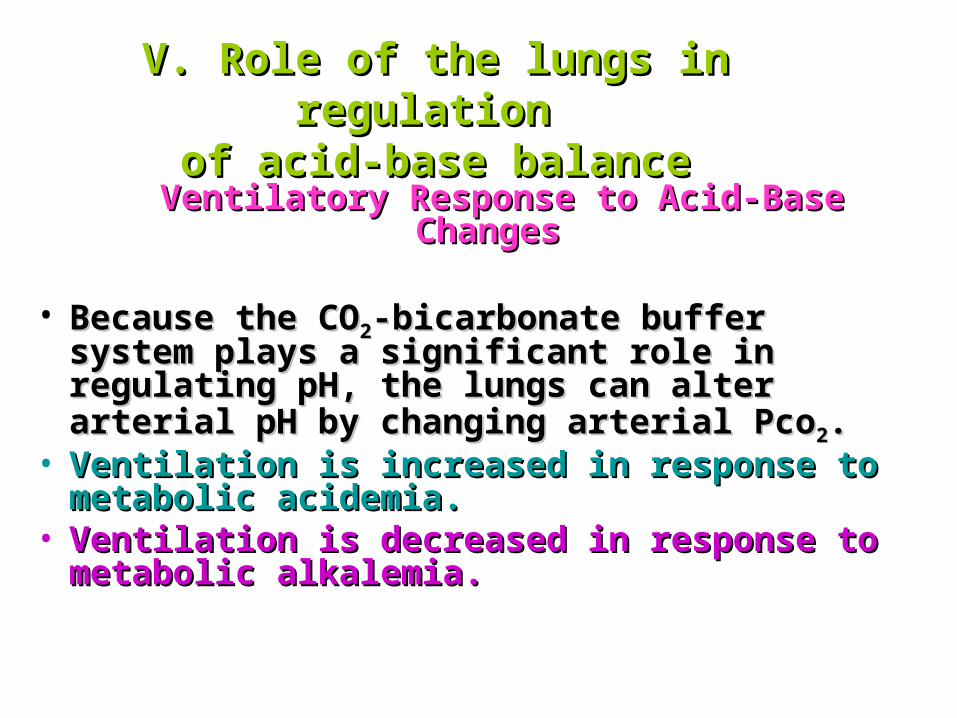
V. Role of the lungs in regulation V. Role of the lungs in regulation of acid-base balanceof acid-base balance
Ventilatory Response to Acid-Base ChangesVentilatory Response to Acid-Base Changes
• Because the COBecause the CO22-bicarbonate buffer system -bicarbonate buffer system plays a significant role in regulating pH, the plays a significant role in regulating pH, the lungs can alter arterial pH by changing arterial lungs can alter arterial pH by changing arterial PcoPco22..
• Ventilation is increased in response to Ventilation is increased in response to metabolic acidemia.metabolic acidemia.
• Ventilation is decreased in response to Ventilation is decreased in response to metabolic alkalemia.metabolic alkalemia.

• The CO2-bicarbonate buffer system is the major way in which the body maintains arterial pH because the lungs regulate the CO2 level of the blood and the kidneys regulate the amount of bicarbonate (chapter4).
• CO2undergoes the following reaction in blood: CO2 + H2O H2CO3 H + + HCO3
- pH=6.1+log ( [HCO3
- ] / Pco2) {deriving from the Henderson-Hasselbalch equation}
• Normal blood values can be substituted for the various parameters. To convert the units of mm Hg for Pco2 to mEq/L, Pco2 is multiplied by 0.03.
7.4=6.1+log [24mEq/L/(0.03×40 mm Hg)]=6.1+log 20/1 This relationship shows that as long as the ratio of bicarbonate
to CO2 is 20:1,pH will be 7.4. The body adjusts the amounts of these two substances in order to maintain a normal pH. The lungs regulate the amount of CO2.
Role of the lungs in regulation Role of the lungs in regulation of acid-base balanceof acid-base balance
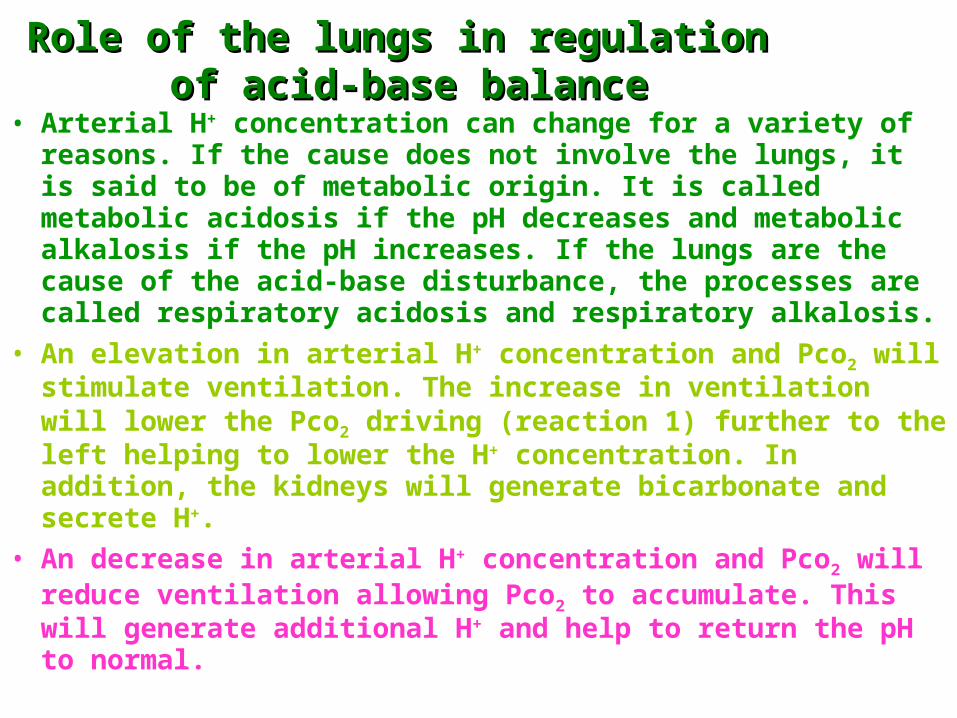
• Arterial H+ concentration can change for a variety of reasons. If the cause does not involve the lungs, it is said to be of metabolic origin. It is called metabolic acidosis if the pH decreases and metabolic alkalosis if the pH increases. If the lungs are the cause of the acid-base disturbance, the processes are called respiratory acidosis and respiratory alkalosis.
• An elevation in arterial H+ concentration and Pco2 will stimulate ventilation. The increase in ventilation will lower the Pco2 driving (reaction 1) further to the left helping to lower the H+ concentration. In addition, the kidneys will generate bicarbonate and secrete H+.
• An decrease in arterial H+ concentration and Pco2 will reduce ventilation allowing Pco2 to accumulate. This will generate additional H+ and help to return the pH to normal.
Role of the lungs in regulation Role of the lungs in regulation of acid-base balanceof acid-base balance

Altered ventilation causes Altered ventilation causes acid-base changesacid-base changes
• CO2 + H2O H2CO3 H + + HCO3-
• An inability of the lungs to remove CO2
results in respiratory acidemia.• Inappropriate removal of CO2 by the lungs
results in respiratory alkalemia.