Gary Van Domselaar University of Alberta gary.vandomselaar ...– Good all around viewer that uses...
54
Lecture 3.4 1 Structure Tools and Visualization † Gary Van Domselaar University of Alberta [email protected] † Slides Adapted from Michel Dumontier, Blueprint Initiative
Transcript of Gary Van Domselaar University of Alberta gary.vandomselaar ...– Good all around viewer that uses...

Lecture 3.4 1
Structure Tools and Visualization†
Gary Van DomselaarUniversity of Alberta
†Slides Adapted from Michel Dumontier, Blueprint Initiative

Lecture 3.4 2
Visualization & Communication
Visualization tools allow us to• see 3D structure data• communicate features about 3-D structures to colleagues• illustrate biological processes (catalytic/binding)• educate laypersons about structural biology
Go beyond Rasmol & communicate other structural features • surface shape
– show the surface, transparent over a backbone
• hydrophobicity / charge – show the binding surfaces or charge complementarity
• mutations– making a simple model (e.g. 1 amino acid change)

Lecture 3.4 3
In this lecture we introduce• Rasmol and CHIME
– Good introductory packages for biomolecule visualization
• Cn3D for Structure Annotation– Good all around viewer that uses OpenGL graphics– Good annotation engine for exchanging information about
3D structure
• Swiss PDB Viewer (Deep View)– Make molecular surfaces– Align multiple proteins– Apply scoring functions– Simple, fast modeling including site-directed mutagenesis– Complex modeling including loop rebuilding
• PyMOL– Python based, can be used for scripting

Lecture 3.4 4
Rasmol & CHIME
• Developed by Roger Sayle• Open source, binaries available
– http://openrasmol.org/
• Widely used, simple to use (menus) for simple operations
• Complex operations require command-line interface

Lecture 3.4 5
Rasmol
• Developed by Roger Sayle• Open source, binaries available
– http://openrasmol.org/
• Widely used, simple to use (menus) for simple operations
• Complex operations require command-line interface

Lecture 3.4 6
Getting Rasmol Structure Files
• Uses PDB files: http://www.rcsb.org/
1
2
3

Lecture 3.4 7
Working With the PDB File
“select cys115.cb”
“select lys116”

Lecture 3.4 8
Hen egg-white lysozyme and tri-N-acetylchitotriose

Lecture 3.4 9
Rasmol Help
• Quick Reference Card:– http://info.bio.cmu.edu/Courses/BiochemMols/RasFrames/REFCARD.PDF
• Rasmol Manual– http://openrasmol.org/doc/rasmol.html
• Tutorials:– http://www.umass.edu/microbio/rasmol/rastut.htm
• Gallery:– http://www.umass.edu/microbio/rasmol/galmz.htm

Lecture 3.4 10
CHIME
• CHIME: “Chemical mIME”• A free molecular viewer web browser plugin
based on Rasmol• Developed by MDL Information Systems Inc:
– http://www.mdl/com/
• Improves on Rasmol:– More commands
– Hypertext button-controlled scripting
– Animations

Lecture 3.4 11
CHIME
• What you need to run CHIME:– Netscape / Mozilla / Internet Explorer
– Windows or Macintosh
– A web page designed to use CHIME
– A PDB file

Lecture 3.4 12
CHIME and Protein Explorer• Protein Explorer: A website that works with
CHIME to help you visualize your structures• http://molvis.sdsc.edu/protexpl/frntdoor.htm

Lecture 3.4 13
Cn3D
• Developed by the NCBI• Open source, binaries available
http://www.ncbi.nlm.nih.gov/Structure/ • Fast OpenGL Graphics• Annotation Engine
– Lets you mark up a protein at the residue level
• Can fetch a structure over the Internet• Can display protein “movies”
– NMR ensembles– Protein folding trajectories
Lecture 3.4 14
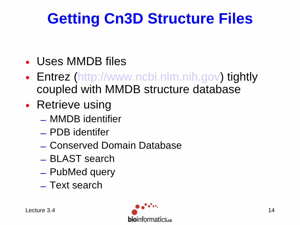
Getting Cn3D Structure Files
• Uses MMDB files• Entrez (http://www.ncbi.nlm.nih.gov) tightly
coupled with MMDB structure database• Retrieve using
– MMDB identifier– PDB identifer– Conserved Domain Database– BLAST search– PubMed query– Text search

Lecture 3.4 15
Hen egg-white lysozyme and tri-N-acetylchitotriose

Lecture 3.4 16
ASN.1 Structure File (ascii)
MMDB-ID
PDB ID
History
Publications
Chemical Graph
- Molecule Graph
- taxonomy
- residue sequence
- inter residue bonds
- heterogens
- solvent
- Inter-molecule bonds
- Annotation
- Camera Settings

Lecture 3.4 17
Global Style Settings

Lecture 3.4 18
Create New Annotation Disulfide Cysteines
1. Select Residues2. Menu>Style>Annotate3. Create New Annotation4. Give Name and Edit Style

Lecture 3.4 19
Edit Style Options

Lecture 3.4 20
Selective Annotation of Cysteine Disulfide Bridges

Lecture 3.4 21
Annotation Menu
Turn On/Off – Show/Hide selected annotation
Move Up/Down – Change priorityShow – Show annotation and
affected residuesEdit – Edit an existing annotationMove – Move the annotation to
selected residuesDelete – Delete the Annotation

Lecture 3.4 22
Shortcuts
Rendering: SpaceFillColoring: Rainbow
Rendering: Ball and StickColoring: Hydrophobicity

Lecture 3.4 23
Another Salient Feature
Show/Hide>Select by distance

Lecture 3.4 24
Imports
• Import sequences – BLAST alignments
• Import structures– Structural Superposition

Lecture 3.4 25
BLAST
• Create alignments between sequences– In this case, an
alignment between the structure sequence and the natural sequence reveals missing N and C termini residues and a loop region, not resolved in structure.

Lecture 3.4 26
Conserved Domain Alignments

Lecture 3.4 27
Folding Simulations

Lecture 3.4 28
NMR Solution Structures

Lecture 3.4 29
Finishing Your Work
• Save– Annotations
– Camera Settings
• Export– PNG image

Lecture 3.4 30
Other Cn3D Resources
• Online Help• Application Help

Lecture 3.4 31
Swiss PDV Viewer at a Glance
• PDB structure viewer with structure utilities
• Superimposition to compare proteins and their components such as active/binding sites
• Measure angles, distances between atoms
• Manual or automated (Swiss-Model) homology modeling including loop modeling
• Threading (Fold recognition)
• Mutations and Energy minimization
• Electron density map reading and model building (crystallography data)
• Interface to POV-Ray rendering software

Lecture 3.4 32
Main Interface
• Tons of menu options reasonably well categorized
• Button bar for image manipulation (center, zoom, move, rotate) and some structure measurement and mutation tools
• Layers window to select from multiple layers

Lecture 3.4 33
Control Panel
• Select– Individual Residues (one or more)– All Residues– Chains– Secondary Structure
• Show– Backbone– Side chains– Labels– Molecular surface (VDW)– Ribbon Cartoons (Helices/Strands)– Colors (specify backbone +/- sidechain,
labels, etc)

Lecture 3.4 34
Alignment and Ramachandran Plot
• Alignment Window shows alignment of sequences to one another (structural superposition, threading / homology modelling)
• Ramachandran plot showing Phi-Psi angles of selected residues. Can move individual residues to new Phi-Psi angles.

Lecture 3.4 35
Molecular Surfaces

Lecture 3.4 36
Electrostatic Potentials
• Useful for evaluating the effects of a potential mutation
• Analysis of binding site

Lecture 3.4 37
SPDBV Home Page
• http://expasy.org/spdbv

Lecture 3.4 38
•http://www.usm.maine.edu/~rhodes/SPVTut/index.html

Lecture 3.4 39
Persistence of Vision(POV) RAY Visualization
• Ray tracing occurs from the camera to the scene
• Specify – Camera location
– Light sources
– Objects
– Surface textures
– Atmospheric media (fog, haze, or fire)

Lecture 3.4 40
POV RAY
http://www.povray.org/documentation
Lecture 3.4 41
PovRay Molecules...
• Armand Tepper ‘s Energy minimized Yeast Cu-metallothionein from an averaged NMR structure. 6 copper atoms are in the reddish metal texture and the sulfurs of the coordinating cysteines appear in yellow. This picture features the new smoother ribbon feature (quality 2) implemented in SPDBV3.6b2. The surface is at detail level 3.
• http://wwwchem.leidenuniv.nl/metprot/armand

Lecture 3.4 42

Lecture 3.4 43
POV Ray scene generators
• Swiss PDB Viewer can save file as POV scene
• PDB to POV converters available– MolPov (windows) http://www.chem.ufl.edu/~der/
der_pov2.htm
– PPOVIT (PERL) http://huron.cem.msu.edu/~rstc/ppovit/

Lecture 3.4 44
PyMOL
• Set of structure tools built on top of Python
• Supports all Standard Features
• Extensible, Scriptable
• Native Ray Tracer• Freely Available:
– http://pymol.sourceforge.net/

Lecture 3.4 45
PyMOL
• Set of structure tools built on top of Python
• Supports all Standard Features
• Extensible, Scriptable
• Native Ray Tracer• Freely Available:
– http://pymol.sourceforge.net/

Lecture 3.4 46
PyMOL External GUI
• Command line interface
• Standard menu bar
• Some Handy Buttons

Lecture 3.4 47
PyMOL External GUI
VIEWING WINDOW
Mouse Bindings
Movie ControlsCommand Line
Named Groups Panel

Lecture 3.4 48
PyMOL Actions Menu (A)
VIEWING WINDOW
Mouse Bindings
Movie ControlsCommand Line
Named Groups Panel

Lecture 3.4 49
PyMOL Show Menu (S)
VIEWING WINDOW
Mouse Bindings
Movie ControlsCommand Line
Named Groups Panel

Lecture 3.4 50
PyMOL Hide Menu (S)
VIEWING WINDOW
Mouse Bindings
Movie ControlsCommand Line
Named Groups Panel

Lecture 3.4 51
PyMOL Labels Menu (L)
VIEWING WINDOW
Mouse Bindings
Movie ControlsCommand Line
Named Groups Panel

Lecture 3.4 52
PyMOL Color Menu (C)
VIEWING WINDOW
Mouse Bindings
Movie ControlsCommand Line
Named Groups Panel

Lecture 3.4 53
Hen egg-white lysozyme and tri-N-acetylchitotriose

Lecture 3.4 54
Conclusions
• Several of the more powerful structure tools with visualization and structure manipulation features are freely available– Rasmol, Cn3D, Swiss PDB Viewer, PyMOL
• Mark up your structures to convey important and useful information
• Ray Trace output scenes for best rendering and artistic flash