HAIJKLMNBCDOPG...GU D ƒ G G G GGUØ ÙDÌ`u›rvbm“ł Kß¡l˘fˇˆu˙qN D˝˛G GGG~ˇŽÓT œ‡

WATERA Level Biology
AQA Topic 1
© Dr Zoë HuggettPage 1 of 1
Structure• Two hydrogen atoms share electrons with one oxygen atom (covalent bonds)• Electrons pulled towards oxygen → hydrogens have a partial positive charge• Oxygen has two lone (unshared) electron pairs → oxygen has a partial negative charge• A polar molecule• Partial charges attract other water molecules → hydrogen bonding
Properties• Good solvent → polar water molecules surround and are attracted to ions or other polar molecules, so
they can dissolve
• Strong cohesion → polar molecules stick together with hydrogen bonds → water flows well e.g it forms an unbroken column in the xylem vessels→ has a high surface tension so forms droplets
• Useful metabolite → used in metabolic reactions e.g condensation and hydrolysis reactions
• High latent heat of vaporisation → lots of energy needed to break hydrogen bonds→ lots of heat energy used to evaporate (vaporise)
• High specific heat capacity → hydrogen bonds can absorb lots of energy→ can lose or gain a lot of energy without changing temperature which
is good for aquatic organisms
Sweat forms droplets on the skin then use heat energy from the skin to evaporate, cooling you down in the process
Ionic compounds are important in metabolic reactions
Specific heat capacity = energy needed to raise temperature of 1g of a substance by 1°C
partial negativeg-← charge
oxygenunsharedelectrons
✗•
• •+• hydrogen bond →
• sit ⑧•←nudeus'
is -
hydrogen + • t
*÷" "
#*:→
⑨• •
•if•
+• .g-•
••too partial
•@Xa- s+ positive charge gt covalentgt
si.St
ybonds
dissolved ,%µg&ggg± ¥.× . ¥:
•
•e
•
ionic *
MHg-igf-GB.EDUcompound%
÷¥¥÷ .÷ ¥. #•÷
•
,
°
,
•

LIPIDSA Level Biology
AQA Topic 1
Triglycerides• One molecule of glycerol bound to three fatty acids• Fatty acids join to glycerol in a condensation reaction → an ester bond is formed and a water molecule
is released• Three water molecules released and three ester bonds formed for each triglyceride• Ester bonds are broken by a hydrolysis reaction• Energy store → hydrocarbon tails release a lot of energy when broken down• Insoluble in water → do not affect the water potential of cells so water is not drawn in by osmosis• Clump together in droplets with the hydrophobic hydrocarbon tails facing inwards
© Dr Zoë HuggettPage 1 of 1
• Lipids contain carbon, hydrogen and oxygen
Fatty Acids• Have a variable Rgroup → the hydrocarbon tail• Saturated fatty acids have no double C=C bonds in
the hydrogen carbon tail• Unsaturated fatty acids have one or more double C=C
bonds in the hydrocarbon tail so the chain kinks• Hydrocarbon tails are hydrophobic (insoluble in water)
Phospholipids• One molecule of glycerol bound to two fatty acids and a
phosphate group• Phosphate groups are hydrophilic, fatty acids are hydrophobic• Form a bilayer in cell membranes → water soluble (polar)
substances cannot pass through the hydrophobic centre of the bilayer
Emulsion Test for Lipids1) Mix food sample with ethanol and shake until dissolved2) Pour mixture into water3) If lipid is present → milky emulsion forms (the more obvious it is, the more lipid there is)4) If no lipid is present → stays clear
Tip: know how to relate structure to function
O variable saturated
Basic X t group H H H
structure,C- R
Ol H l
H"
H
Ho xifyrfyrfyC H H
- H/ H l l l
HO H H H
H Unsaturated
Ol H HC l l
X 11 =H H
f Hygienic" O H double l
H,
- H
bond H H
H Q triglyceride H O1
,C - R Y l H
H - C- O"
fatty H - C- O - C - RO acids -
I Xd / O
H - C- O - C - R 11
/ o S H- C - O- C- R A droplet"
-
H - C- O - C / Oof triglycerides
I'R H
H - C- O- C - R← glycerol
T H ↳3h20
l U(fatty acids
glycerol H esitsornds
hydrophilicone ← heads
phospholipid hydrophobic→ ← tails
fatty acids
← glycerolphosphate←
group
PROTEINSA Level Biology
AQA Topic 1
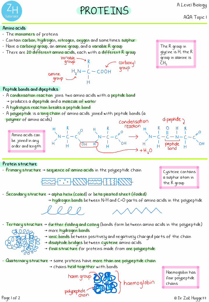
Amino acids• The monomers of proteins• Contain carbon, hydrogen, nitrogen, oxygen and sometimes sulphur• Have a carboxyl group, an amine group, and a variable R group• There are 20 different amino acids, each with a different R group
Peptide bonds and dipeptides• A condensation reaction joins two amino acids with a peptide bond
→ produces a dipeptide and a molecule of water• A hydrolysis reaction breaks a peptide bond• A polypeptide is a long chain of amino acids joined with peptide bonds (a
polymer of amino acids)
© Dr Zoë HuggettPage 1 of 2
Protein structure• Primary structure → sequence of amino acids in the polypeptide chain
• Secondary structure → alpha helix (coiled) or beta pleated sheet (folded)→ hydrogen bonds between N-H and C=O parts of amino acids in the polypeptide
• Tertiary structure → further folding and coiling (bonds form between amino acids in the polypeptide)→ more hydrogen bonds→ ionic bonds between positively and negatively charged parts of the chain→ disulphide bridges between cysteine amino acids→ final structure for proteins made from one polypeptide
• Quaternary structure → some proteins have more than one polypeptide chain→ chains held together with bonds
Amino acids can be joined in any order and length
Cysteine contains a sulphur atom in the R group
Haemoglobin has four polypeptide chains
The R group in glycine is H, the R group in alanine is CH3
variable→
group R
amine Han - § - cojgcarrbuopxy'
groupTH
condensation dipeptide yreaction
:÷÷÷. "÷÷÷:.÷÷÷÷÷÷: .I peptide✓ + H2O
bond
.
-¥-333
lele mm
BBhaemgroupf.ggpolypeptide.in#OB&@ haemoglobin

PROTEINSA Level Biology
AQA Topic 1
© Dr Zoë HuggettPage 2 of 2
Functions of proteins• Enzymes
→ soluble and almost spherical→ tightly folded polypeptides→ catalyse metabolic reactions→ examples: amylase digests starch, lipase
digests lipids
• Structural proteins → provide strength and support→ long polypeptide chains parallel to each
other or twisted round each other into arope shape
→ chains held together with cross-linkse.g disulphide bonds.
→ examples: collagen in connective tissue,and keratin in hair and nails
Biuret test for proteins1) Add a few drops of sodium hydroxide solution to
the sample to make it alkaline.2) Add copper (II) sulphate solution.3) If protein is present: solution turns purple. 4) If no protein is present: solution stays blue.
The colour change is subtle in this test
• Antibodies → made by plasma cells in the immune
response→ two light chains and two heavy
chains→ have constant regions and variable
regions (variable regions make themspecific for one antigen)
• Transport proteins→ found in cell membranes→ channel proteins and carrier proteins→ hydrophobic and hydrophilic regions
of the protein help it to form itsshape
④ active "9"
has:#ein

CARBOHYDRATESA Level Biology
AQA Topic 1
Glucose• A monosaccharide with the formula C6H12O6 • A hexose monosaccharide (six carbon atoms) in a ring structure• Soluble in water → easily transported• Main energy source for animals and plants
→ chemical bonds store lots of energy• Two isomers: α-glucose and β-glucose
Glycosidic bonds and condensation/hydrolysis reactions• Condensation reaction: two molecules join to form a new chemical bond
and a water molecule is released• Condensation reactions form glycosidic bonds between monosaccharides
to create disaccharides and polysaccharides• Hydrolysis reaction: a water molecule is inserted and the chemical bond is
broken (reverse of a condensation reaction → breaks glycosidic bonds)
© Dr Zoë Huggett
Displayed formula: Skeletal formula:
Page 1 of 2
Disaccharides• Two monosaccharides joined together with a glycosidic bond• Maltose = glucose + glucose• Sucrose = glucose + fructose• Lactose = glucose + galactose
Tip: condensation and hydrolysis reactions are really common
• Carbohydrates contain carbon, hydrogen and oxygen, usually with the general formula CnH2nOn
Monosaccharides are small soluble carbohydrate monomers. They also include fructose and galactose.
Monosaccharides and disaccharides are called sugars.
fHZOH a -glucose fHz0H B -glucoseC- O
a -glucose B -glucoseHy '
y H H F- 0 OH H Hl H l l IH Is 1- O H 1- 0 OH
¢ OH H C COH H C
Hol ' ' / LOH ' Il 11 It,C - C HO c - C
l l l l HO OH HOH
H OH H OH
←a -glucose→ ly saccharide
H H (P°
with glycosidic bonds1- O H 1- O H s
-
- O - - O - -
^n
HO
0HH inserted insertedcondensation
maltose v reaction hydrolysis✓ reaction
H tH1- O H 1- O H
yHo - -OH HO - - OH
+ H2O
HO O of,mon accharides ① OH
l l
glycosidicbond

CARBOHYDRATESA Level Biology
AQA Topic 1
© Dr Zoë Huggett
Tip: know how to relate structure to function
Page 2 of 2
Polysaccharides• Large polymers of monosaccharides joined with glycosidic bonds• Starch, glycogen and cellulose
Benedict’s Test for Sugars• Monosaccharides, maltose, and lactose are reducing sugars• Sucrose is a non-reducing sugar
1) Add an excess of blue Benedict’s reagent to liquid food sample in a test tube.
2) Heat the tube in a water bath set to boil.3) If reducing sugars are present: coloured precipitate forms.
End test here.4) If no reducing sugars are present: solution stays blue. Go to
step 5.5) Break down non-reducing sugars to monosaccharides: add dilute
HCl to new sample and heat in a water bath set to boil.6) Neutralise with sodium hydrogencarbonate, then repeat steps
1) and 2).7) If coloured precipitate now forms, non-reducing sugars are
present in the sample.8) If the solution is still blue, neither type of sugars are present.
Blue
Green
Yellow
Orange
Brick Red
Colour of precipitate depends on the concentration of reducing sugars:
You could filter the sample and weigh the precipitate to make more accurate comparisons.
Starch• Glucose storage in plants → broken down when glucose is needed• Insoluble in water → does not affect the water potential of
cells so water is not drawn in by osmosis• Amylose → unbranched α-glucose polysaccharide (1,4 glycosidic
bonds)→ coiled structure (compact)
• Amylopectin → branched α-glucose polysaccharide (1,4 and 1,6glycosidic bonds)
→ branches mean enzymes can easily access more glycosidic bonds = faster glucose release
Glycogen• Excess glucose storage in animals → broken
down when glucose is needed• Insoluble in water → does not affect the
water potential of cells so water is not drawn in by osmosis
• Highly branched α-glucose polysaccharide (1,4 and 1,6 glycosidic bonds) → glucose can be released quickly by enzymes, and it is compact
Cellulose• Unbranched straight β-glucose polymers (1,4
glycosidic bonds)• Chains linked with hydrogen bonds to form
strong myofibrils• Found in plant cell walls to give structural support
Iodine Test for Starch1) Add iodine in potassium iodide
solution to sample.2) If starch is present: goes from
browny-orange to blue-black.
4-glucoseAmylose.
I → Amylopectin,
l
l l -
s-
'
--
,
- -
y- =
I
\
,
i
--
-- -
,
- - Jonecellulose
--
chain
B-glucose-
"
÷"
is ! :"
is,
"
iswehaykarogenbonds
Benedict's T Hsolution f 8is blue
,so blue 8
is a negative 8result f §
§Qt I⇐
f S
V

DNA AND RNAA Level Biology
AQA Topic 1
© Dr Zoë HuggettPage 1 of 2
DNA Nucleotides• The monomers of DNA• Consist of a pentose sugar (deoxyribose), a
nitrogen-containing base, and a phosphate group• The base can be adenine (A), thymine (T), guanine
(G) or cytosine (C)
DNA• Stores genetic information • Polymer of DNA nucleotides• Two antiparallel polynucleotide chains twisted into a double-helix structure• The phosphate groups and pentose sugars form the sugar-phosphate backbone• The bases join by complementary base pairing → A pairs to T with two hydrogen bonds
→ C pairs to G with three hydrogen bonds→ holds the two strands together
• DNA = deoxyribonucleic acid, RNA = ribonucleic acid
RNA Nucleotides• The monomers of RNA• Consist of a pentose sugar (ribose), a nitrogen-
containing base, and a phosphate group• The base can be adenine (A), uracil (U), guanine (G)
or cytosine (C)
• Both DNA and RNA nucleotides form polynucleotides → phosphodiester bonds form between the phosphate group of one and the deoxyribose of the next in a condensation reaction
A pentose sugar has five carbon atoms
Antiparallel means the strands run in opposite directions
The double-helix was discovered by Watson and Crick in 1953
There is always the same amount of A and T and the same amount of C and G in DNA
Bases are organic (contain carbon)
Scientists first doubted that DNA stored complex genetic information because of its simple structure
DNA nucleotide :
RNA nucleotide :
r rphosphate - phosphategroup ) group
-
'
( base ( base )deoxyribose (A.ICORG) ribose
(A. 4. Coro)
hydrogenbonds
b
phosphodiester - A T -
bond
€- c G -
- T A -
Tsuga- phosphate Tbackbones
DNA AND RNAA Level Biology
AQA Topic 1
© Dr Zoë HuggettPage 2 of 2
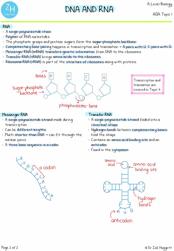
RNA• A single polynucleotide chain• Polymer of RNA nucleotides• The phosphate groups and pentose sugars form the sugar-phosphate backbone• Complementary base pairing happens in transcription and translation → A pairs with U, C pairs with G• Messenger RNA (mRNA) transfers genetic information from DNA to the ribosomes• Transfer RNA (tRNA) brings amino acids to the ribosomes• Ribosomal RNA (rRNA) is part of the structure of ribosomes along with proteins
Messenger RNA• A single polynucleotide strand made during
transcription• Can be different lengths• Much shorter than DNA → can fit through the
nuclear pores• A three base sequence is a codon
Transfer RNA• A single polynucleotide strand folded into a
cloverleaf shape• Hydrogen bonds between complementary bases
hold the shape• Contains an amino acid binding site and an
anticodon• Found in the cytoplasm
Transcription and translation are covered in Topic 4
→
bases A U C G
l l l l
sugar- phosphatebackbone -7
xphosphodiester bond
amino
acid→ amino acidOdon ←
binding site-⇒÷÷
.
€¥Iµ= hydrogen
bonds
PA't's
anticodon

ATPA Level Biology
AQA Topic 1
© Dr Zoë HuggettPage 1 of 1
Structure• ATP = adenosine triphosphate• A nucleotide derivative (a modified nucleotide)• A ribose sugar bound to adenine (a base) and three phosphate groups• Energy stored in high energy bonds between phosphate groups• Made during respiration using energy released from glucose
Function• Diffuses to areas of cells where energy is needed• Broken down by ATP hydrolase to ADP (adenosine diphosphate) and Pi (inorganic phosphate) in a
hydrolysis reaction → energy is released• Energy can be used straight away in a coupled reaction → a reaction requiring energy which is
coupled to ATP hydrolysis• Immediate energy supply → little is lost as heat• Re-synthesised by ATP synthase in a condensation reaction → happens in respiration and
photosynthesis• Pi can be used in a phosphorylation reaction → a phosphate group is added to another molecule
→ phosphorylation can make compounds more reactiveor change their shape
An example of a reaction coupled to ATP hydrolysis is the synthesis of sucrose from glucose and fructose
Remember that water is produced in condensation and used in hydrolysis
“Inorganic” means a molecule does not contain carbon
phosphate groupsadenine-f ! I
.
--
→ itribose
high energy(a pentosesugar)
bonds
inorganicenergy required
phosphateATP synthase
↳p ,
Koindensatjon)--
--
Afp hydrolase ATPADP (hydrolysis)energy released

INORGANIC IONSA Level Biology
AQA Topic 1
© Dr Zoë HuggettPage 1 of 1
• Ions have electric charge → anions have positive charge→ cations have negative charge
• Inorganic ions are soluble → dissolved in the fluids of an organism and in the cytoplasm
Examples• Phosphate ions → attached to other molecules to become a phosphate group (phosphorylation)
→ found in ATP, DNA, RNA and phospholipids→ get transferred through the food chain in the phosphorous cycle→ used in photosynthesis and respiration
• Iron ions → Fe2+ is the part of haemoglobin that binds oxygen (becomes Fe3+ until oxygen is released)
• Sodium ions → used in co-transport to help glucose and amino acids cross membranes e.g glucose absorption in the small intestine
→ needed to create an action potential in neurones
• Hydrogen ions → more H+ ions present means a lower (more acidic) pH→ enzyme activity is affected by pH (and therefore H+ concentration)
You will come across many other ions in biology e.gCa2+ ions at synapses
POE
Fe" [email protected]@ haemoglobin
Nat
Ht


![Final April 11 - abulhasanalinadwi.orgabulhasanalinadwi.org/books/payam04. April_11.pdf · ÔÔÔÔ gggg»»»ÃÃÃÃheee${{{zzz{zÐÐÐ]]]]ÑÑÑÑqqqq{ ... ZggZg~~g~ggg~g‚‚‚‚ŸtttgggzzgzZZZzZ](https://static.fdocuments.us/doc/165x107/5a9bbd637f8b9ad96f8e2737/final-april-11-april11pdf-ggggheeezzzzqqqq-zggzggggggytttgggzzgzzzzzz.jpg)