Fuzzy Cells Extraction and Cell Morphology for the … 11 I 03/1110203-4949 IJVIPNS-IJENS.pdf"Fuzzy...
10
International Journal of Video & Image Processing and Network Security IJVIPNS-IJENS Vol: 11 No: 03 40 1110203-4949 IJVIPNS-IJENS © June 2011 IJENS I J E N S Fuzzy Cells Extraction and Cell Morphology for the Antigen Positivity Determination of Immunohistochemical Image *Lestari Handayani Faculty of Science and Technology UIN SUSKA Riau lestari.handayani@uin- suska.ac.id M. R. Widyanto Faculty of Computer Science University of Indonesia [email protected] Ria Kodariah Faculty of Medicine University of Indonesia Putu W. Handayani Faculty of Computer Science University of Indonesia Abstract -- The accuracy of immunohistochemical image antigen determination is very important in reporting result immunohistochemical examination. The previous studies [5], [10] have not been succeeded to determine such accuracy. The failure is caused by the characters in the image cells vary widely in shape, size, cell type, outward color appearance, and the huddled cells. EsFuMos method that uses fuzzy inference system is proposed to extract the cells, obtain cell morphology, and watershed separating huddled cells. The experimental result of comparing EsFuMoS method with MoFSoTH method [5] and the NN method [10] shows that EsFuMoS method is the most successful method to determine positivity. Meanwhile for huddled cell separation, EsFuMoS method success rate is 77.63%. Lack of EsFuMoS method is having an error in cell separation, and not able to lift the weak negative cell nucleus. According to pathologist, this method works very helpful. But it needs to be improved by paying close attention to the character of the cell membrane and weak cell nucleus. 1. INTRODUCTION According to the World Health Organization (WHO), cancer is the leading cause of death in the world. In 2004, it was recorded 7.4 million deaths or about 13% of all deaths worldwide. Breast cancer ranks the fifth position in the world with 519,000 deaths. The choice of therapy will be given to cancer patients is very important for his recovery. One of the choices in breast carcinoma is hormonal therapy which aimed to prevent hormones and stop the growth of tumor cells.Hormonal therapy can be given if the cancer cells contain receptors for these hormones. Immunohistochemical examination can be used to identify it. Immunohistochemistry is a method of examination of antigen in tissue using specific antibodies for the target of antigen . This method still used widely in pathology because of morphology sustained. In breast cancer, examination of estrogen receptors and progesteron receptors in tissue must be done before hormonal theraphy is given. It is important to ensure the patient receives optimal results from hormonal therapy given. To determine positifitas from that reseptors, it is need to observation with microscop to determine the number of nucleus colored in field of view. The percentage of positive nucleus is counted of 1000 cancer cells. To calculate positive cells some researchers have been conducted using a microscopic image of cells, namely: a) Neural networks (NN) [10]: using the RGB color model as input, the networks of back propagation with two hidden layers and an output layer followed by morphological operations. It works with good results for images with different colors cells+, cell-, and sufficiently high background, but if it does not work, the result will go wrong. b) Fuzzy morphology method based on hue [4]: using HSV color models and method of fuzzy morphology is to segment positive cell nucleus. The result can determine the positive cell, but it is still lack of the ability to distinguish between positive cell and tissue cell that does not have a close value but with the intensity of saturation and different light intensity (brightness). c) Fuzzy morphology method based on hue and threshold (MoFSoTH) [5]: improving the method b) using two steps, i.e. filtering components of hue and saturation component segmentation with fuzzy morphological operations to segment positive cell nucleus. The result can determine the strong and middle positive cell nucleus, but it has not able to lift weak positive cell nucleus and differentiate huddled cells yet. (d) 3D structuring element method based on Multiscale Morphology (3DSEM) [9]: segmenting nucleus and the cytoplasm for the identification of the white blood cells in the spinal column using the morphology of multiple scales, the result can distinguish between the nucleus and the cytoplasm for the image with the difference of the highest cell nucleus and cytoplasm, or the result is bad or even not clear for the nucleus and the cytoplasm. The above methods are simulated by using sample of the immunohistochemical outward appearance image from Danial’ s research [5] is to segment positive and negative cells. There are still the errors from the segmentation of the cell using the above methods with a quite moderate error rate. It can be concluded that it happened, i.e.: having error identification of positive and negative cells, not having identified the weak cell nucleus yet, not able to separate the huddled cells that is considered as a single cell. Lack of segmentation of the cell by the above methods, is caused by cell character and image as results of immunohistochemical outward appearance, namely: 1) varied cell shape (round, elliptical, flattened, dented), 2) varied cell size, 3) the varied characteristics of positive and negative cells, positive cells have strong, moderate, and weak positive cell nucleus, the color tends to brown, and the cell nucleus is greater than the cytoplasm, whereas negative cells tend to be blue, and smaller cell nucleus from the cytoplasm, 4) object color and background image of the results of varied immunohistochemical outward appearance depending on the
Transcript of Fuzzy Cells Extraction and Cell Morphology for the … 11 I 03/1110203-4949 IJVIPNS-IJENS.pdf"Fuzzy...

International Journal of Video & Image Processing and Network Security IJVIPNS-IJENS Vol: 11 No: 03 40
1110203-4949 IJVIPNS-IJENS © June 2011 IJENS I J E N S
Fuzzy Cells Extraction and Cell Morphology for the
Antigen Positivity Determination of
Immunohistochemical Image
*Lestari Handayani
Faculty of Science and
Technology
UIN SUSKA Riau
lestari.handayani@uin-
suska.ac.id
M. R. Widyanto
Faculty of Computer Science
University of Indonesia
Ria Kodariah
Faculty of Medicine
University of Indonesia
Putu W. Handayani
Faculty of Computer Science
University of Indonesia
Abstract-- The accuracy of immunohistochemical image
antigen determination is very important in reporting result
immunohistochemical examination. The previous studies [5],
[10] have not been succeeded to determine such accuracy. The
failure is caused by the characters in the image cells vary widely
in shape, size, cell type, outward color appearance, and the
huddled cells. EsFuMos method that uses fuzzy inference
system is proposed to extract the cells, obtain cell morphology,
and watershed separating huddled cells. The experimental
result of comparing EsFuMoS method with MoFSoTH method
[5] and the NN method [10] shows that EsFuMoS method is the
most successful method to determine positivity. Meanwhile for
huddled cell separation, EsFuMoS method success rate is
77.63%. Lack of EsFuMoS method is having an error in cell
separation, and not able to lift the weak negative cell nucleus.
According to pathologist, this method works very helpful. But it
needs to be improved by paying close attention to the character
of the cell membrane and weak cell nucleus.
1. INTRODUCTION
According to the World Health Organization (WHO), cancer is the leading cause of death in the world. In 2004, it was recorded 7.4 million deaths or about 13% of all deaths worldwide. Breast cancer ranks the fifth position in the world with 519,000 deaths.
The choice of therapy will be given to cancer patients is very important for his recovery. One of the choices in breast carcinoma is hormonal therapy which aimed to prevent hormones and stop the growth of tumor cells.Hormonal therapy can be given if the cancer cells contain receptors for these hormones. Immunohistochemical examination can be used to identify it.
Immunohistochemistry is a method of examination of antigen in tissue using specific antibodies for the target of antigen . This method still used widely in pathology because of morphology sustained. In breast cancer, examination of estrogen receptors and progesteron receptors in tissue must be done before hormonal theraphy is given. It is important to ensure the patient receives optimal results from hormonal therapy given. To determine positifitas from that reseptors, it is need to observation with microscop to determine the number of nucleus colored in field of view. The percentage of positive nucleus is counted of 1000 cancer cells.
To calculate positive cells some researchers have been conducted using a microscopic image of cells, namely:
a) Neural networks (NN) [10]: using the RGB color model as input, the networks of back propagation with two
hidden layers and an output layer followed by morphological operations. It works with good results for images with different colors cells+, cell-, and sufficiently high background, but if it does not work, the result will go wrong.
b) Fuzzy morphology method based on hue [4]: using HSV color models and method of fuzzy morphology is to segment positive cell nucleus. The result can determine the positive cell, but it is still lack of the ability to distinguish between positive cell and tissue cell that does not have a close value but with the intensity of saturation and different light intensity (brightness).
c) Fuzzy morphology method based on hue and threshold (MoFSoTH) [5]: improving the method b) using two steps, i.e. filtering components of hue and saturation component segmentation with fuzzy morphological operations to segment positive cell nucleus. The result can determine the strong and middle positive cell nucleus, but it has not able to lift weak positive cell nucleus and differentiate huddled cells yet.
(d) 3D structuring element method based on Multiscale Morphology (3DSEM) [9]: segmenting nucleus and the cytoplasm for the identification of the white blood cells in the spinal column using the morphology of multiple scales, the result can distinguish between the nucleus and the cytoplasm for the image with the difference of the highest cell nucleus and cytoplasm, or the result is bad or even not clear for the nucleus and the cytoplasm.
The above methods are simulated by using sample of the
immunohistochemical outward appearance image from Danial’ s research [5] is to segment positive and negative cells. There are still the errors from the segmentation of the cell using the above methods with a quite moderate error rate. It can be concluded that it happened, i.e.: having error identification of positive and negative cells, not having identified the weak cell nucleus yet, not able to separate the huddled cells that is considered as a single cell.
Lack of segmentation of the cell by the above methods, is caused by cell character and image as results of immunohistochemical outward appearance, namely: 1) varied cell shape (round, elliptical, flattened, dented), 2) varied cell size, 3) the varied characteristics of positive and negative cells, positive cells have strong, moderate, and weak positive cell nucleus, the color tends to brown, and the cell nucleus is greater than the cytoplasm, whereas negative cells tend to be blue, and smaller cell nucleus from the cytoplasm, 4) object color and background image of the results of varied immunohistochemical outward appearance depending on the
International Journal of Video & Image Processing and Network Security IJVIPNS-IJENS Vol: 11 No: 03 41
1110203-4949 IJVIPNS-IJENS © June 2011 IJENS I J E N S
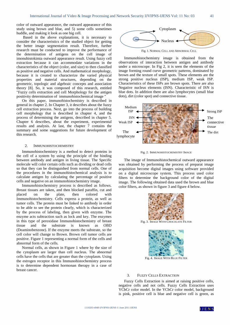
Medium
ISP
Weak ISP
ISN
The
lymphocyte
s
Strong ISP
The dirt
The
connective
tissue
color of outward appearance, the outward appearance of this study using brown and blue, and 5) some cells sometimes huddle, and making it look as one big cell.
Based in the above explanations, it is necessary to consider the characteristics of the studied object for getting the better image segmentation result. Therefore, further research must be conducted to improve the performance of the determination of antigens on the cell image of imonuhistokimia outward appearance result. Using fuzzy cell extraction because it can accommodate variations in the characteristics of the object (color, and size) to then classified as positive and negative cells. And mathematical morphology, because it is created to characterize the varied physical properties and material structures, depending on the geometric, topologic and algebraic concepts and association theory [8]. So, it was composed of this research, entitled "Fuzzy cells extraction and cell Morphology for the antigen positivity determination of immunohistochemical image "
On this paper, immunohistochemistry is described in general in chapter 2. In Chapter 3, it describes about the fuzzy cell extraction process. Next, go into the process of building cell morphology that is described in chapter 4, and the process of determining the antigens, described in chapter 5. Chapter 6 describes, about the experiment, experimental results and analysis. At last, the chapter 7 contains the summary and some suggestions for future development of this research.
2. IMMUNOHISTOCHEMISTRY
Immunohistochemistry is a method to detect proteins in the cell of a system by using the principle of the binding between antibody and antigen in living tissue. The Specific molecule will color certain cells such as dividing or dead cells so that they can be distinguished from normal cells. One of the procedures in the immunohistochemical analysis is to calculate antigen by calculating the percentage of positive cells and negative on an immunohistochemistry image.
Immunohistochemistry process is described as follows.
Breast tissues are taken, and then blocked paraffin, cut and
placed on the plate, then colored with
Immunohistochemistry. Cells express a protein, as well as
tumor cells. The protein must be linked to antibody in order
to be able to see the protein clearly, which is characterized
by the process of labeling, then given with enzyme. The
enzyme acts subtraction such as lock and key. The enzymes
in this type of peroxidase Immunohistochemistry of breast
tissue and the substrate is known as OBD
(Deaminobenzone). If the enzyme meets the substrate, so the
cell color will change to Brown. Brown cell tumor cells are
positive. Figure 1 representing a normal form of the cells and
abnormal form of the cells.
Normal cells, as shown in Figure 1 where by the size of
the cytoplasm are larger than cell nucleus. The abnormal
cells have the cells that are greater than the cytoplasm. Using
the estrogen receptor in this Immunohistochemistry process
is to determine dependent hormonan therapy in a case of
breast cancer.
Fig. 1. NORMAL CELL AND ABNORMAL CELL
Immunohistochemistry image is obtained from the observations of interaction between antigen and antibody under a microscope. In Fig 2, it is seen the elements of the image forming round curve geometric patterns, dominated by brown and the texture of small spots. These elements are the strong positive nucleus (ISP), medium ISP, weak ISP. Characteristics of these ISPs are brown spots. There are also Negative nucleus elements (ISN). Characteristic of ISN is blue dots. In addition there are also lymphocytes (small blue dots), dirt (color spot) and connective tissue.
Fig. 2. IMMUNOHISTOCHEMISTRY IMAGE
The image of Immunohistochemical outward appearance
was obtained by performing the process of preparat image
acquisition become digital images using software provided
on a digital microscope system. This process used color
filters to determine the background color of the digital
image. The following obtained data used the brown and blue
color filters, as shown in figure 3 and figure 4 below.
Fig. 3. IMAGE WITH CHOCOLATE FILTER
Fig. 4. IMAGE WITH BLUE FILTER
3. FUZZY CELLS EXTRACTION
Fuzzy Cells Extraction is aimed at raising positive cells, negative cells and not cells. Fuzzy Cells Extraction uses YCbCr color model. In the YCbCr color model, background is pink, positive cell is blue and negative cell is green, as
Nucleus
Cytoplasm

International Journal of Video & Image Processing and Network Security IJVIPNS-IJENS Vol: 11 No: 03 42
1110203-4949 IJVIPNS-IJENS © June 2011 IJENS I J E N S
shown in Fig 5. According to the theory [6], the color model is properly used for processing, analysis and coding, and linear transformation so that the research uses YCbCr color model.
Fig. 5. IMMUNOHISTOCHEMISTRY IMAGE IN THE YCBCR COLOR MODEL
After converting the image into the YCbCr color model,
then go to the Fuzzy Cells Extraction. Fuzzy cell extraction development processes as follows:
3.1 Issue Specification and Defining Linguistic
Variables
The problem is how to separate the positive cells, negative cells and not cells (background). There are four main linguistic variables, namely variable Y, CB, CR, and variable cell. The range of variables is given in table I. below.
TABLE I
LINGUISTIC VARIABLES AND RANGE OF VALUE
Input Variable
Value Low Low Med MedHigh High
Y =[16-235] [0-115] [113-135] [132-150] [147-235]
Cb = [16-240] [0-120] [118-125] [122-150] [145-240]
Cr = [16-240] [0-115] [113-135] [133-150] [145-240]
Output
Variable\Value
Positive
Cells
Negative
Cells Background
Cells [0-0.4] [0.3-0.7] [0.6-1]
3.2 Defining Fuzzy Sets
Fuzzy sets for each linguistic variable as follows:
1. Fuzzy sets of Y input
Fuzzy set of Y variable is depicted in Fig 6 below.
Fig. 6. FUZZY SET OF Y VARIABLE
2. Fuzzy sets of Cb input
Fuzzy set of Cb variable is depicted in Fig 7 below.
Fig. 7. FUZZY SET OF CB VARIABLE
3. Fuzzy sets of Cr input
Fuzzy set of Cr variable is depicted in Fig 8 below.
Fig. 8. FUZZY SET OF CR VARIABLE
4. Fuzzy sets of Cells output
Fuzzy set of Cells variable is depicted in Fig 9 below
Fig. 9. FUZZY SET OF CELLS VARIABLE
3.3 Create Fuzzy Rules
Fuzzy rules for the extraction of cells are seen in table II below.

International Journal of Video & Image Processing and Network Security IJVIPNS-IJENS Vol: 11 No: 03 43
1110203-4949 IJVIPNS-IJENS © June 2011 IJENS I J E N S
TABLE II FUZZY RULES
No Fuzzy Rules
1 If (Y is Low) and (Cb is MH) and (Cr is Low) then (Cell is
+)
2 If (Y is Low) and (Cb is High) and (Cr is Low) then (Cell is
+)
3 If (Y is Low) and (Cb is MH) and (Cr is LM) then (Cell is +)
4 If (Y is Low) and (Cb is High) and (Cr is LM) then (Cell is
+)
5 If (Y is Low) and (Cb is Low) and (Cr is Low) then (Cell is -
)
6 If (Y is Low) and (Cb is LM) and (Cr is Low) then (Cell is -)
7 If (Y is Low) and (Cb is MH) and (Cr is LM) then (Cell is -)
8 If (Y is Low) and (Cb is LM) and (Cr is LM) then (Cell is -)
9 If (Y is LM) and (Cb is Low) and (Cr is Low) then (Cell is -)
10 If (Y is LM) and (Cb is Low) and (Cr is LM) then (Cell is -)
11 If (Y is LM) and (Cb is LM) and (Cr is Low) then (Cell is -)
12 If (Y is LM) and (Cb is LM) and (Cr is LM) then (Cell is -)
13 If (Y is LM) and (Cb is MH) and (Cr is Low) then (Cell is -)
14 If (Y is LM) and (Cb is MH) and (Cr is LM) then (Cell is -)
15 If (Y is LM) and (Cb is Low) and (Cr is High) then (Cell is -
)
16 If (Y is LM) and (Cb is LM) and (Cr is MH) then (Cell is
Background)
17 If (Y is LM) and (Cb is Low) and (Cr is MH) then (Cell is
Background)
18 If (Y is LM) and (Cb is LM) and (Cr is High) then (Cell is
Background)
19 If (Y is LM) and (Cb is Low) and (Cr is High) then (Cell is
Background)
20 If (Y is MH) and (Cb is Low) and (Cr is MH) then (Cell is
Background)
21 If (Y is MH) and (Cb is Low) and (Cr is High) then (Cell is
Background)
22 If (Y is MH) and (Cb is MH) and (Cr is High) then (Cell is
Background)
3.4 The Fuzzy Inference System
Fuzzy inference system is built using fuzzy inference mamdani. Inference system is built using tools of fuzzy logic applications such as Matlab fuzzy logic toolbox.
3.5 Evaluation of Fuzzy Cells Extraction
Result of fuzzy cells extraction has not been able to raise all the pixels that are considered positive cells, as well as negative cells. As shown in Fig 9 and Fig 10 below. However, these results provide information on the existence of positive cells and negative cells to be used for the determination of antigens.
Fig. 9. THE FUZZY CELLS EXTRACTION RESULTS ON C1 IMAGE
Fig. 10. THE FUZZY CELLS EXTRACTION RESULTS ON B1 IMAGE
4. BUILDING THE MORPHOLOGICAL CELL
Before entering into the process of determining an antigen, it is required to build cell morphology in order to obtain the full cell shape and less noise (pixels that are not part of the cell). In this study, it uses a binary image in order to lift the form of better cell. The steps are as follows:
4.1 Changing the color image into binary image with a
certain level
Level is obtained from the average of YCbCr image sharpness and then multiplied by a scale from 0 to 1, with the algorithm below.
level = mean(reshape(image, [], 1)) / (235-16); Scale=0.85; level=mean(level)*scale; Scale used was generally 0.85. This scale is very useful to set the binary image to be
produced, e.g. for image data with dominant connective tissue, the scale used is lower than 0.5. An example shown in Figure 11 which displays the better images of C26 and C28 (slightly lifted as cells) using a 0.2 scale.
Fig. 11. BINARY IMAGE WITH SCALE= 0.2
4.2 Performing morphological operations namely the
contents of the hole
Fill the holes that are in the cell, so that the cell is full.
The algorithm as follows:
Image2 = imfill(Image1,'holes');
Examples of imfill operation result shown in the picture 12
for the image of C1 and B1.

International Journal of Video & Image Processing and Network Security IJVIPNS-IJENS Vol: 11 No: 03 44
1110203-4949 IJVIPNS-IJENS © June 2011 IJENS I J E N S
Fig. 12. RESULT OF IMFILL ON C1 AND B1 IMAGE
4.3 Performing morphological operation that
eliminates the noise.
In this operation, it uses the basic element that is a disk with one size 1. Using the structure of this element because the image size is not too big and it takes meticulous results by one pixel. Operation used the opening operation is followed by a closing operation.
se = strel('disk',1);
openedImage = imopen(Image2,se);
closeImage = imclose(openedImage,se);
For examples of noise removal operation results shown in
Figure 13 for the image of C1 and B1.
Fig. 13. RESULT OF NOISE REMOVAL ON C1 AND B1 IMAGE
4. 4 Separating the huddled Cells
Result of cell morphology operations as shown in Figure
13, showing cells with some huddled cells so it looks as one
big cell. It is necessary for cell separation. This process uses
Watershed methods, with the algorithm as follows.
T = bwdist(~closeImage,'quasi-
euclidean');
T = -T;
T(~closeImage) = -Inf;
W = watershed(T,8);
For example of watershed operation results shown in Figure
14 for the image of C1 and B1.
Fig. 14. THE RESULT OF WATERSHED ON IMAGE C1 AND B1
5 DETERMINATION OF ANTIGENS
Determination of antigens is aimed to determine whether
the cell is a cell or not, and determine the positive cells and
negative cells. First, conducting an analysis of the results of
watershed that is calculated the number of pixel in each class
of generated cells. Next, make a comparison of the cell size
whether exceeding the maximum or minimum cell. If it
exceeds, it doesn’t suppose to be a cell, on the contrary
stated cell.
From each class expressed as a cell, and then determined
whether the class is a positive cells or negative cells. This
process uses the results of the extraction of fuzzy cell and
morphology cell. If the dominant is positive cell, the class is
expressed as positive cells, whereas if the dominant is
negative cell, so expressed as a negative cell.
Fig. 15. THE RESULT OF ANTIGEN DETERMINATION ON C1 AND B1 IMAGE
5.1 Calculation of Antigen Positivity
Counting antigen positivity is by calculating the class
positive cells and negative cells from the determination of
the above antigens. Positive cells are calculated as
percentage of all detected cells (cells positive and negative
cells).
5.2 Fuzzy Cells Extraction and Cell Morphology
Algorithm
Fuzzy Cells Extraction and Cell Morphology Algorithm
for the antigen positivity determination of
immunohistochemical image, as follows:

International Journal of Video & Image Processing and Network Security IJVIPNS-IJENS Vol: 11 No: 03 45
1110203-4949 IJVIPNS-IJENS © June 2011 IJENS I J E N S
Line Algorithm EsFuMoS(filename)
Input : the image of the immunohistochemistry outward appearance color result
Output: the image of the antigen determination result
Begin :
1 Open and read the filename
2 The image is resized to 225x300 pixels
3 Conversion of RGB to YCbCr image
4 Separating cell +, Cell-l and background with extraction of
fuzzy cell
5 Changing the color image into binary image with a certain level and scale
6 Operation of cell morphology: filling holes
7 Cell morphology operation: removing noise
8 Separating huddled cells with the operation of cell watershed
9 Determination of antigens: - Comparing the cell size with max and min cell size,
- Determining whether the dominant cell is cell + or cells -
10 Calculating the number of positive cell, negative cell, and
positivity
11 Display the result
6 EXPERIMENT AND ANALISYS RESULTS
An experiment is carried out on the image of
immunohistochemical results. 56 images are obtained from
the Laboratory of Immunology Department Anatomical
Pathology, Faculty of Medicine, University of Indonesia.
The image Immunohistochemical with 2 colors outward
appearance of brown and blue, and 4 models of cell state,
namely: the dominant positive cell nucleus, balanced positive
and negative cell nucleus, dominant connective tissue, and
dominant negative cell nucleus. As comparison, it was conducted experiments using the
above image data on the comparative method. Fuzzy morphology method hue and double threshold (MoFSoTH) [1], and neural network methods [8] are used to compare the segment positive cells and negative cells.
Experiments are conducted to test whether the positive
cells and negative cells can be determined by both methods
and subsequently affect the outcome of antigen. Data that are
used for this experiment namely image code C1 to C28 and
B1 to B28.
Experiment is also undertaken to test whether the huddled
cells can be well separated or not. Data that are used for this
experiment are all images data except: C25, C26, C28, B8,
B18, B22, B24, B26, and B27, because huddled cell can it
cannot be founded on that images. The experimental results are shown in Table III. The
proposed method of identified positive cells is highlighted with red, and blue for negative cells. The method of MoFSoTH cells identified was given margins. On the identified positive cells NN is highlighted with yellow, and orange for negative cells.
TABLE III THE EXPERIMENTAL RESULTS
Proposed Method MoFSoTH[1] NN [8]
C1.jpg
C2.jpg
C3.jpg
C20.jpg
C25.jpg
C28.jpg
B1.jpg
B2.jpg
B3.jpg

International Journal of Video & Image Processing and Network Security IJVIPNS-IJENS Vol: 11 No: 03 46
1110203-4949 IJVIPNS-IJENS © June 2011 IJENS I J E N S
Proposed Method MoFSoTH[1] NN [8]
B20.jpg
B25.jpg
B28.jpg
The experimental results of the proposed method and
comparison method are MoFSoTH and NN, and then compared with the observation by the observer with the positive cells (OS +) and negative cell observations (OS-). The comparison is carried out by seeing the true positive cells were identified (TP), false positive cells were identified (FP), false negative cells were identified (TN) and false negative cells were identified (FN). Some results of comparison can be seen in Table IV for identification of positive cells and table V. for identification of negative cells.
TABLE IV COMPARISON OF EXPERIMENTAL IDENTIFICATION OF POSITIVE CELLS
Code OS+ Proposed Method MoFSoTH[1] NN [8]
%TP %FP %TP %FP %TP %FP
C1 150 76,67 8,00 40,00 0,00 52,00 12,36
C2 110 93,64 10,43 35,45 0,00 56,36 11,43
C3 114 73,68 16,00 29,82 8,11 39,47 10,00
C4 136 75,00 11,30 33,09 4,26 29,41 23,08
C5 122 83,61 7,27 42,62 1,89 50,82 16,22
C6 120 64,17 10,47 47,50 5,00 40,83 33,78
C7 119 83,19 13,16 44,54 0,00 43,70 21,21
C8 105 91,43 14,29 44,76 2,08 34,29 30,77
C9 118 79,66 7,84 33,05 2,50 33,90 39,39
C10 101 98,02 18,18 48,51 5,77 42,57 28,33
C11 117 70,94 14,43 33,33 4,88 41,03 23,81
C12 121 67,77 9,89 33,88 2,38 29,75 35,71
C13 97 93,81 11,65 42,27 16,33 41,24 32,20
C14 109 71,56 16,13 26,61 12,12 33,03 28,00
Code OS+ Proposed Method MoFSoTH[1] NN [8]
%TP %FP %TP %FP %TP %FP
C15 131 68,70 10,00 25,95 0,00 24,43 43,86
C16 147 68,71 15,83 31,97 0,00 33,33 25,76
C17 110 76,36 12,50 48,18 5,36 40,00 32,31
C18 114 77,19 13,73 32,46 7,50 39,47 33,82
C19 87 93,10 24,30 29,89 21,21 19,54 57,50
C20 90 86,67 23,53 38,89 5,41 42,22 45,71
C21 53 83,02 42,86 37,74 20,00 30,19 65,96
C22 40 87,50 60,67 22,50 64,00 32,50 61,76
C23 60 90,00 42,55 38,33 14,81 38,33 43,90
C24 32 96,88 27,91 25,00 27,27 9,38 85,00
C25 0 0,00 0,00 0,00 100,00 0,00 100,00
C26 0 0,00 0,00 0,00 125,00 0,00 100,00
C27 13 61,54 60,00 30,77 71,43 53,85 68,18
C28 0 0,00 0,00 0,00 100,00
B1 30 86,67 16,28 13,33 50,00 40,00 75,51
B2 30 100,00 30,23 0,00 53,33 60,00 25,00
B3 34 76,47 27,78 20,59 41,18 38,24 0,00
B4 33 81,82 10,00 18,18 39,39 51,52 0,00
B5 40 90,00 12,20 10,00 40,00 47,50 26,92
B6 34 85,29 3,33 14,71 29,41 35,29 0,00
B7 55 98,18 11,48 1,82 49,09 52,73 17,14
B8 23 78,26 10,00 13,04 17,39 47,83 21,43
B9 33 75,76 13,79 24,24 36,36 45,45 0,00
B10 20 95,00 5,00 5,00 45,00 50,00 16,67
B11 50 88,00 15,38 12,00 16,00 22,00 0,00
B12 33 81,82 26,83 18,18 33,33 39,39 0,00
B13 42 88,10 11,90 11,90 23,81 38,10 23,81
B14 41 73,17 33,33 26,83 41,46 43,90 5,26
B15 15 100,00 34,78 0,00 66,67 60,00 10,00
B16 17 82,35 17,65 17,65 52,94 58,82 0,00
B17 18 94,44 19,05 5,56 50,00 66,67 14,29
B18 11 81,82 35,71 18,18 36,36 63,64 12,50
B19 7 71,43 44,44 28,57 42,86 85,71 0,00
B20 6 83,33 37,50 16,67 16,67 100,00 0,00
B21 18 83,33 11,76 38,89 12,50 38,89 30,00
B22 1 100,00 75,00 0,00 0,00 0,00 0,00
B23 10 60,00 68,42 0,00 0,00 0,00 0,00
B24 2 100,00 33,33 0,00 0,00 0,00 0,00
B25 3 33,33 75,00 0,00 0,00 0,00 0,00
B26 5 60,00 40,00 0,00 0,00 0,00 0,00
B27 6 66,67 60,00 0,00 0,00 0,00 0,00
B28 11 81,82 50,00 45,45 0,00 54,55 0,00
Mean 76,96 23,81 31,48 15,05 37,00 26,58

International Journal of Video & Image Processing and Network Security IJVIPNS-IJENS Vol: 11 No: 03 47
1110203-4949 IJVIPNS-IJENS © June 2011 IJENS I J E N S
In Table IV, it can be seen that the EsFuMoS method obtains the highest TP value% that equals to 76.96, which means that this is the best method for identification of positive cells. As for the FP variables, EsFuMoS method also has the smallest value of% FP in the amount of 23.82 which means that this method has the smallest error to identify the positive cell identification.
TABLE V
COMPARISON OF EXPERIMENTAL IDENTIFICATION OF NEGATIVE CELLS
Code OS- Proposed Method MoFSoTH[1] NN [8]
%TN %FN %TN %FN %TN %FN
C1 18 0 0 0 0 5,56 8,33
C2 22 13,64 50 0 100 13,64 28,57
C3 15 13,33 83,33 0 100 26,67 69,23
C4 22 13,64 75 13,64 25 27,27 40,00
C5 24 0 100 0 0 25,00 57,14
C6 16 12,5 80 0 0 31,25 66,67
C7 18 11,11 71,43 0 100 22,22 66,67
C8 23 4,348 85,71 0 100 21,74 61,54
C9 24 12,5 78,57 0 100 45,83 0,00
C10 25 12 75 0 100 8,00 77,78
C11 29 10,34 83,33 0 100 0,00 100,00
C12 33 9,091 76,92 0 100 6,06 88,89
C13 18 16,67 50 0 0 11,11 86,67
C14 28 17,86 44,44 7,143 33,33 21,43 50,00
C15 20 15 80 0 0 20,00 63,64
C16 28 17,86 58,33 3,571 0 17,86 44,44
C17 16 31,25 66,67 0 100 31,25 50,00
C18 30 33,33 52,38 0 0 33,33 23,08
C19 23 34,78 66,67 0 100 26,09 64,71
C20 21 38,1 68 0 0 19,05 66,67
C21 20 20 100 0 100 0,00 100,00
C22 5 60 95,45 0 0 100,00 54,55
C23 14 42,86 92,77 0 0 28,57 77,78
C24 18 33,33 90,63 5,556 50 11,11 71,43
C25 0 0 0 0 0 0,00 0,00
C26 0 0 0 0 0 0,00 0,00
C27 9 44,44 95,65 0 100 11,11 66,67
C28 0 0 0 0 0 0,00 0,00
B1 80 75 25 5 0 5,00 55,56
B2 59 42,37 21,88 11,86 22,22 76,27 4,26
B3 64 35,94 17,86 23,44 16,67 95,31 3,17
B4 85 35,29 6,25 9,412 20 50,59 6,52
B5 60 46,67 17,65 33,33 4,762 66,67 0,00
B6 68 44,12 0 16,18 0 63,24 0,00
B7 153 98,04 76,08 36,6 12,5 30,72 2,08
B8 93 97,85 31,58 38,71 14,29 62,37 3,33
B9 100 97 25,95 44 22,81 34,00 0,00
Code OS- Proposed Method MoFSoTH[1] NN [8]
%TN %FN %TN %FN %TN %FN
B10 108 96,3 23,53 52,78 17,39 79,63 0,00
B11 97 92,78 32,33 38,14 5,128 65,98 0,00
B12 106 88,68 31,39 38,68 6,818 71,70 0,00
B13 65 92,31 35,48 24,62 0 92,31 0,00
B14 47 74,47 57,83 29,79 26,32 89,36 2,33
B15 40 50 62,26 15 76 95,00 0,00
B16 49 57,14 47,17 12,24 62,5 69,39 0,00
B17 45 66,67 58,9 31,11 44 91,11 2,38
B18 25 52 71,74 28 75,86 80,00 0,00
B19 31 83,87 55,93 54,84 36,36 45,16 41,67
B20 42 54,76 53,7 14,29 50 16,67 22,22
B21 47 42,55 67,74 10,64 64,29 31,91 48,28
B22 46 91,3 42,47 71,74 2,941 58,70 0,00
B23 46 84,78 31,58 73,91 8,108 56,52 0,00
B24 52 94,23 30,99 53,85 12,5 55,77 0,00
B25 82 95,12 36,59 46,34 5 56,10 0,00
B26 74 90,54 23,86 58,11 4,444 2,70 0,00
B27 65 89,23 20,55 41,54 12,9 29,23 0,00
B28 143 93,01 31,09 36,36 13,33 12,59 48,57
Mean 46,07 51,03 17,51 34,74 38,36 30,8
In Table V, it can be seen that the EsFuMoS method
obtains the highest value of% TN in the amount of 49.64,
which means that this is the best method for identification of
negative cells. As for the variable FN, EsFuMoS method has
value% FN between MoFSoTH and NN method that equals
to 51.03, which means that this method is pretty much doing
the wrong identifications of negative cells. This occurs
because the error of huddled cell separation and
identification of many lymphocytes or connective tissue cells
and background whose color is close to negative cell.
Result of experiment to test huddled cells is shown in
Table VI.
TABLE VI
THE EXPERIMENTAL RESULTS OF HUDDLED CELL TEST
Code OSD TSD %TSD Code OSD TSD %TSD
C1 49 33 67,35 B1 19 14 73,68
C2 33 28 84,85 B2 8 6 75,00
C3 62 50 80,65 B3 4 4 100,00
C4 43 37 86,05 B4 8 6 75,00
C5 30 24 80,00 B5 4 4 100,00
C6 26 19 73,08 B6 8 6 75,00
C7 17 16 94,12 B7 72 70 97,22
C8 15 12 80,00 B9 22 20 90,91
C9 38 21 55,26 B10 20 20 100,00
C10 24 21 87,50 B11 35 32 91,43

International Journal of Video & Image Processing and Network Security IJVIPNS-IJENS Vol: 11 No: 03 48
1110203-4949 IJVIPNS-IJENS © June 2011 IJENS I J E N S
Code OSD TSD %TSD Code OSD TSD %TSD
C11 17 14 82,35 B12 41 35 85,37
C12 14 10 71,43 B13 23 17 73,91
C13 8 6 75,00 B14 17 12 70,59
C14 20 16 80,00 B15 7 4 57,14
C15 19 14 73,68 B16 8 4 50,00
C16 28 26 92,86 B17 2 0 0,00
C17 17 12 70,59 B19 2 2 100,00
C18 11 10 90,91 B20 0 0 0,00
C19 21 19 90,48 B21 6 4 66,67
C20 12 11 91,67 B23 2 2 100,00
C21 13 11 84,62 B25 9 7 77,78
C22 9 7 77,78 B28 45 43 95,56
C23 9 8 88,89 Mean 75,24
C24 3 2 66,67
C27 4 3 75,00
Mean 80,03
Description Table VI:
OSD = the Number of huddled cell of observation results
TSD = the Number of huddled cell of experimental results
using EsFuMoS method
% TSD = the percentage of huddled cell obtained from (TSD
/ OSD) x 100%
TSD variable is huddled cell percentage which
successfully separated. The higher the value, the better the
result because many huddled cells can be separated
successfully. From table VI it can be seen that the arithmetic
average of the experimental results of huddled cell separation
is 77.63, it means that EsFuMoS method is good for
separation of the huddled cells.
7 CONCLUSION
This research assists the pathologist in Indonesia to
determine the positivity of immunohistochemical images
antigen which previously has been done manually for a long
period of time. Previous studies [5], [10] have not been
succeeded to determine positivity. The failure was caused by
varieties of characters of the cells such as cell shape, size,
cell type, the appearance of color image outwards, and the
huddled cell. To overcome these variations, it is proposed to
use method with fuzzy cell extraction and cell morphology.
EsFuMos method uses fuzzy system inference for
extracting the cells, and morphology to obtain the cell shape,
and then use a watershed transformation to separate the
huddled cells. By making modifications to some of the above
methods to obtain a better result, then performs the
experiments on immunohistochemical image data.
The experimental results are compared with the method
comparers: EsFuMoS MoFSoTH [5] and the NN [10] show
that the EsFuMoS method is the most successful method to
determine positive and negative cells. For positive cells, it is
obtained value% TP at 76.96. As for the negative cells, it is
obtained value% TN of 49.64. In addition with the
experimental result for the huddled cell separation, EsFuMoS
method is successfully separate with an average of 77.63
success rate.
Error in cell determination of EsFuMoS method is due to
an error cell separation, in which a single cell that should not
be split but divided into several cells. In addition, it is less
able to lift the weak negative cell nucleus, because the color
is close to lymphocytes or connective tissue cells and
background.
The software which is made with the EsFuMoS method
has been tested by a pathologist of FK UI. Based on the test
this program works effectively and antigen positivity can be
used for determining immunohistochemistry image.
Therefore, this software is very useful for pathologist to
assist the implementation of therapy in patients with breast
cancer. In addition, this software can be developed as a
device for the application of therapy aid of all kinds of
diseases carried by immunohistochemical analysis.
Currently EsFuMoS method has been able to determine
the image positivity Immunohistochemistry Antigen
correctly. However, there is a lot of effort to improve its
performance through the development and improvement
methods. One further study that can be undertaken
overcomes the shortcomings of the EsFuMoS method is how
to determine the positive and negative cells by lifting of the
cell membrane character. It is also necessary to improve the
techniques of cell separation, so that there is no error in cell
separation. Other subject is to carry out research on how to
do better cell extraction technique, for example, using the
deconcovution color method.
REFERENCES
[1] Serra, J. (1987). Image Analysis and Mathematical Morphology.
London: Academic Press Inc.
[2] American Cancer Society. (2009). Global Cancer Facts and
Figures. Georgia: Author.
[3] J. A. Ramos-Vara. (2005). Technical Aspects of Immunohistochemistry. Veterinary Pathology Online. 42, 405–426.
[4] Wiwaha, Bobby Alexander (2009). Segmentasi Citra Sel Positif
Pulasan Imunohistokimia pada Kanker Payudara menggunakan Fuzzy Morphologi. Depok: Universitas Indonesia.
[5] Danial, T.Ahmad. (2010). Peningkatan Kinerja Identifikasi Inti Sel Positif pada Diagnosis Kanker Payudara dengan Metode
Morfologi Fuzzy berbasis Saturasi dan Filterisasi Hue. Depok:
Universitas Indonesia. [6] Zhang, Yu-Jin. (2006). Advances in Image and Video
Segmentation. Pennsylvania: IRM Press. 18.
[7] Andreas Koschan, Mongi Abidi. (2008). Digital Color Image Processing. New Jersey: John Wiley & Sons, Inc.
[8] A., Bouchet. Pastore, J., dan Ballarin, V. (3 Oktober 2007).
Segmentation of Medical Images using Fuzzy Mathematical Morphology. JCS&T Vol. 7.
[9] FATICHAH, Chastine., TANGEL, Martin Leonard., WIDYANTO,
M. Rahmat., DONG, Fangyan., HIROTA, Kaoru. (25, 26 Sep 2010). 3D Structuring Element based Multiscale Morphology for
Bone Marrow White Blood Cell Segmentation. The 2010
International Symposium on Intelligent, Tokyo. [10] P. Phukpattaranont, P. Boonyaphiphat. (2006). Segmentation of
Cancer Cells in Microscopic Images using Neural Network and
Mathematical Morphology. SICE-ICASE International Joint Conference 2006 Oct. 18-21, 2006 in Bexco, Busan, Korea.
[11] Darma Putra. (2010). Pengolahan Citra Digital. Andi Offset,
Yogyakarta. [12] Toyohisa Kaneko, Lixu Gu, and Hideyuki Fujimoto. (2000).
Abdominal Organ Recognition using 3D Mathematical
Morphology. IEEE.

International Journal of Video & Image Processing and Network Security IJVIPNS-IJENS Vol: 11 No: 03 49
1110203-4949 IJVIPNS-IJENS © June 2011 IJENS I J E N S
[13] Takuo KIKUCHI, Shuta MURAKAMI. (2001). Characteristic Extraction from An Ambiguous Image Using Fuzzy Mathematical
Morphology with Adaptive Structuring Elements. IEEE
International Fuzzy Systems Conference.
[14] Sugiyono. (2009). Memahami Penelitian Kualitatif. Alfabeta,
Bandung.
[15] Siregar, Syofian. (2010). Statistika Deskriptif untuk Penelitian. Rajawali Pers, Jakarta.
[16] Soille, Pierre. (2004). Morphological Image Analysis principles
and applications. Jerman : Springer. [17] Popov , Antony T. (2007). Fuzzy mathematical morphology and its
applications to colour image processing. Plzen, Czech Republic :
UNION Agency – Science Press. [18] P. Sobrevilla, E. Montseny, J. Keller. (2000). Using a Fuzzy
Morphological Structural Element for Image Segmentation. IEEE.


![Chapter 3: Fuzzy Rules & Fuzzy Reasoning513].pdf · CH. 3: Fuzzy rules & fuzzy reasoning 1 Chapter 3: Fuzzy Rules & Fuzzy Reasoning ... Application of the extension principle to fuzzy](https://static.fdocuments.us/doc/165x107/5b3ed7b37f8b9a3a138b5aa0/chapter-3-fuzzy-rules-fuzzy-513pdf-ch-3-fuzzy-rules-fuzzy-reasoning.jpg)