Fungus Botryodiplodia deccanii form Mohgaonkalan Cherts, …
4
© 2014-15| All right reserved 24 Int. Res. J. of Science & Engineering, 2015; Vol. 3 (1): 24-27 ISSN: 2322-0015 Fungus Botryodiplodia deccanii form Mohgaonkalan Cherts, M.P., India Puranik SD Department of Botany, Shri Shivaji Science college, Nagpur, Maharashtra, India. Email: [email protected] Manuscript Details ABSTRACT Received : 18.12.2014 Revised : 05.01.2015 Revised Received :15.01.2015 Accepted : 18.01.2015 Published: 25.01.2015 ISSN: 2322-0015 Cite this article as: Puranik SD. Fungus Botryodiplodia deccanii form Mohgaonkalan Cherts, M.P., India, Int. Res. J. of Sci. & Engg., 2015; 3(1):24-27. Copyright: © Author(s), This is an open access article under the terms of the Creative Commons Attribution Non-Commercial No Derivs License, which permits use and distribution in any medium, provided the original work is properly cited, the use is non- commercial and no modifications or adaptations are made. Present paper deals with fungi imperfecti from Deccan Intertrappean beds of Mohgaonkalan Cherts. Here Pycnidia black coloured and compact, round halfmoon or semicircular in shape. Semicircular pycnidia open to exterior by ostiole and hyphae branchedseparte and multicelluler forming pseudoparenchymatous funagal tissue, conidiophores branches, conidia bicelled, dark coloured, elongated to ovoid Keywords: Hyphea, Pycnidia, Imperfecti. INTRODUCTION The fossil fungi imperfecti from the Deccan intertrappean beds of Mohgaonkalan. Mahabale (1969) has recorded Diplodia rodei. Four different fossil pycnidia namely Palaephoma intertrappea, Mohgaonidium deccani, Mohgaonidi deccani, Diplodia sahnii and Deccanodia eocenum have been reported by Singhai (1974). Above pycnidia are from the Deccan Intertrappean beds of Mohgaonkalan in M.P. Singh and Patil (1978) reported the pycnidia Palaeocylosphaera intertrappea, Rabenharstinidium intertrappeum, Hendersonula mohgaoense, Sarcophoma deccani belonging to Coelomycetes. Barlinge and Paradkar (1979) reported the Deuteromycetous pycnidia Botryodiplodia mohgaoensis and Ascochytiles intertrappea from the same beds of Mohgaonkalan. Dixit (1984) has reported same type of fructifications from the same beds. Chawhan (1987) described three different fungal pycnidia of fungi imperfecti from Nagpur. They are Palaeosclerotipsis intertrappea, Phutalites deccani and Astermellites deccani. Here a pychidium showing affinities to form order Sphaeropsidales of fungi imperfecti is described. OPEN ACCESS RESEARCH ARTICLE
Transcript of Fungus Botryodiplodia deccanii form Mohgaonkalan Cherts, …
© 2014-15| All right reserved 24
Int. Res. J. of Science & Engineering, 2015; Vol. 3 (1): 24-27 ISSN: 2322-0015
Fungus Botryodiplodia deccanii form Mohgaonkalan Cherts, M.P.,
India
Puranik SD
Department of Botany, Shri Shivaji Science college, Nagpur, Maharashtra, India.
Email: [email protected]
Manuscript Details ABSTRACT
Received : 18.12.2014
Revised : 05.01.2015
Revised Received :15.01.2015
Accepted : 18.01.2015
Published: 25.01.2015
ISSN: 2322-0015
Cite this article as: Puranik SD. Fungus Botryodiplodia
deccanii form Mohgaonkalan Cherts,
M.P., India, Int. Res. J. of Sci. & Engg.,
2015; 3(1):24-27.
Copyright: © Author(s), This is an
open access article under the terms of
the Creative Commons Attribution
Non-Commercial No Derivs License,
which permits use and distribution in
any medium, provided the original
work is properly cited, the use is non-
commercial and no modifications or
adaptations are made.
Present paper deals with fungi imperfecti from Deccan Intertrappean
beds of Mohgaonkalan Cherts. Here Pycnidia black coloured and
compact, round halfmoon or semicircular in shape. Semicircular
pycnidia open to exterior by ostiole and hyphae branchedseparte and
multicelluler forming pseudoparenchymatous funagal tissue,
conidiophores branches, conidia bicelled, dark coloured, elongated to
ovoid
Keywords: Hyphea, Pycnidia, Imperfecti.
INTRODUCTION
The fossil fungi imperfecti from the Deccan intertrappean beds of
Mohgaonkalan. Mahabale (1969) has recorded Diplodia rodei. Four
different fossil pycnidia namely Palaephoma intertrappea,
Mohgaonidium deccani, Mohgaonidi deccani, Diplodia sahnii and
Deccanodia eocenum have been reported by Singhai (1974). Above
pycnidia are from the Deccan Intertrappean beds of Mohgaonkalan in
M.P. Singh and Patil (1978) reported the pycnidia Palaeocylosphaera
intertrappea, Rabenharstinidium intertrappeum, Hendersonula
mohgaoense, Sarcophoma deccani belonging to Coelomycetes. Barlinge
and Paradkar (1979) reported the Deuteromycetous pycnidia
Botryodiplodia mohgaoensis and Ascochytiles intertrappea from the
same beds of Mohgaonkalan. Dixit (1984) has reported same type of
fructifications from the same beds.
Chawhan (1987) described three different fungal pycnidia of fungi
imperfecti from Nagpur. They are Palaeosclerotipsis intertrappea,
Phutalites deccani and Astermellites deccani.
Here a pychidium showing affinities to form order Sphaeropsidales of
fungi imperfecti is described.
OPEN ACCESS
RESEARCH ARTICLE
Fungus Botryodiplodia deccanii form Mohgaonkalan Cherts
Int. Res. J. of Science & Engineering, 2015; Volume 3, No. 1, January –February, 2015. 25
METHODS AND MATERIAL
The Material is black in colour. The preservation is fine
regarding cellular details. The material is studied by
taking peel sections.
RESULTS AND DISCUSSION
Description
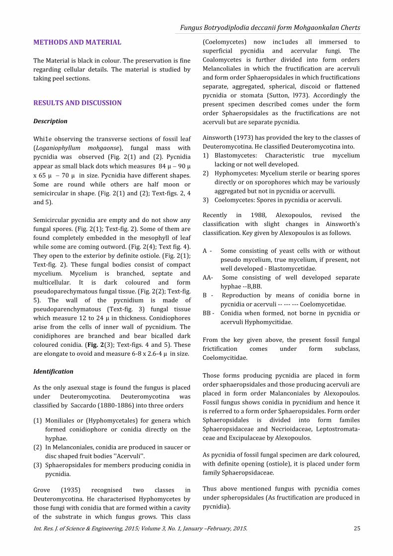
Whi1e observing the transverse sections of fossil leaf
(Loganiophyllum mohgaonse), fungal mass with
pycnidia was observed (Fig. 2(1) and (2). Pycnidia
appear as small black dots which measures 84 µ 90 µ
x 65 µ 70 µ in size. Pycnidia have different shapes.
Some are round while others are half moon or
semicircular in shape. (Fig. 2(1) and (2); Text-figs. 2, 4
and 5).
Semicircular pycnidia are empty and do not show any
fungal spores. (Fig. 2(1); Text-fig. 2). Some of them are
found completely embedded in the mesophyll of leaf
while some are coming outword. (Fig. 2(4); Text fig. 4).
They open to the exterior by definite ostiole. (Fig. 2(1);
Text-fig. 2). These fungal bodies consist of compact
mycelium. Mycelium is branched, septate and
multicellular. It is dark coloured and form
pseudoparechymatous fungal tissue. (Fig. 2(2); Text-fig.
5). The wall of the pycnidium is made of
pseudoparenchymatous (Text-fig. 3) fungal tissue
which measure 12 to 24 µ in thickness. Conidiophores
arise from the cells of inner wall of pycnidium. The
conidiphores are branched and bear bicalled dark
coloured conidia. (Fig. 2(3); Text-figs. 4 and 5). These
are elongate to ovoid and measure 6-8 x 2.6-4 µ in size.
Identification
As the only asexual stage is found the fungus is placed
under Deuteromycotina. Deuteromycotina was
classified by Saccardo (1880-1886) into three orders
(1) Moniliales or (Hyphomycetales) for genera which
formed conidiophore or conidia directly on the
hyphae.
(2) In Melanconiales, conidia are produced in saucer or
disc shaped fruit bodies ''Acervuli''.
(3) Sphaeropsidales for members producing conidia in
pycnidia.
Grove (1935) recognised two classes in
Deuteromycotina. He characterised Hyphomycetes by
those fungi with conidia that are formed within a cavity
of the substrate in which fungus grows. This class
(Coelomycetes) now inc1udes all immersed to
superficial pycnidia and acervular fungi. The
Coalomycetes is further divided into form orders
Melancoliales in which the fructification are acervuli
and form order Sphaeropsidales in which fructifications
separate, aggregated, spherical, discoid or flattened
pycnidia or stomata (Sutton, l973). Accordingly the
present specimen described comes under the form
order Sphaeropsidales as the fructifications are not
acervuli but are separate pycnidia.
Ainsworth (1973) has provided the key to the classes of
Deuteromycotina. He classified Deuteromycotina into.
1) Blastomycetes: Characteristic true mycelium
lacking or not well developed.
2) Hyphomycetes: Mycelium sterile or bearing spores
directly or on sporophores which may be variously
aggregated but not in pycnidia or acervulli.
3) Coelomycetes: Spores in pycnidia or acervuli.
Recently in 1988, Alexopoulos, revised the
classification with slight changes in Ainsworth's
classification. Key given by Alexopoulos is as follows.
A - Some consisting of yeast cells with or without
pseudo mycelium, true mycelium, if present, not
well developed - Blastomycetidae.
AA- Some consisting of well developed separate
hyphae --B,BB.
B - Reproduction by means of conidia borne in
pycnidia or acervuli -- --- --- Coelomycetidae.
BB - Conidia when formed, not borne in pycnidia or
acervuli Hyphomycitidae.
From the key given above, the present fossil fungal
frictification comes under form subclass,
Coelomycitidae.
Those forms producing pycnidia are placed in form
order sphaeropsidales and those producing acervuli are
placed in form order Malanconiales by Alexopoulos.
Fossil fungus shows conidia in pycnidium and hence it
is referred to a form order Sphaeropsidales. Form order
Sphaeropsidales is divided into form familes
Sphaeropsidaceae and Necrioidaceae, Leptostromata-
ceae and Excipulaceae by Alexopoulos.
As pycnidia of fossil fungal specimen are dark coloured,
with definite opening (ostiole), it is placed under form
family Sphaeropsidaceae.
Thus above mentioned fungus with pycnidia comes
under spheropsidales (As fructification are produced in
pycnidia).

Puranik SD, 2015
26 www.irjse.in
In sphaeropsidales, the present fungal pycnidium is
comparable with Ascochyta, diplodia, diplodina and
Botryodiplodia, because of bicelled nature of conidia.
Present fungal specimen differs from Ascochyta in
having small sized condidia and they are hyaline. In
diplodina also the conidia are hyaline. (Conidia are dark
coloured in present fungus).
Present fossil specimen shows presence of dark
coloured conidia like those of Diplodia but in Diplodia
conidia are more longer than 15 µ (6-8 µ long in
present specimen) and conidiophores are needle
shaped (while conidiophores are branched in present
specimen).
After eliminating Ascohyta, Diplodina, Diplodia, present
specimen is more closer to Botryodiplodia in having
1) dark coloured pycnidia.
2) pycnidia with ostiole,
3) branched conidiophore,
4) Conidia dark, 2-celled, ovoid to elongate in shape.
Fossil fungal specimen is compared with following
reported fossil genera.
It differs from Diplodia rodei, (Mahabale, 1969) in the
following characters. Conidia of Diplodia rodei are
larger, 17.5 - 18.0 x 7.5 in size, purple in colour, while
the present fungus shows smaller conidia which
measure 6-8 x 2.6-4µ and they are dark.
In Ascochytiles intertrappea (Barlinge and Paradkar,
1979), pycnidia are ostiolate, mycelium is septate and
branched, conidia are 2-celled, but it differs from
present fossil specimen, in the following respect.
1. Conidia are elongate, hyaline.
2. They are small. 3.5 x 1.0 - 1.5 µ in size.
It is also compared with fossil genus Botryodiplodia
mohgaonensis (Barlinge and Paradkar, 1979) in which
pycnidia are 100-114 x 70-80 µ in size, bicelled, conidia
dark, 7-8 x 3.0-3.5 µ in size, fusiform with striations,
while in present fungus, pycnidia are 84-90 x 65 -70 µ
in size, condiophores are branched, conidia are dark,
bicelled, 6-8 x 2.6-4 µ in size, but without striations,
hence it is named as Botryodiplodia deccanii sp. nov. the
specific name being after the horizon.
Fig. 2: Botryodiplodia deccanli sp. Nov
(1) Leaf showing upper empty half moon shaped
pycnidium and lower round, ostiolate pycnidium
with fungal mass coming out . arrow) X 200;
(2) Round pycnidium showing
pseudoparenchymatous fungal tissue and
bicelled dark coloured conidia with fungal
mycelium. X 750;
(3) Pycnidium with bicelled dark conidia. X 750;
(4) Empty pycnidium completely embedded in
the mesophyll tissue of leaf. X 750.
Text Fig. 1: Botryodiplodia deccanli sp. Nov
(1) T.S. leaf showing pycnidia;
(2) Pycnidium showing distinct ostiole.
(3) Pseudoparenchymatous nature of wall of pycnidium;
(4) Pycnidium showing branched conidiophores;
(5) Pycnidium magnified showing branched conidiophore and
bicelled conidia.

Fungus Botryodiplodia deccanii form Mohgaonkalan Cherts
Int. Res. J. of Science & Engineering, 2015; Volume 3, No. 1, January –February, 2015. 27
Thus the systematic position of the present fossil
fungus will be as follows –
Botryodiplodia deccanii sp. nov.
Division - Eumycota
Subdivision - Deuteromycotina.
Form Class - Deuteromycetes.
Form subclass - Coelomycetidae.
Form order - Sphaeropsidales.
Form family - Sphaeropsidaceae.
Form genus - Botryodiplodia
Form species - Botryodiplodia deccanii sp. Nov.
Diagnosis
Botryodiplodia deccanii sp. nov.
Pycnidia black coloured and compact, round, halfmoon
or semicircular in shape. Semicircular pycnidia open to
exterior by ostiole and measures 84-90 x 65 -70 µ
Hyphae branched septate and multicellular forming
pseudoparenchymatous fungal tissue, wall of
pycnidium measure 12-24 µ in thickness,
conidiophores branched, conidia bicelled, dark
coloured, elongate to ovoid 6-8 x 2.6-4 µ in size.
Holotype : SDP Department of Botany, Shivaji
Science, Nagpur
Horizon : Deccan Intertrappean Series of India.
Locality : Mohgaon-Kalan, Chhindwara
District.
Age : Upper Cretaceous.
REFERENCES
1. Ainsworth GC. In the Fungi : An advance treatise (Eds.
Ainsworth, Sparrow and Sussaman), 1973, Vol.4B,pp.1-
7,academic press,London.
2. Alexopoules CJ. In Introductory mycology.3rd
edition,wiley Eastern Ltd.,New Delhi, 1988.
3. Barlinge SG and Paradkar SA. Record of new fossil
algal and fungal, forms from the Deccan Intertrappean
beds of Mohgaonkalan, M.P., India. The Botanique. 1979;
X : 1-4.
4. Chawhan NJ. Contribution towards Palaeocene flora from
the Intertrappean Series of India Ph.D. Thesis,Nagpur
University,Nagpur, 1987.
5. Dixit VP. Palaeobotanical studies of the Deccan
Intertrappean.Ph.D. Thesis,Nagpur University,Nagpur,
1984.
6. Grove WB. “British stem and leaf Fungi (Coelomycetes)”
I, Cambridge University.Press,London and New York,
1935.
7. Mahabale TS. On a fossil species of Diplodia from the
Deccan Intertrappean series, M.P.,India.Palaeobotanist,
1969; 17(3) :295-297.
8. Saccardo PA. (In Italian) Michelia, 2 : 1-39. Sylloge
Furgorum Omnivum(1886)Huscusque Cognitarum,
Pavia, 1980; 4:1-807.
9. Singh RB and Patil.GV. On remains of Coelomycetes in
Mohgaonkalan Intertrappean M.P., India. The Botanique,
1978; 9 : 13-20.
10. Singhai LC. Fossil fungi from the Deccan Intertrappean
beds of M.P.,India, J.Biol . Sci., 1974; 17 : 92-102.
11. Sutton BC. Coelomycetes in the Fungi, Vol . IVA ,ed ;
Ainsworth,Sparrow and Sussman, 1973; pp. 513-582.
© 2014| Published by IRJSE