Fundamental Types of Neurons Sensory (afferent) neurons –receptors detect changes in body and...
28
Fundamental Types of Neurons • Sensory (afferent) neurons – receptors detect changes in body and external environment – this information is transmitted into brain or spinal cord • Interneurons (association neurons) – lie between sensory & motor pathways in CNS – 90% of our neurons are interneurons – process, store & retrieve information • Motor (efferent) neuron – send signals out to muscles & gland cells – organs that carry out responses called effectors
-
Upload
elvin-simmons -
Category
Documents
-
view
216 -
download
2
Transcript of Fundamental Types of Neurons Sensory (afferent) neurons –receptors detect changes in body and...

Fundamental Types of Neurons
• Sensory (afferent) neurons– receptors detect changes in body and external
environment– this information is transmitted into brain or spinal cord
• Interneurons (association neurons)– lie between sensory & motor pathways in CNS– 90% of our neurons are interneurons– process, store & retrieve information
• Motor (efferent) neuron– send signals out to muscles & gland cells– organs that carry out responses called effectors

Fundamental Types of Neurons

Fundamental Properties of Neurons
• Excitability (irritability)– ability to respond to changes in the body and
external environment called stimuli
• Conductivity– produce traveling electrical signals
• Secretion– when electrical signal reaches end of nerve
fiber, a chemical neurotransmitter is secreted

Structure of a Neuron• Cell body = soma
– single, central nucleus with large nucleolus
– cytoskeleton of microtubules & neurofibrils (bundles of actin filaments)
• compartmentalizes RER into Nissl bodies
– lipofuscin product of breakdown of worn-out organelles -- more with age
• Vast number of short dendrites– for receiving signals
• Singe axon (nerve fiber) arising from axon hillock for rapid conduction– axoplasm & axolemma & synaptic
vesicles

Axonal Transport• Many proteins made in soma must be
transported to axon & axon terminal– repair axolemma, for gated ion channel
proteins, as enzymes or neurotransmitters
• Fast anterograde axonal transport– either direction up to 400 mm/day for
organelles, enzymes, vesicles & small molecules
• Fast retrograde for recycled materials & pathogens
• Slow axonal transport or axoplasmic flow– moves cytoskeletal & new axoplasm at 10
mm/day during repair & regeneration in damaged axons

Electrical Potentials & Currents
• Neuron doctrine -- nerve pathway is not a continuous “wire” but a series of separate cells
• Neuronal communication is based on mechanisms for producing electrical potentials & currents– electrical potential - difference in
concentration of charged particles between different parts of the cell
– electrical current - flow of charged particles from one point to another within the cell
• Living cells are polarized– resting membrane potential is -70 mV
with a relatively negative charge on the inside of nerve cell membranes

Resting Membrane Potential• Unequal electrolytes distribution
– diffusion of ions down their concentration gradients– selective permeability of plasma membrane – electrical attraction of cations and anions
• Explanation for -70 mV resting potential– membrane very permeable to K+
• leaks out until electrical gradient created attracts it back in
– membrane much less permeable to Na+
– Na+/K+ pumps out 3 Na+ for every 2 K+ it brings in• works continuously & requires great deal of ATP• necessitates glucose & oxygen be supplied to nerve
tissue

Be clear on vocabulary
• Polarize = to increase the difference in ion concentration. To move away from 0mV.– Resting potential is polarized (-70mV).– There’s a difference in Na+/K+ conc.
• Depolarize = To move toward no electrical potential. – Allowing Na+/K+ to go where they want.– “Opening flood gates”
• Repolarize = To go back to original potential

Ionic Basis of Resting Membrane Potential
• Na+ more concentrated outside of cell (ECF) • K+ more concentrated inside cell (ICF)

Local Potentials• Local disturbances in membrane potential
– occur when neuron is stimulated by chemicals, light, heat or mechanical disturbance
– depolarization decreases potential across cell membrane due to opening of gated Na+ channels
• Na+ rushes in down concentration and electrical gradients
• Na+ diffuses for short distance inside membrane producing a change in voltage called a local potential
• Differences from action potential– are graded (vary in magnitude with stimulus strength)– are decremental (get weaker the farther they spread)– are reversible as K+ diffuses out, pumps restore balance– can be either excitatory or inhibitory (hyperpolarize)

Chemical Excitation

Action Potentials• More dramatic change in membrane produced where
high density of voltage-gated channels occur– trigger zone has 500 channels/m2 (normal is 75)
• If threshold potential (-55mV) is reached voltage-gated Na+ channels open (Na+ enters causing depolarization)
• Passes 0 mV & Na+ channels close (peaks at +35)• K+ gates fully open, K+ exits
– no longer opposed by electrical gradient
– repolarization occurs
• Negative overshoot produceshyperpolarization
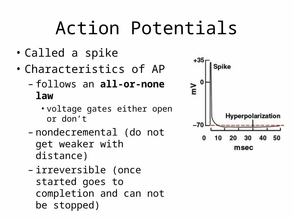
Action Potentials• Called a spike
• Characteristics of AP– follows an all-or-none
law • voltage gates either open or
don’t
– nondecremental (do not get weaker with distance)
– irreversible (once started goes to completion and can not be stopped)

The Refractory Period
• Period of resistance to stimulation• Absolute refractory period
– while Na+ gates are open– no stimulus will trigger AP
• Relative refractory period– as long as K+ gates are open– only especially strong
stimulus will trigger new AP
• Refractory period is occurring only to a small patch of membrane at one time (quickly recovers)

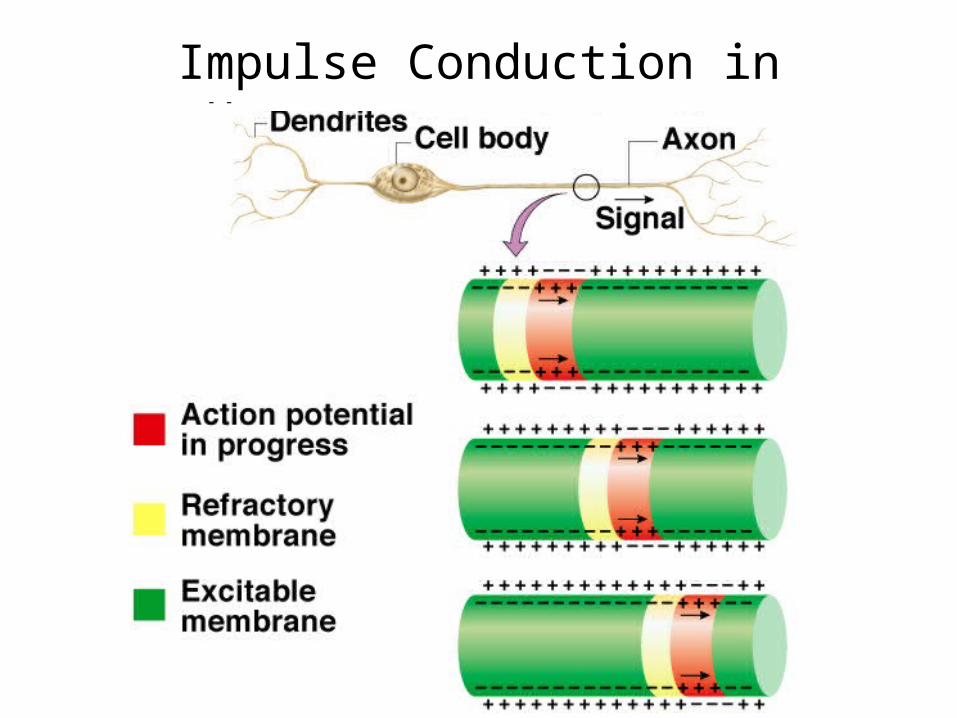
Impulse Conduction in Unmyelinated Fibers
• Threshold voltage in trigger zone begins impulse
• Nerve signal (impulse) - a chain reaction of sequential opening of voltage-gated Na+ channels down entire length of axon
• Nerve signal (nondecremental) travels at 2m/sec
Impulse Conduction in Unmyelinated Fibers

Saltatory Conduction in Myelinated Fibers
• Voltage-gated channels needed for APs– fewer than 25 per m2 in myelin-covered regions – up to 12,000 per m2 in nodes of Ranvier
• Fast Na+ diffusion occurs between nodes

Saltatory Conduction of Myelinated Fiber
• Notice how the action potentials jump from node of Ranvier to node of Ranvier.

Synapses Between Two Neurons
• First neuron in path releases neurotransmitter onto second neuron that responds to it– 1st neuron is presynaptic neuron– 2nd neuron is postsynaptic neuron
• Number of synapses on postsynaptic cell variable– 8000 on spinal motor neuron– 100,000 on neuron in cerebellum

The Discovery of Neurotransmitters
• Histological observations revealed a 20 to 40 nm gap between neurons (synaptic cleft)
• Otto Loewi (1873-1961) first to demonstrate function of neurotransmitters at chemical synapse
– flooded exposed hearts of 2 frogs with saline
– stimulated vagus nerve of one frog --- heart slows
– removed saline from that frog & found it would slow heart of 2nd frog --- “vagus substance”
– later renamed acetylcholine

Chemical Synapse Structure
• Presynaptic neurons have synaptic vesicles with neurotransmitter and postsynaptic have receptors

Postsynaptic Potentials• Excitatory postsynaptic potentials (EPSP)
– a positive voltage change causing postsynaptic cell to be more likely to fire
• result from Na+ flowing into the cell
– glutamate & aspartate are excitatory neurotransmitters
• Inhibitory postsynaptic potentials (IPSP)– a negative voltage change causing postsynaptic cell
to be less likely to fire (hyperpolarize)• result of Cl- flowing into the cell or K+ leaving the cell
– glycine & GABA are inhibitory neurotransmitters
• ACh & norepinephrine vary depending on cell
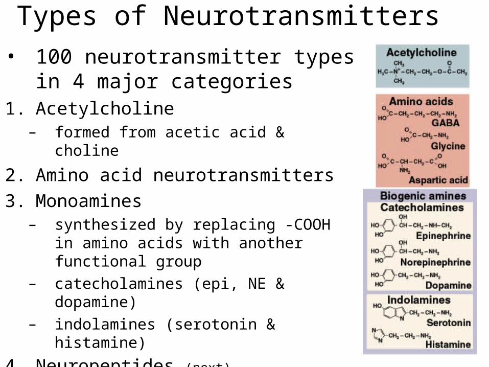
Types of Neurotransmitters
• 100 neurotransmitter types in 4 major categories
1. Acetylcholine – formed from acetic acid & choline
2. Amino acid neurotransmitters
3. Monoamines – synthesized by replacing -COOH in
amino acids with another functional group
– catecholamines (epi, NE & dopamine)– indolamines (serotonin & histamine)
4. Neuropeptides (next)

Neuropeptides
• Chains of 2 to 40 amino acids
• Stored in axon terminal as larger secretory granules Act at lower concentrations
• Longer lasting effects
• Some released from nonneural tissue– gut-brain peptides cause food cravings
• Some function as hormones– modify actions of neurotransmitters

Monamines, • Catecholines: Come from amino acid tyrosine
– Made in adrenal medulla– Blood soluable– Prepare body for activity– High levels in stressed people
• Norepinephrine: raises heart rate, releases E
• Dopamine: elevates mood– Helps with movement, balance– Low levels = Parkinson’s disease