
Functional visual loss ? Malingering - · PDF fileNon-organic visual loss • Malingering:...
37
Prof. Dr. Werner SPILEERS Functional visual loss ? Malingering ?
-
Upload
dangnguyet -
Category
Documents
-
view
222 -
download
0
Transcript of Functional visual loss ? Malingering - · PDF fileNon-organic visual loss • Malingering:...

Prof. Dr. Werner SPILEERS
Functional visual loss ?
Malingering ?

Definitions:
• 5% in general ophthalmological practice ?
• Functional visual loss; non-organic visual
loss; conversion, somatisation, …
• Malingering
• Aggravation: concurrently with organic illness

Functional visual loss (FVL)
• The patient is simulating poor vision (acuity, visual field), when in fact their vision is normal
ptosis, blepharospasm, diplopia, convergence spasm, voluntary
nystagmus, convergence insufficiency are also possible symptoms
may be antecedent mild trauma, …
(children: psychosocial stress, school, home, …)

Non-organic visual loss
• Malingering: a psychologically well patient who feigns visual los for some material benefit with full awareness that the reported complaints are false
• Conversion disorder (“hysteria”): often teenager or young adult with significant psychological or social problems (school or work stress, parental conflicts, …)

Functional visual loss
• The intent is: ruling out of organic pathology
• Try and “trick” the patient into demonstrating normal visual function
• “diagnosis by exclusion” after extensive investigation is not as reliable
- false positive: some patients can accurately and repeatedly “fake” abnormal testing
- false negative: in genuine but early and subtle disease !
• Non-organic overlay !! In addition to genuine organic visual loss

Diseases often misdiagnosed as functional visual loss
• Pituitary tumors with early compressive signs
• Leber’s hereditary optic neuropathy
• Bilateral retrochiasmal disease
• Early cone dystrophy or Stargardt’s disease
• Retrobulbar optic neuropathies
• CAR, MAR, …
• Small occipital infarcts

Unexplained visual loss
• BCVA • Stenopeic vision: pinhole test • Full eye examination: cornea, lens, tear film, … • Pupils: RAPD !! • Colour vision • Exclusion of amblyopia • Eye fundus: macula: OCT, angiography • Appropriate electrophysiology: VEP, ERG • Visual fields of both eyes • Neuro-imaging taking into account the clinical findings

Unexplained visual loss
• First exclude organic visual loss !!
• Be aware of clues that a patient’s visual loss may be non-organic:
RAPD ?!

Relative afferent pupillary defect
• Reliable and sensitive indicator of asymmetrical optic nerve
dysfunction
• Absence of RAPD should prompt reevaluation of a working
diagnosis of optic neuropathy or consideration of bilateral
involvement
• A relatively small lesion of the optic nerve results in a large
RAPD

Relative afferent pupillary defect

Relative afferent pupillary defect
• A retinal lesion must be substantially larger: retinal artery
occlusion; widespread chorioretinal lesions; retinal detachment:
R/ eye fundus examination !
• Chiasmal lesion if fibers of optic nerves are involved
asymmetrically
• Optic tract lesion: mild RAPD in contralateral eye (= eye with
temporal field loss) due to more crossed fibers

Relative afferent pupillary defect
• LGN, radiatio optica, cortical lesion: no RAPD
• An RAPD should never be attributed to media opacities only
• (amblyopia: only very mild RAPD possible, versus important
acuity loss)
• NOT PRESENT in functional visual loss

Visual electrophysiology
• Pattern-VEP: denotes abnormality along the visual pathway BUT DOES NOT give the localisation of the deficit !!
• Flash-ERG: is a mass retinal response; ganglion cell activity does not contribute to the waveforms obtained

Perimetry
• Do not forget teh manual perimeter of Goldmann !!
• (automated perimetry)

Clinical “tricks” to try and demonstrate normal vision
• Bilateral complete blindness (rare)
• Unilateral complete blindness
• Blurred vision in both eyes
• Blurred vision in one eye
• Constricted (altered) visual field in one or both eyes
• The more severe and “one-eyed” the complaint, the
easier it is to diagnose FVL

Bilateral complete blindness (rare)
• Pupils:
bilateral blindness due to severe bilateral retinal, optic nerve or chiasmal disease: both pupils “sluggishly” reactive to light
if both pupils are briskly reactive to light, the only cause for true bilateral blindness is bilateral retrochiasmal disease
• Observation:
in the waiting room; walking out of the hospital, hand shake, …

Bilateral complete blindness
• Tests that are independent of vision: proprioception
(signature test)
• Optokinetic nystagmus
• (Mirror test)

Bilateral complete blindness
• Optokinetic nystagmus:
can be generated in eyes with acuity of count fingers as long as a degree of visual field remains
suppressing the reflex is difficult (but possible)
unquantifiable nature of the response
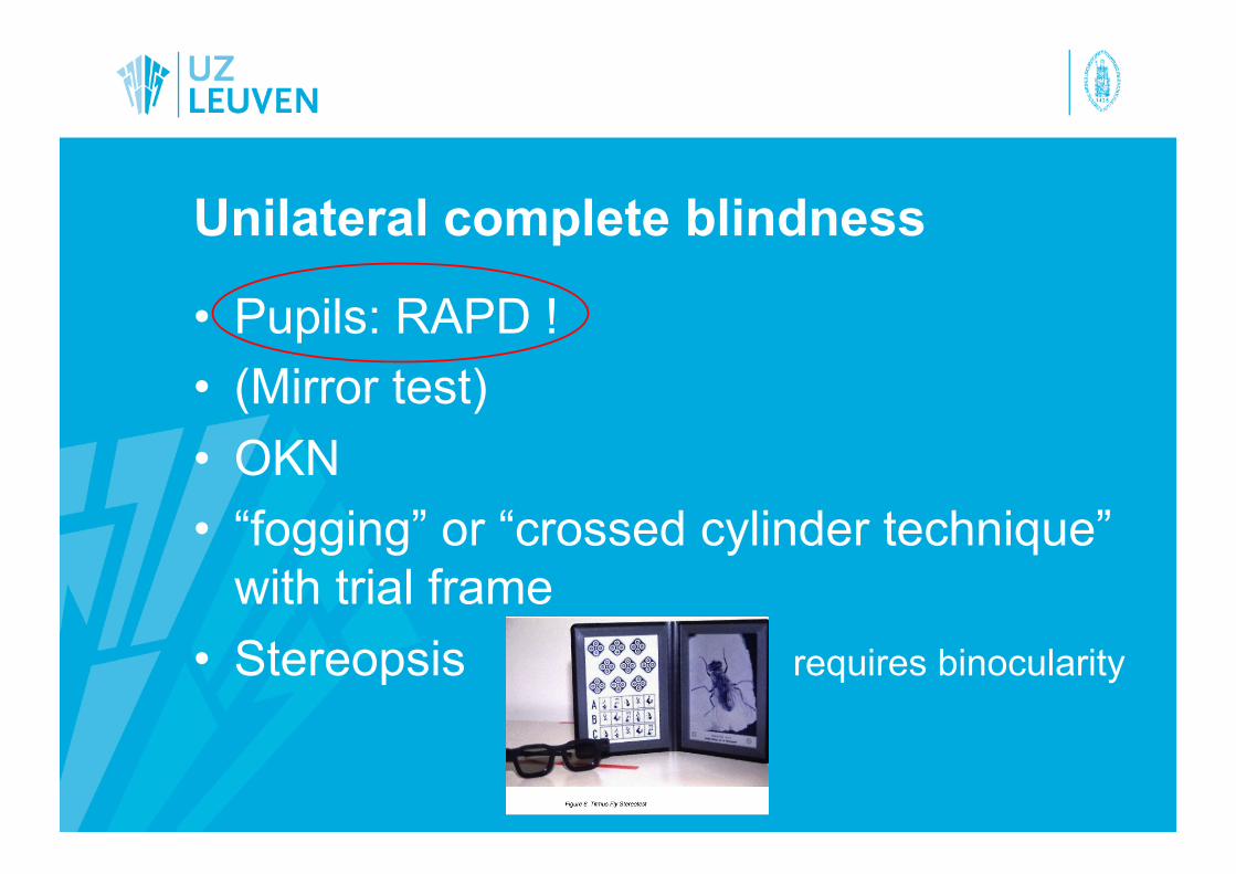
Unilateral complete blindness
• Pupils: RAPD !
• (Mirror test)
• OKN
• “fogging” or “crossed cylinder technique” with trial frame
• Stereopsis requires binocularity

“fogging” or “crossed cylinder technique” with trial frame
• Trial frame • +4 cyl and -4 cyl on the same axis for both eyes:
neutral • Read the VA chart • Slowly rotate one of the cyls in the “good” eye =
blurring the “good” eye
• Patient may close one eye to check what is going on

Stereotest

Prism shift test
• 10 D base-out prism in front of the “blind”eye
• If normal binocular vision: movement of both eyes towards the apex of the prism, followed by a shift of both eyes back to the centre

Diplopia test

Blurred vision in both eyes
• Testing visual acuity at different distances: no improvement at closer distances is inconsistent with organic visual loss
• Reading vision and distance vision
• Ishihara plate color testing: test plate
• Perimetry ! Cave: small central scotoma missed on 30-2/24-2: also perform 10-2

Blurred vision in one eye
• Pupils
• “fogging” or “crossed cylinder technique” with trial frame
• Stereopsis
• Monocular vertical prism dissociation test

Monocular vertical prism dissociation test
• “The good eye is being tested”
• Look at acuity chart with both eyes open
• 4 diopter prism is placed base down in front of the “good” eye
• “What do you see?” • Normal: two equally distinct rows
• Genuine VA loss: one row (or the lower image is less distinct)
• FVL: two equally distinct rows

Constricted visual field in one or both eyes
• Manual perimeter of Goldmann !
Constricted visual field
Spiraling
Crossing of isopters


Constricted visual field in one or both eyes
• Tangent screen visual field at one and two meters
The size of the VF should expand at the 2 meter distance in functional VF constriction, it is frequent to see the VF remain the same size or actually shrink


Central visual field
• Central sotoma:
perform careful fundoscopy, OCT, angiography, ERG to rule out subtle maculopathy
perform neuroimaging: most patients with central sotoma on VF testing, have organic pathology !

Pattern-VEP and « objective visual acuity measurement »
• Holder et al., Graefe’s Arch Clin Exp Ophthalmol, 2007
• Pattern appearance-disappearance 40ms/500 ms
• Different check sizes at different contrast levels
• Minimum check size and contrast level required to elicit a reproducible response of >= 5 µV


Holder et al, 2007
Suspected but unproven functional visual loss
• CAVE: real visual pathway disease being misdiagnosed as non-organic
• Follow-up until you can demonstrate either organic or non-organic disease

Patient with “proven” functional visual loss
• Reassurance
• “good chance to recover” …
• Leave a way out …
• Personalize your strategy to the particular patient