Functional neuroanatomy of altered states of consciousness - UCSD
26
Functional neuroanatomy of altered states of consciousness: The transient hypofrontality hypothesis Arne Dietrich * Department of Psychology, Behavioral Neuroscience Laboratory, Georgia College and State University, Milledgeville, GA 31061, USA Received 1 April 2002 Abstract It is the central hypothesis of this paper that the mental states commonly referred to as altered states of consciousness are principally due to transient prefrontal cortex deregulation. Supportive evidence from psychological and neuroscientific studies of dreaming, endurance running, meditation, daydreaming, hypnosis, and various drug- induced states is presented and integrated. It is proposed that transient hypofrontality is the unifying feature of all altered states and that the phenomenological uniqueness of each state is the result of the differential viability of various frontal circuits. Using an evolutionary approach, consciousness is conceptualized as hierarchically ordered cognitive function. Higher-order structures perform increasingly integrative functions and thus contribute more sophisticated content. Although this implies a holistic approach to consciousness, such a functional hierarchy localizes the most sophisti- cated layers of consciousness in the zenithal higher-order structure: the prefrontal cortex. The hallmark of altered states of consciousness is the subtle modification of behavioral and cognitive functions that are typically ascribed to the prefrontal cortex. The theoretical framework presented yields a number of testable hypotheses. Ó 2002 Elsevier Science (USA). All rights reserved. Keywords: Altered states; Consciousness; Prefrontal cortex; Philosophy of mind; Dreaming; RunnerÕs high; Meditation; Hypnosis; Daydreaming; Drugs Consciousness and Cognition 12 (2003) 231–256 www.elsevier.com/locate/concog Consciousness and Cognition * Fax: 1-912-445-0856. E-mail address: [email protected]. 1053-8100/02/$ - see front matter Ó 2002 Elsevier Science (USA). All rights reserved. doi:10.1016/S1053-8100(02)00046-6
Transcript of Functional neuroanatomy of altered states of consciousness - UCSD

Functional neuroanatomy of altered statesof consciousness: The transient
hypofrontality hypothesis
Arne Dietrich*
Department of Psychology, Behavioral Neuroscience Laboratory,
Georgia College and State University, Milledgeville, GA 31061, USA
Received 1 April 2002
Abstract
It is the central hypothesis of this paper that the mental states commonly referred
to as altered states of consciousness are principally due to transient prefrontal cortex
deregulation. Supportive evidence from psychological and neuroscientific studies of
dreaming, endurance running, meditation, daydreaming, hypnosis, and various drug-
induced states is presented and integrated. It is proposed that transient hypofrontality
is the unifying feature of all altered states and that the phenomenological uniqueness
of each state is the result of the differential viability of various frontal circuits. Using
an evolutionary approach, consciousness is conceptualized as hierarchically orderedcognitive function. Higher-order structures perform increasingly integrative functions
and thus contribute more sophisticated content. Although this implies a holistic
approach to consciousness, such a functional hierarchy localizes the most sophisti-
cated layers of consciousness in the zenithal higher-order structure: the prefrontal
cortex. The hallmark of altered states of consciousness is the subtle modification of
behavioral and cognitive functions that are typically ascribed to the prefrontal cortex.
The theoretical framework presented yields a number of testable hypotheses.
� 2002 Elsevier Science (USA). All rights reserved.
Keywords: Altered states; Consciousness; Prefrontal cortex; Philosophy of mind; Dreaming;
Runner�s high; Meditation; Hypnosis; Daydreaming; Drugs
Consciousness and Cognition 12 (2003) 231–256
www.elsevier.com/locate/concog
ConsciousnessandCognition
* Fax: 1-912-445-0856.
E-mail address: [email protected].
1053-8100/02/$ - see front matter � 2002 Elsevier Science (USA). All rights reserved.
doi:10.1016/S1053-8100(02)00046-6

1. The hierarchy of consciousness
Modern brain research conceptualizes cognitive function as hierarchically or-
dered. Evolutionary pressures forced the development of ever more integrative
neural structures able to process increasingly complex information. This in turn led
to increased behavioral flexibility and adaptability. The cerebral cortex, and inparticular the prefrontal cortex, is at the top of that hierarchy, representing the
neural basis of higher cognitive functions (e.g., Frith & Dolan, 1996; Fuster, 2000).
Historically, consciousness was approached in a similar manner (for a review, see
Markowitsch, 1995). Consciousness was defined by selecting various attributes, such
as self-reflective consciousness, attention, memory, perception, and arousal, which
were ordered in a functional hierarchy with the frontal lobe necessary for the �top�attributes. Recent theories of the neural correlates of consciousness similarly em-
phasize frontal cortex function for higher attributes (e.g., Crick & Koch, 1998;Dehaene & Naccache, 2001).
Perhaps the single most important functional division in the neocortex is the
central sulcus. It demarcates the frontal lobe from the three posterior cortices, the
temporal, the occipital, and the parietal, which will be collectively referred to as
TOP. Functions of TOP are very different from those of the frontal lobe. TOP
neurons are devoted primarily to perception, learning, and memory. The primary
sensory cortices of all sense modalities are located in TOP and its association cortex
further assembles and assimilates sensory information decoded initially in primarycortex. It is generally agreed that TOP is the site of long-term memory storage (e.g.,
Gilbert, 2001). The frontal lobe, located rostal to the central sulcus, neither receives
direct sensory input nor stores long-term memory. The prefrontal cortex, which
comprises approximately half of the frontal lobe in humans, is involved in executive
functions. It integrates perceptual information, formulates plans and strategies for
appropriate behavior in a given situation, and instructs the adjacent motor cortices
to execute its computational product.
At the outset, it should be made clear that it is not suggested that the prefrontalcortex is the seat of consciousness. Rather, the prefrontal cortex enables the top
layers of consciousness by contributing the highest-order cognitive functions to the
conscious experience. A large body of evidence from lesion, imaging, and electro-
physiological studies suggests that the prefrontal cortex, in contrast to various
brainstem nuclei or the intralaminar nuclei, is not necessary for basic awareness (e.g.,
Baars, 1995; Bogen, 1995). Likewise, evidence suggests that initial and much ensuing
information processing on perception, attention, or memory occurs in other brain
areas before further integration in the frontal lobes (e.g., Crick & Koch, 1998;Pollen, 1999; Taylor, 2001). Indeed, information processing on these cognitive
functions appears to be all but complete so that frontal lobe patients do not show
any evidence of dysfunction when tested for them, either directly or with conven-
tional intelligence tests (Brickner, 1936; Hebb, 1939). Rather than adding further
detail to the analysis, the frontal cortex utilizes this highly processed information to
enable still higher cognitive functions such as a self-construct (Keenan, Wheeler,
Gallup, & Pascual-Leone, 2000; Vogeley, Kurthen, Falkai, & Maier, 1999), self-re-
flective consciousness (Courtney, Petit, Haxby, & Ungerleider, 1998; Vogeley et al.,
232 A. Dietrich / Consciousness and Cognition 12 (2003) 231–256
2001), complex social function (Damasio, 1994), abstract thinking (e.g., Rylander,
1948), cognitive flexibility (Lhermitte, 1983; Lhermitte, Pillon, & Serdaru, 1986),
planning (Norman & Shallice, 1986; Shallice & Burgess, 1991), willed action (Frith &
Dolan, 1996), and theory of mind (Frith & Frith, 2001; Povinelli & Preuss, 1995;
Stone, Baron-Cohen, & Knight, 1998).
Three other cognitive functions of the prefrontal cortex, working memory(Fuster, 2000; Goldman-Rakic, 1992) temporal integration (e.g., Fuster, 1995;
Knight & Grabowecky, 1999; Kolb, 1984), and sustained and directed attention
(e.g., Posner, 1994; Sarter, Givens, & Bruno, 2001) provide the infrastructure to
compute these complex cognitive functions by providing a buffer to hold infor-
mation in mind and order it in space–time (Dehaene & Naccache, 2001; Duncan &
Owen, 2000). It is this superimposing of already highly complex mental constructs
that dramatically increases cognitive flexibility and permits a unified phenomeno-
logical experience.The prefrontal cortex is not a single unit. It is functionally divided into ventro-
medial (VM) and dorsolateral (DL) aspects. Lesion and functional imaging studies
have shown that different prefrontal circuits, along with their respective cortical and
subcortical connections, are implicated in specific cognitive abilities. There is also a
discernible hemispheric specialization, further suggesting that these cognitive pro-
cesses are not global functions of the frontal lobe. For instance, semantic memory
retrieval is implemented in the left DL, whereas activation of the right DL is most
closely associated with sustained attention (for a review, see Cabeza & Nyberg, 2000;Duncan & Owen, 2000).
The most common deficit associated with the VM region of the prefrontal cortex
is impaired social function. This deficit was epitomized by the famous case of Phi-
neas Gage, who after a freak accident that damaged his frontal lobe, exhibited what
is now recognized as the typical frontal syndrome: inappropriate social behaviors,
lack of moral judgment, few social inhibitions, few abstract thought processes, an
inability to plan for the future, and/or difficulty to maintain a plan of action. He
showed a lack of concern for himself and others and behaved with little regard forsocial constraints. Damasio (1994) suggested that the VM, with its intricate con-
nections to the limbic system, might assess the personal consequences of one�s be-
havior, and that the resulting emotions are an essential prerequisite to making logical
and rational decisions.
Damage to the DL prefrontal cortex does not involve changes in personality and
emotion. The DL does not receive direct innervation from subcortical structures
such as the amygdala that are involved in affective behavior (Petrides & Pandya,
1999). Its primary input comes from TOP and its primary output is the motorcortices. Primarily, research has implicated the DL in working memory, directed
attention, and temporal integration (Fuster, 2000; Goldman-Rakic, 1992; Knight &
Grabowecky, 1999; Posner, 1994). With its ability to sustain online processing in real
time, the working memory buffer appears to be a prerequisite for cognitive flexibility,
abstract thinking, strategic planning, access to memory, as well as sentience. It has
been suggested that it is in these DL circuits that full-fledged, self-reflective con-
sciousness comes about (e.g., Baddeley, 2000; Courtney et al., 1998; Dehaene &
Naccache, 2001; Posner, 1994).
A. Dietrich / Consciousness and Cognition 12 (2003) 231–256 233

Perseverance, or the inability to shift between modes of thinking, is the most
reliable deficit associated with damage to the DL prefrontal cortex (Brauer Boone,
1999). Perseverance is perhaps most indicative of a lack of cognitive flexibility and
ability to think abstractly. It is commonly tested for and quantified with the
Wisconsin Card Sorting Task (WCST), in which cards are sorted by one of three
characteristics: color, number, or shape. The patient is required to discover thesorting rule empirically using only feedback from the examiner about their se-
lection. When the examiner changes the sorting rule patients are required to adapt
to the new rule. Patients with DL damage show perseverance errors in this task.
They keep sorting according to the old rule even in the face of obvious discrep-
ancy. This is frequently interpreted as a failure of working memory as relevant
past behavior is required to perform the task successfully. A comparable deficit
can be produced in rats. Using a T-maze, a rat is trained over many trials to find
food located in one of the T�s arms. Once the rule (right or left) is acquired ac-cording to some criterion, it is changed to the other arm. After a number of re-
versals, a strategy emerges that is called win/stay loose/shift, or in other words, the
animal acquires the concept of shifting. Prefrontal lesions cause animals to per-
severe and adhere to the previously learned rule (e.g., Dietrich, Taylor, & Pass-
more, 2001).
It appears that the prefrontal cortex exerts inhibitory control over inappropriate
or maladaptive emotional and cognitive behaviors. Lhermitte (1983) and Lhermitte
et al. (1986) documented this tendency by showing that frontal lobe patients areoverly dependent on immediate cues. They tend to act on what they see without
taking into account the �bigger picture.� Similarly, patients show a strong tendency to
imitate inappropriate behaviors modeled by others. As Lhermitte put it: ‘‘the sight of
the movement is perceived in the patient�s mind as an order to imitate; the sight of an
object implies the order to use it’’ (p. 330). Without a fully functional frontal lobe,
the patient can utilize only immediate cues and fails to select behaviors based on
more universal principles. Thus, the frontal lobe provides for cognitive flexibility and
freedom and releases us from the slavery of direct environmental triggers or thememory stored in TOP.
The picture presented thus far is that of a hierarchy of layers of consciousness that
is intimately associated with the functional hierarchical organization of the brain.
This view assumes that full-fledged consciousness is a global function of a fully
operational brain. The concept of a functional hierarchy of consciousness can be
incorporated into the larger framework of global workspace theory (Baars, 1989;
Dehaene & Naccache, 2001). Any pattern of activation that constitutes a given
transient global workspace can be conceptualized as a functional hierarchy in whichprefrontal neurons provide a higher-order computation than, for example, TOP
neurons. Although this is a holistic view of consciousness, it is evident that not all
areas of the brain, not even all areas of the neocortex, contribute equally to con-
sciousness. Damage to any layer will result in an alteration of consciousness, but the
lower the level of malfunctioning the more fundamental the alteration. It is no easy
task to establish such a hierarchy in detail, and perhaps impossible to order the top
layers as they mutually enable each other. Primarily as a visual aid, the hierarchy is
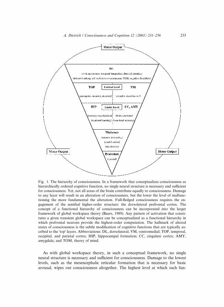
conceptualized as an inverted pyramid (see Fig. 1).
234 A. Dietrich / Consciousness and Cognition 12 (2003) 231–256
As with global workspace theory, in such a conceptual framework, no single
neural structure is necessary and sufficient for consciousness. Damage to the lowest
levels, such as the mesencephalic reticular formation that is necessary for basic
arousal, wipes out consciousness altogether. The highest level at which such fun-
Fig. 1. The hierarchy of consciousness. In a framework that conceptualizes consciousness as
hierarchically ordered cognitive function, no single neural structure is necessary and sufficient
for consciousness. Yet, not all areas of the brain contribute equally to consciousness. Damage
to any layer will result in an alteration of consciousness, but the lower the level of malfunc-
tioning the more fundamental the alteration. Full-fledged consciousness requires the en-
gagement of the zenithal higher-order structure: the dorsolateral prefrontal cortex. The
concept of a functional hierarchy of consciousness can be incorporated into the larger
framework of global workspace theory (Baars, 1989). Any pattern of activation that consti-
tutes a given transient global workspace can be conceptualized as a functional hierarchy in
which prefrontal neurons provide the highest-order computation. The hallmark of altered
states of consciousness is the subtle modification of cognitive functions that are typically as-
cribed to the �top� layers. Abbreviations: DL, dorsolateral; VM, ventromedial; TOP, temporal,
occipital, and parietal cortex; HIP, hippocampal formation; CC, cingulate cortex; AMY,
amygdala; and TOM, theory of mind.
A. Dietrich / Consciousness and Cognition 12 (2003) 231–256 235

damental change can occur is apparently the thalamus (Bogen, 1995). Nonetheless,
the thalamus might better be conceptualized as representing the next level by pro-
viding aspects to consciousness such as binding experiences together (e.g., Llin�aas &
Par�ee, 1991), preliminary processing of perceptual information (e.g., LGN), or dif-
ferential amplification of information (Crick, 1994). Its precise reciprocal connec-
tions with the neocortex and the limbic system also distribute information to theappropriate places for further processing (e.g., LeDoux, 1996). It is suggested that all
stages of processing beyond the thalamus provide some form of content.
It is reasonable to assume that when patients with brain damage fail to display a
particular function, such as fear or abstract thinking, that their phenomenological
experience does not contain its computation. Initial processing of affective content
occurs in various limbic system structures (LeDoux, 1996). Consequently, dysfunc-
tion in structures such as the amygdala changes the emotional content of con-
sciousness profoundly. For instance, lesions to the central nucleus of the amygdalawill eliminate the basic emotion of fear altogether (LeDoux, 1996). The computa-
tional product of these limbic structures is used by the next levels of affective pro-
cessing represented by the cingulate cortex and the VM (Damasio, 1994). Damage or
dysfunction at that level results in more subtle alterations to the emotional content of
consciousness, such as the context in which fear might be triggered.
The thalamus is the starting point of a separate and parallel line of information
processing that is devoid of any salient information. It is represented by another set
of limbic system structures, primarily the hippocampal formation, as well as TOPcortices. These structures process perceptual information and store the computation
as memory. The required level of selective attention to process the information is
also supplied by these structures. Again, the higher the level at which a dysfunction
occurs (e.g., association vs. primary cortex) the more selective the deficit. If the in-
formation required for a cognitive construct is not computed, it cannot be brought
�online� by TOP circuits and is thus unavailable for prefrontal modules to be included
in the computation of higher cognitive functions. Consequently, it would be missing
in the content of consciousness, resulting in distinct phenomenological distortions.Damasio (1999) provided a detailed account of how a complex cognitive function
such as the self-construct is built at various functional neuroanatomical levels in a
hierarchy of increasing complexity. The sense of self has many subcomponents such
as an experience of ownership, body-centered spatial perceptivity, long-term unity of
beliefs (Vogeley et al., 1999) as well as representation in somatic, kinesthetic, and
motor maps (Damasio, 1999). Damage to structures that process information lower
on the hierarchy will produce severe distortions such as unilateral neglect, which
results from specific right parietal damage; whereas, prefrontal dysfunction mightresult in lesser distortion of the self such as delusions of grandeur.
The neural substrate for the highest layers of cognitive ability is the DL. As seen
in Fig. 1, further sorting these layers into an ascending order is avoided. Instead, it is
proposed that they mutually enable and potentiate one another. The framework
presumes that working memory, temporal integration, and focused attention are
global capacities of frontal tissue that provide the computational parameters for all
other higher cognitive functions implemented by the prefrontal cortex. The resulting
fully flavored conscious experience is a function of computations from all structures
236 A. Dietrich / Consciousness and Cognition 12 (2003) 231–256

comprising a given global workspace that are superimposed at the level of the pre-
frontal cortex to provide a unified conscious experience.
It has been suggested that the content in the buffers of working memory is the
current conscious awareness (e.g., Baddeley, 2000; Courtney et al., 1998; Posner,
1994). A normal, healthy brain relies heavily on this buffered information in the
frontal lobe. If information is not represented in the buffers it is unconscious to theextent that we cannot reflect or report on it. At all levels of the hierarchy, including
the spinal cord, neural structures have direct access to activating the motor system.
This makes sense from an evolutionary perspective as these structures developed, at
the time, in order to directly affect behavior. Blindsight and amygdala-driven con-
ditioned emotional responses are good examples of that. In either case, a structure
drives motor output before, if ever, sending the information up the chain for con-
scious processing.
It is evident that any modification of information processing, from the sensorylevel to the prefrontal cortex, alters the content of consciousness. Yet, changes below
the level represented by the DL do not seem to affect the maximal capacity for
conscious experience (provided the arousal system is properly activated). For in-
stance, patients with unilateral neglect, anterograde amnesia, or blindsight, to name
a few, can still self-reflect—just not on the particular information that is missing.
Similarly, Damasio (1994) reported the case of EVR who sustained VM damage and
showed no deficit in hypothetical scenarios of complex moral and social situations.
He still showed the capacity indicative of the highest layers of consciousness. Thefrontal syndrome remained confined to �actual� social behavior. In contrast, damage
to the DL has the tendency to diminish the maximal capacity of consciousness. With
the loss of top layers of consciousness, patients show little evidence for higher
cognitive functions such as abstract thinking, regardless of the information that is
brought �online� by other structures.
In such a view, the prefrontal cortex does not represent a supervisory or control
system. Rather, it actively implements higher cognitive functions. It is further sug-
gested that the prefrontal cortex does not act as an inhibitory agent of older, moreprimitive brain structures. The prefrontal cortex restrains output from older struc-
tures not by suppressing their computational product directly but by elaborating on
it to produce more sophisticated output. If the prefrontal cortex is lost, the person
simply functions on the next highest layer that remains. The structures implementing
these next highest layers are not disinhibited by the loss of the prefrontal cortex.
Rather, their processing is unaffected except that no more sophistication is added to
their processing before a motor output occurs.
2. Altered states of consciousness
It is proposed in this article that altered states of consciousness are due to transient
prefrontal deregulation. Six conscious states that are considered putative altered
states (dreaming, the runner�s high, meditation, hypnosis, daydreaming, and various
drug-induced states) are briefly examined. These altered states share characteristics
whose proper function are regulated by the prefrontal cortex such as time distortions,
A. Dietrich / Consciousness and Cognition 12 (2003) 231–256 237
disinhibition from social constraints, or a change in focused attention. It is further
proposed that the phenomenological uniqueness of each state is the result of the
differential viability of various DL circuits. To give one example, the sense of self is
reported to be lost to a higher degree in meditation than in hypnosis; whereas, the
opposite is often reported for cognitive flexibility and willed action, which are absent
to a higher degree in hypnosis. The neutralization of specific prefrontal contributionsto consciousness has been aptly called ‘‘phenomenological subtraction’’ by Allan
Hobson (2001). The individual in such an altered state operates on what top layers
remain. In altered states that cause severe prefrontal hypofunction, such as non-lucid
dreaming or various drug states, the resulting phenomenological awareness is ex-
traordinarily bizarre. In less dramatic altered states, such as long-distance running,
the change is more subtle. Finally, unlike most cases of mental illness and brain
damage, altered states of consciousness can be characterized as transient in nature.
2.1. Dreaming
Sleep is a profound alteration of consciousness. Not surprisingly, it is instigated
by neural activity in the brainstem, an area near the bottom in the hierarchy of
consciousness (for a review, see Hobson, Pace-Schott, & Stickhold, 2000). Electro-
physiological, neuroimaging, and psychological data strongly suggest that REM
sleep is a state phenomenologically different from NREM sleep (Hobson et al.,
2000). According to Hobson and McCarley�s (1977) activation-synthesis model, thebrainstem contains a dream-state generator that periodically activates a great
number of subcortical and cortical structures during the REM stage. The brainstem-
initiated neural activation is most pronounced in the visual cortex, the motor cortex,
the basal ganglia, and various limbic system structures, particularly the amygdala
(Braun et al., 1997; Maquet et al., 1996; Nofzinger, Mintun, Wiseman, Kupfer, &
Moore, 1997). The pattern of activation during REM corresponds closely with
dream content and matches the neural activity of the same behaviors during waking
consciousness (see Hobson et al., 2000).PET studies have also revealed significant deactivation of a vast area of the DL,
including areas 8, 9, 10, 11, 46, 47 (Braun et al., 1997; Maquet et al., 1996). The
pattern of activation is so striking that ‘‘REM sleep may constitute a state of gen-
eralized brain activity with the specific exclusion of the executive system’’ (Braun et
al., 1997, p. 1190). Phenomenologically, dream stories are void of prefrontal-de-
pendent cognition. Self-reflection is absent (Rechtschaffen, 1978), time is distorted
with past, present, and future freely exchanged (Hobson, 1988), and volitional
control is greatly diminished (Hartman, 1966). There is also little abstract, �biggerpicture� thinking, little active decision making, little consistent logic, a crude self-
construct, and not much indication of theory of mind capabilities or focused at-
tention. The capacity for semantic and episodic retrieval of specific memories, which
heavily relies on DL areas (see Cabeza & Nyberg, 2000), is also greatly decreased.
Furthermore, the extent to which a dream is bizarre is related to the extent of the
prefrontal hypofunction (Hobson et al., 2000). It is evident that the principal dif-
ference between dream mentation and normal waking consciousness is due to the
deactivation of the DL.
238 A. Dietrich / Consciousness and Cognition 12 (2003) 231–256

For the study of consciousness, lucid dreaming is an imperative subjective alter-
ation of the dream state. During a lucid dream, a person becomes aware that the
transpiring events are part of a dream (LaBerge, 1985). Hobson (2001) has defined
lucid dreaming ‘‘as the bolstering of the self-reflective awareness that is normally
diminished or absent in dreaming’’ (p. 93). The degree of lucidity is variable and it is
not necessarily accompanied by dream control, the intentional directing of the dreamstory. Similarly dream control can occur without lucidity (LaBerge, 1985). Hobson
(2001) has proposed that lucid dreaming is the result of DL activation during REM
sleep, or more specifically ‘‘the residual activation of the dorsolateral prefrontal
cortex [that] is amplified by the REM activation’’ (p. 97) from other cortical areas.
Psychological reports from lucid dreamers are indicative of other prefrontal cogni-
tive capacities that are normally absent in dreaming, such as willed action, an un-
distorted sense of self, directed attention, abstract thinking in the form of
constructive creativity, consciously operated memory searches, awareness of com-plex societal standards, and strategic planning. LaBerge (1985) has also shown in his
laboratory that the estimation of the passage of time during a lucid dream is as
accurate as in waking consciousness. Additionally, the degree and quality of the
lucid experience might be a function of the extent and pattern of prefrontal acti-
vation. The observation that dream control and lucidity, although highly correlated,
are independent might underscore this notion. With the top layers of consciousness
activated, lucid dreaming is more akin to waking consciousness, neurally as well as
psychologically. Indeed, according to LaBerge (1985), awakening is one of the mostcommon results of suddenly gaining lucidity in a dream. In that sense, lucid
dreaming might be better thought of as daydreaming during REM sleep.
2.2. Endurance running
An exercise induced altered state of consciousness has long been appreciated by
endurance athletes. The effect has been well documented and subjected to scientific
investigation (e.g., Hoffman, 1997; Mandell, 1981; Pargman & Baker, 1980), andsince the effect is particularly prominent in long-distance running, the phenomenon
was given its contemporary descriptor, the ‘‘runner�s high’’ (Pargman & Baker,
1980). Phenomenologically, the runner�s high has been described as pure happiness,
elation, feelings of unity with one�s self and/or nature, endless peacefulness, time-
lessness, inner harmony, boundless energy, as well as the reduction of pain sensa-
tions (e.g., Farrell, Gustafson, Morgan, & Pert, 1987; Hoffman, 1997). These
subjective descriptions are similar to the claims of distorted perception, atypical
thought patterns, diminished awareness of one�s surroundings, and intensified in-trospective understanding of one�s sense of identity and emotional status made by
people who describe trance states. Moreover, exercise has well-established beneficial
effects on mood states, particularly stress, anxiety, and depression, (for reviews, see
Glenister, 1996; Salmon, 2001; Scully, Kremer, Meade, Graham, & Dudgeon, 1998).
There is extensive literature documenting depression as a result of frontal lobe
damage (for a review, see Starkstein & Robinson, 1999). PET neuroimaging studies
have also demonstrated significant hypometabolism in the DL of patients with
depression (Baxter et al., 1987). This may explain a depressed patient�s persever-
A. Dietrich / Consciousness and Cognition 12 (2003) 231–256 239

ance on negative thoughts and the inability to shift away from negative events.
Without the cognitive flexibility of a fully functional DL, patients might find
themselves unable to escape from negative environmental cues. PET studies have
further demonstrated that the VM, along with the amygdala and the anterior
cingulate, is hyperactive during depression (see Mayberg, 1997). This hyperme-
tabolism may explain why depressed patients overemphasize the importance ofnegative events in their lives, especially with sustained and heightened amygdala
input. Healthy subjects that are asked to think sad thoughts show similar hyper-
metabolism in the VM, while metabolic activity is less than normal for DL circuits
(Damasio et al., 2000). These studies point to an abnormal interaction between the
VM and the DL rather than global prefrontal dysfunction (Starkstein & Robinson,
1999). PET studies of anxiety disorders show a very similar prefrontal pattern
(Ruben & Harris, 1999). In obsessive-compulsive disorder (OCD) for instance, the
VM also exhibits widespread hypermetabolism but without the concomitant limbichypermetabolism (Baxter, 1990).
Based on the prefrontal pathology in depression and anxiety disorders, I have
proposed the hypothesis that exercise induces a state of transient hypofrontality,
which could account for the beneficial effects of exercise on mental health (Dietrich,
in review). A brief reiteration of the main points and the line of reasoning might be
beneficial to the continuity of this article.
Neuroimaging techniques demonstrate that different tasks place different demands
on neural structures, which results in unique patterns of activation. Considering thephysical demands of exercise, it is reasonable to assume that, at a minimum, there is
activation in all neural structures that are required in order to run the motor patterns
to sustain the physical activity: primary motor cortex, secondary motor cortices,
basal ganglia, cerebellum, various midbrain nuclei (red and substantia nigra), as well
as various thalamic nuclei would be involved. Depending on the type of sport, ex-
ercise should result in activity of structures involved in perceptual function and
memory, particularly TOP and the sensory thalamus. Local cerebral glucose me-
tabolism and utilization measured in running rats has confirmed this pattern ofactivity (Vissing, Anderson, & Diemer, 1996). Since such a large part of the brain is
devoted to perception and motor output, there is a widespread activation of nu-
merous structures across the entire brain during exercise. Yet, it is important to
clarify at this point that, despite regional increases, global cerebral blood flow to the
brain during exercise as well as global oxygen uptake is constant (Ide & Secher,
2000).
Research in cognitive psychology has shown that humans have a limited infor-
mation processing capacity, particularly with respect to attentional resources(Broadbent, 1958; Cerry, 1953). This is likely due to the brain�s limited resources and
the inability to sustained activation in all neural structures at once. As Pinker (1999)
has pointed out, there are costs and benefits associated with efficient information
processing. It was suggested that the widespread activation of motor and sensory
systems during exercise comes at the expense of, first and foremost, the higher
cognitive centers of the prefrontal cortex. Also, limbic system structures such as the
amygdala that are not directly required for the exercise might similarly show de-
pressed activity.
240 A. Dietrich / Consciousness and Cognition 12 (2003) 231–256

EEG recordings of exercising individuals seem to be consistent with this hy-
pothesis. Exercise is associated with a-enhancement, particularly in the frontal cortex
(e.g., Kubitz & Pothakos, 1997). The increase in a-activity during and after exercise
has been interpreted in the literature as indicative of decreased brain activation
(Kubitz & Pothakos, 1997). Single cell recording in exercising cats has also provided
support for decreased activation in prefrontal regions. Recording from 63 neurons inthe prefrontal cortex, units associated with the control of the movement showed
increased activity during locomotion, while other prefrontal units decreased their
discharge (Criado, de la Fuente, Heredia, Riolobos, & Yajeya, 1997).
It is currently impossible to test the hypothesis directly with functional imaging
tools because they preclude head movement. However, specific predictions can be
made using cognitive tests that are indicative of a specific brain region�s viability. For
instance, according to the hypothesis, an individual�s ability to perform frontal-de-
pendent cognitive tasks during exercise should be impaired. It was noted early (e.g.,Hebb, 1939) that people with frontal lobe lesions perform normally on conventional
intelligence tests, suggesting that successful performance on IQ tests does not require
a fully functional frontal lobe. The transient hypofrontality hypothesis predicts that
endurance running might produce greater deficits in frontal lobe sensitive tasks than
with conventional IQ tests. This experiment was recently performed in our labora-
tory and it has confirmed that endurance exercise selectively impairs frontal-de-
pendent cognition (Dietrich & Sparling, in review). In addition, other possible tests
of the transient hypofrontality hypothesis might make use of tools such as opticalimaging, single cell recording in animals, EEG recordings in humans, as well as a
variety of different cognitive and emotional test batteries for humans and animals
that test for psychological processes that are prefrontal-dependent.
A state of transient frontal hypofunction can explain a variety of emotional and
cognitive changes that are experienced during exercise. On a psychological level, it
has been suggested that exercise�s mental health benefits might simply be due to
distraction or a �time-out� from life�s stress (Bahrke & Morgan, 1978). Exercise-in-
duced transient hypofrontality could represent a neural origin for this distraction.Given the analytical and attentional capacities of the prefrontal cortex, excessive
activity generates a state of hyper-vigilance and hyper-awareness. In such a state,
there is a tendency to overanalyze and evaluate every event with respect to personal
relevance. Exercise might simply �take the edge off� by neutralizing this circuitry,
producing an inability to focus on life�s worries.
Although the hypothesis of exercise-induced transient hypofrontality was devel-
oped to account for the beneficial effects of exercise on mental health, the prolonged
disengagement of higher cognitive centers in the prefrontal cortex also offers a neuralmechanism that provides insight into the alteration of consciousness known as the
runner�s high. Some of the phenomenologically unique features of this state such as
experiences of timelessness, living in the here and now, reduced awareness of one�ssurroundings, peacefulness (being less analytical), and floating (diminished working
memory and attentional capacities), are consistent with a state of frontal hypo-
function. Even abstruse feelings such as the unity with the self and/or nature might
be more explicable, considering that the prefrontal cortex is the very structure that
provides us with the ability to segregate, differentiate, and analyze the environment.
A. Dietrich / Consciousness and Cognition 12 (2003) 231–256 241

The hypothesis proposes further that the degree to which the prefrontal cortex is
free to disengage from an on-going activity is dependent upon the demands made by
the physical activity on these higher cognitive functions. Thus, clear predictions can
be made about how different types of sports affect brain function. Strategic team
sports such as basketball require a variety of frontal-dependent cognitive processes
while running through a familiar park does not. The hypothesis also predicts thatintensity is important. A minimum level of intensity is required to force the redis-
tribution of resources in the brain. Conversely, exercise that is extremely strenuous
and associated with high levels of discomfort and fatigue lead to ubiquitous
awareness and alarm signals, that forces prefrontal regions to evaluate the person�sstatus and situation.
The transient hypofrontality hypothesis also predicts that the experience of an
‘‘exercise high’’ depends on the skill level and the nature of the movement. It has
been suggested that automatic motor behaviors, or habits, are controlled by thebasal ganglia (Mishkin, Malamut, & Bachevalier, 1984). The more a skill is practiced
and becomes automatic, the less prefrontal cortex activity is required during its
execution. Hence, skill level, or the degree to which a motor pattern is run by the
basal ganglia, has an affect on how much an individual can afford to deregulate the
prefrontal cortex. Similarly, the more natural the movement, the more rapidly
the movement is transferred to the basal ganglia. Thus, the natural activity of
running would more readily produce a �high� than perhaps swimming or yoga.
2.3. Meditation
Meditation has long been regarded as an altered state of consciousness. Although
there are many different forms and practices of it, all share a number of common
features. First, sensory input is gradually diminished in the meditative state, creating
what Carrington (1998) called ‘‘a mental isolation chamber’’ (p. 68). The continuous
sensory bombardment of daily life is ultimately replaced by a non-judgmental, re-
ceptive, quiescent state that deliberately banishes the intrusion of new input. Thisfeature by itself might explain many well-documented meditation effects such as
increased relaxation, reduced anxiety, lowered blood pressure, or changes in cortisol
levels (for a review, see Carrington, 1998; Jevning, Wallace, & Beidebach, 1992).
Second, meditation entails sustained concentration and heightened awareness by
focusing attention on a mantra, breathing rhythm, or a number of other internal or
external events. This increase in attentional effort serves to accomplish the exclusion
of other, intruding information. The attention is directed, deliberate, and sustained,
suggesting the activation of the frontal attentional network. Indeed, all existingstudies using neuroimaging techniques during meditation show converging evidence
of DL activation (Herzog et al., 1990; Lazar et al., 2000; Lou et al., 1999; Newberg et
al., 2001). At first glance, these data appear to contradict the transient hypofrontality
hypothesis of altered states of consciousness.
Paradoxically, early EEG studies of meditation consistently detected a-wave ac-
tivity across the frontal lobe (Anand, China, & Singh, 1961; Banquent, 1972; Ben-
son, Mahlotra, Goldman, Jacobs, & Hopkins, 1990; Corby, Roth, Zarcone, &
Kopell, 1978). a-Activity consists of regular, smooth medium-frequency waves,
242 A. Dietrich / Consciousness and Cognition 12 (2003) 231–256
which are produced when subjects are neither particularly aroused nor excited, and
are not engaged in strenuous mental activity such as problem solving. In short, a-
activity suggests decreased brain activation. Conversely, b-activity is an irregular
wave pattern that is detected when subjects are alert and attentive. Given that
meditation increases attentional focus and awareness, as suggested by the psycho-
logical reports and neuroimaging data, one would expect to detect b-activity acrossthe frontal lobe during meditation. Or in more esoteric terms, if meditation is, as so
often claimed, a vehicle for reaching �higher consciousness� one would expect the
neural structure that enables the highest cognitive functions and thus the top layers
of consciousness to be fully functional. The presence of a-waves in EEG studies are
in stark contrast to the neuroimaging data showing increased neural activity in the
prefrontal cortex.
EEG is a crude method that measures summed postsynaptic electrical activity of a
large area of the cortex. The desynchronized pattern of b-activity is due to the dif-ferential activation of individual but spatially overlapping circuits or modules, re-
sulting in a noisy, overall signal. The neural synchrony that is reflected by a-activity
indicates that large groups of neurons fire in unison. This has been, often pre-
sumptuously, interpreted as the neural correlate of psychological harmony when it is
perhaps more indicative of baseline cortical activity or metabolic depression.
It is evident that more research is needed to resolve the conflicting EEG and
neuroimaging data. Reinterpreting and integrating the limited data from existing
studies, it is proposed that meditation results in transient hypofrontality with thenotable exception of the attentional network in the prefrontal cortex. The resulting
conscious state is one of full alertness and a heightened sense of awareness, but
without content. Since attention appears to be a rather global prefrontal function
(e.g., Cabeza & Nyberg, 2000), PET, SPECT, and fMRI scans showed an overall
increase in DL activity during the practice of meditation. However, the attentional
network is likely to overlap spatially with modules subserving other prefrontal
functions and an increase as measured by fMRI does not inevitably signify the ac-
tivation of all of the region�s modules. Humans appear to have a great deal of controlover what they attend to (Atkinson & Shiffrin, 1968), and in meditation, attentional
resources are used to actively amplify a particular event such as a mantra until it
becomes the exclusive content in the working memory buffer. This intentional,
concentrated effort selectively disengages all other cognitive capacities of the pre-
frontal cortex, accounting for the a-activity. Phenomenologically, meditators report
a state that is consistent with decreased frontal function such as a sense of time-
lessness, denial of self, little if any self-reflection and analysis, little emotional con-
tent, little abstract thinking, no planning, and a sensation of unity. The highlyfocused attention is the most distinguishing feature of the meditative state, while
other altered states of consciousness tend to be more characterized by aimless
drifting.
It is well documented that meditation also reduces anxiety and alleviates mild
depression (Carrington, 1998). As mentioned earlier, depression and anxiety are
believed to be due, in part, to a differential activation of VM and DL circuits
(Starkstein & Robinson, 1999). Treatment with selective serotonin reuptake blockers
normalizes this imbalance (Mayberg, 1997). Although highly speculative at this
A. Dietrich / Consciousness and Cognition 12 (2003) 231–256 243

point, it is proposed that any selective boosting of DL activity, such as in meditation,
should have an antidepressant and anxiolytic effect. In a similar manner, talking to a
friend or therapist about an emotional dilemma involves sustained attention, ab-
stract thinking, and formulating explanations so the person can understand the
situation. This should also require DL activation and thus the reactivation of circuits
that can modulate the perseverance on negative thinking. Normalizing DL activityshould result in increased cognitive flexibility and thus enhance the ability to extract
oneself from the perceived dilemma.
2.4. Hypnosis
Although still viewed with skepticism, hypnosis has gained respectability in
medicine, in large part, due to its demonstrated effects on analgesia. It is less clear
however whether hypnosis constitutes an altered state of consciousness. Forty per-cent of hypnotized subjects describe it as an altered state, while sixty percent com-
pare it to a state of focused attention (Kirsch & Lynn, 1998). While some researchers
postulated a divided stream of consciousness in hypnosis (Hilgard, 1980), others
caution that the evidence only supports a state of high suggestibility (e.g., Barber,
1970; Kirsch & Lynn, 1998; Spanos, 1994). Hypnotized subjects reported or exhib-
ited analgesia, vivid images, hallucinations in all sense modalities, amnesia, time-
lessness, detachment from the self, and a willingness to accept distortions of logic
and reality (e.g., Kihlstrom, 1985; Orne, 1959; Tart, 1979). More commonly, hyp-notized individuals remain fully alert, conversing with people and navigating the
environment normally. The main distinguishing characteristic of the hypnotic state
remains the lack of initiative or willful movement.
Early EEG studies failed to uncover an unambiguous physiological marker of
hypnosis (Benson, Arns, & Hoffman, 1981). Conversely, recent neuroimaging studies
have demonstrated subtle changes in neural activation, particularly in the left
hemisphere (Maquet et al., 1999; Rainville et al., 1999; Wik, Fischer, Bragee, Finer,
& Frederikson, 1999). The observed activation occurred predominantly in TOPcortices without a corresponding increase in DL circuits. Increased activation was
also observed in the ventrolateral prefrontal cortex during hypnotic suggestion,
presumably reflecting the demand on attentional resources in order to process the
verbal input (Rainville et al., 1999; Wik et al., 1999). This was also shown in an
earlier regional blood flow study (Crawford, Gur, Skolnick, Gur, & Benson, 1993).
This pattern of activation was not found in neuroimaging studies using other de-
pendent and independent variables of hypnosis (Faymonville et al., 2000; Ground,
Pawlik, Walter, Lesch, & Heiss, 1995).A number of researchers have postulated that neural structures compete for access
to consciousness (e.g., Baddeley, 1995; Pinker, 1999). The process of hypnotic in-
duction, regardless of how it is implemented, serves to narrow a person�s attention. It
has been suggested that the effects of hypnosis are due to frontal inhibition (for
recent reviews see, Gruzelier, 2000; Kallio, Revonsuo, H€aam€aal€aainen, Markela, &
Gruzelier, 2001). The transient hypofrontality hypothesis suggests further that,
similar to meditation, the focused attention of the hypnotic state is the mechanism by
which the activation of various prefrontal circuits is decreased, eliminating their
244 A. Dietrich / Consciousness and Cognition 12 (2003) 231–256
computation from figuring into immediate conscious experience. It appears that
some cognitive functions supported by the DL prefrontal cortex such as willed ac-
tion, initiative, critical self-reflection, memory accessibility, cognitive flexibility, and
independent thinking and logic are particularly affected in hypnosis.
It is proposed that during hypnosis, the hypnotist�s suggestions, due to the sub-
ject�s narrow focus on them, become the predominant content in the workingmemory buffers without passing through the filter of these other downregulated DL
circuits. Without the added higher cognitive computation provided by these DL
circuits, the person does not have the capacity to critically examine the suggestions
and thus enable behavioral flexibility. It is evident that the hypofunction does not
appear to be a total or global prefrontal event. For instance, it has been demon-
strated that hypnotized subjects cannot be induced to act contrary to their moral
beliefs and values (Kirsch & Lynn, 1998), which is primarily a function of the VM
(Damasio, 1994). This is supported by neuroimaging studies showing VM activation(Maquet et al., 1999; Rainville et al., 1999). It is also suggested that the experience of
a hidden observer and a divided stream of consciousness postulated by Hilgard
(1980) might be due to prefrontal circuits that are not altered by the hypnotic in-
duction. Similar to lucid dreaming, these �indomitable� circuits may provide lucidity
without being able to influence real actions in real life. In hypnosis, suggestions
become executed by directly activating the motor system without being further
scrutinized. This is in accordance with people�s subjective description of the hypnotic
experience, stating that a given behavioral act appeared to happened by itself.As mentioned earlier, Lhermitte (1983) and Lhermitte et al. (1986) documented a
strong tendency for frontal lobe patients to depend on immediate environmental
cues to guide their behavior. They appear to be compelled to use objects they see and
to imitate the behavior they observe in others. Such utilization behavior is as-
toundingly reminiscent of hypnosis; only with frontal lobe patients the suggestions
are environmental triggers and not verbal commands. As with hypnosis, the sus-
ceptibility to suggestions in frontal lobe patients is accompanied by a concomitant
lack of initiative, perseverance of thought, lack of independent thinking, and de-creased behavioral flexibility. It is also interesting to note that children tend to be
more hypnotizable (Plotnick, Payne, & O�Grady, 1991). The prefrontal cortex is still
in the developmental stage and children show imitation behaviors with a much
higher frequency. The willingness to accept apparent contradictions in logic during
hypnosis is also reminiscent of the logic used by children. Children are unable, until a
certain age, to reflect on and integrate logical inconsistencies and do not appear to be
bothered by their existence. Research shows that young adults with frontal brain
damage perform at the level of six- or seven-year-olds on Piaget�s inclusion or othercategorization tasks (Houde & Joyes, 1995), demonstrating the intimate link between
logical reasoning ability and prefrontal cortex viability. In hypnosis, logic is similarly
suspended (Orne, 1959), further implicating prefrontal circuits.
The peculiar properties of hypnotic analgesia further point to the involvement of
the prefrontal cortex in hypnosis. For instance, a patient might experience pain-free
surgery while still complaining about the cold temperature in the operating room
(Hardcastle, 1999). Similarly, hypnotized subjects might report pain in one hand but
not in the another, although both are subjected to the same treatment. Hardcastle
A. Dietrich / Consciousness and Cognition 12 (2003) 231–256 245

(1999) pointed out that, to intentionally ignore some specific pain, one would have to
notice it first in order to know what pain to ignore. In other words, the sensation
must be recognized, distinguished from other sensations, and then selectively
blocked from figuring into conscious awareness. This suggests a top down process of
the highest order. Frontal lobe patients show a strikingly similar analgesic phe-
nomenon in that they report the experience of pain but show little, if any, signs ofbeing distressed by it (Vertosick, 2000). It has been suggested that the increase in
blood flow over the VM prefrontal cortex might reflect the attention system�s effort
to keep the emotional salience of the sensation from reaching consciousness
(Crawford et al., 1993).
These observations are consistent with the frontal hypothesis of hypnosis (e.g.,
Gruzelier, 2000). Direct evidence for this notion has come from testing hypno-
tized subjects using various neuropsychological measures such as the Stroop test
that depend on prefrontal activation. Results from these studies showed thatsubjects perform poorly under hypnosis as compared to baseline (Dixon &
Laurence, 1992; Nordby, Hugdahl, Jasiukaitis, & Spiegel, 1999; Kaiser, Barker,
Haenschel, Baldeweg, & Gruzelier, 1997; Kallio et al., 2001; Sheehan, Donovan,
& MacLeod, 1988). In addition, studies measuring event-related potentials during
hypnosis also indicate decreased prefrontal activation (Nordby et al., 1999; Kaiser
et al., 1997). Further research using neuropsychological tests that are more in-
dicative for various prefrontal functions such as working memory (e.g., WCST or
a delayed non-matching to sample task), sustained attention (e.g., PASAT), orspecific higher cognitive functions such as theory of mind paradigms, could help
identify in more detail specific prefrontal circuits that are affected by the hypnotic
induction.
2.5. Daydreaming
In meditation and hypnosis, subjective experience is intentionally altered through
the ability to control and direct attentional resources. Since these resources arelimited, neural activity not brought into focus is excluded from consciousness. In
contrast, daydreaming is (1) not necessarily intentional and (2) not a redirection of
attentional focus, but simply a loss of attentional power. Consequently, day-
dreaming is not a state of high alertness, but characterized by drifting, ephemeral
thoughts and a sense of timelessness. However, all three alterations of consciousness
share the feature of toning-down external noise as a prerequisite to enable the mental
state.
Humans daydream frequently (Gold, Gold, & Milner, 1987; Ray & Faith, 1995).Given the astronomical amount of sensory information, attention is believed to be
‘‘the result of a limited information processing capacity’’ (Broadbent, 1958, p. 68).
Research in cognitive psychology has shown that focused attention is a taxing
mechanism and cannot be maintained indefinitely. The shifting of attentional style
towards internal events during daydreams (Singer, 1978) might simply be the inev-
itable result of the constant demands placed on the attentional system to selectively
processing novel information. Daydreaming goes dramatically down with age,
suggesting that it might be necessary for normal, healthy brain development (Singer,
246 A. Dietrich / Consciousness and Cognition 12 (2003) 231–256

1975). Given the limited attentional capacity, the brain must downregulate external
awareness in order to assimilate the massive amount of input. Either way, day-
dreaming is an integral part of conscious life (Singer & Pope, 1981).
While daydreaming, humans are capable of an astonishingly high level of pro-
cessing. A common example is the experience of daydreaming while driving a car on
a familiar route. As consciousness is engaged in a daydream scenario, the drivernavigates complex traffic patterns without paying conscious attention to them. If
daydreaming is a loss of attentional power and a toning down of external stimuli,
how can the driver steer through ever-changing traffic situations?
Attention occurs at all levels of information processing (Taylor, 2001) but di-
rected, sustained attention is a prefrontal function (e.g., Posner, 1994). As mentioned
earlier, research has shown that attention at the prefrontal level is a serial process,
and humans are capable of two behaviors simultaneously only if one is automatic
(Broadbent, 1958; Cerry, 1953). Given that automatic motor behaviors are con-trolled by the basal ganglia (Mishkin et al., 1984), the more a skill is practiced and
becomes automatic, the less prefrontal cortex activity is required during its execu-
tion. Hence, while performing a routine task, the prefrontal cortex is permitted to
generate a daydream scenario. At any time, the control can be transferred back to
the prefrontal cortex, for instance, if an unusual event occurs during the drive home.
These and other data strongly suggest that consciousness is a singular process, while
the unconscious brain appears to be a parallel processor (Gazzaniga, Ivry, &
Mangun, 1998). The unity of consciousness might be a result of limited attentionalcapacity (Posner, 1994).
Given the notions that working memory holds in mind the current content of
consciousness, and that attention is the mechanism to select the content, basal
ganglia controlled motor output bypasses consciousness. As mentioned earlier, there
is selectivity of sensory information at all levels of perceptual processing. The pa-
rietal cortex and a number of higher visual cortices such as the inferior temporal
cortex have been implicated in intermediate level or ‘‘central’’ attentional processes
(Taylor, 2001). As suggested by frontal lobe patients, these processes are sufficient tonavigate one�s changing environment. Parietal and temporal cortical areas have di-
rect connections to the basal ganglia and it has been suggested that such input guides
basal ganglia motor output (Ashby, Isen, & Turken, 1999), while the working
memory buffer contains the daydream scenario. In a similar manner, highly so-
phisticated cognitive processes may occur in other altered states of consciousness
such as sleepwalking or hypnosis.
2.6. Drug-induced altered states
Drugs have been used throughout history to alter consciousness. Due to the
known mechanisms of action of many drugs and their widespread use, theories of
altered states remain focused on neurotransmitter modulation (e.g., Hobson, 2001).
Neurochemical explanations of brain function and brain disorders have been equally
popular. From monoamine and b-adrenergic theories of depression to the dopamine
theory of schizophrenia, neurotransmitters have been credited with functions as
diverse as mood, memory, attention, or consciousness.
A. Dietrich / Consciousness and Cognition 12 (2003) 231–256 247

There is a danger of leaning too hard on this concept. Each neurotransmitter
system can be modulated by drugs that do or do not alter consciousness. It is im-
portant to point out that neurotransmitters and neuromodulators do not carry
content in their messages. While chemical neurotransmission and synaptic changes
certainly play an important role in regulating brain function and thus consciousness,
it is similarly important to understand the function of the neural structure in whichthe synaptic change occurs.
Neural structures were molded by evolutionary pressures. Neuronal cells in a
given structure migrated and differentiated according to a genetic code, forming
circuits that execute a specific neural computation. A change in the neural structure
will alter the way the mind operates in a particular way, regardless of the neuro-
transmitter the structure evolved to utilize. In other words, brain chemicals work
globally and the same neurotransmitter/receptor coupling or the same synaptic
modulation in one structure will produce a different effect in another structure. Thus,it is crucial to understand the fundamental task performed by the relevant neural
structure.
Drugs that work on the same neurotransmitter system, even via the same synaptic
mechanism within that system, can have different behavioral effects (Diaz, 1997). For
instance, cocaine and Ritalin are both dopamine reuptake blockers, but while one
drug induces sleepless nights, the other is used to calm children with attention deficit
disorder. Most transmitter systems have multiple receptor types and subtypes. The
density and number of these receptor populations vary widely from structure tostructure, resulting in differential binding profiles for different drugs and thus dif-
ferent functional changes. It is not the molecule per se that determines drug effects,
but also the location of its receptors and thus the neural structure whose function is
altered.
Hence, theories of drug-induced altered consciousness solely based on neuro-
chemical changes are inadequate. A comprehensive theory of these states must
consider functional neuroanatomy. The transient hypofrontality hypothesis pro-
poses that psychoactive drugs that induce an altered state of consciousness do so bytemporarily decreasing prefrontal viability, either as a direct consequence of the
drug�s primary action or as a secondary consequence. Each drug causes a unique
phenomenological awareness that can be discriminated from one caused by another
drug (Siegel, 1985). Yet all drug-induced altered states, regardless of the drug that
generated them, share a number of prominent characteristics that are consistent with
a state frontal inhibition. To various degrees, all generate sensations of timelessness,
decrease attentional and working memory ability, produce a lack of strategic plan-
ning, enhance perseverance, and increase social impropriety (e.g., Diaz, 1997). Ad-ditionally, numerous drugs, particularly the psychedelics, cause distortions of the
self, dream-like detachment from reality, and a lack of cognitive flexibility evidenced
by the inability to extract oneself from the here and now of the experience (Diaz,
1997; Siegel, 1985). In that sense, it is suggested that drugs are not mind-expanding
but rather mind-reducing, as they limit the maximum capacity for consciousness.
The fact that these similarities in cognitive impairment are induced by drugs that
belong to different classes and have different mechanisms of action suggests that
all inhibit prefrontal activity in some way. Of course, this is not to say that all
248 A. Dietrich / Consciousness and Cognition 12 (2003) 231–256

psychoactive drugs deregulate prefrontal activity. For instance, psychoactive drugs
such as Prozac or Ritalin do not alter prefrontal-dependent higher cognitive func-
tions (Diaz, 1997). However, these drugs also do not induce an altered state of
consciousness. Unlike non-chemical induced altered states, drugs may also cause
excessive neural activity. Depending on the affected region, this should result in
phenomenological addition. Thus, the unique phenomenological state that is in-duced by a particular drug might be the result of the drug�s primary effect on a
particular neurotransmitter system as well as the downregulation of a particular set
of prefrontal circuits. For instance, fMRI studies of marijuana users show excessive
activity in the medial forebrain bundle and other mesolimbic circuits, while showing
a decrease in neural activity in the DL (Loeber & Yurgelun-Todd, 1999; Pistis,
Porcu, Melis, Diana, & Gessa, 2001). Cocaine and ecstasy show similar neural
profiles (Bolla, Cadet, & London, 1998; Hegadoren, Baker, & Bourin, 1999; Kalant,
2001). Whatever information is �online� at the time is used by the residual capacity ofthe prefrontal cortex to enable an ontological reality. Consequently, it is suggested
that the greater the differential between prefrontal and other brain activity, the more
bizarre the experience. Given the distribution of individual neurotransmitter systems
in the brain and the specificity of certain drugs to particular receptors, it is evident
that some chemical substances produce a greater disparity and thus a more profound
alteration to consciousness.
3. Concluding remarks
Relevant data and supportive evidence from psychology and neuroscience were
reviewed in order to establish a theoretical basis for the transient hypofrontality
hypothesis of altered states of consciousness. Although different behavioral methods
are used to achieve different states, it is proposed that all altered states share a
common neural mechanism; that is a transient decrease in prefrontal cortex activity.
Furthermore, it is hypothesized that different induction methods target specificprefrontal circuits, removing their computation from the conscious experience. This
distinct phenomenological subtraction accounts for the uniqueness of each altered
state.
For each altered state, a behavioral technique induces neural changes that com-
promise the viability of the prefrontal cortex, and thus produce subtle changes in the
content of consciousness. In meditation, hypnosis, and daydreaming, we make use of
our ability to control attentional resources in order to eliminate extraneous infor-
mation from being processed consciously. This intentional blockage permits specificprefrontal circuits to be run in �safe mode.� While in meditation and hypnosis at-
tention is redirected, daydreaming accomplishes this feat by reducing attentional
ability. Likewise, REM sleep is a state of reduced environmental awareness with the
concomitant inactivity in prefrontal regions, except that in this altered state the event
is much more profound and induced involuntarily by a circadian rhythm. In long-
distance running, the demands of the physical workload force the redistribution of
neural resources in the brain. Given our limited information processing capacity, it is
suggested that sports that do not require frontal-dependent cognition will disengage
A. Dietrich / Consciousness and Cognition 12 (2003) 231–256 249

the prefrontal cortex from on-going activities, making those sports more conducive
to alter consciousness. Finally, evidence strongly suggests that the administration of
drugs deactivates prefrontal regions.
The transient hypofrontality hypothesis attempts to unify the disparate literature
on altered states of consciousness by proposing a common neural mechanism that
can be subjected to vigorous assessment in the laboratory. The hypothesis is basedon considerations of the functional neuroanatomy of the prefrontal cortex and
offers a feasible alternative to neurochemical explanations. Unlike other theories, a
state of diminished metabolism in prefrontal regions can account for a wide variety
of well-documented psychological and neurological effects of altered states. It is
hoped that the heuristic value of the hypothesis will stimulate research and will
encourage researchers from diverse backgrounds to address testable hypotheses
derived from it.
Acknowledgments
I thank Thomas Burkholder, Bill McDaniel, Phil Sparling, and Tony Johnson for
valuable suggestions and comments. I also thank Charles Selye for help with the
figure.
References
Anand, B. K., China, G., & Singh, B. (1961). Some aspects of electroencephalographic studies
in yogies. Electroencephalogry and Clinical Neurophysiology, 13, 452–456.
Ashby, G. F., Isen, A. M., & Turken, A. U. (1999). A neuropsychological theory of positive
affect and its influence on cognition. Psychological Review, 106, 529–550.
Atkinson, R. C., & Shiffrin, R. M. (1968). Human memory: A proposed system and its control
processes. In K. W. Spence & J. T. Spence (Eds.), The psychology of learning and
motivation: Advances in research and theory (Vol. 2, pp. 89–195). New York: Academic
Press.
Baars, B. J. (1989). A cognitive theory of consciousness. Cambridge: Cambridge University
Press.
Baars, B. J. (1995). Tutorial commentary: Surprisingly small subcortical structures are needed
for the state of waking consciousness, while cortical projection areas seem to provide
perceptual content of consciousness. Consciousness and Cognition, 4, 159–162.
Baddeley, A. (1995). Working memory. In M. S. Gazzaniga (Ed.), The cognitive neurosciences
(pp. 755–764). Cambridge, MA: MIT Press.
Baddeley, A. (2000). The episodic buffer: A new component of working memory. Trends in
Cognitive Sciences, 4, 417–423.
Bahrke, M. S., & Morgan, W. P. (1978). Anxiety reduction following exercise and meditation.
Cognitive Therapy and Research, 2, 323–334.
Banquent, J. P. (1972). EEG and meditation. Electroencephalogry and Clinical Neurophysi-
ology, 33, 454.
Barber, T. X. (1970). Who believes in hypnosis? Psychology Today, 4, 20–27.
Baxter, L. R. (1990). Brain imaging as a tool in establishing a theory of brain pathology in
obsessive-compulsive disorder. Journal of Clinical Psychiatry, 51(Suppl.), 22–25.
250 A. Dietrich / Consciousness and Cognition 12 (2003) 231–256

Baxter, L. R., Phelps, M. E., Mazziotta, J. C., Guze, B. H., Schwartz, J. M., & Selin, C. E.
(1987). Local cerebral glucose metabolic rates in obsessive-compulsive disorder: A
comparison with rates in unipolar depression and normal controls. Archives of General
Psychiatry, 44, 211–218.
Benson, H., Arns, P. A., & Hoffman, J. W. (1981). The relaxation response and hypnosis.
International Journal of Clinical and Experimental Hypnosis, 29, 259–270.
Benson, H., Mahlotra, M. S., Goldman, R. F., Jacobs, G. D., & Hopkins, P. J. (1990). Three
case reports of the metabolic and electroencephalographic changes during advanced
Buddhist meditation techniques. Behavioral Medicine, 16, 90–95.
Bogen, J. E. (1995). On the neurophysiology of consciousness. I. An overview. Consciousness
and Cognition, 4, 137–158.
Bolla, K. I., Cadet, J. L., & London, E. D. (1998). The neuropsychiatry of cocaine abuse.
Journal of Neuropsychiatry and Clinical Neuroscience, 10, 280–289.
Brauer Boone, K. (1999). Neuropsychological assessment of executive functions. In B. L.
Miller & J. L. Cummings (Eds.), The human frontal lobes: Functions and disorders (pp. 247–
260). New York: Guilford Press.
Braun, A. R., Balkin, T. J., Wesensten, N. J., Gwadry, F., Carson, R. E., Varga, M., Baldwin,
P., Selbie, S., Belenky, G., & Herscovitch, P. (1997). Regional cerebral blood flow
throughout the sleep–wake cycle. Brain, 120, 1173–1197.
Brickner, R. M. (1936). The intellectual function of the frontal lobe: Studies based upon
observation of a man after partial bilateral frontal lobotomy. New York: Macmillan.
Broadbent, D. A. (1958). Perception and communication. New York: Pergamon.
Cabeza, R., & Nyberg, L. (2000). Imaging cognition II: An empirical review of 275 PET and
fMRI studies. Journal of Cognitive Neuroscience, 12, 1–47.
Carrington, P. (1998). The book of meditation: The complete guide to modern meditation
(revised ed.). New York: Element Books.
Cerry, E. C. (1953). Some experiments on the recognition of speech, with one and two ears.
Journal of Acoustic Society American, 25, 975–979.
Corby, J. C., Roth, W. T., Zarcone, V. P., & Kopell, B. S. (1978). Psychophysiological
correlates of the practice of Tantric Yoga meditation. Archives of General Psychiatry, 35,
571–577.
Courtney, S. M., Petit, L., Haxby, J. V., & Ungerleider, L. G. (1998). The role of prefrontal
cortex in working memory: Examining on the contents of consciousness. Philosophical
Transactions of the Royal Society of London Series B: Biological Sciences, 353, 1819–
1828.
Crawford, H. J., Gur, R. C., Skolnick, B., Gur, R. E., & Benson, D. M. (1993). Effects of
hypnosis on regional blood flow during ischemic pain with and without suggested hypnotic
analgesia. International Journal of Psychophysiology, 15, 181–195.
Criado, J. M., de la Fuente, A., Heredia, M., Riolobos, A. S., & Yajeya, J. (1997).
Electrophysiological study of prefrontal neurons of cats during a motor task. European
Journal of Physiology, 434, 91–96.
Crick, F. H. C. (1994). The astonishing hypothesis. New York: Scribner.
Crick, F. H. C., & Koch, C. (1998). Consciousness and neuroscience. Cerebral Cortex, 8, 97–
107.
Damasio, A. R. (1994). Descartes’ error: Emotion, reason, and the human brain. New York:
G.P. Putnam.
Damasio, A. R. (1999). The feeling of what happens. New York: Hartcourt Brace.
Damasio, A. R., Graboweski, T. J., Bechera, A., Damasio, H., Ponto, L. L. B., Parvizi, J., &
Hichwa, R. D. (2000). Subcortical and cortical brain activity during the feeling of self-
generated emotions. Nature Neuroscience, 3, 1049–1056.
A. Dietrich / Consciousness and Cognition 12 (2003) 231–256 251
Dehaene, S., & Naccache, L. (2001). Towards a cognitive science of consciousness: Basic
evidence and a workspace framework. Cognition, 79, 1–37.
Diaz, J. (1997). How drugs influence behavior: A neurobehavioral approach. Upper Saddle
River, NJ: Prentice-Hall.
Dietrich, A. (in review). Exercise, mental health and neuroscience: A new hypothesis.
Psychological Science.
Dietrich, A., & Sparling, P. B. (in review). Endurance exercise selectively impairs prefrontal-
dependent cognition. Neuropsychologia.
Dietrich, A., Taylor, J. T., & Passmore, C. E. (2001). AVP (4–8) improves concept learning in
PFC-damaged but not hippocampal-damaged rats. Brain Research, 919, 41–47.
Dixon, M., & Laurence, J. R. (1992). Hypnotic susceptibility and verbal automaticity:
Automatic and strategic processing differences in the Stroop color-naming task. Journal of
Abnormal Psychology, 101, 344–347.
Duncan, J., & Owen, A. M. (2000). Common regions of the human frontal lobe recruited by
diverse cognitive demands. Trends in Neuroscience, 23, 475–483.
Farrell, P. A., Gustafson, A. B., Morgan, W. P., & Pert, C. B. (1987). Enkephalins,
catecholamines, and psychological mood alterations: Effects of prolonged exercise.
Medicine and Science in Sports and Exercise, 19, 347–353.
Faymonville, M. E., Laureys, S., Degueldre, C., Del Fiore, G., Luxen, A., Frank, G., Lamy,
M., & Maquet, P. (2000). Neural mechanisms of antinocioceptive effects of hypnosis.
Anesthesiology, 92, 1257–1267.
Frith, C. D., & Dolan, R. (1996). The role of the prefrontal cortex in higher cognitive
functions. Cognitive Brain Research, 5, 175–181.
Frith, C. D., & Frith, U. (2001). Cognitive psychology—interacting minds—a biological basis.
Science, 286, 1692–1695.
Fuster, J. M. (1995). Temporal processing—Structure and function of the human prefrontal
cortex. Annals of the New York Academy of Sciences, 769, 173–181.
Fuster, J. M. (2000). Executive frontal functions. Experimental Brain Research, 133, 66–
70.
Gazzaniga, S. M., Ivry, R. B., & Mangun, G. R. (1998). Cognitive neuroscience. New York:
W.W. Norton.
Gilbert, P. F. C. (2001). An outline of brain function. Cognitive Brain Research, 12, 61–
74.
Glenister, D. (1996). Exercise and mental health: A review. Journal of the Royal Society of
Health, 116, 7–13.
Gold, S. R., Gold, R. G., & Milner, J. S. (1987). Daydreaming and mental health. Imagination
Cognition and Personality, 6, 67–73.
Goldman-Rakic, P. S. (1992). Working memory and the mind. Scientific American, 261, 111–
117.
Ground, M., Pawlik, G., Walter, H., Lesch, O. M., & Heiss, W.-D. (1995). Hypnotic
cataplexy-induced changes of regional cerebral glucose metabolism. Psychiatry Research:
Neuroimaging, 61, 173–179.
Gruzelier, J. H. (2000). Redefining hypnosis: Theory, methods and integration. Contemporary
Hypnosis, 17, 51–70.
Hardcastle, V. G. (1999). The myth of pain. Cambridge, MA: MIT Press.
Hartman, E. (1966). The psychophysiology of free will. In R. Lowenstein, L. Newman, & A.
Solnit (Eds.), Psychoanalysis: A general psychology. New York: International University
Press.
Hebb, D. O. (1939). Intelligence in man after large removal of cerebral tissue: Report of four
left frontal lobe cases. Journal of General Psychology, 21, 73–87.
252 A. Dietrich / Consciousness and Cognition 12 (2003) 231–256

Hegadoren, K. M., Baker, G. B., & Bourin, M. (1999). 3,4-Methylenedioxy analogues of
amphetamine: Defining the risks to humans. Neuroscience and Biobehavioral Reviews, 23,
539–553.
Herzog, H., Lele, V. R., Kuwert, T., Langen, K.-J., Kops, E. R., & Feinendengen, L. E.
(1990). Changed pattern of regional glucose metabolism during yoga meditative relaxation.
Neuropsychobiology, 23, 182–187.
Hilgard, E. R. (1980). Consciousness in contemporary psychology. Annual Review of
Psychology, 31, 1–26.
Hobson, J. A. (1988). The dreaming brain. New York: Basic Books.
Hobson, J. A. (2001). The dream drugstore. Cambridge, MA: MIT Press.
Hobson, J. A., & McCarley, R. (1977). The brain as a dream-state generator: An activation-
synthesis hypothesis of the dream process. American Journal of Psychiatry, 134, 1335–1348.
Hobson, J. A., Pace-Schott, E. F., & Stickhold, R. (2000). Dreaming and the brain: Toward a
cognitive neuroscience of conscious states. Behavioural and Brain Sciences, 23, 793–866.
Hoffman, P. (1997). The endorphin hypothesis. In W. P. Morgan (Ed.), Physical activity &
mental health (pp. 161–177). Washington: Taylor & Francis.
Houde, O., & Joyes, C. (1995). Logicomathematical development, prefrontal cortex, and
inhibition—An example of categorization. Revue De Neuropsychologie, 5, 281–307.
Ide, K., & Secher, N. H. (2000). Cerebral blood flow and metabolism during exercise. Progress
in Neurobiology, 61, 397–414.
Jevning, R., Wallace, R. K., & Beidebach, M. (1992). The physiology of meditation: A review.
A wakeful hypometabolic integrated response. Neuroscience and Biobehavioral Reviews, 16,
415–424.
Kaiser, J., Barker, R., Haenschel, C., Baldeweg, T., & Gruzelier, J. H. (1997). Hypnosis and
event-related potential correlates of error processing in a Stroop-type paradigm: A test of
the frontal hypothesis. International Journal of Psychophysiology, 27, 215–222.
Kalant, H. (2001). The pharmacology and toxicology of ‘‘ecstasy’’ (MDMA) and related
drugs. Canadian Medical Association Journal, 165, 917–928.
Kallio, A., Revonsuo, A., H€aam€aal€aainen, H., Markela, J., & Gruzelier, J. (2001). Anterior brain
functions and hypnosis: A test of the frontal hypothesis. International Journal of Clinical
and Experimental Hypnosis, 49, 95–108.
Keenan, J. P., Wheeler, M. A., Gallup, G. G., & Pascual-Leone, A. (2000). Self-recognition
and the right prefrontal cortex. Trends in Cognitive Sciences, 4, 338–344.
Kihlstrom, J. F. (1985). Hypnosis. Annual Review of Psychology, 36, 385–418.
Kirsch, I., & Lynn, S. J. (1998). Social-cognitive alternatives to dissociation theories of
hypnotic involuntariness. Review of General Psychology, 2, 66–80.
Knight, R. T., & Grabowecky, M. (1999). Prefrontal cortex, time, and consciousness. In M. S.
Gazzaniga (Ed.), The cognitive neurosciences (2nd ed., pp. 1319–1337). Cambridge, MA:
MIT Press.
Kolb, B. (1984). Functions of the prefrontal cortex in the rat: A comparative view. Brain
Research Review, 8, 65–98.
Kubitz, K. A., & Pothakos, K. (1997). Does aerobic exercise decrease brain activation?
Journal of Sport & Exercise Psychology, 19, 291–301.
LaBerge, S. (1985). Lucid dreaming. Los Angels: Jeremy P. Tarcher.
Lazar, S. W., Bush, G., Gollub, R. L., Fricchione, G. L., Khalsa, G., & Benson, H. (2000).
Functional brain mapping of the relaxation response and meditation. NeuroReport, 11,
1581–1585.
LeDoux, J. (1996). The emotional brain. New York: Touchstone.
Lhermitte, F. (1983). ‘‘Utilization behaviour’’ and its relation to lesions of the frontal lobes.
Brain, 106, 237–255.
A. Dietrich / Consciousness and Cognition 12 (2003) 231–256 253

Lhermitte, F., Pillon, B., & Serdaru, M. (1986). Human autonomy and the frontal lobes. Part
I: Imitation and utilization behavior: A neuropsychological study of 75 patients. Annals of
Neurology, 19, 326–334.
Llin�aas, R. R., & Par�ee, D. (1991). Of dreaming and wakefulness. Neuroscience, 44, 521–535.
Loeber, R. T., & Yurgelun-Todd, D. A. (1999). Human neuroimaging of acute and chronic
marijuana use: Implications for frontocerebellar dysfunction. Human Psychopharmacology,
14, 291–304.
Lou, H. C., Kjaer, T. W., Friberg, L., Wildschiodts, G., Holm, S. & Nowak, M. (1999). A15O–H2O PET study of meditation and the resting state of normal consciousness. Human
Brain Mapping, 7, 98–105.
Mandell, A. J. (1981). The second wind. In M. H. Sacks & M. L. Sachs (Eds.), Psychology of
running (pp. 221–223). Champaign, IL: Human Kinetics.
Maquet, P., Peters, J. M., Aerts, J., Delfiore, G., Degueldre, C., Luxen, A., & Franck, G.
(1996). Functional neuroanatomy of human rapid-eye-movement sleep and dreaming.
Nature, 383, 163–166.
Maquet, P., Faymonville, M. E., Degueldre, C., Delfiore, G., Frank, G., Luxen, A., & Lamy,
M. (1999). Functional neuroanatomy of hypnotic state. Biological Psychiatry, 45, 327–333.
Markowitsch, H. J. (1995). Cerebral bases of consciousness: A historical view. Neuropsych-
ologia, 33, 1181–1192.
Mayberg, H. S. (1997). Limbic-cortical dysregulation: A proposed model of depression.
Journal of Neuropsychiatry and Clinical Neuroscience, 9, 471–481.
Mishkin, M., Malamut, B., & Bachevalier, J. (1984). Memory and habit: Two neural systems.
In G. Lynch, J. J. McGaugh, & N. M. Weinberger (Eds.), Neurobiology of learning and
memory (pp. 66–77). New York: Guilford Press.
Newberg, A. B., Alavi, A., Baime, M., Pourdehnad, M., Santanna, J., & d�Aquili, E. G.
(2001). The measurement of regional cerebral blood flow during the cognitive task of
meditation: A preliminary SECT study. Psychiatry Research Neuroimaging Section, 106,
113–122.
Nofzinger, E. A., Mintun, M. A., Wiseman, M. B., Kupfer, D. J., & Moore, R. Y. (1997).
Forebrain activation in REM sleep: An FDG PET study. Brain Research, 770, 192–201.
Nordby, H., Hugdahl, K., Jasiukaitis, P., & Spiegel, D. (1999). Effects of hypnotizability on
performance of a Stroop task and event-related potential. Perceptual and Motor Skills, 88,
819–830.
Norman, D. A., & Shallice, T. (1986). Attention to action: Willed and automatic control of
behavior. In R. S. Davidson, G. E. Schwartz, & D. Shapiro (Eds.), Consciousness and self-
regulation (pp. 1–18). New York: Plenum.
Orne, M. T. (1959). The nature of hypnosis: Artifact and essence. Journal of Abnormal and
Social Psychology, 58, 277–299.
Pargman, D., & Baker, M. (1980). Running high: Enkephalin indicted. Journal of Drug Issues,
10, 341–349.
Petrides, M., & Pandya, D. N. (1999). Dorsolateral prefrontal cortex: Comparative
cytoarchitectonic analysis in the human and the macaque brain. European Journal of
Neuroscience, 11, 1011–1036.
Pinker, S. (1999). How the mind works. New York: W.W. Norton.
Pistis, M., Porcu, G., Melis, M., Diana, M., & Gessa, G. L. (2001). Effects of cannabinoids on
prefrontal neuronal responses to ventral tegmental area stimulation. European Journal of
Neuroscience, 14, 96–102.
Plotnick, A. B., Payne, P. A., & O�Grady, D. J. (1991). Correlates of hypnotizability in
children—Absorption, vividness of imagery, fantasy play, and social desirability. American
Journal of Clinical Hypnosis, 34, 51–58.
254 A. Dietrich / Consciousness and Cognition 12 (2003) 231–256

Pollen, D. (1999). On the neural correlate of visual perception. Cerebral Cortex, 9, 4–19.
Posner, M. (1994). Attention: The mechanism of consciousness. Proceedings of the National
Academy of Sciences, 91, 7398–7403.
Povinelli, D. J., & Preuss, T. M. (1995). Theory of mind: Evolutionary history of a cognitive
specialization. Trends in Neuroscience, 18, 418–424.
Rainville, P., Hofbauer, R. K., Paus, T., Duncan, G. H., Bushnell, M. C., & Price, D. D.
(1999). Cerebral mechanisms of hypnotic induction and suggestion. Journal of Cognitive
Neuroscience, 11, 110–125.
Ray, W. J., & Faith, M. (1995). Dissociate experiences in a college age population: Follow-up
with 1190 subjects. Personality and Individual Differences, 18, 223–230.
Rechtschaffen, A. (1978). The single-mindedness and isolation of dreams. Sleep, 1, 97–
109.
Ruben, R. T., & Harris, G. J. (1999). Obsessive-compulsive disorders and the frontal lobes. In
B. L. Miller & J. L. Cummings (Eds.), The human frontal lobes: Functions and disorders (pp.
247–260). New York: Guilford Press.
Rylander, G. (1948). Personality analysis before and after frontal lobotomy. Research
Publication—Association for Research in Nervous and Mental Disease, 27, 691–705.
Salmon, P. (2001). Effects of physical exercise on anxiety, depression, and sensitivity to stress:
A unifying theory. Clinical Psychological Review, 21, 33–61.
Sarter, M., Givens, B., & Bruno, J. P. (2001). The cognitive neuroscience of sustained
attention: Where top-down meets bottom-up. Brain Research Reviews, 35, 146–160.
Scully, D., Kremer, J., Meade, M. M., Graham, R., & Dudgeon, K. (1998). Physical exercise
and psychological well being: A critical review. British Journal of Sports Medicine, 32, 111–
120.
Shallice, T., & Burgess, W. (1991). Deficits in strategy application following frontal lobe
damage in man. Brain, 114, 727–741.
Sheehan, P. W., Donovan, P., & MacLeod, C. M. (1988). Strategy manipulation and the
Stroop effect in hypnosis. Journal of Abnormal Psychology, 97, 455–460.
Siegel, R. K. (1985). LSD hallucination: From ergot to electric Kool-Aid. Journal of
Psychoactive Drugs, 17, 247–256.
Singer, J. L. (1975). Navigating the stream of consciousness: Research in daydreaming and
related inner experiences. American Psychologist, 30, 727–738.
Singer, J. L. (1978). Experimental studies of daydreaming and the stream of thought. In K. S.
Pope & J. L. Singer (Eds.), The stream of consciousness: Scientific investigations into the flow
of human experience (pp. 209–227). New York: Plenum.
Singer, J., & Pope, K. S. (1981). Daydreaming and imagery skills as predisposing capacities for
self-hypnosis. International Journal of Clinical and Experimental Hypnosis, 29, 271–281.
Spanos, N. P. (1994). Multiple identity enactment and multiple personality disorder: A
sociocognitive perspective. Psychological Bulletin, 116, 143–165.
Starkstein, S. E., & Robinson, R. G. (1999). Depression and frontal lobe disorders. In B. L.
Miller & J. L. Cummings (Eds.), The human frontal lobes: Functions and disorders (pp. 247–
260). New York: Guilford Press.
Stone, V. E., Baron-Cohen, S., & Knight, R. T. (1998). Frontal lobe contributions to the
theory of mind. Journal of Cognitive Neuroscience, 10, 640–656.
Tart, C. T. (1979). Measuring the depth of an altered state of consciousness, with particular
reference to self-report scales of hypnotic depth. In E. Fromm & R. E. Shor (Eds.),
Hypnosis: Developments in research and new perspectives (2nd ed.). New York: Aldine.
Taylor, J. G. (2001). The central role of the parietal lobes in consciousness. Consciousness and
Cognition, 10, 379–417.
Vertosick, F. T. (2000). Why we hurt: A natural history of pain. San Diego: Hartcourt Brace.
A. Dietrich / Consciousness and Cognition 12 (2003) 231–256 255

Vissing, J., Anderson, M., & Diemer, N. H. (1996). Exercise-induced changes in local cerebral
glucose utilization in the rat. Journal of Cerebral Blood Flow and Metabolism, 16, 729–736.
Vogeley, K., Bussfeld, P., Newen, A., Herrman, S., Happe, F., Falkai, P., Maier, W., Shah, N.
J., Fink, G. R., & Zilles, K. (2001). Mind reading: Neural mechanisms of theory of mind
and self-perspective. Neuroimage, 14, 170–181.
Vogeley, K., Kurthen, M., Falkai, P., & Maier, W. (1999). Essential functions of the human
self model are implemented in the prefrontal cortex. Consciousness and Cognition, 8, 343–
363.
Wik, G., Fischer, H., Bragee, B., Finer, B., & Frederikson, M. (1999). Functional anatomy of
hypnotic analgesia: A PET study of patients with fibromyalgia. European Journal of Pain:
London, 3, 7–12.
256 A. Dietrich / Consciousness and Cognition 12 (2003) 231–256