
Functional Magnetic Resonance Imaging. All subatomic particles possess a property called ‘spin’...
15
PSYC 4380 Rhythms of the Brain Cycle 4: Methods Functional Magnetic Resonance Imaging
-
Upload
joseph-fitzgerald -
Category
Documents
-
view
217 -
download
1
Transcript of Functional Magnetic Resonance Imaging. All subatomic particles possess a property called ‘spin’...
PSYC 4380 Rhythms of the BrainCycle 4: Methods
Functional Magnetic Resonance Imaging
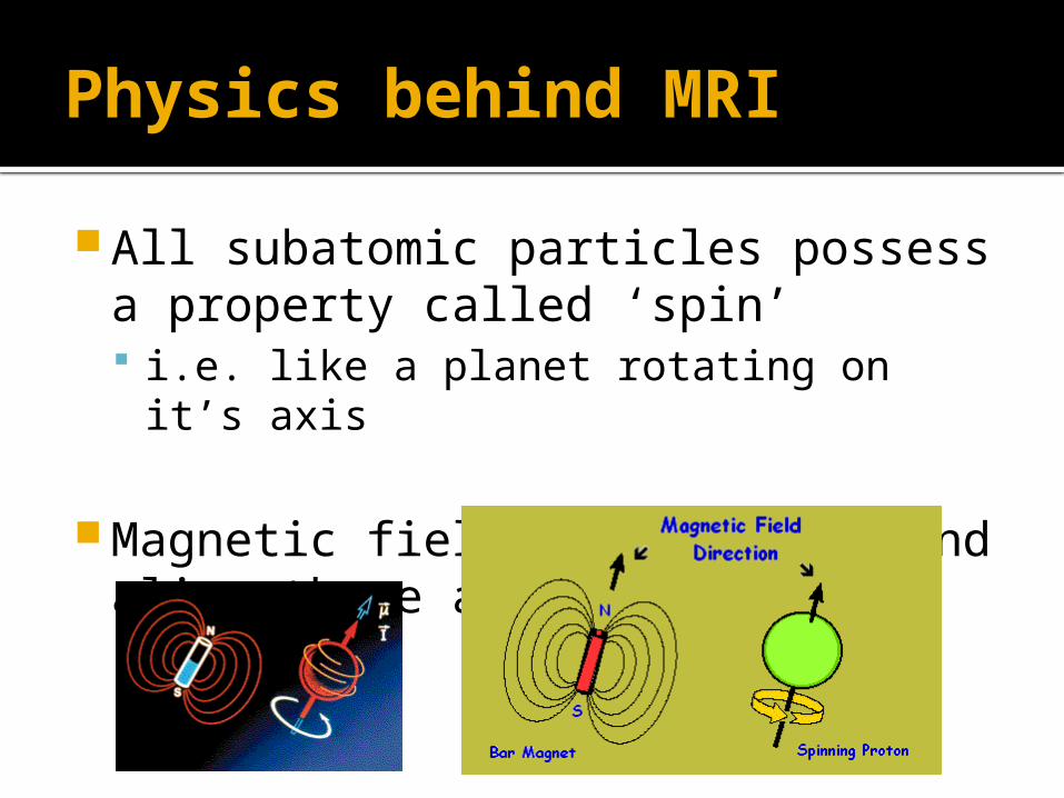
Physics behind MRI
All subatomic particles possess a property called ‘spin’ i.e. like a planet rotating on it’s axis
Magnetic fields can perturb and align these axes of rotation
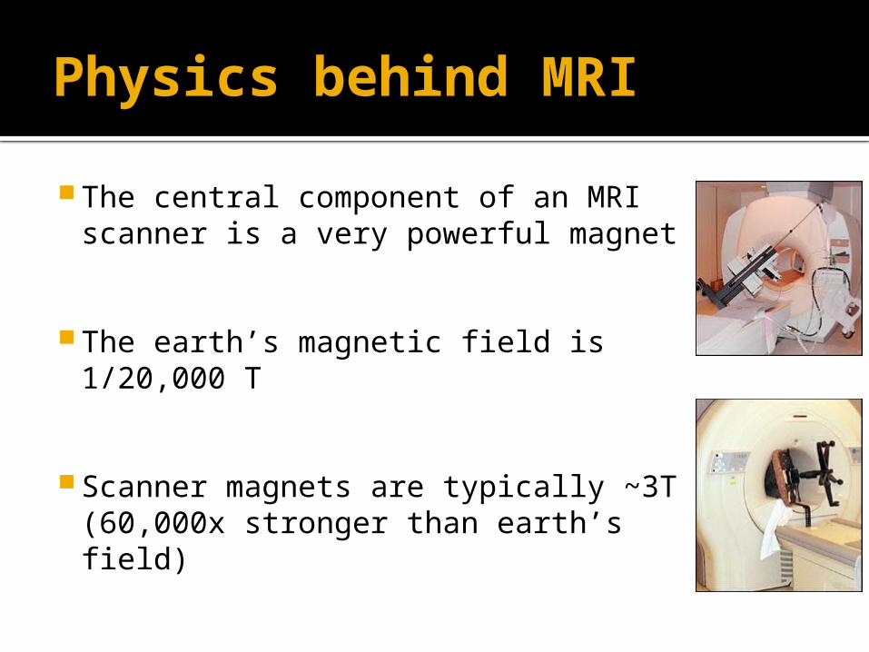
Physics behind MRI
The central component of an MRI scanner is a very powerful magnet
The earth’s magnetic field is 1/20,000 T
Scanner magnets are typically ~3T (60,000x stronger than earth’s field)
MRI – Basic Principles
Use powerful magnet to align hydrogen atoms in biological tissue
Transmit radio-frequency (RF) pulses to perturb the rotational axes of protons
Record RFs emitted by protons as they return the orientation imposed by large magnet, and use this to calculate H+ density
MRI – Basic Principles
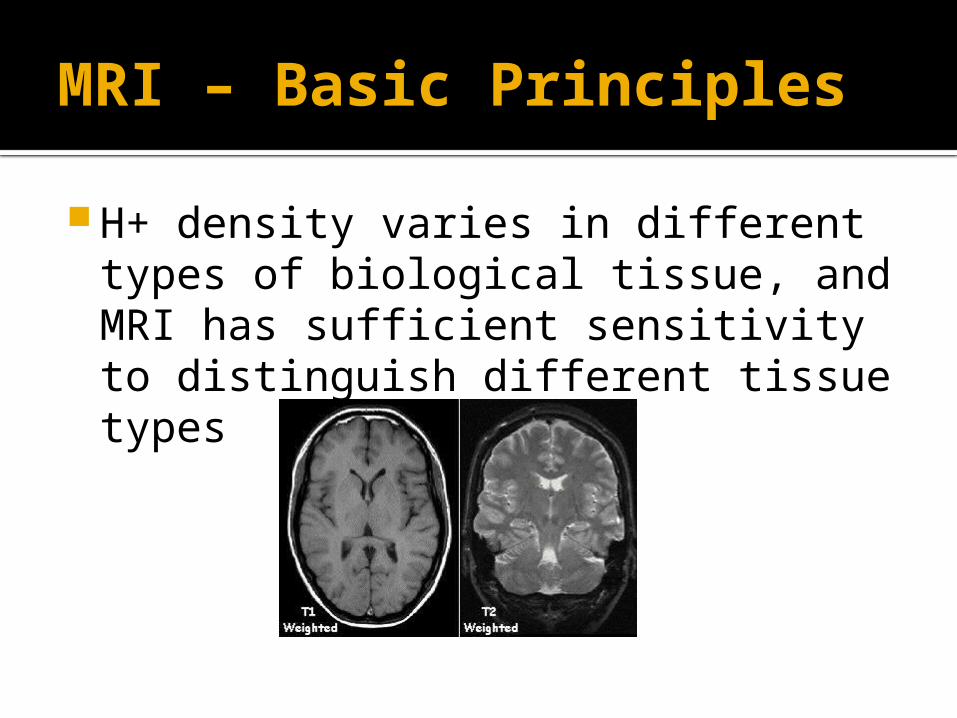
H+ density varies in different types of biological tissue, and MRI has sufficient sensitivity to distinguish different tissue types
MRI – Basic Principles
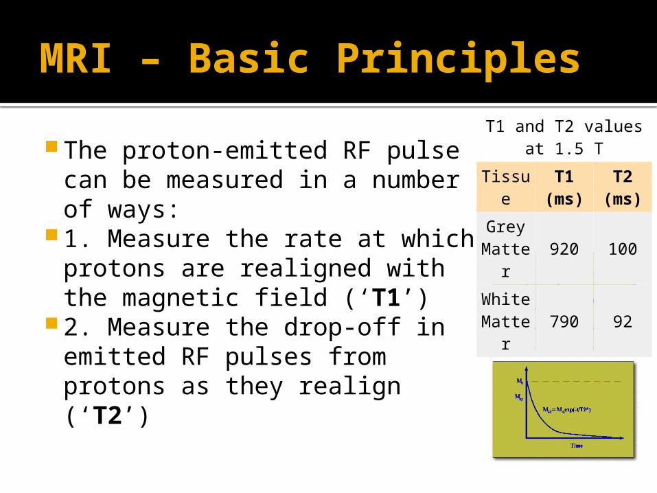
The proton-emitted RF pulse can be measured in a number of ways:
1. Measure the rate at which protons are realigned with the magnetic field (‘T1’)
2. Measure the drop-off in emitted RF pulses from protons as they realign (‘T2’)
T1 and T2 values at 1.5 T
Tissue T1 (ms)
T2 (ms)
Grey Matte
r920 100
White Matte
r790 92
Magnetic Properties of HB Hemoglobin’s magnetic properties
depend on whether it is oxygenated (HbO2) or deoxygenated (Hb)
HbO2 is ‘dimagnetic’ and has no net effect on the magnetic field
Hb is ‘paramagnetic’ and thus increases the strength of the local magnetic field when aligned by the scanner magnet
BOLD Signals
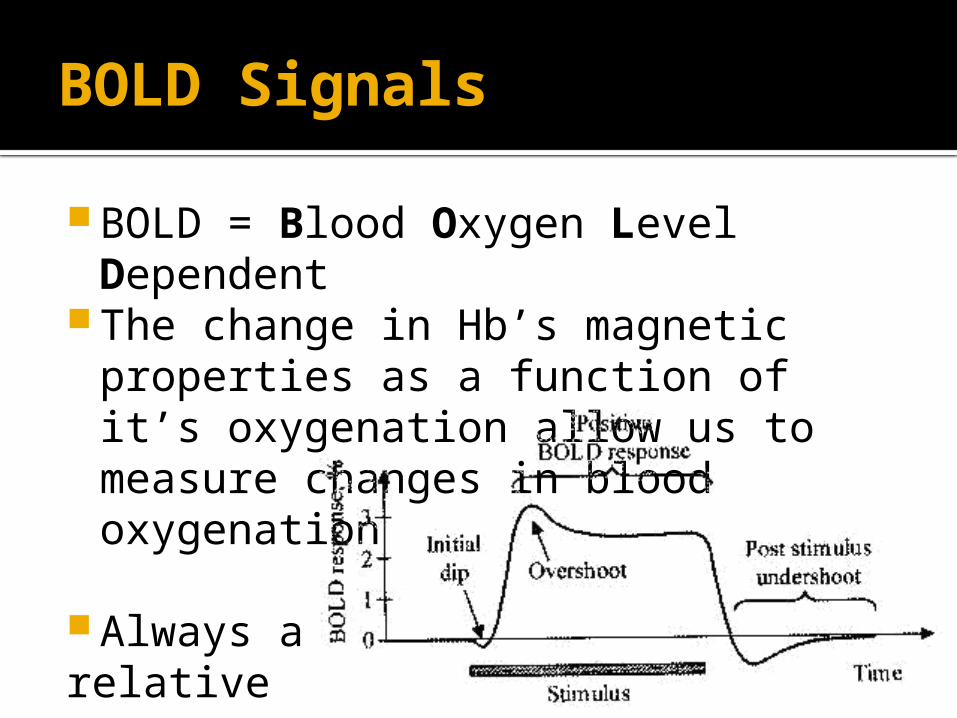
BOLD = Blood Oxygen Level Dependent
The change in Hb’s magnetic properties as a function of it’s oxygenation allow us to measure changes in blood oxygenation
Always a relative measure (A-B)
BOLD Signals
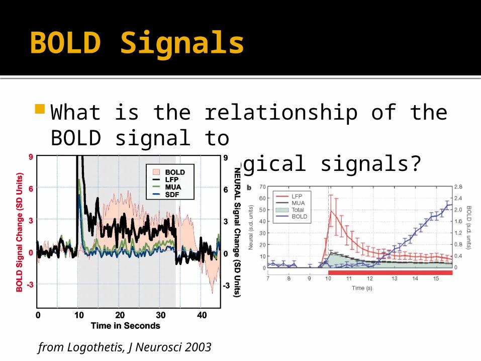
What is the relationship of the BOLD signal to electrophysiological signals?
from Logothetis, J Neurosci 2003
BOLD Signals
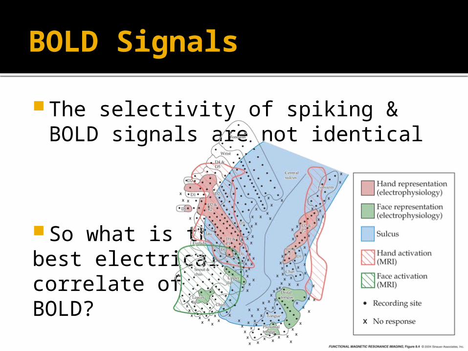
The selectivity of spiking & BOLD signals are not identical
So what is thebest electricalcorrelate of BOLD?
BOLD Signals
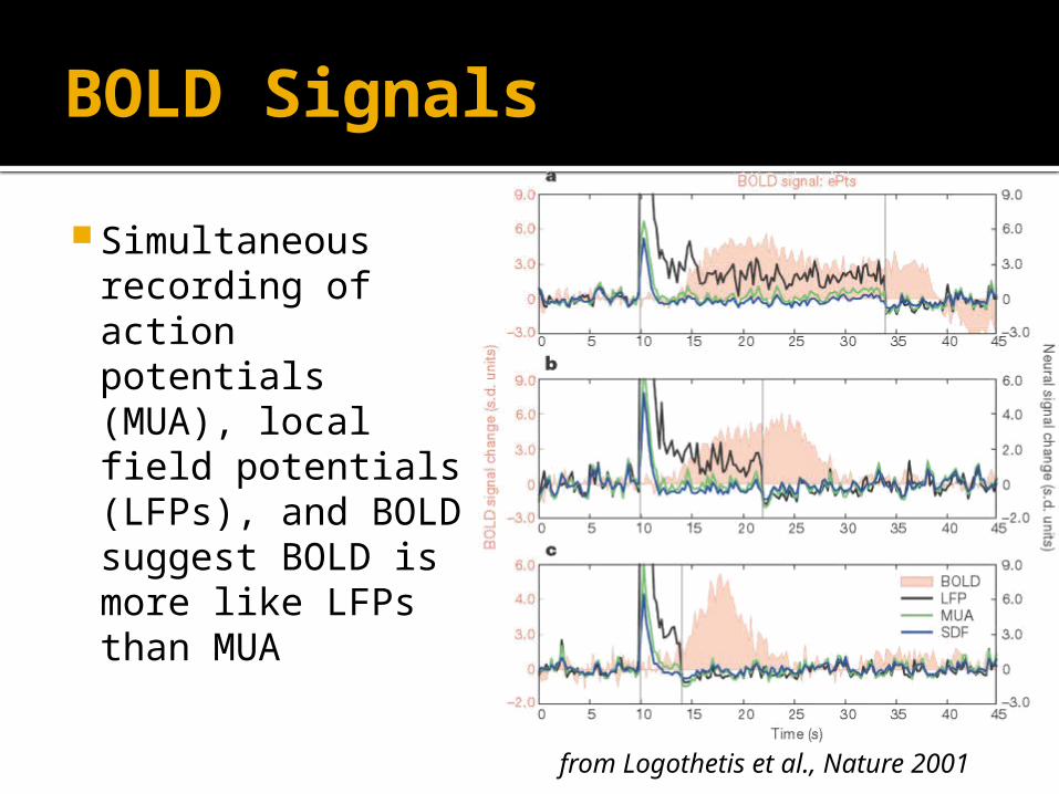
from Logothetis et al., Nature 2001
Simultaneous recording of action potentials (MUA), local field potentials (LFPs), and BOLD suggest BOLD is more like LFPs than MUA
BOLD Signals
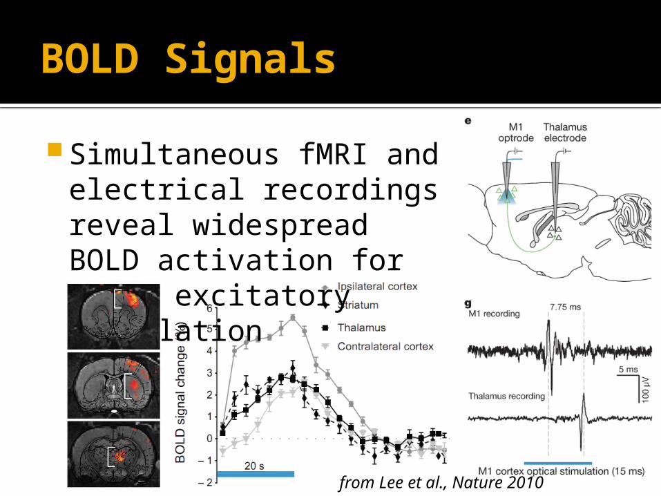
Simultaneous fMRI and electrical recordings reveal widespread BOLD activation for local excitatory stimulation
from Lee et al., Nature 2010
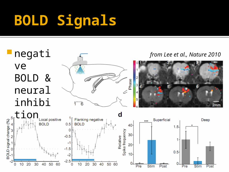
BOLD Signals
negative BOLD & neural inhibition
from Lee et al., Nature 2010
BOLD Signals
What can you infer from the absence of a BOLD response?
Positive BOLD response?
Negative BOLD response?
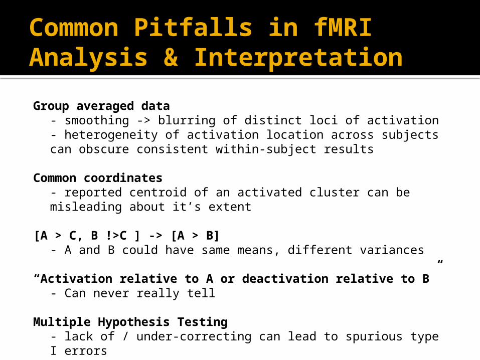
Common Pitfalls in fMRI Analysis & Interpretation
Group averaged data- smoothing -> blurring of distinct loci of activation- heterogeneity of activation location across subjects can obscure consistent within-subject results
Common coordinates- reported centroid of an activated cluster can be misleading about it’s extent
[A > C, B !>C ] -> [A > B]- A and B could have same means, different variances
“Activation relative to A or deactivation relative to B”- Can never really tell
Multiple Hypothesis Testing - lack of / under-correcting can lead to spurious type I errors - parametric over-correction can lead to many type II errors