Functional Cell-Responsive Biomaterials
35
biomedicines Review Integrin-Targeting Peptides for the Design of Functional Cell-Responsive Biomaterials Junwei Zhao , Federica Santino , Daria Giacomini and Luca Gentilucci * Department of Chemistry “G. Ciamician”, University of Bologna, via Selmi 2, 40126 Bologna, Italy; [email protected] (J.Z.); [email protected] (F.S.); [email protected] (D.G.) * Correspondence: [email protected]; Tel.: +39-0512099570; Fax: +39-0512099456 Received: 23 July 2020; Accepted: 23 August 2020; Published: 25 August 2020 Abstract: Integrins are a family of cell surface receptors crucial to fundamental cellular functions such as adhesion, signaling, and viability, deeply involved in a variety of diseases, including the initiation and progression of cancer, of coronary, inflammatory, or autoimmune diseases. The natural ligands of integrins are glycoproteins expressed on the cell surface or proteins of the extracellular matrix. For this reason, short peptides or peptidomimetic sequences that reproduce the integrin-binding motives have attracted much attention as potential drugs. When challenged in clinical trials, these peptides/peptidomimetics let to contrasting and disappointing results. In the search for alternative utilizations, the integrin peptide ligands have been conjugated onto nanoparticles, materials, or drugs and drug carrier systems, for specific recognition or delivery of drugs to cells overexpressing the targeted integrins. Recent research in peptidic integrin ligands is exploring new opportunities, in particular for the design of nanostructured, micro-fabricated, cell-responsive, stimuli-responsive, smart materials. Keywords: integrin ligands; tumor-targeting nanoparticles; peptide conjugates; RGD peptides; drug delivery; smart nanomaterials; cell adhesion; tissue engineering; regenerative medicine 1. Introduction In the last 20 years, many studies focused on the implication of integrins in important cell activities, in the signaling from and to the cell, and in a variety of diseases. After the identification of the peptidic recognition sequences to different kinds of integrins, much effort has been dedicated to identifying peptidomimetic ligands with antagonistic properties, especially for cancer therapy, circulatory, or inflammatory diseases. These issues have been widely discussed earlier [1–5], and will be not proposed again herein. Despite good initial results in vitro and in animal models, clinical trials have not met the expectations. Nevertheless, peptidic integrin ligands still maintain a noteworthy appeal. This review aims at summarizing the more innovative applications of peptide-material conjugates: from nanoparticles (NPs) to new drug delivery systems, to functionalized surfaces, self-assembled monolayers (SAMs) and finally to smart and responsive materials. These conjugates are expected to allow a range of applications in theranostics, disease monitoring, regenerative medicines, and tissue engineering. 2. Integrins Structure and Functions Integrins are cell adhesion receptors of soluble and insoluble glycoproteins of the extracellular matrix (ECM). These glycoproteins include collagens, fibronectins (FN), vitronectin (VT), laminins, fibrinogen (Fib), as well as cell surface receptors, e.g., the vascular cell adhesion molecule-1 (VCAM-1) and the intercellular adhesion molecule (ICAM). Furthermore, integrins are involved in the assembly Biomedicines 2020, 8, 307; doi:10.3390/biomedicines8090307 www.mdpi.com/journal/biomedicines
Transcript of Functional Cell-Responsive Biomaterials
biomedicines
Review
Integrin-Targeting Peptides for the Design ofFunctional Cell-Responsive Biomaterials
Junwei Zhao , Federica Santino , Daria Giacomini and Luca Gentilucci *
Department of Chemistry “G. Ciamician”, University of Bologna, via Selmi 2, 40126 Bologna, Italy;[email protected] (J.Z.); [email protected] (F.S.); [email protected] (D.G.)* Correspondence: [email protected]; Tel.: +39-0512099570; Fax: +39-0512099456
Received: 23 July 2020; Accepted: 23 August 2020; Published: 25 August 2020�����������������
Abstract: Integrins are a family of cell surface receptors crucial to fundamental cellular functionssuch as adhesion, signaling, and viability, deeply involved in a variety of diseases, includingthe initiation and progression of cancer, of coronary, inflammatory, or autoimmune diseases.The natural ligands of integrins are glycoproteins expressed on the cell surface or proteins ofthe extracellular matrix. For this reason, short peptides or peptidomimetic sequences that reproducethe integrin-binding motives have attracted much attention as potential drugs. When challenged inclinical trials, these peptides/peptidomimetics let to contrasting and disappointing results. In thesearch for alternative utilizations, the integrin peptide ligands have been conjugated onto nanoparticles,materials, or drugs and drug carrier systems, for specific recognition or delivery of drugs to cellsoverexpressing the targeted integrins. Recent research in peptidic integrin ligands is exploringnew opportunities, in particular for the design of nanostructured, micro-fabricated, cell-responsive,stimuli-responsive, smart materials.
Keywords: integrin ligands; tumor-targeting nanoparticles; peptide conjugates; RGD peptides; drugdelivery; smart nanomaterials; cell adhesion; tissue engineering; regenerative medicine
1. Introduction
In the last 20 years, many studies focused on the implication of integrins in important cellactivities, in the signaling from and to the cell, and in a variety of diseases. After the identificationof the peptidic recognition sequences to different kinds of integrins, much effort has been dedicatedto identifying peptidomimetic ligands with antagonistic properties, especially for cancer therapy,circulatory, or inflammatory diseases. These issues have been widely discussed earlier [1–5], and willbe not proposed again herein. Despite good initial results in vitro and in animal models, clinical trialshave not met the expectations. Nevertheless, peptidic integrin ligands still maintain a noteworthyappeal. This review aims at summarizing the more innovative applications of peptide-materialconjugates: from nanoparticles (NPs) to new drug delivery systems, to functionalized surfaces,self-assembled monolayers (SAMs) and finally to smart and responsive materials. These conjugates areexpected to allow a range of applications in theranostics, disease monitoring, regenerative medicines,and tissue engineering.
2. Integrins Structure and Functions
Integrins are cell adhesion receptors of soluble and insoluble glycoproteins of the extracellularmatrix (ECM). These glycoproteins include collagens, fibronectins (FN), vitronectin (VT), laminins,fibrinogen (Fib), as well as cell surface receptors, e.g., the vascular cell adhesion molecule-1 (VCAM-1)and the intercellular adhesion molecule (ICAM). Furthermore, integrins are involved in the assembly
Biomedicines 2020, 8, 307; doi:10.3390/biomedicines8090307 www.mdpi.com/journal/biomedicines
Biomedicines 2020, 8, 307 2 of 35
of the actin cytoskeleton and signal transduction pathways of biological and cellular functions:cell adhesion, migration, proliferation, cell differentiation, and apoptosis [6].
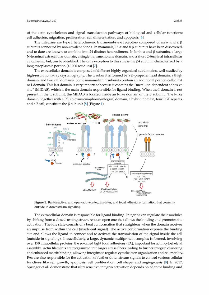
The integrins are type I heterodimeric transmembrane receptors composed of an α and a βsubunits connected by non-covalent bonds. In mammals, 18 α and 8 β subunits have been discovered,and to date are known to combine into 24 distinct heterodimers. In both α and β subunits, a largeN-terminal extracellular domain, a single transmembrane domain, and a short C-terminal intracellularcytoplasmic tail, can be identified. The only exception to this rule is the β4 subunit, characterized by along cytoplasmic portion (>1000 residues) [7].
The extracellular domain is composed of different highly organized subdomains, well-studied byhigh-resolution x-ray crystallography. The α subunit is formed by a β-propeller head domain, a thighdomain, and two calf domains. Some mammalian α subunits contain an additional portion called αAor I-domain. This last domain is very important because it contains the “metal-ion-dependent adhesivesite” (MIDAS), which is the main domain responsible for ligand binding. When the I-domain is notpresent in the α subunit, the MIDAS is located inside an I-like domain of the β subunit. The I-likedomain, together with a PSI (plexin/semaphorin/integrin) domain, a hybrid domain, four EGF repeats,and a B tail, constitute the β subunit [8] (Figure 1).
Biomedicines 2020, 8, x FOR PEER REVIEW 2 of 34
of the actin cytoskeleton and signal transduction pathways of biological and cellular functions: cell
adhesion, migration, proliferation, cell differentiation, and apoptosis [6].
The integrins are type I heterodimeric transmembrane receptors composed of an α and a β
subunits connected by non-covalent bonds. In mammals, 18 α and 8 β subunits have been discovered,
and to date are known to combine into 24 distinct heterodimers. In both α and β subunits, a large N-
terminal extracellular domain, a single transmembrane domain, and a short C-terminal intracellular
cytoplasmic tail, can be identified. The only exception to this rule is the β4 subunit, characterized by
a long cytoplasmic portion (>1000 residues) [7].
The extracellular domain is composed of different highly organized subdomains, well-studied
by high-resolution x-ray crystallography. The α subunit is formed by a β-propeller head domain, a
thigh domain, and two calf domains. Some mammalian α subunits contain an additional portion
called αA or I-domain. This last domain is very important because it contains the “metal-ion-
dependent adhesive site” (MIDAS), which is the main domain responsible for ligand binding. When
the I-domain is not present in the α subunit, the MIDAS is located inside an I-like domain of the β
subunit. The I-like domain, together with a PSI (plexin/semaphorin/integrin) domain, a hybrid
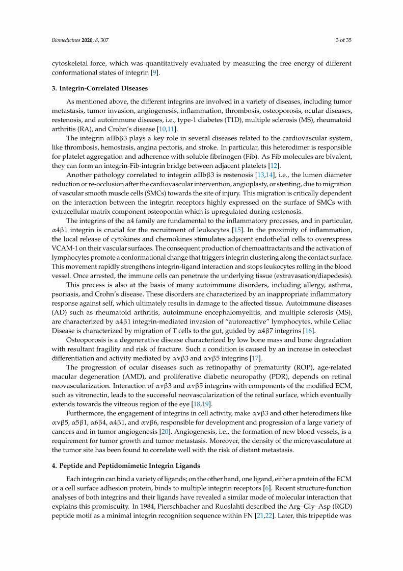
domain, four EGF repeats, and a B tail, constitute the β subunit [8] (Figure 1).
Figure 1. Bent-inactive, and open-active integrin states, and focal adhesions formation that consents
outside-in downstream signaling.
The extracellular domain is responsible for ligand binding. Integrins can regulate their modules
by shifting from a closed-resting structure to an open one that allows the binding and promotes the
activation. The idle state consists of a bent conformation that straightens when the domain receives
an impulse from within the cell (inside-out signal). The active conformation exposes the binding site
and allows the ligand to connect and to activate the transmission of the signal inside the cell (outside-
in signaling). Intracellularly, a large, dynamic multiprotein complex is formed, involving over 150
intracellular proteins, the so-called tight focal adhesions (FA), important for actin cytoskeletal
assembly. Actin filaments are reorganized into larger stress fibers leading to further integrin
clustering and enhanced matrix binding, allowing integrins to regulate cytoskeleton organization and
cell motility. FAs are also responsible for the activation of further downstream signals to control
various cellular functions like cell growth, apoptosis, cell proliferation, cell shape, and angiogenesis
[Error! Bookmark not defined.]. In 2017, Springer et al. demonstrate that ultrasensitive integrin
activation depends on adaptor binding and cytoskeletal force, which was quantitatively evaluated
by measuring the free energy of different conformational states of integrin [9].
Figure 1. Bent-inactive, and open-active integrin states, and focal adhesions formation that consentsoutside-in downstream signaling.
The extracellular domain is responsible for ligand binding. Integrins can regulate their modulesby shifting from a closed-resting structure to an open one that allows the binding and promotes theactivation. The idle state consists of a bent conformation that straightens when the domain receivesan impulse from within the cell (inside-out signal). The active conformation exposes the bindingsite and allows the ligand to connect and to activate the transmission of the signal inside the cell(outside-in signaling). Intracellularly, a large, dynamic multiprotein complex is formed, involvingover 150 intracellular proteins, the so-called tight focal adhesions (FA), important for actin cytoskeletalassembly. Actin filaments are reorganized into larger stress fibers leading to further integrin clusteringand enhanced matrix binding, allowing integrins to regulate cytoskeleton organization and cell motility.FAs are also responsible for the activation of further downstream signals to control various cellularfunctions like cell growth, apoptosis, cell proliferation, cell shape, and angiogenesis [8]. In 2017,Springer et al. demonstrate that ultrasensitive integrin activation depends on adaptor binding and
Biomedicines 2020, 8, 307 3 of 35
cytoskeletal force, which was quantitatively evaluated by measuring the free energy of differentconformational states of integrin [9].
3. Integrin-Correlated Diseases
As mentioned above, the different integrins are involved in a variety of diseases, including tumormetastasis, tumor invasion, angiogenesis, inflammation, thrombosis, osteoporosis, ocular diseases,restenosis, and autoimmune diseases, i.e., type-1 diabetes (T1D), multiple sclerosis (MS), rheumatoidarthritis (RA), and Crohn’s disease [10,11].
The integrin αIIbβ3 plays a key role in several diseases related to the cardiovascular system,like thrombosis, hemostasis, angina pectoris, and stroke. In particular, this heterodimer is responsiblefor platelet aggregation and adherence with soluble fibrinogen (Fib). As Fib molecules are bivalent,they can form an integrin-Fib-integrin bridge between adjacent platelets [12].
Another pathology correlated to integrin αIIbβ3 is restenosis [13,14], i.e., the lumen diameterreduction or re-occlusion after the cardiovascular intervention, angioplasty, or stenting, due to migrationof vascular smooth muscle cells (SMCs) towards the site of injury. This migration is critically dependenton the interaction between the integrin receptors highly expressed on the surface of SMCs withextracellular matrix component osteopontin which is upregulated during restenosis.
The integrins of the α4 family are fundamental to the inflammatory processes, and in particular,α4β1 integrin is crucial for the recruitment of leukocytes [15]. In the proximity of inflammation,the local release of cytokines and chemokines stimulates adjacent endothelial cells to overexpressVCAM-1 on their vascular surfaces. The consequent production of chemoattractants and the activation oflymphocytes promote a conformational change that triggers integrin clustering along the contact surface.This movement rapidly strengthens integrin-ligand interaction and stops leukocytes rolling in the bloodvessel. Once arrested, the immune cells can penetrate the underlying tissue (extravasation/diapedesis).
This process is also at the basis of many autoimmune disorders, including allergy, asthma,psoriasis, and Crohn’s disease. These disorders are characterized by an inappropriate inflammatoryresponse against self, which ultimately results in damage to the affected tissue. Autoimmune diseases(AD) such as rheumatoid arthritis, autoimmune encephalomyelitis, and multiple sclerosis (MS),are characterized by α4β1 integrin-mediated invasion of “autoreactive” lymphocytes, while CeliacDisease is characterized by migration of T cells to the gut, guided by α4β7 integrins [16].
Osteoporosis is a degenerative disease characterized by low bone mass and bone degradationwith resultant fragility and risk of fracture. Such a condition is caused by an increase in osteoclastdifferentiation and activity mediated by ανβ3 and ανβ5 integrins [17].
The progression of ocular diseases such as retinopathy of prematurity (ROP), age-relatedmacular degeneration (AMD), and proliferative diabetic neuropathy (PDR), depends on retinalneovascularization. Interaction of ανβ3 and ανβ5 integrins with components of the modified ECM,such as vitronectin, leads to the successful neovascularization of the retinal surface, which eventuallyextends towards the vitreous region of the eye [18,19].
Furthermore, the engagement of integrins in cell activity, make ανβ3 and other heterodimers likeανβ5, α5β1, α6β4, α4β1, and ανβ6, responsible for development and progression of a large variety ofcancers and in tumor angiogenesis [20]. Angiogenesis, i.e., the formation of new blood vessels, is arequirement for tumor growth and tumor metastasis. Moreover, the density of the microvasculature atthe tumor site has been found to correlate well with the risk of distant metastasis.
4. Peptide and Peptidomimetic Integrin Ligands
Each integrin can bind a variety of ligands; on the other hand, one ligand, either a protein of the ECMor a cell surface adhesion protein, binds to multiple integrin receptors [6]. Recent structure-functionanalyses of both integrins and their ligands have revealed a similar mode of molecular interaction thatexplains this promiscuity. In 1984, Pierschbacher and Ruoslahti described the Arg–Gly–Asp (RGD)peptide motif as a minimal integrin recognition sequence within FN [21,22]. Later, this tripeptide was
Biomedicines 2020, 8, 307 4 of 35
found to bind to several other integrins: ανβ1, ανβ3, ανβ5, ανβ6, ανβ8, α5β1, α8β1, and αIIbβ3.Other integrins interact with their ligands by recognizing different ligand motif. For instance,α4 integrins (α4β1 and α4β7) recognize Leu–Asp–Val–Pro (LDVP) motif in FN, Leu–Asp–Thr–Ser(LDTS) sequence in the mucosal addressing cell adhesion molecule-1 (MAdCAM-1), and Ile–Asp–Ser(IDS) in VCAM-1.
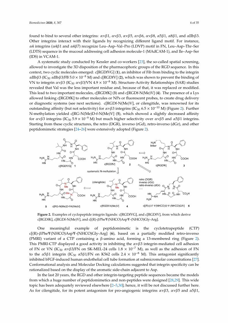
A systematic study conducted by Kessler and co-workers [23], the so-called spatial screening,allowed to investigate the 3D disposition of the pharmacophoric groups of the RGD sequence. In thiscontext, two cyclic molecules emerged: c[RGDfVG] (1), an inhibitor of Fib from binding to the integrinαIIbβ3 (IC50 αIIbβ3/FB 5.0 × 10⁻⁸ M) and c[RGDfV] (2), which was shown to prevent the binding ofVN to integrin ανβ3 (IC50 ανβ3/VN 4.9 × 10⁻⁸ M). Structure-Activity Relationships (SAR) studiesrevealed that Val was the less important residue and, because of that, it was replaced or modified.This lead to two important molecules, c[RGDfK] (3) and c[RGDf-N(Me)V] (4). The presence of a Lysallowed linking c[RGDfK] to other molecules or NPs or fluorescent probes, to create drug deliveryor diagnostic systems (see next sections). c[RGDf-N(Me)V], or cilengitide, was renowned for itsoutstanding affinity (but not selectivity) for ανβ3 integrins (IC50 6.5 × 10⁻¹⁰ M) (Figure 2). FurtherN-methylation yielded c[RG-N(Me)D-f-N(Me)V] (5), which showed a slightly decreased affinityfor ανβ3 integrins (IC50 5.9 × 10⁻⁹ M) but much higher selectivity over ανβ5 and α5β1 integrins.Starting from these cyclic structures, the retro (DGR), inverso (rGd), retro-inverso (dGr), and otherpeptidomimetic strategies [24–26] were extensively adopted (Figure 2).
Biomedicines 2020, 8, x FOR PEER REVIEW 4 of 34
Val–Pro (LDVP) motif in FN, Leu–Asp–Thr–Ser (LDTS) sequence in the mucosal addressing cell
adhesion molecule-1 (MAdCAM-1), and Ile–Asp–Ser (IDS) in VCAM-1.
A systematic study conducted by Kessler and co-workers [23], the so-called spatial screening,
allowed to investigate the 3D disposition of the pharmacophoric groups of the RGD sequence. In this
context, two cyclic molecules emerged: c[RGDfVG] (1), an inhibitor of Fib from binding to the integrin
αIIbβ3 (IC50 αIIbβ3/FB 5.0 × 10⁻⁸ M) and c[RGDfV] (2), which was shown to prevent the binding of
VN to integrin ανβ3 (IC50 ανβ3/VN 4.9 × 10⁻⁸ M). Structure-Activity Relationships (SAR) studies
revealed that Val was the less important residue and, because of that, it was replaced or modified.
This lead to two important molecules, c[RGDfK] (3) and c[RGDf-N(Me)V] (4). The presence of a Lys
allowed linking c[RGDfK] to other molecules or NPs or fluorescent probes, to create drug delivery or
diagnostic systems (see next sections). c[RGDf-N(Me)V], or cilengitide, was renowned for its
outstanding affinity (but not selectivity) for ανβ3 integrins (IC50 6.5 × 10⁻¹⁰ M) (Figure 2). Further N-
methylation yielded c[RG-N(Me)D-f-N(Me)V] (5), which showed a slightly decreased affinity for
ανβ3 integrins (IC50 5.9 × 10⁻⁹ M) but much higher selectivity over ανβ5 and α5β1 integrins. Starting
from these cyclic structures, the retro (DGR), inverso (rGd), retro-inverso (dGr), and other
peptidomimetic strategies [24–26] were extensively adopted (Figure 2).
Figure 2. Examples of cyclopeptide integrin ligands: c[RGDfVG], and c[RGDfV], from which derive
c[RGDfK], c[RGDf-N(Me)V], and c[(R)-βPheΨ(NHCO)AspΨ-(NHCO)Gly-Arg].
One meaningful example of peptidomimetic is the cyclotetrapeptide (CTP) c[(R)-
βPheΨ(NHCO)AspΨ-(NHCO)Gly-Arg] (6), based on a partially modified retro-inverso (PMRI)
variant of a CTP containing a β-amino acid, forming a 13-membered ring (Figure 2). This PMRI-CTP
displayed a good activity in inhibiting the ανβ3 integrin-mediated cell adhesion of FN or VN (IC50
ανβ3/FN on SK-MEL-24 cells 1.8 × 10⁻⁷ M), as well as the adhesion of FN to the α5β1 integrin (IC50
α5β1/FN on K562 cells 2.4 × 10⁻⁸ M). This antagonist significantly inhibited bFGF-induced human
endothelial cell tube formation at submicromolar concentrations [27]. Conformational analysis and
Molecular Docking calculations suggested that integrin specificity can be rationalized based on the
display of the aromatic side-chain adjacent to Asp.
In the last 20 years, the RGD and other integrin-targeting peptide sequences became the models
from which a huge number of peptidomimetics and non-peptides were designed [28,29]. This wide
topic has been adequately reviewed elsewhere [2–5,30]; hence, it will be not discussed further here.
As for cilengitide, for its potent antagonism for pro-angiogenic integrins ανβ3, ανβ5 and α5β1, it
became one of the most investigated anticancer peptides in vitro and in vivo. Preclinical in vitro
studies confirmed that cilengitide was a potent inhibitor of angiogenesis, and promoted apoptosis in
cancer cells obstructing the interaction between integrins and their ECM ligands [31,32]. Furthermore,
numerous in vivo experiments confirmed the previous results supporting the ability of cilengitide to
block tumor growth in a dose-dependent fashion [33]. After these promising results, phase I and II
Figure 2. Examples of cyclopeptide integrin ligands: c[RGDfVG], and c[RGDfV], from which derivec[RGDfK], c[RGDf-N(Me)V], and c[(R)-βPheΨ(NHCO)AspΨ-(NHCO)Gly-Arg].
One meaningful example of peptidomimetic is the cyclotetrapeptide (CTP)c[(R)-βPheΨ(NHCO)AspΨ-(NHCO)Gly-Arg] (6), based on a partially modified retro-inverso(PMRI) variant of a CTP containing a β-amino acid, forming a 13-membered ring (Figure 2).This PMRI-CTP displayed a good activity in inhibiting the ανβ3 integrin-mediated cell adhesionof FN or VN (IC50 ανβ3/FN on SK-MEL-24 cells 1.8 × 10⁻⁷ M), as well as the adhesion of FNto the α5β1 integrin (IC50 α5β1/FN on K562 cells 2.4 × 10⁻⁸ M). This antagonist significantlyinhibited bFGF-induced human endothelial cell tube formation at submicromolar concentrations [27].Conformational analysis and Molecular Docking calculations suggested that integrin specificity can berationalized based on the display of the aromatic side-chain adjacent to Asp.
In the last 20 years, the RGD and other integrin-targeting peptide sequences became the modelsfrom which a huge number of peptidomimetics and non-peptides were designed [28,29]. This widetopic has been adequately reviewed elsewhere [2–5,30]; hence, it will be not discussed further here.As for cilengitide, for its potent antagonism for pro-angiogenic integrins ανβ3, ανβ5 and α5β1,
Biomedicines 2020, 8, 307 5 of 35
it became one of the most investigated anticancer peptides in vitro and in vivo. Preclinical in vitrostudies confirmed that cilengitide was a potent inhibitor of angiogenesis, and promoted apoptosis incancer cells obstructing the interaction between integrins and their ECM ligands [31,32]. Furthermore,numerous in vivo experiments confirmed the previous results supporting the ability of cilengitideto block tumor growth in a dose-dependent fashion [33]. After these promising results, phase I andII trials in solid tumors and glioblastoma started, with cilengitide alone or in combination withradiotherapy and/or chemotherapy with temozolomide [34–37]. As no dose-limiting toxicities (DLTs)were observed, the CENTRIC EIRTC phase II trial was started, but the latter lead to disappointingresults. Cilengitide failed to demonstrate survival advantage and that it had a short plasma half-life.Subsequently, Massabeau, Khalifa, and co-workers [38] proposed continuous exposure for optimalefficacy. This idea was pursued in a phase I clinical study that combined cilengitide and chemotherapyfor stage III NSCLC (non-small cell lung cancer) patients. The results evidenced the safety profile ofthe administration of cilengitide as continuous infusion and suggested possible future investigations.
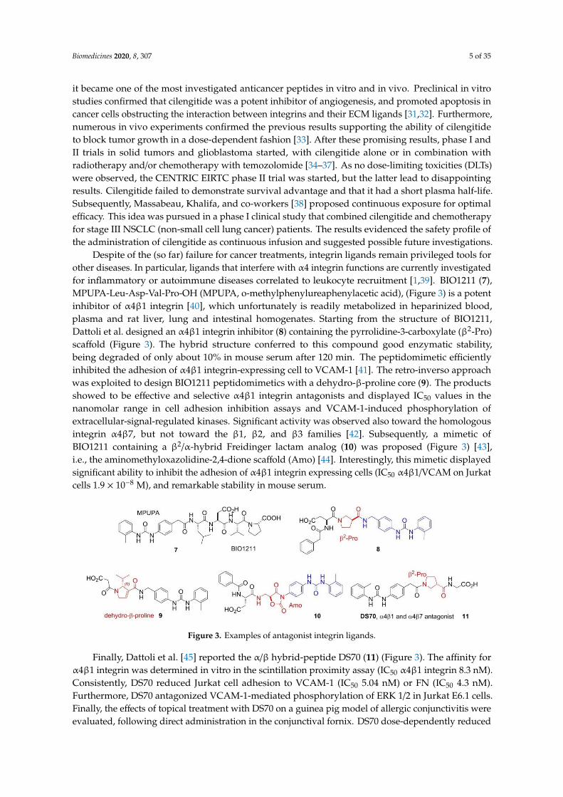
Despite of the (so far) failure for cancer treatments, integrin ligands remain privileged tools forother diseases. In particular, ligands that interfere with α4 integrin functions are currently investigatedfor inflammatory or autoimmune diseases correlated to leukocyte recruitment [1,39]. BIO1211 (7),MPUPA-Leu-Asp-Val-Pro-OH (MPUPA, o-methylphenylureaphenylacetic acid), (Figure 3) is a potentinhibitor of α4β1 integrin [40], which unfortunately is readily metabolized in heparinized blood,plasma and rat liver, lung and intestinal homogenates. Starting from the structure of BIO1211,Dattoli et al. designed an α4β1 integrin inhibitor (8) containing the pyrrolidine-3-carboxylate (β2-Pro)scaffold (Figure 3). The hybrid structure conferred to this compound good enzymatic stability,being degraded of only about 10% in mouse serum after 120 min. The peptidomimetic efficientlyinhibited the adhesion of α4β1 integrin-expressing cell to VCAM-1 [41]. The retro-inverso approachwas exploited to design BIO1211 peptidomimetics with a dehydro-β-proline core (9). The productsshowed to be effective and selective α4β1 integrin antagonists and displayed IC50 values in thenanomolar range in cell adhesion inhibition assays and VCAM-1-induced phosphorylation ofextracellular-signal-regulated kinases. Significant activity was observed also toward the homologousintegrin α4β7, but not toward the β1, β2, and β3 families [42]. Subsequently, a mimetic ofBIO1211 containing a β2/α-hybrid Freidinger lactam analog (10) was proposed (Figure 3) [43],i.e., the aminomethyloxazolidine-2,4-dione scaffold (Amo) [44]. Interestingly, this mimetic displayedsignificant ability to inhibit the adhesion of α4β1 integrin expressing cells (IC50 α4β1/VCAM on Jurkatcells 1.9 × 10⁻⁸ M), and remarkable stability in mouse serum.
Biomedicines 2020, 8, x FOR PEER REVIEW 5 of 34
trials in solid tumors and glioblastoma started, with cilengitide alone or in combination with
radiotherapy and/or chemotherapy with temozolomide [34–37]. As no dose-limiting toxicities (DLTs)
were observed, the CENTRIC EIRTC phase II trial was started, but the latter lead to disappointing
results. Cilengitide failed to demonstrate survival advantage and that it had a short plasma half-life.
Subsequently, Massabeau, Khalifa, and co-workers [38] proposed continuous exposure for optimal
efficacy. This idea was pursued in a phase I clinical study that combined cilengitide and
chemotherapy for stage III NSCLC (non-small cell lung cancer) patients. The results evidenced the
safety profile of the administration of cilengitide as continuous infusion and suggested possible
future investigations.
Despite of the (so far) failure for cancer treatments, integrin ligands remain privileged tools for
other diseases. In particular, ligands that interfere with α4 integrin functions are currently
investigated for inflammatory or autoimmune diseases correlated to leukocyte recruitment [1,39].
BIO1211 (7), MPUPA-Leu-Asp-Val-Pro-OH (MPUPA, o-methylphenylureaphenylacetic acid),
(Figure 3) is a potent inhibitor of α4β1 integrin [40], which unfortunately is readily metabolized in
heparinized blood, plasma and rat liver, lung and intestinal homogenates. Starting from the structure
of BIO1211, Dattoli et al. designed an α4β1 integrin inhibitor (8) containing the pyrrolidine-3-
carboxylate (β2-Pro) scaffold (Figure 3). The hybrid structure conferred to this compound good
enzymatic stability, being degraded of only about 10% in mouse serum after 120 min. The
peptidomimetic efficiently inhibited the adhesion of α4β1 integrin-expressing cell to VCAM-1 [41].
The retro-inverso approach was exploited to design BIO1211 peptidomimetics with a dehydro-β-
proline core (9). The products showed to be effective and selective α4β1 integrin antagonists and
displayed IC50 values in the nanomolar range in cell adhesion inhibition assays and VCAM-1-induced
phosphorylation of extracellular-signal-regulated kinases. Significant activity was observed also
toward the homologous integrin α4β7, but not toward the β1, β2, and β3 families [42]. Subsequently,
a mimetic of BIO1211 containing a β2/α-hybrid Freidinger lactam analog (10) was proposed (Figure
3) [43], i.e., the aminomethyloxazolidine-2,4-dione scaffold (Amo) [44]. Interestingly, this mimetic
displayed significant ability to inhibit the adhesion of α4β1 integrin expressing cells (IC50
α4β1/VCAM on Jurkat cells 1.9 × 10⁻⁸ M), and remarkable stability in mouse serum.
Figure 3. Examples of antagonist integrin ligands.
Finally, Dattoli et al. [45] reported the α/β hybrid-peptide DS70 (11) (Figure 3). The affinity for
α4β1 integrin was determined in vitro in the scintillation proximity assay (IC50 α4β1 integrin 8.3 nM).
Consistently, DS70 reduced Jurkat cell adhesion to VCAM-1 (IC50 5.04 nM) or FN (IC50 4.3 nM).
Furthermore, DS70 antagonized VCAM-1-mediated phosphorylation of ERK 1/2 in Jurkat E6.1 cells.
Finally, the effects of topical treatment with DS70 on a guinea pig model of allergic conjunctivitis
were evaluated, following direct administration in the conjunctival fornix. DS70 dose-dependently
reduced clinical aspects of allergic conjunctivitis, conjunctival mast cell and eosinophil infiltration,
α4 integrin expression, and levels of mRNAs for IL-1β, IL-8 (CXCL8), CCL5, and CCL11, thus
representing an alternative to antihistamines and mast cell-stabilizing agents.
Addressing Integrins with Agonist Ligands
Figure 3. Examples of antagonist integrin ligands.
Finally, Dattoli et al. [45] reported the α/β hybrid-peptide DS70 (11) (Figure 3). The affinity forα4β1 integrin was determined in vitro in the scintillation proximity assay (IC50 α4β1 integrin 8.3 nM).Consistently, DS70 reduced Jurkat cell adhesion to VCAM-1 (IC50 5.04 nM) or FN (IC50 4.3 nM).Furthermore, DS70 antagonized VCAM-1-mediated phosphorylation of ERK 1/2 in Jurkat E6.1 cells.Finally, the effects of topical treatment with DS70 on a guinea pig model of allergic conjunctivitis wereevaluated, following direct administration in the conjunctival fornix. DS70 dose-dependently reduced
Biomedicines 2020, 8, 307 6 of 35
clinical aspects of allergic conjunctivitis, conjunctival mast cell and eosinophil infiltration, α4 integrinexpression, and levels of mRNAs for IL-1β, IL-8 (CXCL8), CCL5, and CCL11, thus representing analternative to antihistamines and mast cell-stabilizing agents.
Addressing Integrins with Agonist Ligands
Even though most of the efforts have been directed to the development of integrin antagonists,the discovery of agonists could represent a new perspective [1]. In recent years, some agonist moleculeshave been disclosed, targeting different types of integrins. Celik and co-workers [46] studied thecapacity of leukadherin-1 to act as an agonist for αMβ2, and thus be able to reduce neutrophilmigration and inflammatory response. Using the structure of the well-known antagonist TBC3486,Dixon and co-workers [47] designed and tested in vitro a new α4β1 agonist, with the aim of increasecell retention and so improve progenitor cell therapy. Because previous works have shown thatchemotherapy can affect more metastatic melanomas when cells are adherent to ECM instead ofsuspended. Schwartz and co-workers [48] tested in vivo the chemotherapeutic drug combined or notwith contortrostatin, a disintegrin from snake venom, and proved that stimulate integrin may help theefficacy of chemotherapy and help to reduce tumor growth. As the interaction between αLβ2 andICAM-1 plays an important role in the immune responses and leukocytes adhesion, this integrin hasbeen the target of several studies. In particular, small molecule agonists were developed and tested [49]to demonstrate that stimulation of αLβ2 integrin facilitates cell signaling.
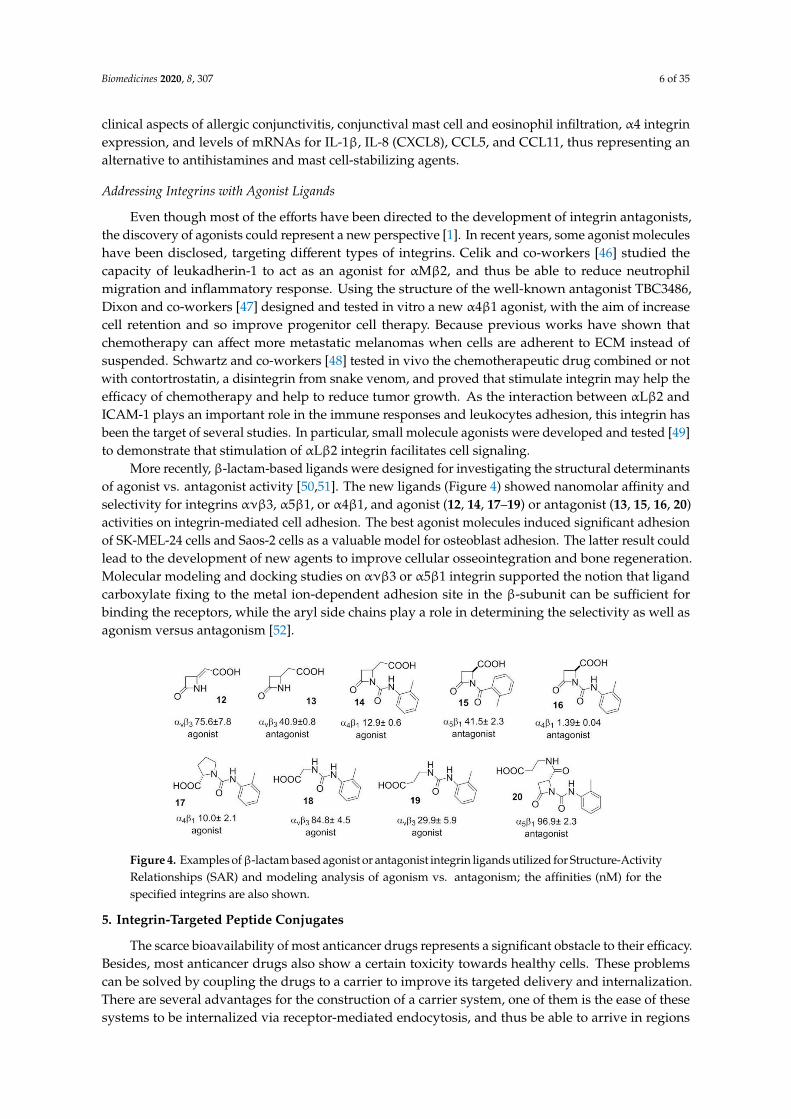
More recently, β-lactam-based ligands were designed for investigating the structural determinantsof agonist vs. antagonist activity [50,51]. The new ligands (Figure 4) showed nanomolar affinity andselectivity for integrins ανβ3, α5β1, or α4β1, and agonist (12, 14, 17–19) or antagonist (13, 15, 16, 20)activities on integrin-mediated cell adhesion. The best agonist molecules induced significant adhesionof SK-MEL-24 cells and Saos-2 cells as a valuable model for osteoblast adhesion. The latter result couldlead to the development of new agents to improve cellular osseointegration and bone regeneration.Molecular modeling and docking studies on ανβ3 or α5β1 integrin supported the notion that ligandcarboxylate fixing to the metal ion-dependent adhesion site in the β-subunit can be sufficient forbinding the receptors, while the aryl side chains play a role in determining the selectivity as well asagonism versus antagonism [52].
Biomedicines 2020, 8, x FOR PEER REVIEW 6 of 34
Even though most of the efforts have been directed to the development of integrin antagonists,
the discovery of agonists could represent a new perspective [Error! Bookmark not defined.]. In recent
years, some agonist molecules have been disclosed, targeting different types of integrins. Celik and
co-workers [46] studied the capacity of leukadherin-1 to act as an agonist for αMβ2, and thus be able
to reduce neutrophil migration and inflammatory response. Using the structure of the well-known
antagonist TBC3486, Dixon and co-workers [47] designed and tested in vitro a new α4β1 agonist,
with the aim of increase cell retention and so improve progenitor cell therapy. Because previous
works have shown that chemotherapy can affect more metastatic melanomas when cells are adherent
to ECM instead of suspended. Schwartz and co-workers [48] tested in vivo the chemotherapeutic
drug combined or not with contortrostatin, a disintegrin from snake venom, and proved that
stimulate integrin may help the efficacy of chemotherapy and help to reduce tumor growth. As the
interaction between αLβ2 and ICAM-1 plays an important role in the immune responses and
leukocytes adhesion, this integrin has been the target of several studies. In particular, small molecule
agonists were developed and tested [49] to demonstrate that stimulation of αLβ2 integrin facilitates
cell signaling.
More recently, β-lactam-based ligands were designed for investigating the structural
determinants of agonist vs. antagonist activity [50,51]. The new ligands (Figure 4) showed nanomolar
affinity and selectivity for integrins ανβ3, α5β1, or α4β1, and agonist (12, 14, 17–19) or antagonist (13,
15, 16, 20) activities on integrin-mediated cell adhesion. The best agonist molecules induced
significant adhesion of SK-MEL-24 cells and Saos-2 cells as a valuable model for osteoblast adhesion.
The latter result could lead to the development of new agents to improve cellular osseointegration
and bone regeneration. Molecular modeling and docking studies on ανβ3 or α5β1 integrin supported
the notion that ligand carboxylate fixing to the metal ion-dependent adhesion site in the β-subunit
can be sufficient for binding the receptors, while the aryl side chains play a role in determining the
selectivity as well as agonism versus antagonism [52].
Figure 4. Examples of β-lactam based agonist or antagonist integrin ligands utilized for Structure-
Activity Relationships (SAR) and modeling analysis of agonism vs. antagonism; the affinities (nM)
for the specified integrins are also shown.
5. Integrin-Targeted Peptide Conjugates
The scarce bioavailability of most anticancer drugs represents a significant obstacle to their
efficacy. Besides, most anticancer drugs also show a certain toxicity towards healthy cells. These
problems can be solved by coupling the drugs to a carrier to improve its targeted delivery and
internalization. There are several advantages for the construction of a carrier system, one of them is
the ease of these systems to be internalized via receptor-mediated endocytosis, and thus be able to
arrive in regions away from blood vessels, the ones previously impossible to reach [53]. Active and
inactive integrins undergo a clathrin-mediated or caveolin-mediated endocytosis-recycling cycle,
involved in different processes, such as cell migration, by detaching integrins from the extracellular
matrix, integrin recycling and activation of different receptors (Figure 5) [54]. Considering this,
interest has focused on the construction of RGD conjugates with anticancer drugs, diagnostic probes,
Figure 4. Examples ofβ-lactam based agonist or antagonist integrin ligands utilized for Structure-ActivityRelationships (SAR) and modeling analysis of agonism vs. antagonism; the affinities (nM) for thespecified integrins are also shown.
5. Integrin-Targeted Peptide Conjugates
The scarce bioavailability of most anticancer drugs represents a significant obstacle to their efficacy.Besides, most anticancer drugs also show a certain toxicity towards healthy cells. These problemscan be solved by coupling the drugs to a carrier to improve its targeted delivery and internalization.There are several advantages for the construction of a carrier system, one of them is the ease of thesesystems to be internalized via receptor-mediated endocytosis, and thus be able to arrive in regions

Biomedicines 2020, 8, 307 7 of 35
away from blood vessels, the ones previously impossible to reach [53]. Active and inactive integrinsundergo a clathrin-mediated or caveolin-mediated endocytosis-recycling cycle, involved in differentprocesses, such as cell migration, by detaching integrins from the extracellular matrix, integrin recyclingand activation of different receptors (Figure 5) [54]. Considering this, interest has focused on theconstruction of RGD conjugates with anticancer drugs, diagnostic probes, NPs, or nanocarriers forcancer therapy or imaging [2,55,56]. All of these drugs are designed to be more effective and reducecollateral damages.
Biomedicines 2020, 8, x FOR PEER REVIEW 7 of 34
NPs, or nanocarriers for cancer therapy or imaging [2,55,56]. All of these drugs are designed to be
more effective and reduce collateral damages.
Figure 5. Integrin-targeted internalization of the Arg–Gly–Asp (RGD) peptide-drug conjugates via
clathrin or caveolin mediated endocytosis.
Among these approaches, antibody-drug conjugates (ADCs) have met great success. However,
they often suffer from limitations ascribable to their dimensions and their possible immunogenicity
[57]. Small molecule-drug conjugates (SMDCs) are an alternative approach to ADCs, as they are
usually designed to include a drug and a targeting ligand linked by a spacer [58]. To develop tumor-
targeting conjugates, it is suggested to introduce a cleavable bridge, which must be stable in human
fluids, while being able to cleave and release the therapeutic payload after penetrating the tumor
cells. Most SMDCs are internalized by the cell through receptor-mediated endocytosis; the
internalized conjugate is then transferred to the early endosomes or in the lysosomes. As ADCs and
SMDCs have been duly reviewed elsewhere [2,56,58,59], these topics will be not analyzed in detail in
this review.
6. Integrin-Targeted NPs
Most NPs used in biomedicine are inorganic, organic, or mixed, particles with dimensions
comprised between 1 and 100 nm. Therapeutic NPs can be coupled to diagnostic probes, giving
multimodal agents. In particular, the combination of NPs and RGD peptides has been widely
explored in cancer and cardiovascular diseases [2,56].
Nanoparticle delivery systems can be exploited to passively target the tumor and able to
penetrate cancer cells, taking advantage of the enhanced permeation and retention (EPR) effect. NPs
functionalized with properly disposed of peptide ligands onto the surface can promote the
multivalent targeting of integrin receptors. The cargo-bearing NPs approach the receptor-embedded
plasma membrane. The ligands bind the recognition domain of the receptors and trigger receptors
clustering, and the cargo is internalized for subsequent intracellular trafficking. Binding NP to
integrins can activate signaling pathways and subsequently affect cell proliferation, differentiation,
or migration. Integrins synergize with other cell surface receptors, such as receptor protein tyrosine
kinases, to activate signaling via ERK1/2 cascade [60].
NPs can be adsorbed onto plasma proteins in the bloodstream, then mononuclear phagocyte
system may recognize and remove them from circulation. NPs surfaces can be coated with
polyethyleneglycols (PEGs), to confer stealth properties with respect to non-specific uptake by the
reticuloendothelial system (RES). Pegylation is a widely adopted strategy to increase circulating time
in vivo, mostly due to the ability to evading macrophage-mediated uptake and removal from the
systemic circulation. In addition, PEG prevents other molecules to bind by steric effects, as well as
non-specific binding to proteins and cells [61].
Figure 5. Integrin-targeted internalization of the Arg–Gly–Asp (RGD) peptide-drug conjugates viaclathrin or caveolin mediated endocytosis.
Among these approaches, antibody-drug conjugates (ADCs) have met great success. However,they often suffer from limitations ascribable to their dimensions and their possible immunogenicity [57].Small molecule-drug conjugates (SMDCs) are an alternative approach to ADCs, as they are usuallydesigned to include a drug and a targeting ligand linked by a spacer [58]. To develop tumor-targetingconjugates, it is suggested to introduce a cleavable bridge, which must be stable in human fluids,while being able to cleave and release the therapeutic payload after penetrating the tumor cells.Most SMDCs are internalized by the cell through receptor-mediated endocytosis; the internalizedconjugate is then transferred to the early endosomes or in the lysosomes. As ADCs and SMDCs havebeen duly reviewed elsewhere [2,56,58,59], these topics will be not analyzed in detail in this review.
6. Integrin-Targeted NPs
Most NPs used in biomedicine are inorganic, organic, or mixed, particles with dimensions comprisedbetween 1 and 100 nm. Therapeutic NPs can be coupled to diagnostic probes, giving multimodal agents.In particular, the combination of NPs and RGD peptides has been widely explored in cancer andcardiovascular diseases [2,56].
Nanoparticle delivery systems can be exploited to passively target the tumor and able topenetrate cancer cells, taking advantage of the enhanced permeation and retention (EPR) effect.NPs functionalized with properly disposed of peptide ligands onto the surface can promote themultivalent targeting of integrin receptors. The cargo-bearing NPs approach the receptor-embeddedplasma membrane. The ligands bind the recognition domain of the receptors and trigger receptorsclustering, and the cargo is internalized for subsequent intracellular trafficking. Binding NP to integrinscan activate signaling pathways and subsequently affect cell proliferation, differentiation, or migration.Integrins synergize with other cell surface receptors, such as receptor protein tyrosine kinases, to activatesignaling via ERK1/2 cascade [60].
NPs can be adsorbed onto plasma proteins in the bloodstream, then mononuclear phagocyte systemmay recognize and remove them from circulation. NPs surfaces can be coated with polyethyleneglycols
Biomedicines 2020, 8, 307 8 of 35
(PEGs), to confer stealth properties with respect to non-specific uptake by the reticuloendothelialsystem (RES). Pegylation is a widely adopted strategy to increase circulating time in vivo, mostly dueto the ability to evading macrophage-mediated uptake and removal from the systemic circulation.In addition, PEG prevents other molecules to bind by steric effects, as well as non-specific binding toproteins and cells [61].
Many protocols have been proposed for NP functionalization. The coupling strategy to be adopteddepends on the stability of the NPs, the functional groups, the bioconjugation conditions, and thebiomolecule to attach. Besides, depending on the conjugated biomolecule, it is important to control theorientation, so that the biomolecule remains active once conjugated to the NP. Biofunctionalizationcan be achieved by covalent coupling, including conjugate maleimide to thiols, or the azide-alkynecycloaddition (CuAAC) reaction catalyzed by copper (I) to introduce functional groups, PEG orproteins. Alternatively, non-covalently physical interactions are also described but compared to covalentfunctionalization, they show some weaknesses, such as scarce stability, and the unreproducible anduncontrollable amount and orientation of the functionalization [62].
6.1. Integrin-Targeted Organic NPs
6.1.1. Liposomes
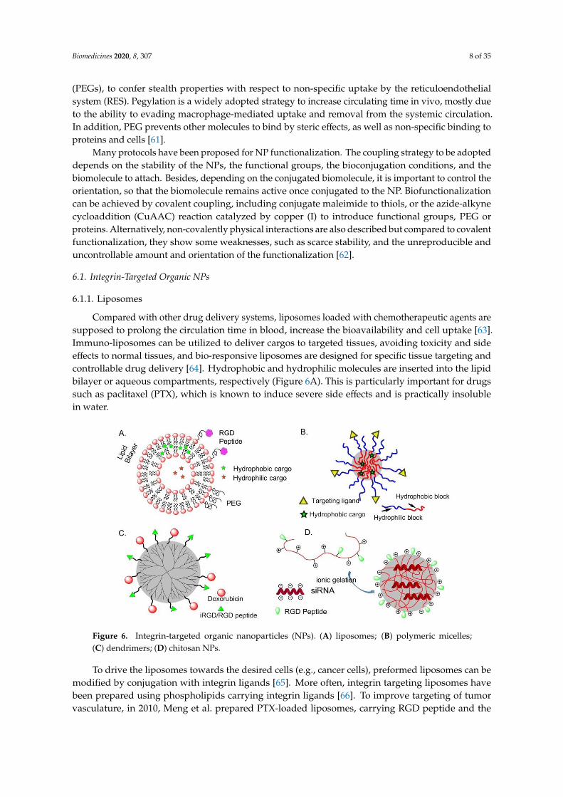
Compared with other drug delivery systems, liposomes loaded with chemotherapeutic agents aresupposed to prolong the circulation time in blood, increase the bioavailability and cell uptake [63].Immuno-liposomes can be utilized to deliver cargos to targeted tissues, avoiding toxicity and sideeffects to normal tissues, and bio-responsive liposomes are designed for specific tissue targeting andcontrollable drug delivery [64]. Hydrophobic and hydrophilic molecules are inserted into the lipidbilayer or aqueous compartments, respectively (Figure 6A). This is particularly important for drugssuch as paclitaxel (PTX), which is known to induce severe side effects and is practically insolublein water.
Biomedicines 2020, 8, x FOR PEER REVIEW 8 of 34
Many protocols have been proposed for NP functionalization. The coupling strategy to be
adopted depends on the stability of the NPs, the functional groups, the bioconjugation conditions,
and the biomolecule to attach. Besides, depending on the conjugated biomolecule, it is important to
control the orientation, so that the biomolecule remains active once conjugated to the NP.
Biofunctionalization can be achieved by covalent coupling, including conjugate maleimide to thiols,
or the azide-alkyne cycloaddition (CuAAC) reaction catalyzed by copper (I) to introduce functional
groups, PEG or proteins. Alternatively, non-covalently physical interactions are also described but
compared to covalent functionalization, they show some weaknesses, such as scarce stability, and the
unreproducible and uncontrollable amount and orientation of the functionalization [62].
6.1. Integrin-Targeted Organic NPs
6.1.1. Liposomes
Compared with other drug delivery systems, liposomes loaded with chemotherapeutic agents
are supposed to prolong the circulation time in blood, increase the bioavailability and cell uptake
[63]. Immuno-liposomes can be utilized to deliver cargos to targeted tissues, avoiding toxicity and
side effects to normal tissues, and bio-responsive liposomes are designed for specific tissue targeting
and controllable drug delivery [64]. Hydrophobic and hydrophilic molecules are inserted into the
lipid bilayer or aqueous compartments, respectively (Figure 6A). This is particularly important for
drugs such as paclitaxel (PTX), which is known to induce severe side effects and is practically
insoluble in water.
To drive the liposomes towards the desired cells (e.g., cancer cells), preformed liposomes can be
modified by conjugation with integrin ligands [65]. More often, integrin targeting liposomes have
been prepared using phospholipids carrying integrin ligands [66]. To improve targeting of tumor
vasculature, in 2010, Meng et al. prepared PTX-loaded liposomes, carrying RGD peptide and the
ATWLPPR sequence, which is the ligand of the VEGF receptor neuropilin-1 (NRP-1). The bimodal
liposomes enabled a greater internalization of PTX than either each of two single-targeted liposomes [67].
Figure 6. Integrin-targeted organic nanoparticles (NPs). (A) liposomes; (B) polymeric micelles; (C)
dendrimers; (D) chitosan NPs.
6.1.2. Polymeric Micelles (PM)
Amphiphilic block or graft copolymers tend to self-assemble in aqueous media giving globular
colloidal polymeric micelles (PM). Their core-shell architecture consents to load lipophilic antitumor
agents into the hydrophobic core and the outer hydrophilic shell allows NPs to be stable in aqueous
solution (Figure 6B). Generally, the copolymers are composed by hydrophobic materials that easily
Figure 6. Integrin-targeted organic nanoparticles (NPs). (A) liposomes; (B) polymeric micelles;(C) dendrimers; (D) chitosan NPs.
To drive the liposomes towards the desired cells (e.g., cancer cells), preformed liposomes can bemodified by conjugation with integrin ligands [65]. More often, integrin targeting liposomes havebeen prepared using phospholipids carrying integrin ligands [66]. To improve targeting of tumorvasculature, in 2010, Meng et al. prepared PTX-loaded liposomes, carrying RGD peptide and the
Biomedicines 2020, 8, 307 9 of 35
ATWLPPR sequence, which is the ligand of the VEGF receptor neuropilin-1 (NRP-1). The bimodalliposomes enabled a greater internalization of PTX than either each of two single-targeted liposomes [67].
6.1.2. Polymeric Micelles (PM)
Amphiphilic block or graft copolymers tend to self-assemble in aqueous media giving globularcolloidal polymeric micelles (PM). Their core-shell architecture consents to load lipophilic antitumoragents into the hydrophobic core and the outer hydrophilic shell allows NPs to be stable in aqueoussolution (Figure 6B). Generally, the copolymers are composed by hydrophobic materials that easilyundergo hydrolytic or enzymatic degradation, such as PCL (poly (ε-caprolactone)), PLA (poly (lacticacid)), PLGA (poly(lactic-co-glycolic acid)), or temperature or pH variations, whereas PEG is commonlyutilized for the hydrophilic segment [68].
In 2004, Gao and co-workers first prepared micelles composed of a PEG-PCL block copolymerending by a maleimide, to which a thiolated-cRGDfK was conjugated. These micelles were ableto deliver into SLK tumor cells [69]. The same authors subsequently prepared RGD-functionalizedPEG-PLA micelles loaded with Dox and superparamagnetic iron oxide NPs (SPIONs), for combiningtherapy and imaging (ultrasensitive MRI) [70]. In these examples, the payload is released upon structuredegradation. In some cases, the drug is covalently conjugated to the block copolymer by a hydrazonebond or an amide bond [71].
RGD-functionalized biocompatible synthetic polymers such as PEG, PLA, PLGA, PLL (poly (lacticlysine)), or natural polymers, chitosan, albumin, collagen, have been utilized to prepare integrintargeting biodegradable polymeric NPs. Drugs such as Paclitaxel, Mitoxantrone, and Fluorouracilcan be incorporated in micelles without chemical modification or with conjugation to the polymers.Then, it can be released in a controlled manner by diffusion through the polymer matrix, by polymerdegradation [72].
6.1.3. Dendrimers
In 2009, Waite et al. found that conjugating cyclic RGD to a poly(amidoamine) (PAMAM) dendrimerenhanced the penetration and delivery of short-interfering RNA (siRNA) through tumors in a mannerthat depended on the targeting ligand density [73].
In 2014, an effective anti-tumor drug delivery system PPCD (PEG-PAMAM-cis-aconityl-DOX)was prepared by covalently bonding or simply mixing the tumor penetrating peptide iRGD(internalizing-RGD, CRGDK/RGPD/EC) and the PAMAM dendrimer. Experiments have shownthat it can increase the permeability of the tumor blood vessel and the accumulation of drugs in thetumor tissue [74] (Figure 6C).
6.1.4. Chitosan
A positively charged chitosan NP is a talented siRNA delivery vehicle because of its advantage incellular membranes transportation and endocytosis (Figure 6D). In 2010, Sood et al. conjugate RGD peptideto chitosan NP by thiolation reaction for targeted siRNA delivery. This strategy significantly enhancedsiRNA delivery to tumor tissues and vasculature, binding efficiency on ανβ3 integrin-expressingtumor cells, and therapeutic efficacy of gene silencing [75].
6.1.5. Other Organic NPs
Doxorubicin-loaded human serum albumin NPs conjugated with an antibody targetingανβ3-positive M21 melanoma cells exhibited enhanced cytotoxicity as compared to free doxorubicin [76].In 2013, splice-switching oligonucleotides (SSOs) was utilized to bond RGD peptide to serum albuminto prepare a nanoconjugate with a diameter of 13 nm. Because of the small size of nanoparticle andgrafting of the tumor-targeting peptide, the ability to penetrate receptor-specific cells was 61 timeshigher than the control. Compared with other siRNA delivery vehicles, this nanoconjugate exhibits
Biomedicines 2020, 8, 307 10 of 35
the advantages of high loading rate, increment tumor specificity, strong tumor permeability, and hightherapeutic oligonucleotide activity [77].
Poly (cystaminebisacrylamide-diaminohexane) [poly (CBA-DAH)] (CD) is a biodegradable andlow-toxic polymer, that was bond to a tumor homing peptide c[RGDfC] by biofunctionalized PEG,c[RGDfC]-PEG-CD shielded on adenovirus (Ad) for reducing cytotoxicity and improving transductionefficiency. This oncolytic Ad expressing short hairpin RNA (shRNA) against interleukin-8 (IL-8) mRNA,after Ad/CD-PEG500-RGD was introduced in HT1080 cells. The expression of IL-8 and VEGF wasinhibited and then induce apoptosis [78].
6.2. Integrin-Targeted Inorganic NPs and QDs
Inorganic NPs of gold, silver semiconductors, magnetic compounds, alloys, silica, etc., displayunusual size-dependent optical and/or magnetic properties, drastically different from those of their bulkmaterials, exploitable for detection and imaging, and the targeting of multifunctional therapeutics [79].
6.2.1. Silica NPs
Silica NPs as tools to develop targeting probes and drug delivery systems have several advantagesover other nanomaterial and self-organized systems. Indeed, silica is photophysically inert, is anintrinsically non-toxic material, and there are many synthetic approaches available to tune thesenanosystems in terms of size and functionalization. The luminescence emission of these systemsdepends on the doping dye so that a large variety of emission properties can be achieved by justchoosing the right doping dye(s). The inclusion of dye molecules in rigid matrix-like silica oftenincreases the quantum yield of the dyes and also their photostability, because of the rigidification ofdye structure and the protection towards quenching molecules present in the environment. These lasttwo features are of prominent importance to univocally assign the recorded fluorescent signal to thepresence of the NPs and to control the local concentration of the cytotoxic compound during therecognition event toward the targeted receptor.
In 2018, Jia et al. generated bifunctional 40 nm-sized silica NPs coated with controlled amounts of thepeptides cRGD and ATWLPPR (neuropilin-1 (NRP1), a co-receptor of VEGFR2) and studied their affinity,selectivity and biological activity in HUVECs (Human Umbilical Vein Endothelial Cells). The resultssupported evidence for a complex cross-talk generated by the binding of the heteromultivalent NPs withανβ3-integrin and with NRP1. In particular, the NPs exerted dose-dependent pro-survival activity.This study demonstrated the difficulties in designing targeted silica-based NPs for antiangiogenictherapies and the possible risks posed by undesirable side effects [80].
In 2020, Juthani et al. introduced ultrasmall fluorescent core-shell silica NPs, Cornell prime dots(C’ dots), were functionalized with cRGD peptide, and PET (Positron Emission Tomography) labels(124I, 89Zr) to investigate the utility of dual-modality cRGD-C’ dots for enhancing accumulation,distribution, and retention (ADR) in a genetically engineered mouse model of glioblastoma (mGBM).The results showed improvements in brain tumor delivery and penetration, as well as enhancement inthe ADR, were observed following administration of integrin-targeted C’ dots, as compared with anontargeted control [81].
Very recently, peptide-functionalized silica NPs mimicking the proapoptotic protein Smac/DIABLOwere prepared and validated in vitro. These NPs were constituted by a fluorescent silica core doped withrhodamine B (RhB), coated with a PEG shell, and carrying the AVPI peptide and/or a tumor-homingcRGD peptide. The preparation started from micellar NPs composed of the tri-block surfactantcopolymer Pluronic® F127 (PF127) (polyethylene glycol-polypropylene oxide -polyethylene glycol,PEG100–PPO65–PEG100) followed by condensation of the silica core (Figure 7).
For the purpose of peptide conjugation, the NPs were prepared from a mixture of PF127 and itsdiazide derivative PF127-(N3)2, and submitted to click chemistry with AVPI-alkyne, containing thepro-apoptotic AVPI N-terminus of Smac/DIABLO (21), or the integrin-targeting cRGD-alkyne (22), or a1:1 mixture of both. At low µM concentration, the bifunctional AVPI/RGD-NPs showed superior toxicity

Biomedicines 2020, 8, 307 11 of 35
towards A549, U373, and HeLa cancer cells and modest toxicity towards other integrin-expressingcells, correlated with integrin-mediated cell uptake and consequent highly increased levels of apoptoticactivity, without perturbing cells not expressing the α5 integrin subunit [82].Biomedicines 2020, 8, x FOR PEER REVIEW 11 of 34
Figure 7. Preparation of AVPI/cRGD-NPs from micelles composed of PF127 (PEG100–PPO65–PEG100)
and PF127-(N3)2, followed by polymerization of the silica core to embed the dye RhB. Peptide
functionalization is achieved by click chemistry.
6.2.2. Magnetic NPs
In 2009, the Ruoslahti group reported a cyclic peptide combining the tumor-homing RGD
sequence with a tissue penetration motif. The homing sequence directs the peptide to the tumor
vascular endothelium, while the tissue penetration motif, once activated by a protease, binds to a
different receptor (neuropilin-1), which mediates extravasation and tissue penetration. As a proof of
concept, iRGD peptide-linked iron oxide nanoworms could be detected by MRI throughout a tumor
once injected in vivo to mice [83]. In 2011, the same group combined two different peptides with the
magnetic nanoworms to image and treat mice with glioblastoma, one of the most difficult tumors to
treat. While the CGKRG peptide targets the NPs to tumor vascular cells and into their mitochondria,
the other peptide acts as a pro-apoptotic drug. By co-injecting these NPs with iRGD, most of the
tumors were eradicated or their development delayed in two glioblastoma mouse models [84].
PEGylated copolymer-coated iron oxide NPs conjugated with an RGD-containing cyclic peptide
c(RGDyK) was administrated in a mouse model for targeting αvβ3 integrins. Successful tumor
homing was perceived in a subcutaneous U87MG glioblastoma xenograft model by magnetic
resonance imaging (MRI) [85] (Figure 8A). Similar integrin specific binding was achieved on HUVEC
cells by using paramagnetic liposomes conjugated with the cyclic RGD peptide [86].
Figure 8. Integrin-targeted inorganic NPs and quantum dots (QDs): (A) magnetic NPs; (B) gold NPs;
(C) QDs.
Figure 7. Preparation of AVPI/cRGD-NPs from micelles composed of PF127 (PEG100–PPO65–PEG100)and PF127-(N3)2, followed by polymerization of the silica core to embed the dye RhB. Peptidefunctionalization is achieved by click chemistry.
6.2.2. Magnetic NPs
In 2009, the Ruoslahti group reported a cyclic peptide combining the tumor-homing RGDsequence with a tissue penetration motif. The homing sequence directs the peptide to the tumorvascular endothelium, while the tissue penetration motif, once activated by a protease, binds to adifferent receptor (neuropilin-1), which mediates extravasation and tissue penetration. As a proof ofconcept, iRGD peptide-linked iron oxide nanoworms could be detected by MRI throughout a tumoronce injected in vivo to mice [83]. In 2011, the same group combined two different peptides with themagnetic nanoworms to image and treat mice with glioblastoma, one of the most difficult tumors totreat. While the CGKRG peptide targets the NPs to tumor vascular cells and into their mitochondria,the other peptide acts as a pro-apoptotic drug. By co-injecting these NPs with iRGD, most of the tumorswere eradicated or their development delayed in two glioblastoma mouse models [84].
PEGylated copolymer-coated iron oxide NPs conjugated with an RGD-containing cyclic peptidec(RGDyK) was administrated in a mouse model for targeting αvβ3 integrins. Successful tumor homingwas perceived in a subcutaneous U87MG glioblastoma xenograft model by magnetic resonanceimaging (MRI) [85] (Figure 8A). Similar integrin specific binding was achieved on HUVEC cells byusing paramagnetic liposomes conjugated with the cyclic RGD peptide [86].
Biomedicines 2020, 8, x FOR PEER REVIEW 11 of 34
Figure 7. Preparation of AVPI/cRGD-NPs from micelles composed of PF127 (PEG100–PPO65–PEG100)
and PF127-(N3)2, followed by polymerization of the silica core to embed the dye RhB. Peptide
functionalization is achieved by click chemistry.
6.2.2. Magnetic NPs
In 2009, the Ruoslahti group reported a cyclic peptide combining the tumor-homing RGD
sequence with a tissue penetration motif. The homing sequence directs the peptide to the tumor
vascular endothelium, while the tissue penetration motif, once activated by a protease, binds to a
different receptor (neuropilin-1), which mediates extravasation and tissue penetration. As a proof of
concept, iRGD peptide-linked iron oxide nanoworms could be detected by MRI throughout a tumor
once injected in vivo to mice [83]. In 2011, the same group combined two different peptides with the
magnetic nanoworms to image and treat mice with glioblastoma, one of the most difficult tumors to
treat. While the CGKRG peptide targets the NPs to tumor vascular cells and into their mitochondria,
the other peptide acts as a pro-apoptotic drug. By co-injecting these NPs with iRGD, most of the
tumors were eradicated or their development delayed in two glioblastoma mouse models [84].
PEGylated copolymer-coated iron oxide NPs conjugated with an RGD-containing cyclic peptide
c(RGDyK) was administrated in a mouse model for targeting αvβ3 integrins. Successful tumor
homing was perceived in a subcutaneous U87MG glioblastoma xenograft model by magnetic
resonance imaging (MRI) [85] (Figure 8A). Similar integrin specific binding was achieved on HUVEC
cells by using paramagnetic liposomes conjugated with the cyclic RGD peptide [86].
Figure 8. Integrin-targeted inorganic NPs and quantum dots (QDs): (A) magnetic NPs; (B) gold NPs;
(C) QDs.
Figure 8. Integrin-targeted inorganic NPs and quantum dots (QDs): (A) magnetic NPs; (B) gold NPs;(C) QDs.
Biomedicines 2020, 8, 307 12 of 35
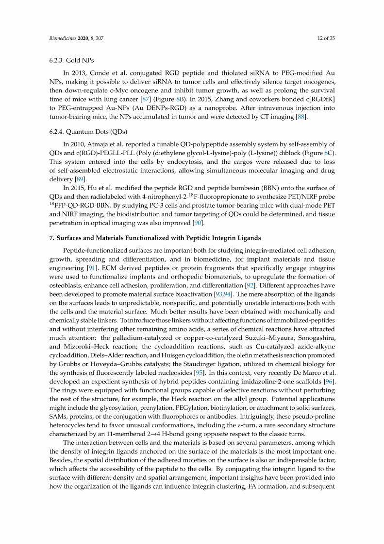
6.2.3. Gold NPs
In 2013, Conde et al. conjugated RGD peptide and thiolated siRNA to PEG-modified AuNPs, making it possible to deliver siRNA to tumor cells and effectively silence target oncogenes,then down-regulate c-Myc oncogene and inhibit tumor growth, as well as prolong the survivaltime of mice with lung cancer [87] (Figure 8B). In 2015, Zhang and coworkers bonded c[RGDfK]to PEG-entrapped Au-NPs (Au DENPs-RGD) as a nanoprobe. After intravenous injection intotumor-bearing mice, the NPs accumulated in tumor and were detected by CT imaging [88].
6.2.4. Quantum Dots (QDs)
In 2010, Atmaja et al. reported a tunable QD-polypeptide assembly system by self-assembly ofQDs and c(RGD)-PEGLL-PLL (Poly (diethylene glycol-L-lysine)-poly (L-lysine)) diblock (Figure 8C).This system entered into the cells by endocytosis, and the cargos were released due to lossof self-assembled electrostatic interactions, allowing simultaneous molecular imaging and drugdelivery [89].
In 2015, Hu et al. modified the peptide RGD and peptide bombesin (BBN) onto the surface ofQDs and then radiolabeled with 4-nitrophenyl-2-18F-fluoropropionate to synthesize PET/NIRF probe18FFP-QD-RGD-BBN. By studying PC-3 cells and prostate tumor-bearing mice with dual-mode PETand NIRF imaging, the biodistribution and tumor targeting of QDs could be determined, and tissuepenetration in optical imaging was also improved [90].
7. Surfaces and Materials Functionalized with Peptidic Integrin Ligands
Peptide-functionalized surfaces are important both for studying integrin-mediated cell adhesion,growth, spreading and differentiation, and in biomedicine, for implant materials and tissueengineering [91]. ECM derived peptides or protein fragments that specifically engage integrinswere used to functionalize implants and orthopedic biomaterials, to upregulate the formation ofosteoblasts, enhance cell adhesion, proliferation, and differentiation [92]. Different approaches havebeen developed to promote material surface bioactivation [93,94]. The mere absorption of the ligandson the surfaces leads to unpredictable, nonspecific, and potentially unstable interactions both withthe cells and the material surface. Much better results have been obtained with mechanically andchemically stable linkers. To introduce those linkers without affecting functions of immobilized-peptidesand without interfering other remaining amino acids, a series of chemical reactions have attractedmuch attention: the palladium-catalyzed or copper-co-catalyzed Suzuki–Miyaura, Sonogashira,and Mizoroki–Heck reaction; the cycloaddition reactions, such as Cu-catalyzed azide-alkynecycloaddition, Diels–Alder reaction, and Huisgen cycloaddition; the olefin metathesis reaction promotedby Grubbs or Hoveyda–Grubbs catalysts; the Staudinger ligation, utilized in chemical biology forthe synthesis of fluorescently labeled nucleosides [95]. In this context, very recently De Marco et al.developed an expedient synthesis of hybrid peptides containing imidazoline-2-one scaffolds [96].The rings were equipped with functional groups capable of selective reactions without perturbingthe rest of the structure, for example, the Heck reaction on the allyl group. Potential applicationsmight include the glycosylation, prenylation, PEGylation, biotinylation, or attachment to solid surfaces,SAMs, proteins, or the conjugation with fluorophores or antibodies. Intriguingly, these pseudo-prolineheterocycles tend to favor unusual conformations, including the ε-turn, a rare secondary structurecharacterized by an 11-membered 2→4 H-bond going opposite respect to the classic turns.
The interaction between cells and the materials is based on several parameters, among whichthe density of integrin ligands anchored on the surface of the materials is the most important one.Besides, the spatial distribution of the adhered moieties on the surface is also an indispensable factor,which affects the accessibility of the peptide to the cells. By conjugating the integrin ligand to thesurface with different density and spatial arrangement, important insights have been provided intohow the organization of the ligands can influence integrin clustering, FA formation, and subsequent
Biomedicines 2020, 8, 307 13 of 35
adhesion and spreading of cells. In particular, it has been discovered that there is a critical lateralspacing of approximately 60–70 nm between integrin ligands. When the spacing is out of range,integrin clustering and FA formation are hindered, thereby limiting cell attachment and diffusion [97].The limitations of spatial distance and peptide arrangement may be related to physiology becauseordered structures happen in the native ECM.
7.1. Self-Assembled Monolayers (SAMs)
SAMs of GRGDS peptides on gold were fabricated by using thiolated PEG linkers containing3 to 6 monomers. An increase in the length of the polyethylene glycol chains resulted in a decrease inSwiss 3T3 fibroblast cell adhesion and spreading, especially for lowest ligand density [98]. SAMs ofcyclic RGD (1 mol.%) were further used to investigate the dynamics of cell migration in the presenceof a linear RGD antagonist. In a definite concentration range, cell migration speed increased uponincreasing the concentration of the antagonist [99].
SAMs of RGD peptides were prepared on silicon surfaces with peptide spacing ranging fromnanometers to micrometers. The silicon materials were modified with undecenoic acid and mixtures of1-amino hexa(ethylene oxide) monomethyl ether and 1-amino hexa(ethylene oxide) in various ratios.The alcohol terminus of the hexa(ethylene oxide) was activated to a succinimide ester to consent thepentapeptide RGD to be coupled to the surface. Endothelial cells adhered to and spread on surfacesindependently of RGD spacing. However, the formation of FAs was particularly sensitive to the ligandspacing and the optimal spacing for RGD was found to be 44 nm [100].
7.2. Interfaces for Studying of Cell Adhesion, Spreading, and Differentiation
To promote cell binding, two domain-peptide ligands were simply absorbed onto gold. One domainis an anchoring domain for AuΦ3 gold binding, and the other is an IKVAV or RGD motif present inECM proteins as a cell-binding domain. Compared with the sequence containing only the cell-bindingdomain, the sequence with the anchoring domain had higher adsorption strength, which can inducecell polarization and larger mature FA area. These correspond to the high forces exerted on the interfaceand they enhance cell interaction with the material [101].
In 2009, Garcia et al. prepared supported lipid monolayers (SLMs) directly on octadecyltrichlorosilane-coated substrates. These surfaces were utilized to study the adhesion of hematopoieticprogenitor cell lines to a peptide derived from FN [102]. In 2001, Sackmann and co-workersprepared artificial membrane giant vesicles which incorporated 1–10 mol.% of c[RGDfK]-lipopeptide.After seeding, endothelial cells remained adherent and spread on RGD-SLM, while cells remainedround on control SLMs [103].
Supported lipid bilayers (SLBs) are biological interfaces mimicking cell membrane, with easilytunable characteristics. The head group of the lipids can be functionalized with integrin-bindingpeptides. In 2017, in Jonkheijm’s group, biotinylated 1, 2-dioleoyl-sn-glycero-3-phosphocholine(biot-DOPC) and 1,2-dipalmitoyl-sn-glycero-3-phosphocholine (DPPC) were utilized to obtainligand-mobile SLBs, which was functionalized with linear biotinylated-RGD thanks to the intermediacyof neutravidin. The resulting RGD-mobile SLBs were employed to study short term cell adhesion andlonger-term cell differentiation of human mesenchymal stem cells (hMSCs), and the result showed thatcell adhesion and differentiation positively correlated to ligand density and mobility [104].
7.3. Application in Regenerative Medicine and Tissue Engineering
In 2013, Rechenmacher et al. utilized click chemistry to immobilize peptidomimetics of α5β1-or ανβ3-selective RGD peptides on Ti-based materials via phosphonic acid-containing anchoringunits, and the resulting surfaces promoted the selective binding of α5β1- or αvβ3-expressingfibroblasts [105]. In 2015, Fraioli et al. reported that these Ti-peptide surfaces also allowed theadhesion, proliferation, and differentiation of OB-like cells, hence representing prototypes for implantmaterials with osteoinductive properties [106].
Biomedicines 2020, 8, 307 14 of 35
Track-etched (TE) microporous membranes of polyethylene terephthalate were grafted withGRGDS peptide or peptidomimetic ligands of ανβ3 integrin, via trifluorotriazine activation.These devices were described by Rémy and his coworkers in 2013 and showed improved adhesionof human endothelial cells under shear stress mimicking arterial conditions. The optimal numberof peptide molecules grafted on the surface was about 50 pmol/cm2 [107], whereas cells were notobserved on the surface of non-grafted PET.
Aiming at improving the adhesion of bone-forming osteoblasts at the surface of implants forregenerative medicine, in 1999, Kessler et al. proposed a method for the coating of the implantsusing integrin-specific ligands. Osteoblasts were found to be effectively bound to the materialpoly(methyl methacrylate) (PMMA), which anchored the cRGD peptide with N-succinylcysteamide ora 3-sulfanylpropionic acid linkers [108].
The functionalization of collagen scaffolds with RGD ligands that support cellular attachmenthas been extensively studied by Schussler and his coworkers in 2009, for cardiac tissue engineering inthe treatment of diseased myocardium or cardiac malformations [109]. In 2012, in the Kilian group,SAMs of cRGD ligands were obtained by conjugating the c[RGDfC]/GRGDSC peptides to Au-SAMsusing thiolated tri-(ethylene glycol) linkers. The differentiation of MSCs was affected by the affinityand density of an immobilized ligand for the integrin receptors. As a result, MSCs on monolayers ofc[RGDfC]-SAM-Au showed increased expression of osteogenic markers, while cells on monolayers ofGRGDSC-SAM-Au expressed early markers of myogenesis at a high density and neurogenesis at a lowdensity of the ligand [110].
7.4. Fabrication Methods of Integrin Ligand Immobilized Nanostructured Surfaces
The random presence of cell adhesive ligands on a material surface alone is not sufficient toelicit a full cell adhesion response, being the nanoscale distribution of these ligands on the surfacealso critically important. Indeed, to promote the formation of FAs, the integrin receptors must beclustered within the cell membrane. Integrin clustering can be favored by culturing cells on surfacesfunctionalized with multivalent ligands. A variety of fabrication methods have been developed tocontrol the nanoscale presentation of integrin-binding ligands on biomaterial substrates, includingblending, electron beam patterns, photolithography, and nanolithography, electrospinning, 3D printing,etc. [111].
7.4.1. Blending Strategy for Surfaces Functionalization
In 2008, Becker and Simon fabricated fibroin and synthetic RGD-containing spidroin(RGD-spidroin) on glass coverslips in different proportions of RGD-spidroin from 0% to 70%. The higherratio of RGD-spidroin is related to the high content of the β-sheet, which has a positive correlationwith film stability and cell adhesion, but an insignificantly negative correlation with differentiation.It was also found that the optimal proportion of RGD-spidroin was 10% for film stability, osteoblastadhesion, and differentiation [112].
In 2014, Yang et al. prepared bio-fibers by blending RGD-containing peptides functionalizedmussel adhesion protein (MAP-RGD) into silk fibroin (SF). It was determined that MAP-RGD-SF notonly improved the attachment, proliferation, and spread of mammalian cells but also promoted theadhesion of carbohydrates and proteins. Compared with SF, MAP-RGD-SF had high hydrophilicity,biodegradability, and wettability, which made it have greater potential in the application of tissueengineering and regeneration medicine [113]. In 2018, Janani et al. prepared a 3D porous silk scaffoldby blending mulberry silk fibroin protein and RGD-containing non-mulberry silk fibroin protein todevelop bioartificial liver constructs. It was found that the blend scaffold increased the density ofhepatocyte clusters and retained liver-specific functions for 3 weeks [114].
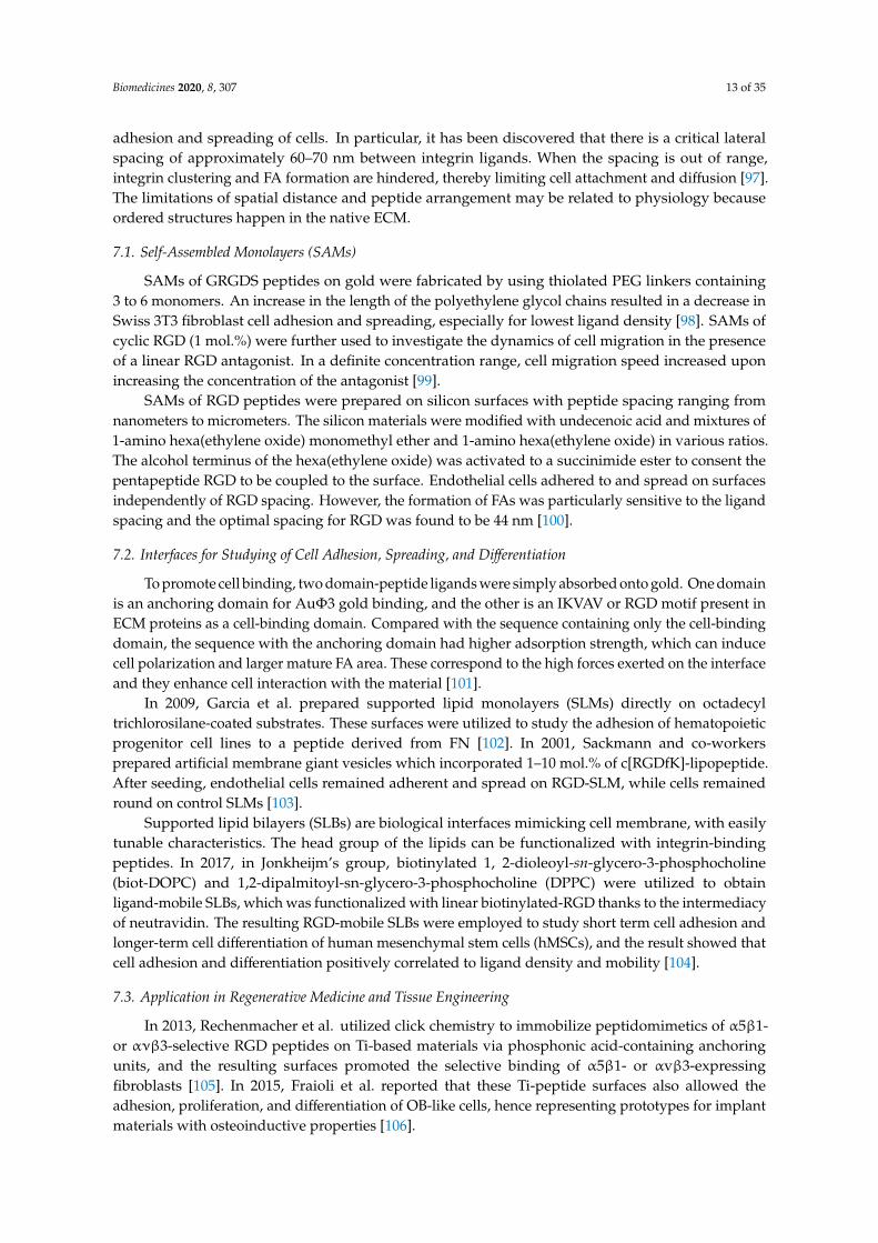
In 2000, Maheshwari et al. reported a Star-shaped polymer containing many PEO arms.The YGRGD attached star-shaped PEO was blended with the unfunctionalized star-shaped polymercovalently tethered on the PEG hydrogel-modified coverslip to independently control the peptide
Biomedicines 2020, 8, 307 15 of 35
density and spatial distribution of the surface. It was found that NR6 fibroblast cells could only migrateon the YGRGD peptide adhered surfaces (Figure 9A) [115].
A cRGD-conjugated micellar system was prepared by blending pluronics L121 and F127 to increasedocetaxel-loading capacity and particle stability (>1 week). It was also found that the enhancementof cellular uptake improved anticancer activity against U87MG cancer cells, and tumor-targetingaccumulation of blending micellar systems in vivo (Figure 9B) [116].
Biomedicines 2020, 8, x FOR PEER REVIEW 15 of 34
enhancement of cellular uptake improved anticancer activity against U87MG cancer cells, and tumor-
targeting accumulation of blending micellar systems in vivo (Figure 9B) [116].
Figure 9. Blending strategy for surface functionalization. (A) Star PEO was blended on the surface.
The surface density of star PEO and special distribution of RGD peptides can be controlled. (B)
Preparation of cRGD-conjugated tumor-targeting drug delivery platform by blending of L121 and
F127 and encapsulating docetaxel.
7.4.2. Electron Beam Fabricated Patterns
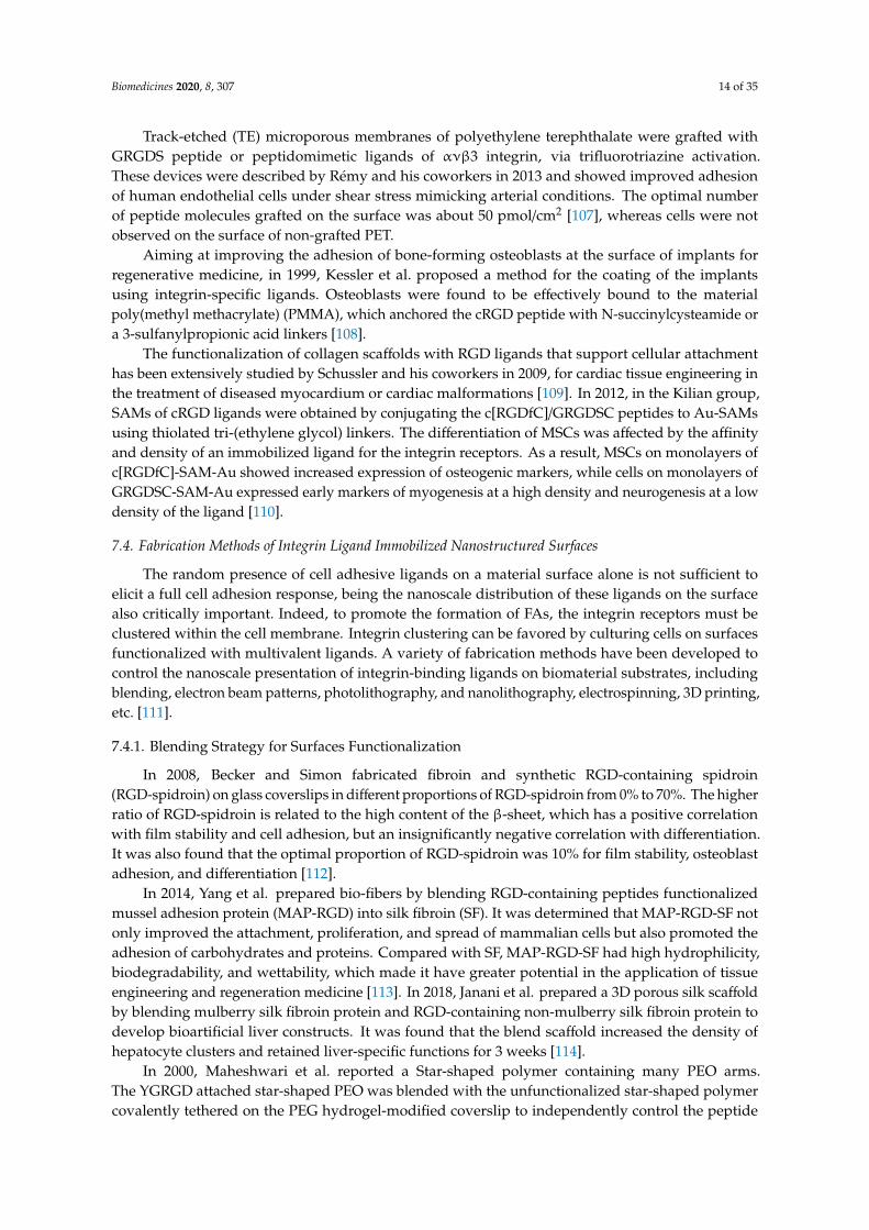
In 2007, Rundqvist et al. proposed an electron beam as a high-fidelity approach for surface
coating with proteins [117]. A silicon substrate was coated with protein fibronectin, which was then
inactivated by an electron beam. The area exposed to the electron beam lost the ability of Ab binding
and cells spreading. The inactivation was dependent on the dose of electron and accelerating voltage.
In contrast to the ablation observed in other organic thin films, this patterning approach allowed local
inactivation, and the level of protein patterning could be controlled to a single molecule.
Using electron-beam lithography, in 2012, Maynard [118] functionalized substrates with both
Lev-GRGDSPG peptide and basic fibroblast growth factor (bFGF). First, 8-arm PEG-OH was coated
on a silicon wafer and passivated by thermal annealing, then a 1:1 blend of PEG-AO (8-arm
aminooxy-terminated PEG) and pSS-co-PEGMA (Poly(styrene-4-sulfonate-co-poly(ethylene glycol)
methacrylate)) was patterned by electron beam lithography. Finally, GRGDSPG and bFGF were
immobilized on the surface. The presence of RGD promoted cell adhesion and the formation of FAs.
Compared with the surface containing only RGD, the surface containing both RGD and bFGF
increased the cell area of HUVEC (Figure 10).
Figure 10. Electron beam lithography. Before immobilizing Lev-GRGDSPG and bFGF on the surface,
pSS-co-PEGMA and PEG-AO were coated and cross-linked on passivated silicon wafers. Then
HUVEC cells were cultured on the surface.
7.4.3. Photolithography Strategy for Surfaces Functionalization
Using photolithography technology, polydimethylsiloxane (PDMS) microgrooves were
prepared in 2017 by Kim and his coworkers and then immobilized by filamentous phages, which
were genetically functionalized with RGD peptide. The presence of RGD-phages in PDMS-
microgrooves enhanced adhesion, proliferation, and orientation of H9c2 cardiomyocyte, which has
application prospects in stent coating materials [119].
In 2017, in the Bilem group, RGD peptide and bone morphogenetic protein (BMP) mimetic
peptide were immobilized on glass surfaces by geometrically controlled photolithography
patterning. hMSCs osteogenic differentiation-related to the spatial distribution of two different
Figure 9. Blending strategy for surface functionalization. (A) Star PEO was blended on the surface.The surface density of star PEO and special distribution of RGD peptides can be controlled.(B) Preparation of cRGD-conjugated tumor-targeting drug delivery platform by blending of L121 andF127 and encapsulating docetaxel.
7.4.2. Electron Beam Fabricated Patterns
In 2007, Rundqvist et al. proposed an electron beam as a high-fidelity approach for surface coatingwith proteins [117]. A silicon substrate was coated with protein fibronectin, which was then inactivatedby an electron beam. The area exposed to the electron beam lost the ability of Ab binding and cellsspreading. The inactivation was dependent on the dose of electron and accelerating voltage. In contrastto the ablation observed in other organic thin films, this patterning approach allowed local inactivation,and the level of protein patterning could be controlled to a single molecule.
Using electron-beam lithography, in 2012, Maynard [118] functionalized substrates with bothLev-GRGDSPG peptide and basic fibroblast growth factor (bFGF). First, 8-arm PEG-OH was coatedon a silicon wafer and passivated by thermal annealing, then a 1:1 blend of PEG-AO (8-armaminooxy-terminated PEG) and pSS-co-PEGMA (Poly(styrene-4-sulfonate-co-poly(ethylene glycol)methacrylate)) was patterned by electron beam lithography. Finally, GRGDSPG and bFGF wereimmobilized on the surface. The presence of RGD promoted cell adhesion and the formation of FAs.Compared with the surface containing only RGD, the surface containing both RGD and bFGF increasedthe cell area of HUVEC (Figure 10).
Biomedicines 2020, 8, x FOR PEER REVIEW 15 of 34
enhancement of cellular uptake improved anticancer activity against U87MG cancer cells, and tumor-
targeting accumulation of blending micellar systems in vivo (Figure 9B) [116].
Figure 9. Blending strategy for surface functionalization. (A) Star PEO was blended on the surface.
The surface density of star PEO and special distribution of RGD peptides can be controlled. (B)
Preparation of cRGD-conjugated tumor-targeting drug delivery platform by blending of L121 and
F127 and encapsulating docetaxel.
7.4.2. Electron Beam Fabricated Patterns
In 2007, Rundqvist et al. proposed an electron beam as a high-fidelity approach for surface
coating with proteins [117]. A silicon substrate was coated with protein fibronectin, which was then
inactivated by an electron beam. The area exposed to the electron beam lost the ability of Ab binding
and cells spreading. The inactivation was dependent on the dose of electron and accelerating voltage.
In contrast to the ablation observed in other organic thin films, this patterning approach allowed local
inactivation, and the level of protein patterning could be controlled to a single molecule.
Using electron-beam lithography, in 2012, Maynard [118] functionalized substrates with both
Lev-GRGDSPG peptide and basic fibroblast growth factor (bFGF). First, 8-arm PEG-OH was coated
on a silicon wafer and passivated by thermal annealing, then a 1:1 blend of PEG-AO (8-arm
aminooxy-terminated PEG) and pSS-co-PEGMA (Poly(styrene-4-sulfonate-co-poly(ethylene glycol)
methacrylate)) was patterned by electron beam lithography. Finally, GRGDSPG and bFGF were
immobilized on the surface. The presence of RGD promoted cell adhesion and the formation of FAs.
Compared with the surface containing only RGD, the surface containing both RGD and bFGF
increased the cell area of HUVEC (Figure 10).
Figure 10. Electron beam lithography. Before immobilizing Lev-GRGDSPG and bFGF on the surface,
pSS-co-PEGMA and PEG-AO were coated and cross-linked on passivated silicon wafers. Then
HUVEC cells were cultured on the surface.
7.4.3. Photolithography Strategy for Surfaces Functionalization
Using photolithography technology, polydimethylsiloxane (PDMS) microgrooves were
prepared in 2017 by Kim and his coworkers and then immobilized by filamentous phages, which
were genetically functionalized with RGD peptide. The presence of RGD-phages in PDMS-
microgrooves enhanced adhesion, proliferation, and orientation of H9c2 cardiomyocyte, which has
application prospects in stent coating materials [119].
In 2017, in the Bilem group, RGD peptide and bone morphogenetic protein (BMP) mimetic
peptide were immobilized on glass surfaces by geometrically controlled photolithography
patterning. hMSCs osteogenic differentiation-related to the spatial distribution of two different
Figure 10. Electron beam lithography. Before immobilizing Lev-GRGDSPG and bFGF on the surface,pSS-co-PEGMA and PEG-AO were coated and cross-linked on passivated silicon wafers. Then HUVECcells were cultured on the surface.
7.4.3. Photolithography Strategy for Surfaces Functionalization
Using photolithography technology, polydimethylsiloxane (PDMS) microgrooves were preparedin 2017 by Kim and his coworkers and then immobilized by filamentous phages, which were geneticallyfunctionalized with RGD peptide. The presence of RGD-phages in PDMS-microgrooves enhanced
Biomedicines 2020, 8, 307 16 of 35
adhesion, proliferation, and orientation of H9c2 cardiomyocyte, which has application prospects instent coating materials [119].
In 2017, in the Bilem group, RGD peptide and bone morphogenetic protein (BMP) mimeticpeptide were immobilized on glass surfaces by geometrically controlled photolithography patterning.hMSCs osteogenic differentiation-related to the spatial distribution of two different peptides and shapeof peptide micropatterns. Compared with the BMP-2 functionalized surface only, the RGD/BMP-2modified surface could significantly promote hMSC osteogenesis [120]. In 2018, the same groupdescribed that spatial patterning surfaces of RGD-TAMRA did not change the stemness character ofhMSCs, but the presentation of BMP-2-FITC peptide on material surfaces promoted the differentiationof hMSCs into osteoblasts. Triangular and square peptide patterns with an aspect ratio of 1 and0.7 significantly enhanced hMSCs osteogenesis [121].
In 2011, Using photolithography, RGD-immobilized nanoporous alumina membrane wasimmobilized with PEG hydrogel micropatterns to construct microwells with different sizes in Koh’slab. Fibroblasts adhered and proliferated in RGD-containing microwells instead of PEG hydrogel wallsto form a cellular micropattern. The formation of filopodia and its penetration into nanopores was alsoobserved (Figure 11) [122].
Biomedicines 2020, 8, x FOR PEER REVIEW 16 of 34
peptides and shape of peptide micropatterns. Compared with the BMP-2 functionalized surface only,
the RGD/BMP-2 modified surface could significantly promote hMSC osteogenesis [120]. In 2018, the
same group described that spatial patterning surfaces of RGD-TAMRA did not change the stemness
character of hMSCs, but the presentation of BMP-2-FITC peptide on material surfaces promoted the
differentiation of hMSCs into osteoblasts. Triangular and square peptide patterns with an aspect ratio
of 1 and 0.7 significantly enhanced hMSCs osteogenesis [121].
In 2011, Using photolithography, RGD-immobilized nanoporous alumina membrane was
immobilized with PEG hydrogel micropatterns to construct microwells with different sizes in Koh’s
lab. Fibroblasts adhered and proliferated in RGD-containing microwells instead of PEG hydrogel
walls to form a cellular micropattern. The formation of filopodia and its penetration into nanopores
was also observed (Figure 11) [122].
Figure 11. Photolithography strategy for surface functionalization. The photoinitiator-containing gel
precursor solution was added on RGD peptides immobilized nanoporous alumina membrane to form
a thin layer. Then it was covered by photomask and irradiated by UV light to get polymerization.
Fibroblasts cells were seeded in the nanowells.
7.4.4. Nanolithography Strategy for Surfaces Functionalization
A substrate-patterning strategy was based on the self-assembly of diblock copolymer micelles.
After the assembly of the Au-dot-containing micelles, the polymer was entirely removed by a gas
plasma treatment, which resulted in extended and highly regular Au nanodots, deposited into a
nearly perfect hexagonal pattern on substrates such as glass. c[RGDfX] peptides were bound to gold
NPs structured surfaces via polyproline, polyethylene glycol, or aminohexanoic acid-containing
spacers of different lengths. Changes of the ligand’s spacer chemistry and length reveal significant
differences in cell adhesion activation and FA formation. An increase in spacer length resulted in an
increase in FA density in fibroblasts as well as an increase in the average cell area. Longer and more
hydrophilic spacers induced more stress fiber formation. Short hydrophobic spacers induced the
slowest cellular spreading rate; and for all spacers, increasing their length led to an increase in the
rate of cell spreading. Polyproline-based peptides demonstrated improved cell adhesion kinetics and
FA formation compared with common aminohexanoic acid or polyethylene glycol spacers, correlated
to the much higher extension and rigidity of the former. Binding activity was additionally improved,
inducing a higher and faster cell spreading, by ligand dimerization, obtained by joining two cRGD
peptides with a lysine branch [123].
In 2014, in Cavalcanti–Adam’s lab, Gold NPs were deposited on β-type Ti-40Nb alloy discs by
block copolymer micelle nanolithography and were subsequently functionalized with thiolated
cRGD ligands in defined patterns. When seeded with human MSC, these nanostructured discs
showed reduced cell heterogeneity, while their adhesion, in terms of cell size and FA formation, was
optimized [124].
In 2013, gold nanopatterns on persistently non-fouling PEG hydrogels were prepared by Wang
et al. via block copolymer micelle nanolithography plus transfer nanolithography. Specifically,
micelles of amphiphilic block copolymer polystyrene-block-poly(2-vinyl pyridine) (PS-b-P2VP) were
reacted with HAuCl4 to give Au-enriched micellar cores. The process of preparation of a gold
nanopattern on glass exploited dip-coating and oxygen plasma, then the Au nanopatterns were
transferred from glass to a PEG hydrogel, which was formed via photopolymerization of PEG-
Figure 11. Photolithography strategy for surface functionalization. The photoinitiator-containing gelprecursor solution was added on RGD peptides immobilized nanoporous alumina membrane to forma thin layer. Then it was covered by photomask and irradiated by UV light to get polymerization.Fibroblasts cells were seeded in the nanowells.
7.4.4. Nanolithography Strategy for Surfaces Functionalization
A substrate-patterning strategy was based on the self-assembly of diblock copolymer micelles.After the assembly of the Au-dot-containing micelles, the polymer was entirely removed by a gasplasma treatment, which resulted in extended and highly regular Au nanodots, deposited into a nearlyperfect hexagonal pattern on substrates such as glass. c[RGDfX] peptides were bound to gold NPsstructured surfaces via polyproline, polyethylene glycol, or aminohexanoic acid-containing spacers ofdifferent lengths. Changes of the ligand’s spacer chemistry and length reveal significant differencesin cell adhesion activation and FA formation. An increase in spacer length resulted in an increase inFA density in fibroblasts as well as an increase in the average cell area. Longer and more hydrophilicspacers induced more stress fiber formation. Short hydrophobic spacers induced the slowest cellularspreading rate; and for all spacers, increasing their length led to an increase in the rate of cell spreading.Polyproline-based peptides demonstrated improved cell adhesion kinetics and FA formation comparedwith common aminohexanoic acid or polyethylene glycol spacers, correlated to the much higherextension and rigidity of the former. Binding activity was additionally improved, inducing a higherand faster cell spreading, by ligand dimerization, obtained by joining two cRGD peptides with a lysinebranch [123].
In 2014, in Cavalcanti–Adam’s lab, Gold NPs were deposited onβ-type Ti-40Nb alloy discs by blockcopolymer micelle nanolithography and were subsequently functionalized with thiolated cRGD ligandsin defined patterns. When seeded with human MSC, these nanostructured discs showed reduced cellheterogeneity, while their adhesion, in terms of cell size and FA formation, was optimized [124].
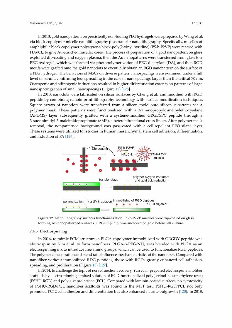
Biomedicines 2020, 8, 307 17 of 35
In 2013, gold nanopatterns on persistently non-fouling PEG hydrogels were prepared by Wang et al.via block copolymer micelle nanolithography plus transfer nanolithography. Specifically, micelles ofamphiphilic block copolymer polystyrene-block-poly(2-vinyl pyridine) (PS-b-P2VP) were reacted withHAuCl4 to give Au-enriched micellar cores. The process of preparation of a gold nanopattern on glassexploited dip-coating and oxygen plasma, then the Au nanopatterns were transferred from glass to aPEG hydrogel, which was formed via photopolymerization of PEG-diacrylate (DA), and then RGDmotifs were grafted onto the gold nanodots to eventually obtain an RGD nanopattern on the surface ofa PEG hydrogel. The behaviors of MSCs on diverse pattern nanospacings were examined under a fulllevel of serum, confirming less spreading in the case of nanospacings larger than the critical 70 nm.Osteogenic and adipogenic inductions resulted in higher differentiation extents on patterns of largenanospacings than of small nanospacings (Figure 12) [125].
In 2013, nanodots were fabricated on silicon surfaces by Cheng et al. and modified with RGDpeptide by combining nanoimprint lithography technology with surface modification techniques.Square arrays of nanodots were transferred from a silicon mold onto silicon substrates via apolymer mask. These patterns were functionalized with a 3-aminopropyldimethylethoxysilane(APDMS) layer subsequently grafted with a cysteine-modified GRGDSPC peptide through a3-succinimidyl-3-maleimidopropionate (SMP), a heterobifunctional cross-linker. After polymer maskremoval, the nonpatterned background was passivated with a cell-repellent PEO-silane layer.These systems were utilized for studies in human mesenchymal stem cell adhesion, differentiation,and induction of FA [126].
Biomedicines 2020, 8, x FOR PEER REVIEW 17 of 34
diacrylate (DA), and then RGD motifs were grafted onto the gold nanodots to eventually obtain an
RGD nanopattern on the surface of a PEG hydrogel. The behaviors of MSCs on diverse pattern
nanospacings were examined under a full level of serum, confirming less spreading in the case of
nanospacings larger than the critical 70 nm. Osteogenic and adipogenic inductions resulted in higher
differentiation extents on patterns of large nanospacings than of small nanospacings (Figure 12) [125].
In 2013, nanodots were fabricated on silicon surfaces by Cheng et al. and modified with RGD
peptide by combining nanoimprint lithography technology with surface modification techniques.
Square arrays of nanodots were transferred from a silicon mold onto silicon substrates via a polymer
mask. These patterns were functionalized with a 3-aminopropyldimethylethoxysilane (APDMS)
layer subsequently grafted with a cysteine-modified GRGDSPC peptide through a 3-succinimidyl-3-
maleimidopropionate (SMP), a heterobifunctional cross-linker. After polymer mask removal, the
nonpatterned background was passivated with a cell-repellent PEO-silane layer. These systems were
utilized for studies in human mesenchymal stem cell adhesion, differentiation, and induction of FA [126].
Figure 12. Nanolithography surfaces functionalization. PS-b-P2VP micelles were dip-coated on glass,
forming Au-nanopatterned surfaces. c[RGDfK]-thiol was anchored on gold before cell culture.
7.4.5. Electrospinning
In 2016, to mimic ECM structure, a PLGA copolymer immobilized with GRGDY peptide was
electrospun by Kim et al. to form nanofibers. PLGA-b-PEG-NH2 was blended with PLGA as an
electrospinning ink to introduce free amino groups, which can be used to functionalize RGD
peptides. The polymer concentration and blend ratio influence the characteristics of the nanofiber.
Compared with nanofiber without immobilized RDG peptides, those with RGDs greatly enhanced
cell adhesion, spreading, and proliferation (Figure 13) [127].
In 2014, to challenge the topic of nerve function recovery, Yun et al. prepared electrospun
nanofiber scaffolds by electrospinning a mixed solution of RGD-functionalized poly(serinol-
hexamethylene urea) (PSHU-RGD) and poly-ε-caprolactone (PCL). Compared with laminin-coated
surfaces, no cytotoxicity of PSHU-RGD/PCL nanofiber scaffolds was found in the MTT test. PSHU-
RGD/PCL not only promoted PC12 cell adhesion and differentiation but also enhanced neurite
outgrowth [128]. In 2018, Madhavan et al. demonstrated that RGD functionalized PSHU/PCL
biomaterial could be also used in vascular grafts [129].
In 2014, to overcome the shortcoming of difficult infiltration of cells into electrospun scaffolds,
Jeong et al. proposed that electrospun mats with high porosity, thickness, and small-fiber diameter
should be considered. To achieve this purpose, high humidity conditions and ultra-sonication were
introduced in the preparation process of RGD-modified alginate mats [130]. In 2016, Antonova et al.
reported that electrospinning of a blend of poly(3-hydroxybutyrate-co-3-hydroxy valerate) (PHBV)
and poly(caprolactone) (PCL) gave small-diameter biomaterial grafts for vascular tissue engineering.
In the rat’s study of grafts implantation, it was found that RGD or VEGF (vascular endothelial growth
Figure 12. Nanolithography surfaces functionalization. PS-b-P2VP micelles were dip-coated on glass,forming Au-nanopatterned surfaces. c[RGDfK]-thiol was anchored on gold before cell culture.
7.4.5. Electrospinning
In 2016, to mimic ECM structure, a PLGA copolymer immobilized with GRGDY peptide waselectrospun by Kim et al. to form nanofibers. PLGA-b-PEG-NH2 was blended with PLGA as anelectrospinning ink to introduce free amino groups, which can be used to functionalize RGD peptides.The polymer concentration and blend ratio influence the characteristics of the nanofiber. Compared withnanofiber without immobilized RDG peptides, those with RGDs greatly enhanced cell adhesion,spreading, and proliferation (Figure 13) [127].
In 2014, to challenge the topic of nerve function recovery, Yun et al. prepared electrospun nanofiberscaffolds by electrospinning a mixed solution of RGD-functionalized poly(serinol-hexamethylene urea)(PSHU-RGD) and poly-ε-caprolactone (PCL). Compared with laminin-coated surfaces, no cytotoxicityof PSHU-RGD/PCL nanofiber scaffolds was found in the MTT test. PSHU-RGD/PCL not onlypromoted PC12 cell adhesion and differentiation but also enhanced neurite outgrowth [128]. In 2018,
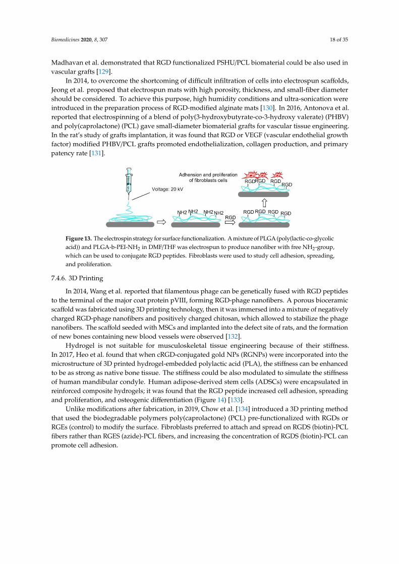
Biomedicines 2020, 8, 307 18 of 35
Madhavan et al. demonstrated that RGD functionalized PSHU/PCL biomaterial could be also used invascular grafts [129].
In 2014, to overcome the shortcoming of difficult infiltration of cells into electrospun scaffolds,Jeong et al. proposed that electrospun mats with high porosity, thickness, and small-fiber diametershould be considered. To achieve this purpose, high humidity conditions and ultra-sonication wereintroduced in the preparation process of RGD-modified alginate mats [130]. In 2016, Antonova et al.reported that electrospinning of a blend of poly(3-hydroxybutyrate-co-3-hydroxy valerate) (PHBV)and poly(caprolactone) (PCL) gave small-diameter biomaterial grafts for vascular tissue engineering.In the rat’s study of grafts implantation, it was found that RGD or VEGF (vascular endothelial growthfactor) modified PHBV/PCL grafts promoted endothelialization, collagen production, and primarypatency rate [131].
Biomedicines 2020, 8, x FOR PEER REVIEW 18 of 34
factor) modified PHBV/PCL grafts promoted endothelialization, collagen production, and primary
patency rate [131].
Figure 13. The electrospin strategy for surface functionalization. A mixture of PLGA (poly(lactic-co-
glycolic acid)) and PLGA-b-PEI-NH2 in DMF/THF was electrospun to produce nanofiber with free
NH2-group, which can be used to conjugate RGD peptides. Fibroblasts were used to study cell
adhesion, spreading, and proliferation.
7.4.6. 3D Printing
In 2014, Wang et al. reported that filamentous phage can be genetically fused with RGD
peptides to the terminal of the major coat protein pVIII, forming RGD-phage nanofibers. A porous
bioceramic scaffold was fabricated using 3D printing technology, then it was immersed into a mixture
of negatively charged RGD-phage nanofibers and positively charged chitosan, which allowed to
stabilize the phage nanofibers. The scaffold seeded with MSCs and implanted into the defect site of
rats, and the formation of new bones containing new blood vessels were observed [132].
Hydrogel is not suitable for musculoskeletal tissue engineering because of their stiffness. In 2017,
Heo et al. found that when cRGD-conjugated gold NPs (RGNPs) were incorporated into the
microstructure of 3D printed hydrogel-embedded polylactic acid (PLA), the stiffness can be enhanced
to be as strong as native bone tissue. The stiffness could be also modulated to simulate the stiffness
of human mandibular condyle. Human adipose-derived stem cells (ADSCs) were encapsulated in
reinforced composite hydrogels; it was found that the RGD peptide increased cell adhesion,
spreading and proliferation, and osteogenic differentiation (Figure 14) [133].
Unlike modifications after fabrication, in 2019, Chow et al. [134] introduced a 3D printing
method that used the biodegradable polymers poly(caprolactone) (PCL) pre-functionalized with
RGDs or RGEs (control) to modify the surface. Fibroblasts preferred to attach and spread on RGDS
(biotin)-PCL fibers rather than RGES (azide)-PCL fibers, and increasing the concentration of RGDS
(biotin)-PCL can promote cell adhesion.
Figure 14. 3D printing for surface functionalization. 3D PLA microstructure was prepared via 3D
printing technology; GelMA was obtained by methacrylation of gelatin; peptide-conjugated Gold NPs
(RGNPs) were prepared by reduction of chloroauric acid before conjugating RGD peptides.
Figure 13. The electrospin strategy for surface functionalization. A mixture of PLGA (poly(lactic-co-glycolicacid)) and PLGA-b-PEI-NH2 in DMF/THF was electrospun to produce nanofiber with free NH2-group,which can be used to conjugate RGD peptides. Fibroblasts were used to study cell adhesion, spreading,and proliferation.
7.4.6. 3D Printing
In 2014, Wang et al. reported that filamentous phage can be genetically fused with RGD peptidesto the terminal of the major coat protein pVIII, forming RGD-phage nanofibers. A porous bioceramicscaffold was fabricated using 3D printing technology, then it was immersed into a mixture of negativelycharged RGD-phage nanofibers and positively charged chitosan, which allowed to stabilize the phagenanofibers. The scaffold seeded with MSCs and implanted into the defect site of rats, and the formationof new bones containing new blood vessels were observed [132].
Hydrogel is not suitable for musculoskeletal tissue engineering because of their stiffness.In 2017, Heo et al. found that when cRGD-conjugated gold NPs (RGNPs) were incorporated into themicrostructure of 3D printed hydrogel-embedded polylactic acid (PLA), the stiffness can be enhancedto be as strong as native bone tissue. The stiffness could be also modulated to simulate the stiffnessof human mandibular condyle. Human adipose-derived stem cells (ADSCs) were encapsulated inreinforced composite hydrogels; it was found that the RGD peptide increased cell adhesion, spreadingand proliferation, and osteogenic differentiation (Figure 14) [133].
Unlike modifications after fabrication, in 2019, Chow et al. [134] introduced a 3D printing methodthat used the biodegradable polymers poly(caprolactone) (PCL) pre-functionalized with RGDs orRGEs (control) to modify the surface. Fibroblasts preferred to attach and spread on RGDS (biotin)-PCLfibers rather than RGES (azide)-PCL fibers, and increasing the concentration of RGDS (biotin)-PCL canpromote cell adhesion.
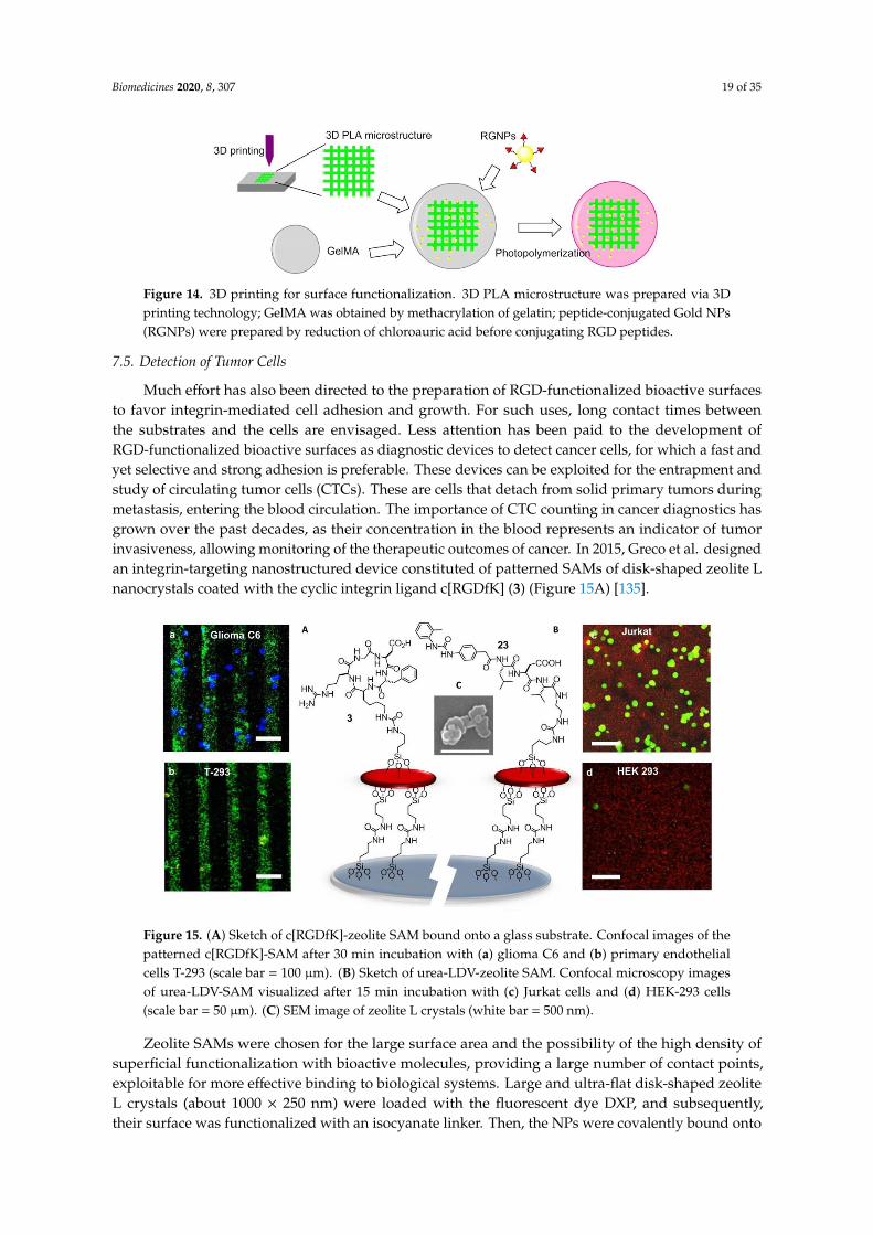
Biomedicines 2020, 8, 307 19 of 35
Biomedicines 2020, 8, x FOR PEER REVIEW 18 of 34
factor) modified PHBV/PCL grafts promoted endothelialization, collagen production, and primary patency rate [131].
Figure 13. The electrospin strategy for surface functionalization. A mixture of PLGA (poly(lactic-co-glycolic acid)) and PLGA-b-PEI-NH2 in DMF/THF was electrospun to produce nanofiber with free NH2-group, which can be used to conjugate RGD peptides. Fibroblasts were used to study cell adhesion, spreading, and proliferation.
7.4.6. 3D Printing
In 2014, Wang et al. reported that filamentous phage can be genetically fused with RGD peptides to the terminal of the major coat protein pVIII, forming RGD-phage nanofibers. A porous bioceramic scaffold was fabricated using 3D printing technology, then it was immersed into a mixture of negatively charged RGD-phage nanofibers and positively charged chitosan, which allowed to stabilize the phage nanofibers. The scaffold seeded with MSCs and implanted into the defect site of rats, and the formation of new bones containing new blood vessels were observed [132].
Hydrogel is not suitable for musculoskeletal tissue engineering because of their stiffness. In 2017, Heo et al. found that when cRGD-conjugated gold NPs (RGNPs) were incorporated into the microstructure of 3D printed hydrogel-embedded polylactic acid (PLA), the stiffness can be enhanced to be as strong as native bone tissue. The stiffness could be also modulated to simulate the stiffness of human mandibular condyle. Human adipose-derived stem cells (ADSCs) were encapsulated in reinforced composite hydrogels; it was found that the RGD peptide increased cell adhesion, spreading and proliferation, and osteogenic differentiation (Figure 14) [133].
Unlike modifications after fabrication, in 2019, Chow et al. [134] introduced a 3D printing method that used the biodegradable polymers poly(caprolactone) (PCL) pre-functionalized with RGDs or RGEs (control) to modify the surface. Fibroblasts preferred to attach and spread on RGDS (biotin)-PCL fibers rather than RGES (azide)-PCL fibers, and increasing the concentration of RGDS (biotin)-PCL can promote cell adhesion.
Figure 14. 3D printing for surface functionalization. 3D PLA microstructure was prepared via 3D printing technology; GelMA was obtained by methacrylation of gelatin; peptide-conjugated Gold NPs (RGNPs) were prepared by reduction of chloroauric acid before conjugating RGD peptides.
Figure 14. 3D printing for surface functionalization. 3D PLA microstructure was prepared via 3Dprinting technology; GelMA was obtained by methacrylation of gelatin; peptide-conjugated Gold NPs(RGNPs) were prepared by reduction of chloroauric acid before conjugating RGD peptides.
7.5. Detection of Tumor Cells
Much effort has also been directed to the preparation of RGD-functionalized bioactive surfacesto favor integrin-mediated cell adhesion and growth. For such uses, long contact times betweenthe substrates and the cells are envisaged. Less attention has been paid to the development ofRGD-functionalized bioactive surfaces as diagnostic devices to detect cancer cells, for which a fast andyet selective and strong adhesion is preferable. These devices can be exploited for the entrapment andstudy of circulating tumor cells (CTCs). These are cells that detach from solid primary tumors duringmetastasis, entering the blood circulation. The importance of CTC counting in cancer diagnostics hasgrown over the past decades, as their concentration in the blood represents an indicator of tumorinvasiveness, allowing monitoring of the therapeutic outcomes of cancer. In 2015, Greco et al. designedan integrin-targeting nanostructured device constituted of patterned SAMs of disk-shaped zeolite Lnanocrystals coated with the cyclic integrin ligand c[RGDfK] (3) (Figure 15A) [135].
Biomedicines 2020, 8, x FOR PEER REVIEW 19 of 34
7.5. Detection of Tumor Cells
Much effort has also been directed to the preparation of RGD-functionalized bioactive surfaces
to favor integrin-mediated cell adhesion and growth. For such uses, long contact times between the
substrates and the cells are envisaged. Less attention has been paid to the development of RGD-
functionalized bioactive surfaces as diagnostic devices to detect cancer cells, for which a fast and yet
selective and strong adhesion is preferable. These devices can be exploited for the entrapment and
study of circulating tumor cells (CTCs). These are cells that detach from solid primary tumors during
metastasis, entering the blood circulation. The importance of CTC counting in cancer diagnostics has
grown over the past decades, as their concentration in the blood represents an indicator of tumor
invasiveness, allowing monitoring of the therapeutic outcomes of cancer. In 2015, Greco et al.
designed an integrin-targeting nanostructured device constituted of patterned SAMs of disk-shaped
zeolite L nanocrystals coated with the cyclic integrin ligand c[RGDfK] (3) (Figure 15A) [135].
Figure 15. (A) Sketch of c[RGDfK]-zeolite SAM bound onto a glass substrate. Confocal images of the
patterned c[RGDfK]-SAM after 30 min incubation with (a) glioma C6 and (b) primary endothelial
cells T-293 (scale bar = 100 μm). (B) Sketch of urea-LDV-zeolite SAM. Confocal microscopy images of
urea-LDV-SAM visualized after 15 min incubation with (c) Jurkat cells and (d) HEK-293 cells (scale
bar = 50 μm). (C) SEM image of zeolite L crystals (white bar = 500 nm).
Zeolite SAMs were chosen for the large surface area and the possibility of the high density of
superficial functionalization with bioactive molecules, providing a large number of contact points,
exploitable for more effective binding to biological systems. Large and ultra-flat disk-shaped zeolite
L crystals (about 1000 × 250 nm) were loaded with the fluorescent dye DXP, and subsequently, their
surface was functionalized with an isocyanate linker. Then, the NPs were covalently bound onto the
silica plated functionalized with amino groups on the surface, by the formation of urea linkages.
Patterning into stripes was done to better highlight the specific attachment of the cells only on the
peptide-functionalized regions of the zeolites. Once prepared, the printed substrate was coupled with
the integrin ligand c[RGDfK]. Adhesion experiments were performed with the integrin-expressing
cancer cell lines HeLa and Glioma C6. Confocal microscopy (Figure 15a,b) showed that the
population of adherent HeLa and Glioma C6 cells was much higher than that of T-293 cells (negative
control)
7.6. Ligands Other than RGD
The next advances in integrin-binding surfaces are likely to emerge from expanding to other
ligands than RGD. For instance, a device for the identification and quantification of the leucocytes
expressing active α4β1 integrins could be utilized for monitoring ongoing inflammatory activity. For
the detection and counting of leucocytes, attention was turned to peptide-coated SAMs of Zeolite L
Figure 15. (A) Sketch of c[RGDfK]-zeolite SAM bound onto a glass substrate. Confocal images of thepatterned c[RGDfK]-SAM after 30 min incubation with (a) glioma C6 and (b) primary endothelialcells T-293 (scale bar = 100 µm). (B) Sketch of urea-LDV-zeolite SAM. Confocal microscopy imagesof urea-LDV-SAM visualized after 15 min incubation with (c) Jurkat cells and (d) HEK-293 cells(scale bar = 50 µm). (C) SEM image of zeolite L crystals (white bar = 500 nm).
Zeolite SAMs were chosen for the large surface area and the possibility of the high density ofsuperficial functionalization with bioactive molecules, providing a large number of contact points,exploitable for more effective binding to biological systems. Large and ultra-flat disk-shaped zeoliteL crystals (about 1000 × 250 nm) were loaded with the fluorescent dye DXP, and subsequently,their surface was functionalized with an isocyanate linker. Then, the NPs were covalently bound onto
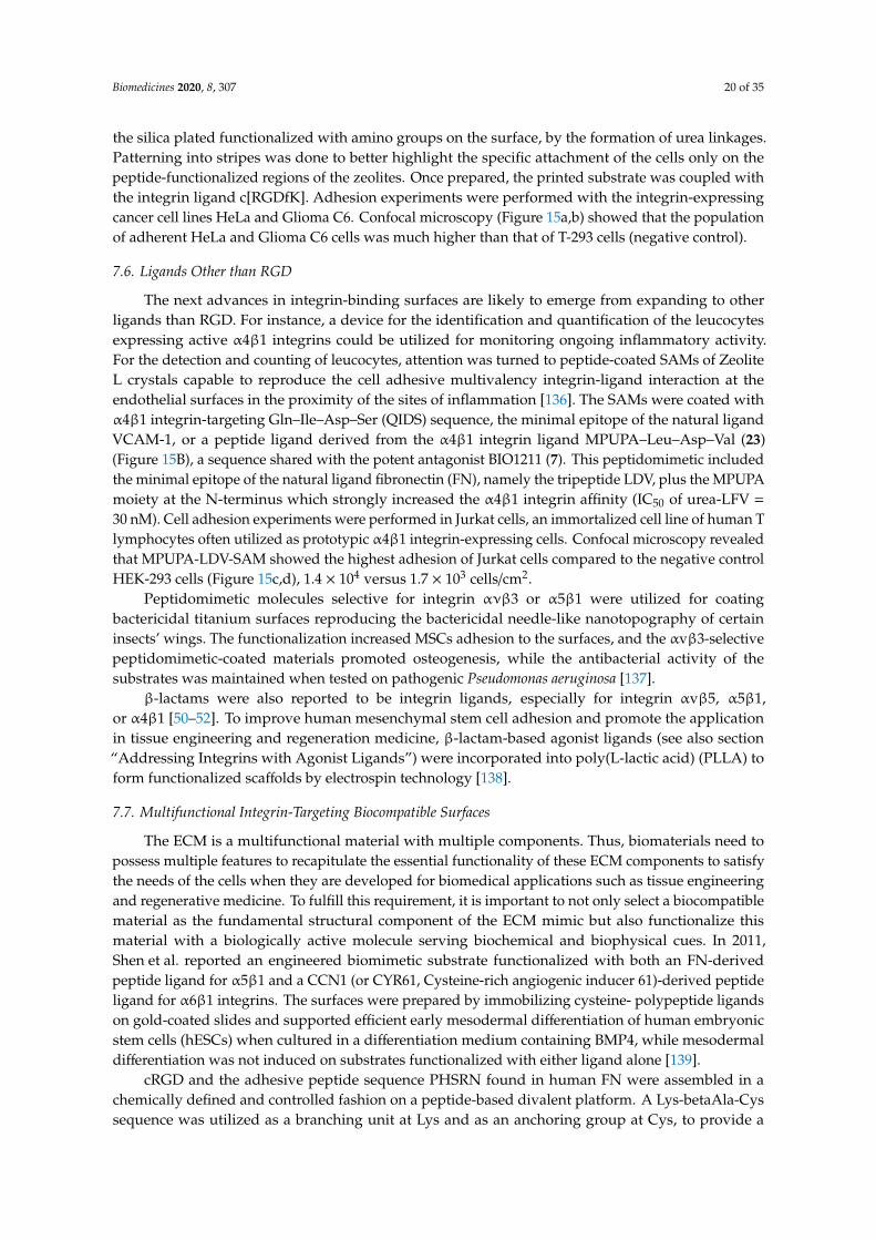
Biomedicines 2020, 8, 307 20 of 35
the silica plated functionalized with amino groups on the surface, by the formation of urea linkages.Patterning into stripes was done to better highlight the specific attachment of the cells only on thepeptide-functionalized regions of the zeolites. Once prepared, the printed substrate was coupled withthe integrin ligand c[RGDfK]. Adhesion experiments were performed with the integrin-expressingcancer cell lines HeLa and Glioma C6. Confocal microscopy (Figure 15a,b) showed that the populationof adherent HeLa and Glioma C6 cells was much higher than that of T-293 cells (negative control).
7.6. Ligands Other than RGD
The next advances in integrin-binding surfaces are likely to emerge from expanding to otherligands than RGD. For instance, a device for the identification and quantification of the leucocytesexpressing active α4β1 integrins could be utilized for monitoring ongoing inflammatory activity.For the detection and counting of leucocytes, attention was turned to peptide-coated SAMs of ZeoliteL crystals capable to reproduce the cell adhesive multivalency integrin-ligand interaction at theendothelial surfaces in the proximity of the sites of inflammation [136]. The SAMs were coated withα4β1 integrin-targeting Gln–Ile–Asp–Ser (QIDS) sequence, the minimal epitope of the natural ligandVCAM-1, or a peptide ligand derived from the α4β1 integrin ligand MPUPA–Leu–Asp–Val (23)(Figure 15B), a sequence shared with the potent antagonist BIO1211 (7). This peptidomimetic includedthe minimal epitope of the natural ligand fibronectin (FN), namely the tripeptide LDV, plus the MPUPAmoiety at the N-terminus which strongly increased the α4β1 integrin affinity (IC50 of urea-LFV =
30 nM). Cell adhesion experiments were performed in Jurkat cells, an immortalized cell line of human Tlymphocytes often utilized as prototypic α4β1 integrin-expressing cells. Confocal microscopy revealedthat MPUPA-LDV-SAM showed the highest adhesion of Jurkat cells compared to the negative controlHEK-293 cells (Figure 15c,d), 1.4 × 10⁴ versus 1.7 × 10³ cells/cm2.
Peptidomimetic molecules selective for integrin ανβ3 or α5β1 were utilized for coatingbactericidal titanium surfaces reproducing the bactericidal needle-like nanotopography of certaininsects’ wings. The functionalization increased MSCs adhesion to the surfaces, and the αvβ3-selectivepeptidomimetic-coated materials promoted osteogenesis, while the antibacterial activity of thesubstrates was maintained when tested on pathogenic Pseudomonas aeruginosa [137].
β-lactams were also reported to be integrin ligands, especially for integrin αvβ5, α5β1,or α4β1 [50–52]. To improve human mesenchymal stem cell adhesion and promote the applicationin tissue engineering and regeneration medicine, β-lactam-based agonist ligands (see also section“Addressing Integrins with Agonist Ligands”) were incorporated into poly(L-lactic acid) (PLLA) toform functionalized scaffolds by electrospin technology [138].
7.7. Multifunctional Integrin-Targeting Biocompatible Surfaces
The ECM is a multifunctional material with multiple components. Thus, biomaterials need topossess multiple features to recapitulate the essential functionality of these ECM components to satisfythe needs of the cells when they are developed for biomedical applications such as tissue engineeringand regenerative medicine. To fulfill this requirement, it is important to not only select a biocompatiblematerial as the fundamental structural component of the ECM mimic but also functionalize thismaterial with a biologically active molecule serving biochemical and biophysical cues. In 2011,Shen et al. reported an engineered biomimetic substrate functionalized with both an FN-derivedpeptide ligand for α5β1 and a CCN1 (or CYR61, Cysteine-rich angiogenic inducer 61)-derived peptideligand for α6β1 integrins. The surfaces were prepared by immobilizing cysteine- polypeptide ligandson gold-coated slides and supported efficient early mesodermal differentiation of human embryonicstem cells (hESCs) when cultured in a differentiation medium containing BMP4, while mesodermaldifferentiation was not induced on substrates functionalized with either ligand alone [139].
cRGD and the adhesive peptide sequence PHSRN found in human FN were assembled in achemically defined and controlled fashion on a peptide-based divalent platform. A Lys-betaAla-Cyssequence was utilized as a branching unit at Lys and as an anchoring group at Cys, to provide a
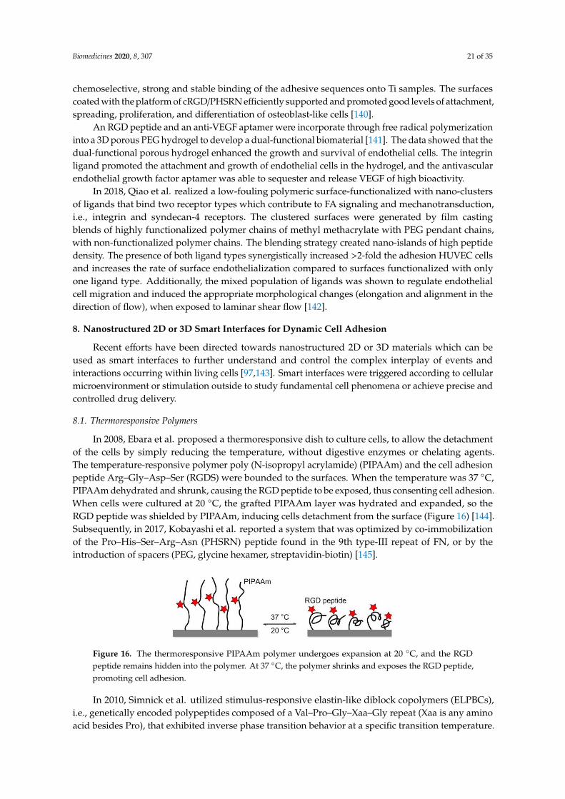
Biomedicines 2020, 8, 307 21 of 35
chemoselective, strong and stable binding of the adhesive sequences onto Ti samples. The surfacescoated with the platform of cRGD/PHSRN efficiently supported and promoted good levels of attachment,spreading, proliferation, and differentiation of osteoblast-like cells [140].
An RGD peptide and an anti-VEGF aptamer were incorporate through free radical polymerizationinto a 3D porous PEG hydrogel to develop a dual-functional biomaterial [141]. The data showed that thedual-functional porous hydrogel enhanced the growth and survival of endothelial cells. The integrinligand promoted the attachment and growth of endothelial cells in the hydrogel, and the antivascularendothelial growth factor aptamer was able to sequester and release VEGF of high bioactivity.
In 2018, Qiao et al. realized a low-fouling polymeric surface-functionalized with nano-clustersof ligands that bind two receptor types which contribute to FA signaling and mechanotransduction,i.e., integrin and syndecan-4 receptors. The clustered surfaces were generated by film castingblends of highly functionalized polymer chains of methyl methacrylate with PEG pendant chains,with non-functionalized polymer chains. The blending strategy created nano-islands of high peptidedensity. The presence of both ligand types synergistically increased >2-fold the adhesion HUVEC cellsand increases the rate of surface endothelialization compared to surfaces functionalized with onlyone ligand type. Additionally, the mixed population of ligands was shown to regulate endothelialcell migration and induced the appropriate morphological changes (elongation and alignment in thedirection of flow), when exposed to laminar shear flow [142].
8. Nanostructured 2D or 3D Smart Interfaces for Dynamic Cell Adhesion
Recent efforts have been directed towards nanostructured 2D or 3D materials which can beused as smart interfaces to further understand and control the complex interplay of events andinteractions occurring within living cells [97,143]. Smart interfaces were triggered according to cellularmicroenvironment or stimulation outside to study fundamental cell phenomena or achieve precise andcontrolled drug delivery.
8.1. Thermoresponsive Polymers
In 2008, Ebara et al. proposed a thermoresponsive dish to culture cells, to allow the detachmentof the cells by simply reducing the temperature, without digestive enzymes or chelating agents.The temperature-responsive polymer poly (N-isopropyl acrylamide) (PIPAAm) and the cell adhesionpeptide Arg–Gly–Asp–Ser (RGDS) were bounded to the surfaces. When the temperature was 37 ◦C,PIPAAm dehydrated and shrunk, causing the RGD peptide to be exposed, thus consenting cell adhesion.When cells were cultured at 20 ◦C, the grafted PIPAAm layer was hydrated and expanded, so theRGD peptide was shielded by PIPAAm, inducing cells detachment from the surface (Figure 16) [144].Subsequently, in 2017, Kobayashi et al. reported a system that was optimized by co-immobilizationof the Pro–His–Ser–Arg–Asn (PHSRN) peptide found in the 9th type-III repeat of FN, or by theintroduction of spacers (PEG, glycine hexamer, streptavidin-biotin) [145].
Biomedicines 2020, 8, x FOR PEER REVIEW 21 of 34
of highly functionalized polymer chains of methyl methacrylate with PEG pendant chains, with non-
functionalized polymer chains. The blending strategy created nano-islands of high peptide density.
The presence of both ligand types synergistically increased >2-fold the adhesion HUVEC cells and
increases the rate of surface endothelialization compared to surfaces functionalized with only one
ligand type. Additionally, the mixed population of ligands was shown to regulate endothelial cell
migration and induced the appropriate morphological changes (elongation and alignment in the
direction of flow), when exposed to laminar shear flow [142].
8. Nanostructured 2D or 3D Smart Interfaces for Dynamic Cell Adhesion
Recent efforts have been directed towards nanostructured 2D or 3D materials which can be used
as smart interfaces to further understand and control the complex interplay of events and interactions
occurring within living cells [97,143]. Smart interfaces were triggered according to cellular
microenvironment or stimulation outside to study fundamental cell phenomena or achieve precise
and controlled drug delivery.
8.1. Thermoresponsive Polymers
In 2008, Ebara et al. proposed a thermoresponsive dish to culture cells, to allow the detachment
of the cells by simply reducing the temperature, without digestive enzymes or chelating agents. The
temperature-responsive polymer poly (N-isopropyl acrylamide) (PIPAAm) and the cell adhesion
peptide Arg–Gly–Asp–Ser (RGDS) were bounded to the surfaces. When the temperature was 37 °C,
PIPAAm dehydrated and shrunk, causing the RGD peptide to be exposed, thus consenting cell adhesion.
When cells were cultured at 20 °C, the grafted PIPAAm layer was hydrated and expanded, so the RGD
peptide was shielded by PIPAAm, inducing cells detachment from the surface (Figure 16) [144].
Subsequently, in 2017, Kobayashi et al. reported a system that was optimized by co-immobilization
of the Pro–His–Ser–Arg–Asn (PHSRN) peptide found in the 9th type-III repeat of FN, or by the
introduction of spacers (PEG, glycine hexamer, streptavidin-biotin) [145].
Figure 16. The thermoresponsive PIPAAm polymer undergoes expansion at 20 °C, and the RGD
peptide remains hidden into the polymer. At 37 °C, the polymer shrinks and exposes the RGD
peptide, promoting cell adhesion.
In 2010, Simnick et al. utilized stimulus-responsive elastin-like diblock copolymers (ELPBCs),
i.e., genetically encoded polypeptides composed of a Val–Pro–Gly–Xaa–Gly repeat (Xaa is any amino
acid besides Pro), that exhibited inverse phase transition behavior at a specific transition temperature.
The ELPBCs were exploited as a scaffold to bind and present the GRGDS peptide, an integrin ligand
characterized by low affinity (IC50 = 1 mM) for the ανβ3 integrin. Despite the low affinity of this
ligand, it has been observed that the avidity of K562 human leukemia cells transformed by ανβ3
receptor (K562/ανβ3) is higher as compared with a wild-type K562 control, through multivalent
presentation induced by the thermally triggered self-assembly of the ELPBCs [146].
8.2. Enzyme-Triggered Polymers
To manage adhesion/cytoskeletal balance and to initiate differentiation of MSCs to prolong cell
culture, in 2016, Robert et al. prepared surfaces immobilized by RGD-containing polymer monolayer,
which can be enzyme-activated. This surface consisted of RGD peptide or RGE peptide (control), an
elastase cleavable dialanine (AA) linker, and steric blocking fluorenylmethyloxycarbonyl (FMOC)
Figure 16. The thermoresponsive PIPAAm polymer undergoes expansion at 20 ◦C, and the RGDpeptide remains hidden into the polymer. At 37 ◦C, the polymer shrinks and exposes the RGD peptide,promoting cell adhesion.
In 2010, Simnick et al. utilized stimulus-responsive elastin-like diblock copolymers (ELPBCs),i.e., genetically encoded polypeptides composed of a Val–Pro–Gly–Xaa–Gly repeat (Xaa is any aminoacid besides Pro), that exhibited inverse phase transition behavior at a specific transition temperature.
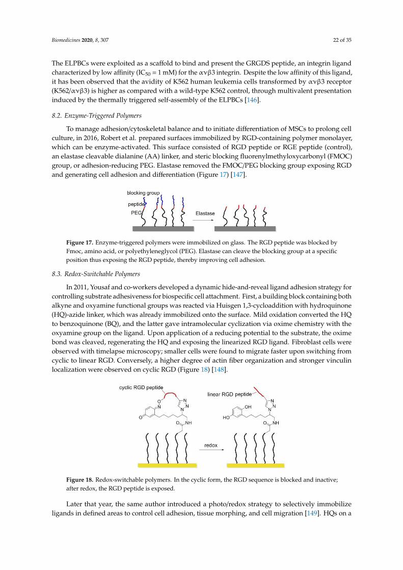
Biomedicines 2020, 8, 307 22 of 35
The ELPBCs were exploited as a scaffold to bind and present the GRGDS peptide, an integrin ligandcharacterized by low affinity (IC50 = 1 mM) for the ανβ3 integrin. Despite the low affinity of this ligand,it has been observed that the avidity of K562 human leukemia cells transformed by ανβ3 receptor(K562/ανβ3) is higher as compared with a wild-type K562 control, through multivalent presentationinduced by the thermally triggered self-assembly of the ELPBCs [146].
8.2. Enzyme-Triggered Polymers
To manage adhesion/cytoskeletal balance and to initiate differentiation of MSCs to prolong cellculture, in 2016, Robert et al. prepared surfaces immobilized by RGD-containing polymer monolayer,which can be enzyme-activated. This surface consisted of RGD peptide or RGE peptide (control),an elastase cleavable dialanine (AA) linker, and steric blocking fluorenylmethyloxycarbonyl (FMOC)group, or adhesion-reducing PEG. Elastase removed the FMOC/PEG blocking group exposing RGDand generating cell adhesion and differentiation (Figure 17) [147].
Biomedicines 2020, 8, x FOR PEER REVIEW 22 of 34
group, or adhesion-reducing PEG. Elastase removed the FMOC/PEG blocking group exposing RGD
and generating cell adhesion and differentiation (Figure 17) [147].
Figure 17. Enzyme-triggered polymers were immobilized on glass. The RGD peptide was blocked by
Fmoc, amino acid, or polyethyleneglycol (PEG). Elastase can cleave the blocking group at a specific
position thus exposing the RGD peptide, thereby improving cell adhesion.
8.3. Redox-Switchable Polymers
In 2011, Yousaf and co-workers developed a dynamic hide-and-reveal ligand adhesion strategy
for controlling substrate adhesiveness for biospecific cell attachment. First, a building block
containing both alkyne and oxyamine functional groups was reacted via Huisgen 1,3-cycloaddition
with hydroquinone (HQ)-azide linker, which was already immobilized onto the surface. Mild
oxidation converted the HQ to benzoquinone (BQ), and the latter gave intramolecular cyclization via
oxime chemistry with the oxyamine group on the ligand. Upon application of a reducing potential to
the substrate, the oxime bond was cleaved, regenerating the HQ and exposing the linearized RGD
ligand. Fibroblast cells were observed with timelapse microscopy; smaller cells were found to migrate
faster upon switching from cyclic to linear RGD. Conversely, a higher degree of actin fiber
organization and stronger vinculin localization were observed on cyclic RGD (Figure 18) [148].
Figure 18. Redox-switchable polymers. In the cyclic form, the RGD sequence is blocked and inactive;
after redox, the RGD peptide is exposed.
Later that year, the same author introduced a photo/redox strategy to selectively immobilize
ligands in defined areas to control cell adhesion, tissue morphing, and cell migration [149]. HQs on a
SAM were uncaged by UV illumination and then electrochemically converted to BQs, which in turn
were reactive toward oxyamine-modified RGD ligands. Adhered fibroblasts were first confined to
hydrophobic patterns; after installing RGD ligands, cells migrated from the hydrophobic patterns
toward the RGD presenting patterns. When a gradient of RGD ligands was formed, cells were
observed to move faster toward lower ligand densities instead of higher ones.
8.4. Potential Responsive Polymers
In 2012, Gooding and coworkers prepared two-component SAMs comprising a protein-resistant
ethylene glycol chain containing a charged moiety at its distal end, and a terminated RGD component
on which cellular adhesion receptors, integrins can bind. The electro-switchable surfaces were able
to control cell adhesion under different electrical potential. In one case, the surface was constituted
by an RGD and hexa(ethylene glycol)-sulfonate species (EG6) mixed SAM that promoted cell
adhesion under a positive potential (+0.3 V), while in the other case the surface comprises an RGD
Figure 17. Enzyme-triggered polymers were immobilized on glass. The RGD peptide was blocked byFmoc, amino acid, or polyethyleneglycol (PEG). Elastase can cleave the blocking group at a specificposition thus exposing the RGD peptide, thereby improving cell adhesion.
8.3. Redox-Switchable Polymers
In 2011, Yousaf and co-workers developed a dynamic hide-and-reveal ligand adhesion strategy forcontrolling substrate adhesiveness for biospecific cell attachment. First, a building block containing bothalkyne and oxyamine functional groups was reacted via Huisgen 1,3-cycloaddition with hydroquinone(HQ)-azide linker, which was already immobilized onto the surface. Mild oxidation converted the HQto benzoquinone (BQ), and the latter gave intramolecular cyclization via oxime chemistry with theoxyamine group on the ligand. Upon application of a reducing potential to the substrate, the oximebond was cleaved, regenerating the HQ and exposing the linearized RGD ligand. Fibroblast cells wereobserved with timelapse microscopy; smaller cells were found to migrate faster upon switching fromcyclic to linear RGD. Conversely, a higher degree of actin fiber organization and stronger vinculinlocalization were observed on cyclic RGD (Figure 18) [148].
Biomedicines 2020, 8, x FOR PEER REVIEW 22 of 34
group, or adhesion-reducing PEG. Elastase removed the FMOC/PEG blocking group exposing RGD
and generating cell adhesion and differentiation (Figure 17) [147].
Figure 17. Enzyme-triggered polymers were immobilized on glass. The RGD peptide was blocked by
Fmoc, amino acid, or polyethyleneglycol (PEG). Elastase can cleave the blocking group at a specific
position thus exposing the RGD peptide, thereby improving cell adhesion.
8.3. Redox-Switchable Polymers
In 2011, Yousaf and co-workers developed a dynamic hide-and-reveal ligand adhesion strategy
for controlling substrate adhesiveness for biospecific cell attachment. First, a building block
containing both alkyne and oxyamine functional groups was reacted via Huisgen 1,3-cycloaddition
with hydroquinone (HQ)-azide linker, which was already immobilized onto the surface. Mild
oxidation converted the HQ to benzoquinone (BQ), and the latter gave intramolecular cyclization via
oxime chemistry with the oxyamine group on the ligand. Upon application of a reducing potential to
the substrate, the oxime bond was cleaved, regenerating the HQ and exposing the linearized RGD
ligand. Fibroblast cells were observed with timelapse microscopy; smaller cells were found to migrate
faster upon switching from cyclic to linear RGD. Conversely, a higher degree of actin fiber
organization and stronger vinculin localization were observed on cyclic RGD (Figure 18) [148].
Figure 18. Redox-switchable polymers. In the cyclic form, the RGD sequence is blocked and inactive;
after redox, the RGD peptide is exposed.
Later that year, the same author introduced a photo/redox strategy to selectively immobilize
ligands in defined areas to control cell adhesion, tissue morphing, and cell migration [149]. HQs on a
SAM were uncaged by UV illumination and then electrochemically converted to BQs, which in turn
were reactive toward oxyamine-modified RGD ligands. Adhered fibroblasts were first confined to
hydrophobic patterns; after installing RGD ligands, cells migrated from the hydrophobic patterns
toward the RGD presenting patterns. When a gradient of RGD ligands was formed, cells were
observed to move faster toward lower ligand densities instead of higher ones.
8.4. Potential Responsive Polymers
In 2012, Gooding and coworkers prepared two-component SAMs comprising a protein-resistant
ethylene glycol chain containing a charged moiety at its distal end, and a terminated RGD component
on which cellular adhesion receptors, integrins can bind. The electro-switchable surfaces were able
to control cell adhesion under different electrical potential. In one case, the surface was constituted
by an RGD and hexa(ethylene glycol)-sulfonate species (EG6) mixed SAM that promoted cell
adhesion under a positive potential (+0.3 V), while in the other case the surface comprises an RGD
Figure 18. Redox-switchable polymers. In the cyclic form, the RGD sequence is blocked and inactive;after redox, the RGD peptide is exposed.
Later that year, the same author introduced a photo/redox strategy to selectively immobilizeligands in defined areas to control cell adhesion, tissue morphing, and cell migration [149]. HQs on a
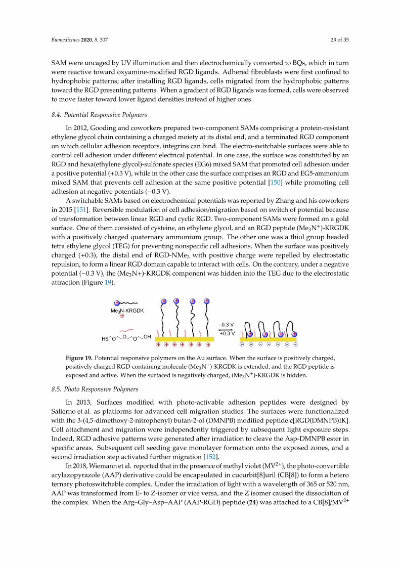
Biomedicines 2020, 8, 307 23 of 35
SAM were uncaged by UV illumination and then electrochemically converted to BQs, which in turnwere reactive toward oxyamine-modified RGD ligands. Adhered fibroblasts were first confined tohydrophobic patterns; after installing RGD ligands, cells migrated from the hydrophobic patternstoward the RGD presenting patterns. When a gradient of RGD ligands was formed, cells were observedto move faster toward lower ligand densities instead of higher ones.
8.4. Potential Responsive Polymers
In 2012, Gooding and coworkers prepared two-component SAMs comprising a protein-resistantethylene glycol chain containing a charged moiety at its distal end, and a terminated RGD componenton which cellular adhesion receptors, integrins can bind. The electro-switchable surfaces were able tocontrol cell adhesion under different electrical potential. In one case, the surface was constituted by anRGD and hexa(ethylene glycol)-sulfonate species (EG6) mixed SAM that promoted cell adhesion undera positive potential (+0.3 V), while in the other case the surface comprises an RGD and EG5-ammoniummixed SAM that prevents cell adhesion at the same positive potential [150] while promoting celladhesion at negative potentials (−0.3 V).
A switchable SAMs based on electrochemical potentials was reported by Zhang and his coworkersin 2015 [151]. Reversible modulation of cell adhesion/migration based on switch of potential becauseof transformation between linear RGD and cyclic RGD. Two-component SAMs were formed on a goldsurface. One of them consisted of cysteine, an ethylene glycol, and an RGD peptide (Me3N+)-KRGDKwith a positively charged quaternary ammonium group. The other one was a thiol group headedtetra ethylene glycol (TEG) for preventing nonspecific cell adhesions. When the surface was positivelycharged (+0.3), the distal end of RGD-NMe3 with positive charge were repelled by electrostaticrepulsion, to form a linear RGD domain capable to interact with cells. On the contrary, under a negativepotential (−0.3 V), the (Me3N+)-KRGDK component was hidden into the TEG due to the electrostaticattraction (Figure 19).
Biomedicines 2020, 8, x FOR PEER REVIEW 23 of 34
and EG5-ammonium mixed SAM that prevents cell adhesion at the same positive potential
[150]while promoting cell adhesion at negative potentials (−0.3 V).
A switchable SAMs based on electrochemical potentials was reported by Zhang and his
coworkers in 2015 [151]. Reversible modulation of cell adhesion/migration based on switch of
potential because of transformation between linear RGD and cyclic RGD. Two-component SAMs
were formed on a gold surface. One of them consisted of cysteine, an ethylene glycol, and an RGD
peptide (Me3N+)-KRGDK with a positively charged quaternary ammonium group. The other one was
a thiol group headed tetra ethylene glycol (TEG) for preventing nonspecific cell adhesions. When the
surface was positively charged (+0.3), the distal end of RGD-NMe3 with positive charge were repelled
by electrostatic repulsion, to form a linear RGD domain capable to interact with cells. On the contrary,
under a negative potential (−0.3 V), the (Me3N+)-KRGDK component was hidden into the TEG due
to the electrostatic attraction (Figure 19).
Figure 19. Potential responsive polymers on the Au surface. When the surface is positively charged,
positively charged RGD-containing molecule (Me3N+)-KRGDK is extended, and the RGD peptide is
exposed and active. When the surfaced is negatively charged, (Me3N+)-KRGDK is hidden.
8.5. Photo Responsive Polymers
In 2013, Surfaces modified with photo-activable adhesion peptides were designed by Salierno et
al. as platforms for advanced cell migration studies. The surfaces were functionalized with the 3-(4,5-
dimethoxy-2-nitrophenyl) butan-2-ol (DMNPB) modified peptide c[RGD(DMNPB)fK]. Cell
attachment and migration were independently triggered by subsequent light exposure steps. Indeed,
RGD adhesive patterns were generated after irradiation to cleave the Asp-DMNPB ester in specific
areas. Subsequent cell seeding gave monolayer formation onto the exposed zones, and a second
irradiation step activated further migration [152].
In 2018, Wiemann et al. reported that in the presence of methyl violet (MV2+), the photo-
convertible arylazopyrazole (AAP) derivative could be encapsulated in cucurbit[8]uril (CB[8]) to
form a hetero ternary photoswitchable complex. Under the irradiation of light with a wavelength of
365 or 520 nm, AAP was transformed from E- to Z-isomer or vice versa, and the Z isomer caused the
dissociation of the complex. When the Arg–Gly–Asp–AAP (AAP-RGD) peptide (24) was attached to
a CB[8]/MV2+ complex bounded biologically active surface, cell adhesion, and release could be
controlled by UV radiation (Figure 20) [153].
Figure 20. Photo-responsive polymers. AAP-RGD is isomerized from E- to Z-isomer under UV
irradiation at 365 nm, resulting in dissociation of AAP-RGD from MV2+/AAP-RGD/CB[8] complex.
Figure 19. Potential responsive polymers on the Au surface. When the surface is positively charged,positively charged RGD-containing molecule (Me3N+)-KRGDK is extended, and the RGD peptide isexposed and active. When the surfaced is negatively charged, (Me3N+)-KRGDK is hidden.
8.5. Photo Responsive Polymers
In 2013, Surfaces modified with photo-activable adhesion peptides were designed bySalierno et al. as platforms for advanced cell migration studies. The surfaces were functionalizedwith the 3-(4,5-dimethoxy-2-nitrophenyl) butan-2-ol (DMNPB) modified peptide c[RGD(DMNPB)fK].Cell attachment and migration were independently triggered by subsequent light exposure steps.Indeed, RGD adhesive patterns were generated after irradiation to cleave the Asp-DMNPB ester inspecific areas. Subsequent cell seeding gave monolayer formation onto the exposed zones, and asecond irradiation step activated further migration [152].
In 2018, Wiemann et al. reported that in the presence of methyl violet (MV2+), the photo-convertiblearylazopyrazole (AAP) derivative could be encapsulated in cucurbit[8]uril (CB[8]) to form a heteroternary photoswitchable complex. Under the irradiation of light with a wavelength of 365 or 520 nm,AAP was transformed from E- to Z-isomer or vice versa, and the Z isomer caused the dissociation ofthe complex. When the Arg–Gly–Asp–AAP (AAP-RGD) peptide (24) was attached to a CB[8]/MV2+
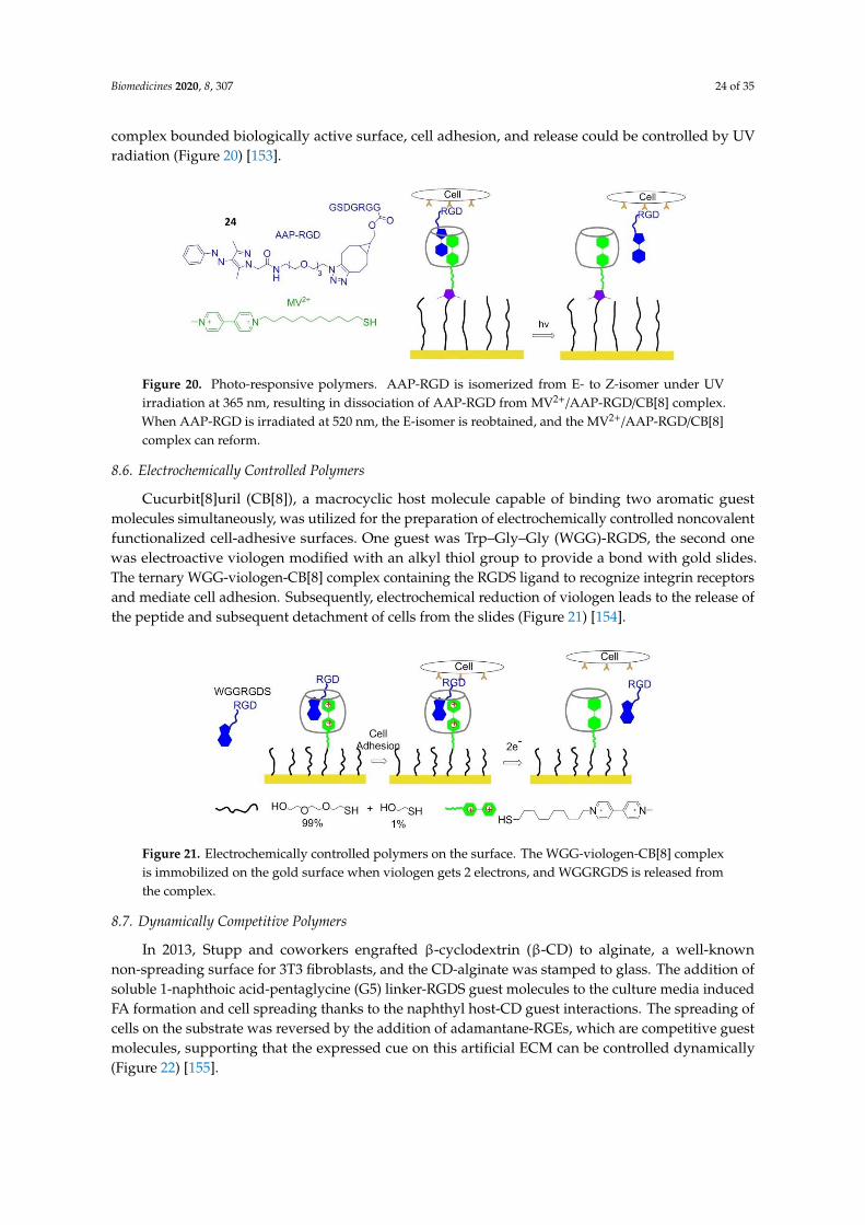
Biomedicines 2020, 8, 307 24 of 35
complex bounded biologically active surface, cell adhesion, and release could be controlled by UVradiation (Figure 20) [153].
Biomedicines 2020, 8, x FOR PEER REVIEW 23 of 34
and EG5-ammonium mixed SAM that prevents cell adhesion at the same positive potential
[150]while promoting cell adhesion at negative potentials (−0.3 V).
A switchable SAMs based on electrochemical potentials was reported by Zhang and his
coworkers in 2015 [151]. Reversible modulation of cell adhesion/migration based on switch of
potential because of transformation between linear RGD and cyclic RGD. Two-component SAMs
were formed on a gold surface. One of them consisted of cysteine, an ethylene glycol, and an RGD
peptide (Me3N+)-KRGDK with a positively charged quaternary ammonium group. The other one was
a thiol group headed tetra ethylene glycol (TEG) for preventing nonspecific cell adhesions. When the
surface was positively charged (+0.3), the distal end of RGD-NMe3 with positive charge were repelled
by electrostatic repulsion, to form a linear RGD domain capable to interact with cells. On the contrary,
under a negative potential (−0.3 V), the (Me3N+)-KRGDK component was hidden into the TEG due
to the electrostatic attraction (Figure 19).
Figure 19. Potential responsive polymers on the Au surface. When the surface is positively charged,
positively charged RGD-containing molecule (Me3N+)-KRGDK is extended, and the RGD peptide is
exposed and active. When the surfaced is negatively charged, (Me3N+)-KRGDK is hidden.
8.5. Photo Responsive Polymers
In 2013, Surfaces modified with photo-activable adhesion peptides were designed by Salierno et
al. as platforms for advanced cell migration studies. The surfaces were functionalized with the 3-(4,5-
dimethoxy-2-nitrophenyl) butan-2-ol (DMNPB) modified peptide c[RGD(DMNPB)fK]. Cell
attachment and migration were independently triggered by subsequent light exposure steps. Indeed,
RGD adhesive patterns were generated after irradiation to cleave the Asp-DMNPB ester in specific
areas. Subsequent cell seeding gave monolayer formation onto the exposed zones, and a second
irradiation step activated further migration [152].
In 2018, Wiemann et al. reported that in the presence of methyl violet (MV2+), the photo-
convertible arylazopyrazole (AAP) derivative could be encapsulated in cucurbit[8]uril (CB[8]) to
form a hetero ternary photoswitchable complex. Under the irradiation of light with a wavelength of
365 or 520 nm, AAP was transformed from E- to Z-isomer or vice versa, and the Z isomer caused the
dissociation of the complex. When the Arg–Gly–Asp–AAP (AAP-RGD) peptide (24) was attached to
a CB[8]/MV2+ complex bounded biologically active surface, cell adhesion, and release could be
controlled by UV radiation (Figure 20) [153].
Figure 20. Photo-responsive polymers. AAP-RGD is isomerized from E- to Z-isomer under UV
irradiation at 365 nm, resulting in dissociation of AAP-RGD from MV2+/AAP-RGD/CB[8] complex.
Figure 20. Photo-responsive polymers. AAP-RGD is isomerized from E- to Z-isomer under UVirradiation at 365 nm, resulting in dissociation of AAP-RGD from MV2+/AAP-RGD/CB[8] complex.When AAP-RGD is irradiated at 520 nm, the E-isomer is reobtained, and the MV2+/AAP-RGD/CB[8]complex can reform.
8.6. Electrochemically Controlled Polymers
Cucurbit[8]uril (CB[8]), a macrocyclic host molecule capable of binding two aromatic guestmolecules simultaneously, was utilized for the preparation of electrochemically controlled noncovalentfunctionalized cell-adhesive surfaces. One guest was Trp–Gly–Gly (WGG)-RGDS, the second onewas electroactive viologen modified with an alkyl thiol group to provide a bond with gold slides.The ternary WGG-viologen-CB[8] complex containing the RGDS ligand to recognize integrin receptorsand mediate cell adhesion. Subsequently, electrochemical reduction of viologen leads to the release ofthe peptide and subsequent detachment of cells from the slides (Figure 21) [154].
Biomedicines 2020, 8, x FOR PEER REVIEW 24 of 34
When AAP-RGD is irradiated at 520 nm, the E-isomer is reobtained, and the MV2+/AAP-RGD/CB[8]
complex can reform.
8.6. Electrochemically Controlled Polymers
Cucurbit[8]uril (CB[8]), a macrocyclic host molecule capable of binding two aromatic guest
molecules simultaneously, was utilized for the preparation of electrochemically controlled
noncovalent functionalized cell-adhesive surfaces. One guest was Trp–Gly–Gly (WGG)-RGDS, the
second one was electroactive viologen modified with an alkyl thiol group to provide a bond with
gold slides. The ternary WGG-viologen-CB[8] complex containing the RGDS ligand to recognize
integrin receptors and mediate cell adhesion. Subsequently, electrochemical reduction of viologen leads
to the release of the peptide and subsequent detachment of cells from the slides (Figure 21) [154].
Figure 21. Electrochemically controlled polymers on the surface. The WGG-viologen-CB[8] complex
is immobilized on the gold surface when viologen gets 2 electrons, and WGGRGDS is released from
the complex.
8.7. Dynamically Competitive Polymers
In 2013, Stupp and coworkers engrafted β-cyclodextrin (β-CD) to alginate, a well-known non-
spreading surface for 3T3 fibroblasts, and the CD-alginate was stamped to glass. The addition of
soluble 1-naphthoic acid-pentaglycine (G5) linker-RGDS guest molecules to the culture media
induced FA formation and cell spreading thanks to the naphthyl host-CD guest interactions. The
spreading of cells on the substrate was reversed by the addition of adamantane-RGEs, which are
competitive guest molecules, supporting that the expressed cue on this artificial ECM can be
controlled dynamically (Figure 22) [155].
Figure 22. Dynamically competitive polymers. Guest molecular naphthyl-RGDS is captured by β-CD,
which is grafted on alginate. Cell adhesion and spreading can be controlled dynamically because the
addition of Ada-RGES can reverse cell adhesion due to it competed with naphthyl-RGDS to form a
complex with β-CD.
8.8. Dynamically Controlled Smart Interfaces for 3D Cell Culture
The dynamic cell-instructive biointerfaces represent a newly proposed concept as smart
interfaces, aimed at mimicking native 3D microenvironment that surround encapsulated cells to
Figure 21. Electrochemically controlled polymers on the surface. The WGG-viologen-CB[8] complexis immobilized on the gold surface when viologen gets 2 electrons, and WGGRGDS is released fromthe complex.
8.7. Dynamically Competitive Polymers
In 2013, Stupp and coworkers engrafted β-cyclodextrin (β-CD) to alginate, a well-knownnon-spreading surface for 3T3 fibroblasts, and the CD-alginate was stamped to glass. The addition ofsoluble 1-naphthoic acid-pentaglycine (G5) linker-RGDS guest molecules to the culture media inducedFA formation and cell spreading thanks to the naphthyl host-CD guest interactions. The spreading ofcells on the substrate was reversed by the addition of adamantane-RGEs, which are competitive guestmolecules, supporting that the expressed cue on this artificial ECM can be controlled dynamically(Figure 22) [155].
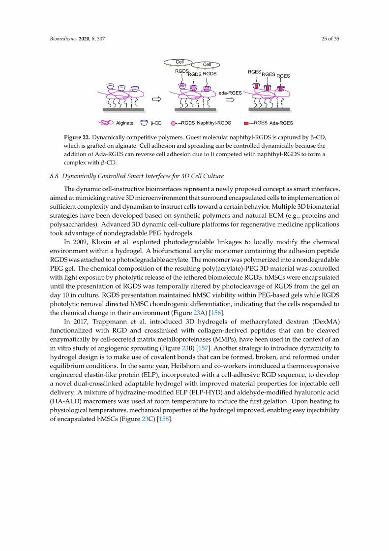
Biomedicines 2020, 8, 307 25 of 35
Biomedicines 2020, 8, x FOR PEER REVIEW 24 of 34
When AAP-RGD is irradiated at 520 nm, the E-isomer is reobtained, and the MV2+/AAP-RGD/CB[8]
complex can reform.
8.6. Electrochemically Controlled Polymers
Cucurbit[8]uril (CB[8]), a macrocyclic host molecule capable of binding two aromatic guest
molecules simultaneously, was utilized for the preparation of electrochemically controlled
noncovalent functionalized cell-adhesive surfaces. One guest was Trp–Gly–Gly (WGG)-RGDS, the
second one was electroactive viologen modified with an alkyl thiol group to provide a bond with
gold slides. The ternary WGG-viologen-CB[8] complex containing the RGDS ligand to recognize
integrin receptors and mediate cell adhesion. Subsequently, electrochemical reduction of viologen leads
to the release of the peptide and subsequent detachment of cells from the slides (Figure 21) [154].
Figure 21. Electrochemically controlled polymers on the surface. The WGG-viologen-CB[8] complex
is immobilized on the gold surface when viologen gets 2 electrons, and WGGRGDS is released from
the complex.
8.7. Dynamically Competitive Polymers
In 2013, Stupp and coworkers engrafted β-cyclodextrin (β-CD) to alginate, a well-known non-
spreading surface for 3T3 fibroblasts, and the CD-alginate was stamped to glass. The addition of
soluble 1-naphthoic acid-pentaglycine (G5) linker-RGDS guest molecules to the culture media
induced FA formation and cell spreading thanks to the naphthyl host-CD guest interactions. The
spreading of cells on the substrate was reversed by the addition of adamantane-RGEs, which are
competitive guest molecules, supporting that the expressed cue on this artificial ECM can be
controlled dynamically (Figure 22) [155].
Figure 22. Dynamically competitive polymers. Guest molecular naphthyl-RGDS is captured by β-CD,
which is grafted on alginate. Cell adhesion and spreading can be controlled dynamically because the
addition of Ada-RGES can reverse cell adhesion due to it competed with naphthyl-RGDS to form a
complex with β-CD.
8.8. Dynamically Controlled Smart Interfaces for 3D Cell Culture
The dynamic cell-instructive biointerfaces represent a newly proposed concept as smart
interfaces, aimed at mimicking native 3D microenvironment that surround encapsulated cells to
Figure 22. Dynamically competitive polymers. Guest molecular naphthyl-RGDS is captured by β-CD,which is grafted on alginate. Cell adhesion and spreading can be controlled dynamically because theaddition of Ada-RGES can reverse cell adhesion due to it competed with naphthyl-RGDS to form acomplex with β-CD.
8.8. Dynamically Controlled Smart Interfaces for 3D Cell Culture
The dynamic cell-instructive biointerfaces represent a newly proposed concept as smart interfaces,aimed at mimicking native 3D microenvironment that surround encapsulated cells to implementation ofsufficient complexity and dynamism to instruct cells toward a certain behavior. Multiple 3D biomaterialstrategies have been developed based on synthetic polymers and natural ECM (e.g., proteins andpolysaccharides). Advanced 3D dynamic cell-culture platforms for regenerative medicine applicationstook advantage of nondegradable PEG hydrogels.
In 2009, Kloxin et al. exploited photodegradable linkages to locally modify the chemicalenvironment within a hydrogel. A biofunctional acrylic monomer containing the adhesion peptideRGDS was attached to a photodegradable acrylate. The monomer was polymerized into a nondegradablePEG gel. The chemical composition of the resulting poly(acrylate)-PEG 3D material was controlledwith light exposure by photolytic release of the tethered biomolecule RGDS. hMSCs were encapsulateduntil the presentation of RGDS was temporally altered by photocleavage of RGDS from the gel onday 10 in culture. RGDS presentation maintained hMSC viability within PEG-based gels while RGDSphotolytic removal directed hMSC chondrogenic differentiation, indicating that the cells responded tothe chemical change in their environment (Figure 23A) [156].
In 2017, Trappmann et al. introduced 3D hydrogels of methacrylated dextran (DexMA)functionalized with RGD and crosslinked with collagen-derived peptides that can be cleavedenzymatically by cell-secreted matrix metalloproteinases (MMPs), have been used in the context of anin vitro study of angiogenic sprouting (Figure 23B) [157]. Another strategy to introduce dynamicity tohydrogel design is to make use of covalent bonds that can be formed, broken, and reformed underequilibrium conditions. In the same year, Heilshorn and co-workers introduced a thermoresponsiveengineered elastin-like protein (ELP), incorporated with a cell-adhesive RGD sequence, to developa novel dual-crosslinked adaptable hydrogel with improved material properties for injectable celldelivery. A mixture of hydrazine-modified ELP (ELP-HYD) and aldehyde-modified hyaluronic acid(HA-ALD) macromers was used at room temperature to induce the first gelation. Upon heating tophysiological temperatures, mechanical properties of the hydrogel improved, enabling easy injectabilityof encapsulated hMSCs (Figure 23C) [158].
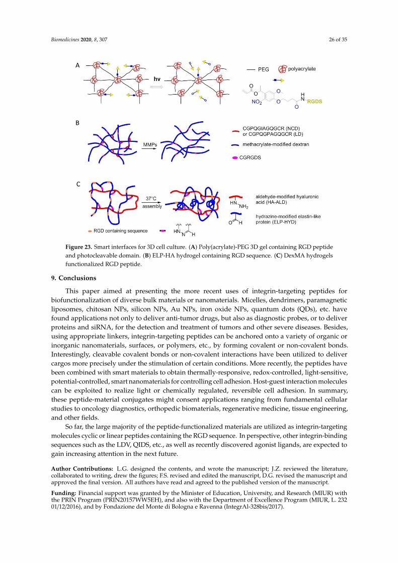
Biomedicines 2020, 8, 307 26 of 35
Biomedicines 2020, 8, x FOR PEER REVIEW 25 of 34
implementation of sufficient complexity and dynamism to instruct cells toward a certain behavior.
Multiple 3D biomaterial strategies have been developed based on synthetic polymers and natural
ECM (e.g., proteins and polysaccharides). Advanced 3D dynamic cell-culture platforms for
regenerative medicine applications took advantage of nondegradable PEG hydrogels.
In 2009, Kloxin et al. exploited photodegradable linkages to locally modify the chemical
environment within a hydrogel. A biofunctional acrylic monomer containing the adhesion peptide
RGDS was attached to a photodegradable acrylate. The monomer was polymerized into a
nondegradable PEG gel. The chemical composition of the resulting poly(acrylate)-PEG 3D material
was controlled with light exposure by photolytic release of the tethered biomolecule RGDS. hMSCs
were encapsulated until the presentation of RGDS was temporally altered by photocleavage of RGDS
from the gel on day 10 in culture. RGDS presentation maintained hMSC viability within PEG-based
gels while RGDS photolytic removal directed hMSC chondrogenic differentiation, indicating that the
cells responded to the chemical change in their environment (Figure 23A) [156].
Figure 23. Smart interfaces for 3D cell culture. (A) Poly(acrylate)-PEG 3D gel containing RGD peptide
and photocleavable domain. (B) ELP-HA hydrogel containing RGD sequence. (C) DexMA hydrogels
functionalized RGD peptide.
In 2017, Trappmann et al. introduced 3D hydrogels of methacrylated dextran (DexMA)
functionalized with RGD and crosslinked with collagen-derived peptides that can be cleaved
enzymatically by cell-secreted matrix metalloproteinases (MMPs), have been used in the context of
an in vitro study of angiogenic sprouting (Figure 23B) [157]. Another strategy to introduce
dynamicity to hydrogel design is to make use of covalent bonds that can be formed, broken, and
reformed under equilibrium conditions. In the same year, Heilshorn and co-workers introduced a
thermoresponsive engineered elastin-like protein (ELP), incorporated with a cell-adhesive RGD
sequence, to develop a novel dual-crosslinked adaptable hydrogel with improved material properties
for injectable cell delivery. A mixture of hydrazine-modified ELP (ELP-HYD) and aldehyde-modified
hyaluronic acid (HA-ALD) macromers was used at room temperature to induce the first gelation.
Upon heating to physiological temperatures, mechanical properties of the hydrogel improved,
enabling easy injectability of encapsulated hMSCs (Figure 23C) [158].
Figure 23. Smart interfaces for 3D cell culture. (A) Poly(acrylate)-PEG 3D gel containing RGD peptideand photocleavable domain. (B) ELP-HA hydrogel containing RGD sequence. (C) DexMA hydrogelsfunctionalized RGD peptide.
9. Conclusions
This paper aimed at presenting the more recent uses of integrin-targeting peptides forbiofunctionalization of diverse bulk materials or nanomaterials. Micelles, dendrimers, paramagneticliposomes, chitosan NPs, silicon NPs, Au NPs, iron oxide NPs, quantum dots (QDs), etc. havefound applications not only to deliver anti-tumor drugs, but also as diagnostic probes, or to deliverproteins and siRNA, for the detection and treatment of tumors and other severe diseases. Besides,using appropriate linkers, integrin-targeting peptides can be anchored onto a variety of organic orinorganic nanomaterials, surfaces, or polymers, etc., by forming covalent or non-covalent bonds.Interestingly, cleavable covalent bonds or non-covalent interactions have been utilized to delivercargos more precisely under the stimulation of certain conditions. More recently, the peptides havebeen combined with smart materials to obtain thermally-responsive, redox-controlled, light-sensitive,potential-controlled, smart nanomaterials for controlling cell adhesion. Host-guest interaction moleculescan be exploited to realize light or chemically regulated, reversible cell adhesion. In summary,these peptide-material conjugates might consent applications ranging from fundamental cellularstudies to oncology diagnostics, orthopedic biomaterials, regenerative medicine, tissue engineering,and other fields.
So far, the large majority of the peptide-functionalized materials are utilized as integrin-targetingmolecules cyclic or linear peptides containing the RGD sequence. In perspective, other integrin-bindingsequences such as the LDV, QIDS, etc., as well as recently discovered agonist ligands, are expected togain increasing attention in the next future.
Author Contributions: L.G. designed the contents, and wrote the manuscript; J.Z. reviewed the literature,collaborated to writing, drew the figures; F.S. revised and edited the manuscript, D.G. revised the manuscript andapproved the final version. All authors have read and agreed to the published version of the manuscript.
Funding: Financial support was granted by the Minister of Education, University, and Research (MIUR) withthe PRIN Program (PRIN20157WW5EH), and also with the Department of Excellence Program (MIUR, L. 23201/12/2016), and by Fondazione del Monte di Bologna e Ravenna (IntegrAl-328bis/2017).
Biomedicines 2020, 8, 307 27 of 35
Conflicts of Interest: The authors declare no conflict of interest.
References
1. Baiula, M.; Spampinato, S.; Gentilucci, L.; Tolomelli, A. Novel ligands targeting α4β1 integrin: Therapeuticapplications and perspectives. Front. Chem. 2019, 7, 489. [CrossRef]
2. Danhier, F.; Le Breton, A.; Préat, V. RGD-based strategies to target ανβ3 integrin in cancer therapy anddiagnosis. Mol. Pharm. 2012, 9, 2961–2973. [CrossRef] [PubMed]
3. De Marco, R.; Tolomelli, A.; Juaristi, E.; Gentilucci, L. Integrin ligands with α/β-hybrid peptide structure:Design, bioactivity, and conformational aspects. Med. Res. Rev. 2016, 36, 389–424. [CrossRef]
4. Curley, G.P.; Blum, H.; Humphries, M.J. Integrin antagonists. Cell. Mol. Life Sci. 1999, 56, 427–441. [CrossRef]5. Auzzas, L.; Zanardi, F.; Battistini, L.; Burreddu, P.; Carta, P.; Rassu, G.; Curti, C.; Casiraghi, G. Targeting ανβ3
integrin: Design and applications of mono- and multifunctional RGD-based peptides and semipeptides.Curr. Med. Chem. 2010, 17, 1255–1299. [CrossRef]
6. Humphries, J.D.; Byron, A.; Humphries, M.J. Integrin ligands at a glance. J. Cell Sci. 2006, 119, 3901–3903.[CrossRef]
7. Campbell, I.D.; Humphries, M.J. Integrin structure, activation and interaction. Cold Spring Harb. Perspect. Biol.2011, 3, a004994. [CrossRef]
8. Zent, R.; Pozzi, A. Cell-Extracellular Matrix Interaction in Cancer; Springer: New York, NY, USA, 2010.[CrossRef]
9. Li, J.; Springer, T.A. Integrin extension enables ultrasensitive regulation by cytoskeletal force. Proc. Natl.Acad. Sci. USA 2017, 114, 4685–4690. [CrossRef]
10. Hillis, G.S.; MacLeod, A.M. lntegrins and disease. Clin. Sci. 1996, 91, 639–650. [CrossRef]11. Winograd-Katz, S.E.; Fassler, R.; Geiger, B.; Legate, K.R. The integrin adhesome: From genes and proteins to
human disease. Nat. Rev. Mol. Cell Biol. 2014, 15, 273–288. [CrossRef]12. Ni, H.; Freedman, J. Platelets in hemostasis and thrombosis: Role of integrins and their ligands. Transfus. Apher.
Sci. 2003, 28, 257–264. [CrossRef]13. Topol, E.J.; Califf, R.M.; Weisman, H.F.; Ellis, S.G.; Tcheng, J.E.; Worley, S.; Ivanhoe, R.; George, B.S.; Fintel, D.;
Weston, M.; et al. Randomised trial of coronary intervention with antibody against platelet IIb/IIIa iritegrinfor reduction of clinical restenosis: Results at six months. Lancet 1994, 343, 881–886. [CrossRef]
14. Chico, T.J.A.; Chamberlain, J.; Gunn, J.; Arnold, N.; Bullens, S.L.; Gadek, T.R.; Francis, S.E.; Bunting, S.;Horton, M.; Shepherd, L.; et al. Effect of selective or combined inhibition of integrins αIIbβ3 and ανβ3 onthrombosis and neointima after oversized porcine coronary angioplasty. Circulation 2001, 103, 1135–1141.[CrossRef] [PubMed]
15. Herter, J.; Zarbock, A. Integrin regulation during leukocytes recruitment. J. Immunol. 2013, 190, 4451–4457.[CrossRef]
16. Von Andrian, U.H.; Engelhardt, B. Alpha(4) integrins as therapeutic targets in autoimmune disease. N. Engl.J. Med. 2003, 348, 68–72. [CrossRef]
17. Teitelbaum, S.L. Osteoporosis and integrins. J. Bone Miner. Metab. 2000, 18, 344–349. [CrossRef]18. Friedlander, M.; Theesfeld, C.L.; Sugita, M.; Fruttiger, M.; Thomas, M.A.; Chang, S.; Cheresh, D.A. Involvement
of integrins ανβ3 and ανβ5 in ocular neovascular diseases. Proc. Natl. Acad. Sci. USA 1996, 93, 9764–9769.[CrossRef]
19. Lee, J.; Kim, K.E.; Choi, D.K.; Jang, J.Y.; Jung, J.J.; Kiyonari, H.; Shioi, G.; Chang, W.; Suda, T.;Mochizuki, N.; et al. Angiopoietin-1 guides directional angiogenesis through integrin ανβ5 signalingfor recovery of ischemic retinopathy. Sci. Transl. Med. 2013, 5, 203ra127. [CrossRef]
20. Avraamides, C.J.; Garmy-Susini, B.; Varner, J.A. Integrins in angiogenesis and lymphangiogenesis. Nat. Rev.Cancer 2008, 8, 604–617. [CrossRef]
21. Ruoslahti, E.; Pierschbacher, M.D. Arg-Gly-Asp: A versatile cell recognition signal. Cell 1986, 44, 517–518.[CrossRef]
22. Ruoslahti, E.; Pierschbacher, M.D. New perspectives in cell adhesion: RGD and integrins. Science 1987, 238,491–497. [CrossRef] [PubMed]
Biomedicines 2020, 8, 307 28 of 35
23. Haubner, R.; Gratias, R.; Diefenbach, B.; Goodman, S.L.; Jonczyk, A.; Kessler, H. Structural and functionalaspects of RGD-containing cyclic pentapeptides as highly potent and selective integrin ανβ3 antagonists.J. Am. Chem. Soc. 1996, 118, 7461–7472. [CrossRef]
24. Gentilucci, L.; De Marco, R.; Cerisoli, L. Chemical modifications designed to improve peptide stability:Incorporation of non-natural amino acids, pseudo-peptide bonds, and cyclization. Curr. Pharm. Des. 2010,16, 3185–3203. [CrossRef] [PubMed]
25. Gentilucci, L.; Tolomelli, A.; Squassabia, F. Peptides and peptidomimetics in medicine, surgery andbiotechnology. Curr. Med. Chem. 2006, 13, 2449–2466. [CrossRef]
26. Gentilucci, L.; Tosi, P.; Bauer, A.; De Marco, R. Modern tools for the chemical ligation and synthesis ofmodified peptides and proteins. Future Med. Chem. 2016, 8, 2287–2304. [CrossRef] [PubMed]
27. Gentilucci, L.; Cardillo, G.; Spampinato, S.; Tolomelli, A.; Squassabia, F.; De Marco, R.; Bedini, A.; Baiula, M.;Belvisi, L.; Civera, M. Antiangiogenic effect of dual/selective α5β1/ανβ3 integrin antagonists designed onpartially modified retro-inverso cyclotetrapeptide mimetics. J. Med. Chem. 2010, 53, 106–118. [CrossRef]
28. Tolomelli, A.; Gentilucci, L.; Mosconi, E.; Viola, A.; Dattoli, S.D.; Baiula, M.; Spampinato, S.; Belvisi, L.;Civera, M. Development of isoxazoline-containing peptidomimetics as dual ανβ3 and αν5β1 integrinligands. ChemMedChem 2011, 6, 2264–2272. [CrossRef]
29. Tolomelli, A.; Baiula, M.; Belvisi, L.; Viola, A.; Gentilucci, L.; Troisi, S.; Dattoli, S.D.; Spampinato, S.; Civera, M.;Juaristi, E.; et al. Modulation of ανβ3- and α5β1-integrin-mediated adhesion by dehydro-β-amino acidscontaining peptidomimetics. Eur. J. Med. Chem. 2013, 66, 258–268. [CrossRef]
30. De Marco, R.; Mazzotti, G.; Greco, A.; Gentilucci, L. Heterocyclic scaffolds in the design of peptidomimeticintegrin ligands: Synthetic strategies, structural aspects, and biological activity. Curr. Top. Med. Chem. 2016,16, 343–359. [CrossRef]
31. Mitjans, F.; Meyer, T.; Fittschen, C.; Goodman, S.; Jonczyk, A.; Marshall, J.F.; Reyes, G.; Piulats, J. In Vivotherapy of malignant melanoma by means of antagonists of αν integrins. Int. J. Cancer 2000, 87, 716–723.[CrossRef]
32. Lomonaco, S.L.; Finniss, S.; Xiang, C.; Lee, H.K.; Jiang, W.; Lemke, N.; Rempel, S.A.; Mikkelsen, T.;Brodie, C. Cilengitide induces autophagy-mediated cell death in glioma cells. Neurooncology 2011, 13, 857–865.[CrossRef] [PubMed]
33. Taga, T.; Suzuki, A.; Gonzalez-Gomez, I.; Gilles, F.H.; Stins, M.; Shimada, H.; Barsky, L.; Weinberg, K.I.;Laug, W.E. Alpha(ν)-integrin antagonist EMD 121974 induces apoptosis in brain tumor cells growing onvitronectin and tenascin. Int. J. Cancer 2002, 98, 690–697. [CrossRef] [PubMed]
34. Nabors, L.B.; Mikkelsen, T.; Rosenfeld, S.S.; Hochberg, F.; Akella, N.S.; Fisher, J.D.; Cloud, G.A.; Zhang, Y.;Carson, K.; Wittemer, S.M.; et al. Phase I and correlative biology study of cilengitide in patients with recurrentmalignant glioma. J. Clin. Oncol. 2007, 25, 1651–1657. [CrossRef] [PubMed]
35. MacDonald, T.J.; Stewart, C.F.; Kocak, M.; Goldman, S.; Ellenbogen, R.G.; Phillips, P.; Lafond, D.;Poussaint, T.Y.; Kieran, M.W.; Boyett, J.M.; et al. Phase I clinical trial of cilengitide in children withrefractory brain tumors: Pediatric brain tumor consortium study PBTC-012. J. Clin. Oncol. 2008, 26, 919–924.[CrossRef]
36. Fink, K.; Mikkelsen, T.; Nabors, L.B.; Ravin, P.; Plotkin, S.R.; Schiff, D.; Hicking, C.; Picard, M.; Reardon, D.A.Long-term effects of cilengitide, a novel integrin inhibitor, in recurrent glioblastoma: A randomized phaseIIa study. J. Clin. Oncol. 2010, 28, 15. [CrossRef]
37. Gilbert, M.R.; Kuhn, J.; Lamborn, K.R.; Lieberman, F.; Wen, P.Y.; Mehta, M.; Cloughesy, T.; Lassman, A.B.;Deangelis, L.M.; Chang, S.; et al. Cilengitide in patients with recurrent glioblastoma: The results of NABTC03-02, a phase II trial with measures of treatment delivery. J. Neurooncol. 2012, 106, 147–153. [CrossRef]
38. Massabeau, C.; Khalifa, J.; Filleron, T.; Modesto, A.; Bigay-Gamé, L.; Plat, G.; Dierickx, L.; Aziza, R.;Rouquette, I.; Gomez-Roca, C.; et al. Continuous infusion of cilengitide plus chemoradiotherapy for patientswith stage III non–small-cell lung cancer: A phase I study. Clin. Lung Cancer 2018, 19, e277–e285. [CrossRef]
39. Yang, G.X.; Hagmann, W.K. VLA-4 antagonists: Potent inhibitors of lymphocyte migration. Med. Res. Rev.2003, 23, 369–392. [CrossRef]
40. Lin, K.C.; Ateeq, H.S.; Hsiung, S.H.; Chong, L.T.; Zimmerman, C.N.; Castro, A.; Lee, W.C.; Hammond, C.E.;Kalkunte, S.; Chen, L.L.; et al. Selective, tight-binding inhibitors of integrin α4β1 that inhibit allergic airwayresponses. J. Med. Chem. 1999, 42, 920–934. [CrossRef]
Biomedicines 2020, 8, 307 29 of 35
41. Dattoli, S.D.; De Marco, R.; Baiula, M.; Spampinato, S.; Greco, A.; Tolomelli, A.; Gentilucci, L. Synthesis andassay of retro-α4β1 integrin-targeting motifs. Eur. J. Med. Chem. 2014, 12, 225–232. [CrossRef]
42. Tolomelli, A.; Baiula, M.; Viola, A.; Ferrazzano, L.; Gentilucci, L.; Dattoli, S.D.; Spampinato, S.; Juaristi, E.;Escudero, M. Dehydro-β-proline containing α4β1 integrin antagonists: Stereochemical recognition inligand-receptor interplay. ACS Med. Chem. Lett. 2015, 6, 701–706. [CrossRef] [PubMed]
43. De Marco, R.; Mazzotti, G.; Dattoli, S.D.; Baiula, M.; Spampinato, S.; Greco, A.; Gentilucci, L.5-Aminomethyloxazolidine-2,4-dione hybrid α/β-dipeptide scaffolds as inductors of constrainedconformations: Applications to the synthesis of integrin antagonists. Biopolymers 2015, 104, 636–649.[CrossRef] [PubMed]
44. Greco, A.; Tani, S.; De Marco, R.; Gentilucci, L. Synthesis and analysis of the conformational preferences of5-aminomethyloxazolidine-2,4-dione scaffolds: First examples of β(2)-and β(2,2)-homo-freidinger lactamanalogues. Chem. Eur. J. 2014, 20, 13390–13404. [CrossRef]
45. Dattoli, S.D.; Baiula, M.; De Marco, R.; Bedini, A.; Anselmi, M.; Gentilucci, L.; Spampinato, S. DS-70, a noveland potent α4 integrin antagonist, is an effective treatment for experimental allergic conjunctivitis in guineapigs. Br. J. Pharmacol. 2018, 175, 3891–3910. [CrossRef] [PubMed]
46. Celik, E.; Faridi, M.H.; Kumar, V.; Deep, S.; Moy, V.T.; Gupta, V. Agonist leukadherin-1 increasesCD11b/CD18-dependent adhesion via membrane tethers. Biophys. J. 2013, 105, 2517–2527. [CrossRef]
47. Vanderslice, P.; Biediger, R.J.; Woodside, D.G.; Brown, W.S.; Khounlo, S.; Warier, N.D.; Gundlach, C.W.;Caivano, A.R.; Bornmann, W.G.; Maxwell, D.S.; et al. Small molecule agonist of very late antigen-4 (VLA-4)integrin induces progenitor cell adhesion. J. Biol. Chem. 2013, 288, 19414–19428. [CrossRef]
48. Schwartz, M.A.; McRoberts, K.; Coyner, M.; Andarawewa, K.L.; Frierson, H.F.; Sanders, J.M.; Swenson, S.;Markland, F.; Conaway, M.R.; Theodorescu, D. Integrin agonists as adjuvants in chemotherapy for melanoma.Clin. Cancer Res. 2008, 14, 6193–6197. [CrossRef]
49. Yang, W.; Carman, C.V.; Kim, M.; Salas, A.; Shimaoka, M.; Springer, T.A. A small molecule agonist of anintegrin, αLβ2. J. Biol. Chem. 2006, 281, 37904–37912. [CrossRef]
50. Galletti, P.; Soldati, R.; Pori, M.; Durso, M.; Tolomelli, A.; Gentilucci, L.; Dattoli, S.D.; Baiula, M.; Spampinato, S.;Giacomini, D. Targeting integrins ανβ3 and α5β1 with new β-lactam derivatives. Eur. J. Med. Chem. 2014,83, 284–293. [CrossRef]
51. Baiula, M.; Galletti, P.; Martelli, G.; Soldati, R.; Belvisi, L.; Civera, M.; Dattoli, S.D.; Spampinato, S.M.;Giacomini, D. New β-lactam derivatives modulate cell adhesion and signaling mediated by RGD-bindingand leukocyte integrins. J. Med. Chem. 2016, 59, 9721–9742. [CrossRef]
52. Martelli, G.; Baiula, M.; Caligiana, A.; Galletti, P.; Gentilucci, L.; Arta, R.; Spampinato, S.; Giacomini, D.Could dissecting the molecular framework of β-lactam integrin ligands enhance selectivity? J. Med. Chem.2019, 62, 10156–10166. [CrossRef] [PubMed]
53. Majumder, P. Integrin-Mediated delivery of drugs and nucleic acids for anti-angiogenic cancer therapy:Current landscape and remaining challenges. Bioengineering 2018, 5, 76. [CrossRef] [PubMed]
54. Bridgewater, R.E.; Norman, J.C.; Caswell, P.T. Integrin trafficking at a glance. J. Cell Sci. 2012, 125, 3695–3701.[CrossRef]
55. Temming, K.; Schiffelers, R.M.; Molemad, G.; Kok, R.J. RGD-based strategies for selective delivery oftherapeutics and imaging agents to the tumour vasculature. Drug Resist. Updat. 2005, 8, 381–402. [CrossRef][PubMed]
56. Arosio, D.; Casagrande, C.; Manzoni, L. Integrin-mediated drug delivery in cancer and cardiovasculardiseases with peptide-functionalized nanoparticles. Curr. Med. Chem. 2012, 19, 3128–3151. [CrossRef][PubMed]
57. Pysz, I.; Jackson, P.J.M.; Thurston, D.E. Introduction to antibody-drug conjugates (ADCs). In CytotoxicPayloads for Antibody-Drug Conjugates; Thurston, D.E., Jackson, P.J.M., Eds.; Royal Society of Chemistry:London, UK, 2019. [CrossRef]
58. Dal Corso, A.; Pignataro, L.; Belvisi, L.; Gennari, C. Alpha(ν)beta(3) Integrin-targetedpeptide/peptidomimetic-drug conjugates: In-Depth analysis of the linker technology. Curr. Top. Med.Chem. 2016, 16, 1–16. [CrossRef]
59. Zhuang, C.; Guan, X.; Ma, H.; Cong, H.; Zhang, W.; Miao, Z. Small molecule-drug conjugates: A novelstrategy for cancer-targeted treatment. Eur. J. Med. Chem. 2019, 163, 883–895. [CrossRef] [PubMed]
Biomedicines 2020, 8, 307 30 of 35
60. Xu, S.; Olenyuk, B.Z.; Okamoto, C.T.; Hamm-Alvarez, S.F. Targeting receptor-mediated endocytotic pathwayswith nanoparticles: Rationale and advances. Adv. Drug Deliv. Rev. 2013, 65, 121–138. [CrossRef]
61. Suk, J.S.; Xu, Q.G.; Kim, N.; Hanes, J.; Ensign, L.M. Gylation as a strategy for improving nanoparticle-baseddrug and gene delivery. Adv. Drug Deliv. Rev. 2016, 99, 28–51. [CrossRef]
62. Spicer, C.D.; Pashuck, E.T.; Stevens, M.M. Achieving controlled biomolecule-biomaterial conjugation.Chem. Rev. 2018, 118, 7702–7743. [CrossRef]
63. Gabizon, A.; Papahadjopoulos, D. Liposome formulations with prolonged circulation time in blood andenhanced uptake by tumors. Proc. Natl. Acad. Sci. USA 1988, 85, 6949–6953. [CrossRef] [PubMed]
64. Madni, A.; Sarfraz, M.; Rehman, M.; Ahmad, M.; Akhtar, N.; Ahmad, S.; Tahir, N.; Ijaz, S.; Al-Kassas, R.;Lobenberg, R. Liposomal drug delivery: A versatile platform for challenging clinical applications. J. Pharm.Pharm. Sci. 2014, 17, 401–426. [CrossRef] [PubMed]
65. Schiffeler, R.M.; Koning, G.A.; ten Hagen, T.L.M.; Fens, M.H.A.M.; Schraa, A.J.; Janssen, A.P.C.A.; Kok, R.J.;Molema, G.; Storm, G. Antitumor efficacy of tumor vasculature-targeted liposomal doxorubicin. J. Control.Release 2003, 91, 115–122. [CrossRef]
66. Bianchini, F.; De Santis, A.; Portioli, E.; Krauss, I.R.; Battistini, L.; Curti, C.; Peppicelli, S.; Calorini, L.;D’Errico, G.; Zanardi, F.; et al. Integrin-Targeted AmpRGD sunitinib liposomes as integrated antiangiogenictools. Nanomedicine 2019, 18, 135–145. [CrossRef]
67. Meng, S.; Su, B.; Li, W.; Ding, Y.; Tang, L.; Zhou, W.; Song, Y.; Li, H.; Zhou, C. Enhanced antitumor effect ofnovel dual-targeted paclitaxel liposomes. Nantecnology 2010, 21, 1–7. [CrossRef]
68. Manavitehrani, I.; Fathi, A.; Badr, H.; Daly, S.; Shirazi, A.N.; Dehghani, F. Biomedical applications ofbiodegradable polyesters. Polymers 2016, 8, 20. [CrossRef]
69. Nasongkla, N.; Shuai, X.; Ai, H.; Weinberg, B.D.; Pink, J.; Boothman, D.A.; Gao, J. cRGD-functionalizedpolymer micelles for targeted doxorubicin delivery. Angew. Chem. Int. Ed. 2004, 43, 6323–6327. [CrossRef]
70. Nasongkla, N.; Bey, E.; Ren, J.; Ai, H.; Khemtong, C.; Guthi, J.S.; Chin, S.F.; Sherry, A.D.; Boothman, D.A.;Gao, J. Multifunctional polymeric micelles as cancer-targeted, MRI-ultrasensitive drug delivery systems.Nano Lett. 2006, 6, 2427–2430. [CrossRef]
71. Xiong, X.B.; Ma, Z.; Lai, L.; Lavasanifar, A. The therapeutic response to multifunctional polymeric nanoconjugates in the targeted cellular and subcellular delivery of doxorubicin. Biomaterials 2010, 31, 757–768.[CrossRef]
72. Danhier, F.; Vroman, B.; Lecouturier, N.; Crokart, N.; Pourcelle, V.; Freichels, H.; Jérôme, C.; Marchand-Brynaert, J.;Feron, O.; Préat, V. Targeting of tumor endothelium by RGD-grafted PLGA-nanoparticles loaded with Paclitaxel.J. Control. Release 2009, 140, 166–173. [CrossRef]
73. Waite, C.L.; Roth, C.M. PAMAM-RGD conjugates enhance siRNA delivery through a multicellular spheroidmodel of malignant glioma. Bioconjugate Chem. 2009, 20, 1908–1916. [CrossRef]
74. Wang, K.; Zhang, X.F.; Liu, Y.; Liu, C.; Jiang, B.H.; Jiang, Y.Y. Tumor penetrability and anti-angiogenesis usingiRGD-mediated delivery of doxorubicin-polymer conjugates. Biomaterials 2014, 35, 8735–8747. [CrossRef][PubMed]
75. Han, H.D.; Mangala, L.S.; Lee, J.W.; Shahzad, M.M.; Kim, H.S.; Shen, D.; Nam, E.J.; Mora, E.M.; Stone, R.L.;Lu, C.; et al. Targeted gene silencing using RGD-labeled chitosan nanoparticles. Clin. Cancer Res. 2010, 16,3910–3922. [CrossRef] [PubMed]
76. Wagner, S.; Rothweiler, F.; Anhorn, M.G.; Sauer, D.; Riemann, I.; Weiss, E.C.; Katsen-Globa, A.; Michaelis, M.;Cinatl Jr., J.; Schwartz, D.; et al. Enhanced drug targeting by attachment of an anti αν integrin antibodyto doxorubicin loaded human serum albumin nanoparticles. Biomaterials 2010, 31, 2388–2398. [CrossRef][PubMed]
77. Ming, X.; Carver, K.; Wu, L. Albumin-Based nanoconjugates for targeted delivery of therapeuticoligonucleotides. Biomaterials 2013, 34, 7939–7949. [CrossRef] [PubMed]
78. Kim, J.; Nam, H.Y.; Kim, T.I.; Kim, P.H.; Ryu, J.; Yun, C.O.; Kim, S.W. Active targeting of RGD-conjugatedbioreducible polymer for delivery of oncolytic adenovirus expressing shRNA against IL-8 mRNA. Biomaterials2011, 32, 5158–5166. [CrossRef]
79. Conde, J.; Dias, J.T.; Grazú, V.; Moros, M.; Baptista, P.V.; de la Fuente, J.M. Revisiting 30 years ofbiofunctionalization and surface chemistry of inorganic nanoparticles for nanomedicine. Front. Chem.2014, 2, 1–27. [CrossRef]
Biomedicines 2020, 8, 307 31 of 35
80. Jia, T.; Choi, J.; Ciccione, J.; Henry, M.; Mehdi, A.; Martinez, J.; Eymin, B.; Subra, G.; Coll, J.L. Heteromultivalenttargeting of integrin ανβ3 and neuropilin 1 promotes cell survival via the activation of the IGF-1/insulinreceptors. Biomaterials 2018, 155, 64–79. [CrossRef]
81. Juthani, R.; Madajewski, B.; Yoo, B.; Zhang, L.; Chen, P.M.; Chen, F.; Turker, M.Z.; Ma, K.; Overholtzer, M.;Longo, V.A.; et al. Ultrasmall core-shell silica nanoparticles for precision drug delivery in a high-grademalignant brain tumor model. Clin. Cancer Res. 2020, 26, 147–158. [CrossRef]
82. De Marco, R.; Rampazzo, E.; Zhao, J.; Prodi, L.; Paolillo, M.; Picchetti, P.; Gallo, F.; Calonghi, N.; Gentilucci, L.Integrin-targeting dye-doped PEG-shell/silica-core nanoparticles mimicking the proapoptotic smac/DIABLOprotein. Nanomaterials 2020, 10, 1211. [CrossRef]
83. Sugahara, K.N.; Teesalu, T.; Karmali, P.P.; Kotamraju, V.R.; Agemy, L.; Girard, O.M.; Hanahan, D.; Mattrey, R.F.;Ruoslahti, E. Tissue-Penetrating delivery of compounds and nanoparticles into tumors. Cancer Cell 2009, 16,510–520. [CrossRef] [PubMed]
84. Agemy, L.; Friedmann-Morvinski, D.; Kotamraju, V.R.; Roth, L.; Sugahara, K.N.; Girard, O.M.; Mattrey, R.F.;Verma, I.M.; Ruoslahti, E. Targeted nanoparticle enhanced proapoptotic peptide as potential therapy forglioblastoma. Proc. Natl. Acad. Sci. USA 2011, 108, 17450–17455. [CrossRef] [PubMed]
85. Chen, K.; Xie, J.; Xu, H.; Behera, D.; Michalski, M.H.; Biswal, S.; Wang, A.; Chen, X. Triblock copolymer coatediron oxide nanoparticle conjugate for tumor integrin targeting. Biomaterials 2009, 30, 6912–6919. [CrossRef][PubMed]
86. Kluza, E.; van der Schaft, D.W.; Hautvast, P.A.; Mulder, W.J.; Mayo, K.H.; Griffioen, A.W.; Strijkers, G.J.;Nicolay, K. Synergistic targeting of ανβ3 integrin and galectin-1 with heteromultivalent paramagneticliposomes for combined MR imaging and treatment of angiogenesis. Nano Lett. 2010, 10, 52–58. [CrossRef][PubMed]
87. Conde, J.; Tian, F.; Hernandez, Y.; Bao, C.; Cui, D.; Janssen, K.P.; Ibarra, M.R.; Baptista, P.V.; Stoeger, T.;de la Fuente, J.M. In vivo tumor targeting via nanoparticle-mediated therapeutic siRNA coupled toinflammatory response in lung cancer mouse models. Biomaterials 2013, 34, 7744–7753. [CrossRef]
88. Li, K.G.; Zhang, Z.L.; Zheng, L.F.; Liu, H.; Wei, W.; Li, Z.Y.; He, Z.Y.; Larson, A.C.; Zhang, G.X.Arg-Gly-Asp-D-Phe-Lys peptide-modified PEGylated dendrimer-entrapped gold nanoparticles for targetedcomputed tomography imaging of breast carcinoma. Nanomedicine 2015, 10, 2185–2197. [CrossRef]
89. Atmaja, B.; Lui, B.H.; Hu, Y.H.; Beck, S.E.; Frank, C.W.; Cochran, J.R. Targeting of cancer cells using quantumdot-polypeptide hybrid assemblies that function as molecular imaging agents and carrier systems. Adv. Funct.Mater. 2010, 20, 4091–4097. [CrossRef]
90. Hu, K.Z.; Wang, H.L.; Tang, G.H.; Huang, T.T.; Tang, X.L.; Liang, X.; Yao, S.B.; Nie, D.H. In vivo cancerdual-targeting and dual-modality imaging with functionalized quantum dots. J. Nucl. Med. 2015, 56,1278–1284. [CrossRef]
91. Hirano, Y.; Mooney, D.J. Peptide and protein presenting materials for tissue engineering. Adv. Mater. 2004,16, 17–25. [CrossRef]
92. Shekaran, A.; García, A.J. Extracellular matrix-mimetic adhesive biomaterials for bone repair. J. Biomed.Mater. Res. A 2011, 96, 261–272. [CrossRef]
93. Meyers, S.R.; Grinstaff, M.W. Biocompatible and bioactive surface modifications for prolonged in vivoefficacy. Chem. Rev. 2012, 112, 1615–1632. [CrossRef] [PubMed]
94. Elbert, D.L.; Hubbell, J.A. Surface treatments of polymers for biocompatibility. Annu. Rev. Mater. Sci. 1996,26, 365–394. [CrossRef]
95. Spicer, C.D.; Davis, B.G. Selective chemical protein modification. Nat. Commun. 2014, 5, 4740–4753. [CrossRef][PubMed]
96. De Marco, R.; Zhao, J.; Greco, A.; Ioannone, S.; Gentilucci, L. In-Peptide synthesis of imidazolidin-2-onescaffolds, equippable with proteinogenic or taggable/linkable side chains, general promoters of unusualsecondary structures. J. Org. Chem. 2019, 84, 4992–5004. [CrossRef] [PubMed]
97. Mendes, P.M. Cellular nanotechnology: Making biological interfaces smarter. Chem. Soc. Rev. 2013, 42,9207–9218. [CrossRef]
98. Houseman, B.T.; Mrksich, M. The microenvironment of immobilized Arg-Gly-Asp peptides is an importantdeterminant of cell adhesion. Biomaterials 2001, 22, 943–955. [CrossRef]
99. Shabbir, S.H.; Eisenberg, J.L.; Mrksich, M. An inhibitor of a cell adhesion receptor stimulates cell migration.Angew. Chem. Int. Ed. 2010, 49, 7706–7709. [CrossRef]

Biomedicines 2020, 8, 307 32 of 35
100. Le Saux, G.; Magenau, A.; Gunaratnam, K.; Kilian, K.A.; Böcking, T.; Gooding, J.J.; Gaus, K. Spacing ofintegrin ligands influences signal transduction in endothelial cells. Biophys. J. 2011, 101, 764–773. [CrossRef]
101. Cosenza, C.; Lettera, V.; Causa, F.; Scognamiglio, P.L.; Battista, E.; Netti, P.A. Cell mechanosensory recognizesligand compliance at biomaterial interface. Biomaterials 2016, 76, 282–291. [CrossRef]
102. Garcia, A.S.; Dellatore, S.M.; Messersmith, P.B.; Miller, W.M. Effects of supported lipid monolayer fluidity onthe adhesion of hematopoietic progenitor cell lines to fibronectin-derived peptide ligands for α5β1 and α4β1integrins. Langmuir 2009, 25, 2994–3002. [CrossRef]
103. Marchi-Artzner, V.; Lorz, B.; Hellerer, U.; Kantlehner, M.; Kessler, H.; Sackmann, E. Selective adhesion ofendothelial cells to artificial membranes with a synthetic RGD-lipopeptide. Chem. Eur. J. 2001, 7, 1095–1101.[CrossRef]
104. Koçer, G.; Jonkheijm, P. Guiding hMSC adhesion and differentiation on supported lipid bilayers. Adv. Healthc.Mater. 2017, 6, 1600862. [CrossRef] [PubMed]
105. Rechenmacher, F.; Neubauer, S.; Mas-Moruno, C.; Dorfner, P.M.; Polleux, J.; Guasch, J.; Conings, B.;Boyen, H.G.; Bochen, A.; Sobahi, T.R.; et al. A molecular toolkit for the functionalization of titanium-basedbiomaterials that selectively control integrin-mediated cell adhesion. Chem. Eur. J. 2013, 19, 9218–9223.[CrossRef] [PubMed]
106. Fraioli, R.; Rechenmacher, F.; Neubauer, S.; Manero, J.M.; Gil, J.; Kessler, H.; Mas-Moruno, C. Mimicking boneextracellular matrix: Integrin-binding peptidomimetics enhance osteoblast-like cells adhesion, proliferationand differentiation on titanium. Colloids Surf. B Biointerfaces 2015, 128, 191–200. [CrossRef] [PubMed]
107. Rémy, M.; Bareille, R.; Rerat, V.; Bourget, C.; Marchand-Brynaert, J.; Bordenave, L. Polyethylene terephthalatemembrane grafted with peptidomimetics: Endothelial cell compatibility and retention under shear stress.J. Biomater. Sci. Polym. Ed. 2013, 24, 269–286. [CrossRef] [PubMed]
108. Kantlehner, M.; Finsinger, D.; Meyer, J.; Schaffner, P.; Jonczyk, A.; Diefenbach, B.; Nies, B.; Kessler, H. SelectiveRGD mediated adhesion of osteoblasts at surfaces of implants. Angew. Chem. Int. Ed. 1999, 38, 560–562.[CrossRef]
109. Schussler, O.; Coirault, C.; Louis-Tisserand, M.; Al-Chare, W.; Oliviero, P.; Menard, C.; Michelot, R.; Bochet, P.;Salomon, D.R.; Chachques, J.C.; et al. Use of Arginine-Glycine-Aspartic acid adhesion peptides coupledwith a new collagen scaffold to engineer a myocardium-like tissue graft. Nat. Clin. Pract. Cardiovasc. Med.2009, 6, 240–249. [CrossRef]
110. Kilian, K.A.; Mrksich, M. Directing stem cell fate by controlling the affinity and density of ligand-receptorinteractions at the biomaterials interface. Angew. Chem. Int. Ed. 2012, 51, 4891–4895. [CrossRef]
111. Karimi, F.; O’Connor, A.J.; Qiao, G.G.; Heath, D.E. Integrin clustering matters: A review of biomaterialsfunctionalized with multivalent integrin-binding ligands to improve cell adhesion, migration, differentiation,angiogenesis, and biomedical device integration. Adv. Healthc. Mater. 2018, 7, 1701324. [CrossRef]
112. Morgan, A.W.; Roskov, K.E.; Lin-Gibson, S.; Kaplan, D.L.; Becker, M.L.; Simon, C.G. Characterization andoptimization of RGD-containing silk blends to support osteoblastic differentiation. Biomaterials 2008, 29,2556–2563. [CrossRef]
113. Yang, Y.J.; Kwon, Y.; Choi, B.H.; Jung, D.; Seo, J.H.; Lee, K.H.; Cha, H.J. Multifunctional adhesive silkfibroin with blending of RGD bioconjugated mussel adhesive protein. Biomacromolecules 2014, 15, 1390–1398.[CrossRef] [PubMed]
114. Janani, G.; Nandi, S.K.; Mandal, B.B. Functional hepatocyte clusters on bioactive blend silk matrices towardsgenerating bioartificial liver constructs. Acta Biomater. 2018, 67, 167–182. [CrossRef] [PubMed]
115. Maheshwari, G.; Brown, G.; Lauffenburger, D.A.; Wells, A.; Griffith, L.G. Cell adhesion and motility dependon nanoscale RGD clustering. J. Cell Sci. 2000, 113, 1677–1686. Available online: https://jcs.biologists.org/
content/113/10/1677.short (accessed on 25 August 2020). [PubMed]116. Lim, C.; Moon, J.; Sim, T.; Hoang, N.H.; Won, W.R.; Lee, E.S.; Youn, Y.S.; Choi, H.G.; Oh, K.; Oh, K.T. Cyclic
RGD-conjugated Pluronic® blending system for active, targeted drug delivery. Int. J. Nanomed. 2018, 13,4627–4639. [CrossRef]
117. Rundqvist, J.; Mendoza, B.; Werbin, J.L.; Heinz, W.F.; Lemmon, C.; Romer, L.H.; Haviland, D.B.; Hoh, J.H.High fidelity functional patterns of an extracellular matrix protein by electron beam-based inactivation.J. Am. Chem. Soc. 2007, 129, 59–67. [CrossRef] [PubMed]
Biomedicines 2020, 8, 307 33 of 35
118. Kolodziej, C.M.; Kim, S.H.; Broyer, R.M.; Saxer, S.S.; Decker, C.G.; Maynard, H.D. Combination ofintegrin-binding peptide and growth factor promotes cell adhesion on electron-beam-fabricated patterns.J. Am. Chem. Soc. 2012, 134, 247–255. [CrossRef] [PubMed]
119. Kim, Y.; Kwon, C.; Jeon, H. Genetically engineered phage induced selective H9c2 cardiomyocytes patterningin PDMS microgrooves. Materials 2017, 10, 973. [CrossRef]
120. Bilem, I.; Chevallier, P.; Plawinski, L.; Sone, E.D.; Durrieu, M.C.; Laroche, G. Interplay of geometric cues andRGD/BMP-2 crosstalk in directing stem cell fate. ACS Biomater. Sci. Eng. 2017, 3, 2514–2523. [CrossRef]
121. Bilem, I.; Plawinski, L.; Chevallier, P.; Ayela, C.; Sone, E.D.; Laroche, G.; Durrieu, M.C. The spatial patterningof RGD and BMP-2 mimetic peptides at the subcellular scale modulates human mesenchymal stem cellsosteogenesis. J. Biomed. Mater. Res. A 2018, 106, 959–970. [CrossRef]
122. Lee, H.J.; Kim, D.N.; Park, S.; Lee, Y.; Koh, W.G. Micropatterning of a nanoporous alumina membrane withpoly(ethylene glycol) hydrogel to create cellular micropatterns on nanotopographic substrates. Acta Biomater.2011, 7, 1281–1289. [CrossRef]
123. Pallarola, D.; Bochen, A.; Boehm, H.; Rechenmacher, F.; Sobahi, T.R.; Spatz, J.P.; Kessler, H. Interfaceimmobilization chemistry of cRGD-based peptides regulates integrin mediated cell adhesion. Adv. Funct.Mater. 2014, 24, 943–956. [CrossRef] [PubMed]
124. Medda, R.; Helth, A.; Herre, P.; Pohl, D.; Rellinghaus, B.; Perschmann, N.; Neubauer, S.; Kessler, H.;Oswald, S.; Eckert, J.; et al. Investigation of early cell-surface interactions of human mesenchymal stemcells on nanopatterned β-type titanium-niobium alloy surfaces. Interface Focus 2014, 4, 20130046. [CrossRef][PubMed]
125. Wang, X.; Yan, C.; Ye, K.; He, Y.; Li, Z.; Ding, J. Effect of RGD nanospacing on differentiation of stem cells.Biomaterials 2013, 34, 2865–2874. [CrossRef] [PubMed]
126. Cheng, Z.A.; Zouani, O.F.; Glinel, K.; Jonas, A.M.; Durrieu, M.C. Bioactive chemical nanopatterns impacthuman mesenchymal stem cell fate. Nano Lett. 2013, 13, 3923–3929. [CrossRef] [PubMed]
127. Kim, T.G.; Park, T.G. Biomimicking extracellular matrix: Cell adhesive RGD peptide modified electrospunpoly (D, L-lactic-co-glycolic acid) nanofiber mesh. Tissue Eng. 2006, 12, 221–233. [CrossRef] [PubMed]
128. Yun, D.H.; Famili, A.; Lee, Y.M.; Jenkins, P.M.; Freed, C.R.; Park, D. Biomimetic poly(serinol hexamethyleneurea) for promotion of neurite outgrowth and guidance. J. Biomater. Sci. Polym. Ed. 2014, 25, 354–369. [CrossRef]
129. Madhavan, K.; Frid, M.G.; Hunter, K.; Shandas, R.; Stenmark, K.R.; Park, D. Development of an electrospunbiomimetic polyurea scaffold suitable for vascular grafting. J. Biomed. Mater. Res. Part. B Appl. Biomater.2018, 106, 278–290. [CrossRef]
130. Jeong, S.I.; Burns, N.A.; Bonino, C.A.; Kwon, I.K.; Khan, S.A.; Alsberg, E. Improved cell infiltration of highlyporous D nanofibrous scaffolds formed by combined fiber-fiber charge repulsions and ultra-sonication.J. Mater. Chem. B 2014, 2, 8116–8122. [CrossRef]
131. Antonova, L.V.; Seifalian, A.M.; Kutikhin, A.G.; Sevostyanova, V.V.; Matveeva, V.G.; Velikanova, E.A.;Mironov, A.V.; Shabaev, A.R.; Glushkova, T.V.; Senokosova, E.A.; et al. Conjugation with RGD peptidesand incorporation of vascular endothelial growth factor are equally efficient for biofunctionalization oftissue-engineered vascular grafts. Int. J. Mol. Sci. 2016, 17, 1920. [CrossRef]
132. Wang, J.L.; Yang, M.Y.; Zhu, Y.; Wang, L.; Tomsia, A.P.; Mao, C.B. Phage nanofibers induce vascularizedosteogenesis in D printed bone scaffolds. Adv. Mater. 2014, 26, 4961–4966. [CrossRef]
133. Heo, D.N.; Castro, N.J.; Lee, S.J.; Noh, H.; Zhu, W.; Zhang, L.G. Enhanced bone tissue regeneration using a Dprinted microstructure incorporated with hybrid nano hydrogel. Nanoscale 2017, 9, 5055–5062. [CrossRef][PubMed]
134. Camacho, P.; Busari, H.; Seims, K.B.; Schwarzenberg, P.; Dailey, H.L.; Chow, L.W. D printing withpeptide-polymer conjugates for single-step fabrication of spatially functionalized scaffolds. Biomater. Sci.2019, 7, 4237–4247. [CrossRef] [PubMed]
135. Greco, A.; Maggini, L.; De Cola, L.; De Marco, R.; Gentilucci, L. Diagnostic implementation of fast and selectiveintegrin-mediated adhesion of cancer cells on functionalized zeolite L monolayers. Bioconjugate Chem. 2015,26, 1873–1878. [CrossRef] [PubMed]
136. De Marco, R.; Greco, A.; Calonghi, N.; Dattoli, S.D.; Baiula, M.; Spampinato, S.; Picchetti, P.; De Cola, L.;Anselmi, M.; Cipriani, F.; et al. Selective detection of α4β1 integrin (VLA-4)-expressing cells usingpeptide-functionalized nanostructured materials mimicking endothelial surfaces adjacent to inflammatorysites. Pept. Sci. 2018, 110, e23081. [CrossRef]
Biomedicines 2020, 8, 307 34 of 35
137. Fraioli, R.; Tsimbouri, P.M.; Fisher, L.E.; Nobbs, A.H.; Su, B.; Neubauer, S.; Rechenmacher, F.; Kessler, H.;Ginebra, M.P.; Dalby, M.J.; et al. Towards the cell-instructive bactericidal substrate: Exploring the combinationof nanotopographical features and integrin selective synthetic ligands. Sci. Rep. 2017, 7, 16363. [CrossRef]
138. Martelli, G.; Bloise, N.; Merlettini, A.; Bruni, G.; Visai, L.; Focarete, M.L.; Giacomini, D. Combining biologicallyactive β-lactams integrin agonists with poly(L-lactic acid) nanofibers: Enhancement of human mesenchymalstem cell adhesion. Biomacromolecules 2020, 21, 1157–1170. [CrossRef]
139. Liu, Y.; Wang, X.; Kaufman, D.S.; Shen, W. A synthetic substrate to support early mesodermal differentiationof human embryonic stem cells. Biomaterials 2011, 32, 8058–8066. [CrossRef]
140. Mas-Moruno, C.; Fraioli, R.; Albericio, F.; Manero, J.M.; Gil, F.J. Novel peptide-based platform for the dualpresentation of biologically active peptide motifs on biomaterials. ACS Appl. Mater. Interfaces 2014, 6,6525–6536. [CrossRef]
141. Zhao, N.; Battig, M.R.; Xu, M.; Wang, X.; Xiong, N.; Wang, Y. Development of a dual-functional hydrogelusing RGD and anti-VEGF aptamer. Macromol. Biosci. 2017, 17, 1700201. [CrossRef]
142. Karimi, F.; Thombare, V.J.; Hutton, C.A.; O’Connor, A.J.; Qiao, G.G.; Heath, D.E. Beyond RGD; nanoclustersof syndecan- and integrin-binding ligands synergistically enhance cell/material interactions. Biomaterials2018, 187, 81–92. [CrossRef]
143. Koçer, G.; Jonkheijm, P. About chemical strategies to fabricate cell-instructive biointerfaces with static anddynamic complexity. Adv. Healthc. Mater. 2018, 7, 1701192. [CrossRef] [PubMed]
144. Ebara, M.; Yamato, M.; Aoyagi, T.; Kikuchi, A.; Sakai, K.; Okano, T. A novel approach to observing synergyeffects of PHSRN on integrin-RGD binding using intelligent surfaces. Adv. Mater. 2008, 20, 3034–3038.[CrossRef]
145. Kobayashi, J.; Yamato, M.; Okano, T. On-Off affinity binding modulation on thermoresponsive polymer-graftedsurfaces for capture and release of proteins and cells. J. Biomater. Sci. Polym. Ed. 2017, 28, 939–957. [CrossRef][PubMed]
146. Simnick, A.J.; Valencia, C.A.; Liu, R.; Chilkoti, A. Morphing low-affinity ligands into high-aviditynanoparticles by thermally triggered self-assembly of a genetically encoded polymer. ACS Nano 2010,4, 2217–2227. [CrossRef] [PubMed]
147. Roberts, J.N.; Sahoo, J.K.; McNamara, L.E.; Burgess, K.V.; Yang, J.L.; Alakpa, E.V.; Anderson, H.J.; Hay, J.;Turner, L.A.; Yarwood, S.J.; et al. Dynamic surfaces for the study of mesenchymal stem cell growth throughadhesion regulation. ACS Nano 2016, 10, 6667–6679. [CrossRef]
148. Lamb, B.M.; Yousaf, M.N. Redox-Switchable surface for controlling peptide structure. J. Am. Chem. Soc.2011, 133, 8870–8873. [CrossRef]
149. Luo, W.; Yousaf, M.N. Tissue morphing control on dynamic gradient surfaces. J. Am. Chem. Soc. 2011, 133,10780–10783. [CrossRef]
150. Ng, C.C.A.; Magenau, A.; Ngalim, S.H.; Ciampi, S.; Chockalingham, M.; Harper, J.B.; Gaus, K.; Gooding, J.J.Using an electrical potential to reversibly switch surfaces between two states for dynamically controlling celladhesion. Angew. Chem. Int. Ed. 2012, 51, 7706–7710. [CrossRef]
151. Li, J.; Lei, Y.F.; Sun, C.L.; Zheng, W.F.; Jiang, X.Y.; Zhang, H.L. Rationally designed peptide interface forpotential modulated cell adhesion and migration. Adv. Mater. Interfaces 2015, 2, 1500335. [CrossRef]
152. Salierno, M.J.; García, A.J.; del Campo, A. Photo-Activatable surfaces for cell migration assays. Adv. Funct.Mater. 2013, 23, 5974–5980. [CrossRef]
153. Wiemann, M.; Niebuhr, R.; Juan, A.; Cavatorta, E.; Ravoo, B.J.; Jonkheijm, P. Photo-Responsive bioactivesurfaces based on cucurbit[8]uril-mediated host–guest interactions of arylazopyrazoles. Chem. Eur. J. 2018,24, 813–817. [CrossRef] [PubMed]
154. An, Q.; Brinkmann, J.; Huskens, J.; Krabbenborg, S.; de Boer, J.; Jonkheijm, P. A supramolecular system forthe electrochemically controlled release of cells. Angew. Chem. Int. Ed. 2012, 51, 12233–12237. [CrossRef][PubMed]
155. Boekhoven, J.; Rubert Pérez, C.M.; Sur, S.; Worthy, A.; Stupp, S.I. Dynamic display of bioactivity throughhost-guest chemistry. Angew. Chem. Int. Ed. 2013, 52, 12077–12080. [CrossRef] [PubMed]
156. Kloxin, A.M.; Kasko, A.M.; Salinas, C.N.; Anseth, K.S. Photodegradable hydrogels for dynamic tuning ofphysical and chemical properties. Science 2009, 324, 59–63. [CrossRef]

Biomedicines 2020, 8, 307 35 of 35
157. Trappmann, B.; Baker, B.M.; Polacheck, W.J.; Choi, C.K.; Burdick, J.A.; Chen, C.S. Matrix degradabilitycontrols multicellularity of D cell migration. Nat. Commun. 2017, 8, 371. [CrossRef]
158. Wang, H.; Zhu, D.; Paul, A.; Cai, L.; Enejder, A.; Yang, F.; Heilshorn, S.C. Covalently adaptable elastin-likeprotein-hyaluronic acid (ELP-HA) hybrid hydrogels with secondary thermoresponsive crosslinking forinjectable stem cell delivery. Adv. Funct. Mater. 2017, 27, 1605609. [CrossRef]
© 2020 by the authors. Licensee MDPI, Basel, Switzerland. This article is an open accessarticle distributed under the terms and conditions of the Creative Commons Attribution(CC BY) license (http://creativecommons.org/licenses/by/4.0/).