Functional Association between Eyegone and HP1a...
9
Functional Association between Eyegone and HP1a Mediates wingless Transcriptional Repression during Development Lara Salvany,* David Requena, and Natalia Azpiazu Centro de Biología Molecular Severo Ochoa, CSIC-UAM, Universidad Autónoma de Madrid, Madrid, Spain The eyegone (eyg) gene encodes Eyg, a transcription factor of the Pax family with multiple roles during Drosophila development. Although Eyg has been shown to act as a repressor, nothing is known about the mechanism by which it represses its target genes. Here, we show that Eyg forms a protein complex with heterochromatin protein 1a (HP1a). Both proteins bind to the same chro- matin regions on polytene chromosomes and act cooperatively to suppress variegation and mediate gene silencing. In addition, Eyg binds to a wingless (wg) enhancer region, recruiting HP1a to assemble a closed, heterochromatin-like conformation that represses transcription of the wg gene. We describe here the evidence that suggests that Eyg, encoded by eyegone (eyg), represses wingless (wg) during eye development by association with HP1a. We show that Eyg forms a protein complex with HP1a and both proteins colocalize on salivary gland polytene chromosomes. Using position effect variegation (PEV) experiments, we demon- strated that eyg has a dose-dependent effect on heterochromatin gene silencing and identified a genetic interaction with HP1a in this process. We further demonstrated that HP1a binds to the same wg enhancer element as Eyg. DNase I sensitivity assays indi- cated that this enhancer region has a closed heterochromatin-like conformation, which becomes open in eyg mutants. In these mutants, much less HP1a binds to the wg enhancer region, as shown by ChIP experiments. Furthermore, as previously described for Eyg, a reduction in the amount of HP1a in the eye imaginal disc derepresses wg. Together, our results suggest a model in which Eyg specifically binds to the wg enhancer region, recruiting HP1a to that site. The recruitment of HP1a prevents transcrip- tion by favoring a closed, heterochromatin-like structure. Thus, for the first time, we show that HP1a plays a direct role in the repression of a developmentally regulated gene, wg, during Drosophila eye development. O ne mechanism used by organisms from yeast to mammals to repress gene expression is gene silencing. Silencing inacti- vates chromosome domains that contain key regulatory genes by packaging them into a specialized chromatin structure that is in- accessible to DNA-binding proteins (14, 32). Several Drosophila genes are required for silencing, including the Su(var)205 gene, which encodes heterochromatin protein 1a (HP1a) (19). HP1a has been shown to bind to methylated histone H3 lysine 9 (H3K9) and to recruit H3K9 methyltransferase to chromatin (1, 24, 41). This sequential process is thought to mediate the spreading of H3K9 methylation and the formation of heterochromatin. HP1- like proteins, in addition to HP1a, have been described in Dro- sophila, but whereas HP1b and HP1c are ubiquitously expressed, HP1d (Rhino) and HP1e are expressed only in the germ line (45). eyegone (eyg) encodes a homeodomain Pax protein (22). Pax proteins, which are present throughout the animal kingdom, are transcription factors that bind DNA through their paired domains (44). In vertebrates, nine Pax genes that fulfill different roles in development have been described to date. Mutations in these genes lead to several diseases, including a variety of cancers (for a review, see references 4 and 31). In Drosophila, eyg is part of the genetic network activated during eye development (16, 17); it also is involved in the development of the salivary gland ducts (21) and plays a role in the genetic subdivision of the thorax (3). Although the functions of Pax proteins during Drosophila development have been studied extensively, very little is known about their molecu- lar modes of action. Nevertheless, in all cases studied so far, they have been shown to act as transcriptional repressors (47). The most extensively studied aspect of eyg is its role in eye development. Proliferative growth in the eye-antennal disc is con- tinuous from the late first-instar to the late second-/early third- instar stages. A key proliferative signal is provided by the Notch pathway, which is active only along the dorsoventral compart- ment boundary of the eye disc. It has been shown that the action of Notch in stimulating eye growth is mediated by Eyg (10). Thus, eyg is expressed from the second instar in a wedge within the eye primordium straddling the dorsal-ventral boundary; this expres- sion is under the control of Notch and is a downstream require- ment for eye growth (10). wingless (wg) is also expressed at these stages at the anterior-dorsal and anterior-ventral disc margins. Thus, Eyg represses wg expression, thereby promoting eye growth and preventing head capsule formation (16, 20). As a result, viable eyg alleles develop without eyes and with expanded head tissue (18). Here, we describe the repression mechanism of Eyg. We show that Eyg acts as a suppressor of variegation, suggesting a role for Eyg in heterochromatin-mediated gene silencing. We show that Eyg colocalizes with HP1a to heterochromatin regions on poly- tene chromosomes. Eyg and HP1a also colocalize to some euchro- matin regions, including the wg genomic region. We demonstrate that Eyg and HP1a bind to the wg enhancer, where they silence wg activity by generating a closed, transcriptionally inactive chroma- Received 21 September 2011 Returned for modification 27 October 2011 Accepted 17 April 2012 Published ahead of print 30 April 2012 Address correspondence to Natalia Azpiazu, [email protected]. * Present address: Lara Salvany, Institute of Molecular and Cell Biology, Proteos, Singapore, Singapore. L.S and D.R. contributed equally to this work. Copyright © 2012, American Society for Microbiology. All Rights Reserved. doi:10.1128/MCB.06311-11 July 2012 Volume 32 Number 13 Molecular and Cellular Biology p. 2407–2415 mcb.asm.org 2407
Transcript of Functional Association between Eyegone and HP1a...

Functional Association between Eyegone and HP1a Mediates winglessTranscriptional Repression during Development
Lara Salvany,* David Requena, and Natalia Azpiazu
Centro de Biología Molecular Severo Ochoa, CSIC-UAM, Universidad Autónoma de Madrid, Madrid, Spain
The eyegone (eyg) gene encodes Eyg, a transcription factor of the Pax family with multiple roles during Drosophila development.Although Eyg has been shown to act as a repressor, nothing is known about the mechanism by which it represses its target genes.Here, we show that Eyg forms a protein complex with heterochromatin protein 1a (HP1a). Both proteins bind to the same chro-matin regions on polytene chromosomes and act cooperatively to suppress variegation and mediate gene silencing. In addition,Eyg binds to a wingless (wg) enhancer region, recruiting HP1a to assemble a closed, heterochromatin-like conformation thatrepresses transcription of the wg gene. We describe here the evidence that suggests that Eyg, encoded by eyegone (eyg), represseswingless (wg) during eye development by association with HP1a. We show that Eyg forms a protein complex with HP1a and bothproteins colocalize on salivary gland polytene chromosomes. Using position effect variegation (PEV) experiments, we demon-strated that eyg has a dose-dependent effect on heterochromatin gene silencing and identified a genetic interaction with HP1a inthis process. We further demonstrated that HP1a binds to the same wg enhancer element as Eyg. DNase I sensitivity assays indi-cated that this enhancer region has a closed heterochromatin-like conformation, which becomes open in eyg mutants. In thesemutants, much less HP1a binds to the wg enhancer region, as shown by ChIP experiments. Furthermore, as previously describedfor Eyg, a reduction in the amount of HP1a in the eye imaginal disc derepresses wg. Together, our results suggest a model inwhich Eyg specifically binds to the wg enhancer region, recruiting HP1a to that site. The recruitment of HP1a prevents transcrip-tion by favoring a closed, heterochromatin-like structure. Thus, for the first time, we show that HP1a plays a direct role in therepression of a developmentally regulated gene, wg, during Drosophila eye development.
One mechanism used by organisms from yeast to mammals torepress gene expression is gene silencing. Silencing inacti-
vates chromosome domains that contain key regulatory genes bypackaging them into a specialized chromatin structure that is in-accessible to DNA-binding proteins (14, 32). Several Drosophilagenes are required for silencing, including the Su(var)205 gene,which encodes heterochromatin protein 1a (HP1a) (19). HP1ahas been shown to bind to methylated histone H3 lysine 9 (H3K9)and to recruit H3K9 methyltransferase to chromatin (1, 24, 41).This sequential process is thought to mediate the spreading ofH3K9 methylation and the formation of heterochromatin. HP1-like proteins, in addition to HP1a, have been described in Dro-sophila, but whereas HP1b and HP1c are ubiquitously expressed,HP1d (Rhino) and HP1e are expressed only in the germ line (45).
eyegone (eyg) encodes a homeodomain Pax protein (22). Paxproteins, which are present throughout the animal kingdom, aretranscription factors that bind DNA through their paired domains(44). In vertebrates, nine Pax genes that fulfill different roles indevelopment have been described to date. Mutations in thesegenes lead to several diseases, including a variety of cancers (for areview, see references 4 and 31). In Drosophila, eyg is part of thegenetic network activated during eye development (16, 17); it alsois involved in the development of the salivary gland ducts (21) andplays a role in the genetic subdivision of the thorax (3). Althoughthe functions of Pax proteins during Drosophila development havebeen studied extensively, very little is known about their molecu-lar modes of action. Nevertheless, in all cases studied so far, theyhave been shown to act as transcriptional repressors (47).
The most extensively studied aspect of eyg is its role in eyedevelopment. Proliferative growth in the eye-antennal disc is con-tinuous from the late first-instar to the late second-/early third-instar stages. A key proliferative signal is provided by the Notch
pathway, which is active only along the dorsoventral compart-ment boundary of the eye disc. It has been shown that the action ofNotch in stimulating eye growth is mediated by Eyg (10). Thus,eyg is expressed from the second instar in a wedge within the eyeprimordium straddling the dorsal-ventral boundary; this expres-sion is under the control of Notch and is a downstream require-ment for eye growth (10). wingless (wg) is also expressed at thesestages at the anterior-dorsal and anterior-ventral disc margins.Thus, Eyg represses wg expression, thereby promoting eye growthand preventing head capsule formation (16, 20). As a result, viableeyg alleles develop without eyes and with expanded head tissue(18).
Here, we describe the repression mechanism of Eyg. We showthat Eyg acts as a suppressor of variegation, suggesting a role forEyg in heterochromatin-mediated gene silencing. We show thatEyg colocalizes with HP1a to heterochromatin regions on poly-tene chromosomes. Eyg and HP1a also colocalize to some euchro-matin regions, including the wg genomic region. We demonstratethat Eyg and HP1a bind to the wg enhancer, where they silence wgactivity by generating a closed, transcriptionally inactive chroma-
Received 21 September 2011 Returned for modification 27 October 2011Accepted 17 April 2012
Published ahead of print 30 April 2012
Address correspondence to Natalia Azpiazu, [email protected].
* Present address: Lara Salvany, Institute of Molecular and Cell Biology, Proteos,Singapore, Singapore.
L.S and D.R. contributed equally to this work.
Copyright © 2012, American Society for Microbiology. All Rights Reserved.
doi:10.1128/MCB.06311-11
July 2012 Volume 32 Number 13 Molecular and Cellular Biology p. 2407–2415 mcb.asm.org 2407

tin structure. Our data thus support a role for Eyg in repressing itstargets through a heterochromatin gene-silencing mechanism.
MATERIALS AND METHODSEye pigmentation measurement. The heads of 25 female flies (raised at25°C) of each genotype were homogenized in methanol (1 ml, acidifiedwith 0.1% HCl). Eye pigmentation was expressed as the absorbance of thesupernatant at 480 nm.
ChIP assays. Chromatin immunoprecipitation (ChIP) assays werecarried out with DNA obtained from 200 yw eye-antennal discs or 0.6 g ofyw Drosophila melanogaster embryos and 200 eyg20MD1/� heterozygousmutant eye-antennal discs, as previously described (29), with some minormodifications. Homogenized discs were sonicated five times (10-s contin-uous pulses; power amplitude, 7 �m) in an MSE Soniprep 150 Sonifierwith a microtip probe at 4°C, with 30 s cooling on ice between pulses,yielding predominantly 200- to 700-bp DNA fragments. For immunopre-cipitation, the following antibodies were used: a 1:20 dilution of poly-clonal guinea pig anti-Eyg antibody (3) and its respective preimmuneserum, a 1:25 dilution of monoclonal mouse anti-HP1a (DevelopmentalHybridoma Bank), and anti-2meH3K9 (Cell Signaling) at a 1:25 dilution.The immunoprecipitated DNA was used for PCR amplification of 320 bpof the 1360 satellite sequence using the primers 1360fw (5=-TGT ATCGTT TTT AAA AAA TTG TC-3=) and 1360rv (5=-GTG GAC CTG TAATAT ATG CTC T-3=). A 410-bp wg enhancer region was amplified usingthe primers wg1fw (5=-GCG TGT AGT TCG AGG CCT AAG C-3=) andwg1rv (5=-GCT TGA CGG CCA AAC GGG GCT TG-3=). A 210-bp wgpromoter region was amplified using the following primers: wgpromfw(5=-GCG GAA TTA ATC GCA CAA AT-3=) and wgpromrv (5=-TTT ATCTGT TCG ACG GCA CA-3).
The control labial region was amplified using the primers labfw (5=-GGC GGG AAG TGC CCC ATC CCA AC-3=) and labrv (5=-CGC GTCAAG TAG CGA TTG AAG TGG-3=).
Quantitative PCR (qPCR) was performed using Roche Light Cyclerequipment and accessories, as described previously (5). To ensure thatonly a single product was amplified, we evaluated PCR products by aga-rose gel electrophoresis and performed dissociation curve analyses. Foreach ChIP assay, the reference sample, termed Mock, corresponds to aChIP assay performed at the same time without the addition of any spe-cific antibody; negative controls used preimmune serum. Data are pre-sented as the amount of DNA enrichment normalized to the input (100%value) diluted 1:100. All ChIP experiments were performed as three inde-pendent events, and the results are presented as the average of each exper-iment � the standard deviation.
DNase I sensitivity assay. DNase I sensitivity assays were performedas previously described (50) with minor modifications. Samples (2 mleach) were analyzed by qPCR using Roche Light Cycler equipment. Thecycle threshold (CT) value for each locus was obtained using Roche Mo-lecular Biochemical-Light Cycler Software (version 3.5). The euchroma-tin actin (act5c) locus was amplified using the primers Ac5Cfw (5=-CACGGT ATC GTG ACC AAC TG-3=) and Ac5Crv (5=-GCC ATC TCC TGCTCA AAG TC-3=), and the heterochromatin locus was amplified using theprimers H23fw (5=-CCA AGT TGG CCA GTT TTG AT-3=) and H23rv(5=-AGT TCA AGC CCG GGT ATT CT-3=). The wg enhancer region wasamplified using the primers described above.
Fly strains and genetic crosses. All crosses were carried out at 25°C onstandard cornmeal-agar medium, unless otherwise specified. The yw Dro-sophila strain was used as the wild-type (WT) strain in this study. Forposition-effect variegation (PEV) analyses, P[white�] (from M. Calleja),DX1 (from L. Wallrath), Su(var)205 and His2AV810 (from BloomingtonStock Center), and T(2;3)Sb[V] (from J. A. Birchler) strains were used.The eyg mutant lines used were eygSA2 and eyg20MD1 (3, 10). The followingtransgenes and Gal4 lines were used: HP1aRNAi (from Vienna Drosoph-ila RNAi Center), UAS-shmiR-dppmutant (15), 248GAL4 (37), ywhsFLP;act�y��Gal4UAS-GFP (from Bloomington), and P{PZ}wg[r0727](wglacZ). UAS transgenes were expressed in random clones by heat shock-
ing the larvae for 10 min at 34°C and dissecting them at the third-instarlarval stage. The P{PZ}wg[r0727] line was used for fluorescence in situhybridization (FISH) experiments on polytene chromosomes.
Antibodies and immunostaining. Polytene chromosomes were pre-pared according to standard procedures (40). The antibodies used weremouse monoclonal anti-HP1a (C1A9; 1:75; Developmental HybridomaBank) and guinea pig polyclonal anti-Eyg (1:200). Topro3 was used tocounterstain nuclei. Combined FISH and immunostaining of polytenechromosomes was performed as described previously (25) using the PZvector as a biotin-labeled probe. For staining of eye-antennal imaginaldiscs, third-instar larvae were fixed in 4% paraformaldehyde for 25 min,washed, and blocked in BBT (phosphate-buffered saline [25 mM NaCl]containing 0.1% Triton X-100 and 1% bovine serum albumen). The pri-mary antibodies used were mouse monoclonal anti-Wg (CD4; 1:50; De-velopmental Hybridoma Bank) and chicken anti-�-galactosidase (1:200);incubations with primary antibodies were performed overnight in BBT at4°C. After 1-h washes in BBT, the appropriate fluorescent secondary an-tibody was added and incubated for 1 h at room temperature. After ex-tensive washes in BBT, the imaginal discs were mounted in Vectashield(Vector Laboratories). Images were obtained with a SPE Leica Confocalmicroscope and subsequently processed using Adobe Photoshop.
Western blotting and immunoprecipitation experiments. For coim-munoprecipitation experiments, salivary gland extracts (1 mg protein)from yw larvae were homogenized in lysis buffer (50 mM Tris, pH 8.0, 150mM NaCl, 1% Triton X-100, 0.5% sodium deoxycholate, 1 mM EDTA)containing protease inhibitors (Roche). The cell pellet was discarded, andthe protein extract was incubated overnight with 6 mg protein A-Sephar-ose (Sigma). Precleared lysates were incubated with 6 mg protein A-Sep-harose for 2 h, and antibody (1:500 guinea pig anti-Eyg, 1:500 guinea pigpreimmune serum, 1:75 mouse anti-HP1a, 1:1,000 mouse anti-�-tubulin,or 1:2,000 mouse antihemagglutinin (anti-HA) (HA.11 from Covance)was added and incubated overnight at 4°C. After two washes in lysis buf-fer, proteins in immunoprecipitated extracts were resolved by sodiumdodecyl sulfate-polyacrylamide gel electrophoresis (SDS-PAGE), trans-ferred to a nitrocellulose membrane, and incubated with guinea pig anti-Eyg antibody (1:750) and mouse anti-HP1a antibody (1:500). Immuno-reactive proteins were detected with the appropriate horseradishperoxidase-conjugated secondary antibodies, and the signal was devel-oped using the ECL Western Blotting Analysis System (Amersham Phar-macia).
Generation of eygPSSLN and stable S2 cells. eygPSSLN was generated byPCR using the primers PSNfw (5=-GGT CCG CCA CCT TCG TCG CTGAAT CAC CCA CTG CAT GGT-3=) and PSNrv (5=-ACC ATG CAG TGGGTG ATT CAG CGA CGA AGG TGG CGG ACC-3=). The template vec-tor tubulin�Flag-HA-dAgo1 was kindly provided by S. Cohen (11a). ThedAgo1 coding sequence was excised using NotI/XhoI and replaced witheygPSVLL and eygPSSLN. Transgenic Schneider S2 cells were established asdescribed by Easow et al. (11a) with slight modifications. Briefly, an emptypRmHA vector containing a puromycin resistance gene was cotransfectedwith Flag-HA-eygPSVLL or -eygPSSLN, and puromycin was applied 24 h aftertransfection. Puromycin-resistant cells were grown to confluence andused for coimmunoprecipitation experiments as described above.
RESULTSEyg is a suppressor of variegation. Eyg, a Pax protein encoded bythe eyg gene, has been reported to act as a transcriptional repressor(46–48). We therefore assessed whether Eyg might repress its tar-gets through heterochromatic gene silencing. The effects of reduc-ing the eyg dosage on heterochromatic gene silencing were exam-ined by PEV using two independent variegated lines: DX1 andT(2;3)Sb[V]. DX1 contains seven tandem copies of a P[white�]reporter transgene inserted in a euchromatic region that inducesheterochromatin formation at the insertion locus (Fig. 1a) (11).Reducing the eyg dosage using a null allele (eygSA2) (see Materialsand Methods) strongly suppressed variegation (Fig. 1b), causing a
Salvany et al.
2408 mcb.asm.org Molecular and Cellular Biology
marked derepression of the white gene and a substantial increasein eye pigmentation.
One of the best-studied proteins involved in heterochromaticgene silencing is HP1, encoded by Su(var)205 in Drosophila (8, 23,26). HP1 also acts as a dosage-dependent modifier of PEV; there-fore, we analyzed its relationship with eyg. The suppression ofvariegation observed in heterozygous HP1�/� flies (Fig. 1c) wasenhanced when combined with eygSA2/� heterozygous flies in theDX1 variegated line (Fig. 1d). Derepression of the white gene wasquantified by measuring red pigment absorbance levels at 480 nm(Fig. 1e) (35a). We found a statistically significant difference un-der each mutant condition compared to the control situation.Heterozygosity of eyg did not affect the expression of a controlP(white�) transgene (compare Fig. 1f with g). The same result wasobtained in HP1�/� (Fig. 1h) and HP1�/� eygSA2/� flies (Fig. 1i),and quantification of eye pigment levels showed no statisticallysignificant difference from the control situation (Fig. 1j).
We also examined the influence of halving the eyg dosage onthe T(2;3)Sb[V] variegated line. T(2;3)Sb[V] is caused by a chro-mosomal translocation that juxtaposes the Sb gene to a hetero-chromatin region (7). Silencing of Sb caused a variegated increasein bristle length toward the wild-type phenotype (Fig. 1k). Flies
heterozygous for eygSA2/� developed more Sb bristles in the no-tum than did control flies (compare Fig. 1k with l). HP1�/�
heterozygous flies also suppressed variegation of Sb (Fig. 1m), andagain, double-mutant HP1�/� eygSA2/� flies displayed enhancedsuppression of variegation (Fig. 1n). The ratio between Sb andwild-type bristles was calculated for all phenotypes and was shownto be statistically significant (Fig. 1o) for each heterozygous mu-tant condition. Similar results were obtained with the eyg20MD1/�
allele (not shown).We did not observe any genetic interaction of eyg with His2Av,
a gene known to be essential for heterochromatin assembly inDrosophila (reference 43 and data not shown), suggesting that Eygacts specifically with HP1a and not in general with heterochroma-tin proteins. Taken together, these results indicate that Eyg is asuppressor of variegation that plays a role in heterochromatin-mediated gene silencing and suggest that Eyg represses gene ex-pression through heterochromatic gene silencing. We also foundthat eyg and HP1 variegating phenotypes were additive, suggestingpossible cooperation in the gene-silencing process.
Eyg colocalizes and coimmunoprecipitates with HP1a. Wenext examined the localization of Eyg and HP1a in wild-type sal-ivary gland polytene chromosomes. Eyg was located at the chro-
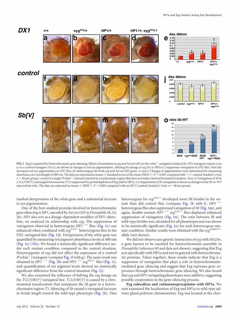
FIG 1 Eyg is required for heterochromatic gene silencing. Effects of mutations in eyg and Su(var)205 on the white� variegation induced by DX1 transgene repeats (a toe) or a control transgene (f to j), are shown as changes in red eye pigmentation. Altering the dosage of eyg (b) or HP1a (c) suppresses variegation in DX1 flies. Note theincreased red eye pigmentation in DX1 flies (d) heterozygous for both eyg and Su(var)205 genes. (e and j) Changes in pigmentation were determined by measuringabsorbance at a wavelength of 480 nm. The data are expressed as means � standard errors of the mean (SEM) (*, P � 0.001 compared with �/� control; Student’s t test;n 60 per group). control is a single P[white�] element inserted in a euchromatic region that does not induce heterochromatin formation. (k to o) Variegation of Sb ina T(2;3)Sb[V] rearranged chromosome (7) is suppressed by partial depletion of Eyg and/or HP1a. (o) Suppression of Sb variegation is shown as changes in the Sb-to-WTmacrochete ratio. The data are expressed as means � SEM (*, P � 0.005 compared with an Sb[V] control; Student’s t test; n 40 per group).
HP1a and Eyg Interact during Eye Development
July 2012 Volume 32 Number 13 mcb.asm.org 2409

mocenter (Fig. 2c, arrow, and c=), where the HP1a protein was alsodetected (Fig. 2b, arrow, and b=), and was also found in otherheterochromatin regions, such as that on chromosome 4 (notshown). We also detected colocalization of Eyg with HP1a in somebands along the chromosomal arms (Fig. 2e and f, arrows) thatcould be targets of Eyg. In this context, we found that HP1a local-izes to the wg genomic region, a known target of Eyg during Dro-sophila eye development (16, 20) (Fig. 2g to i).
To further confirm that Eyg is associated with heterochroma-tin, we performed ChIP assays using the transposable 1360 ele-ment as a heterochromatin marker (42). We used this transpos-able element because the terminal inverted-repeat 1360 elementinitiates heterochromatic domains (9, 42). Enrichment of HP1binding to 1360 sequences was described previously (9), and wefound similar enrichment of Eyg binding to these 1360 elements(expressed as a percentage of the input) (Fig. 2j), suggesting thatboth Eyg and HP1a bind to the 1360 sequence.
A similar degree of enrichment was found in the heterochro-matic H23 locus (region 22000 to 24000 of chromosome 2 [chr2]heterochromatin) (Fig. 2j) (41). We used the promoter region oflabial as a negative control, because neither Eyg nor HP1a binds toit (Fig. 2j).
We also found that Eyg and HP1a coimmunoprecipitated fromsalivary gland extracts (Fig. 2k), indicating that Eyg and HP1a areparts of the same protein complex. The consensus pentapeptidesufficient for interaction with HP1 has been identified as (PL)(WRY)V(MIV)(MLV) (39). Eyg does not contain an exact con-sensus motif but does contain a PSVLL domain in the C-terminalpart of the protein (residues 472 to 476). A previous characteriza-tion of Eyg functional domains showed that the C-terminal regionis not necessary for the repressive function of Eyg (47). In order totest if the PSVLL domain of Eyg was necessary for binding toHP1a, we generated an Eyg mutant in which PSVLL was replacedby PSSLN (as described in reference 28) and assayed its binding toHP1a in stably transfected S2 cells. High endogenous levels ofHP1a and undetectable expression of Eyg in S2 were confirmedusing the Drosophila Schneider S2 cell microarray data set gener-ated and described previously (35). We found that both EygPSSLN
and EygPSVLL coimmunoprecipitated with HP1a, indicating thatthe PSVLL of Eyg is dispensable for the binding of HP1a (Fig. 2l).
HP1a mediates wg repression in the eye imaginal discthrough heterochromatic gene silencing. Because Eyg is a knowntranscriptional repressor, we speculated that its repressor activitymight be executed by recruiting HP1a to chromatin, triggeringgene silencing. The wg gene is a known target of Eyg during eyedevelopment (16, 20), and the minimal wg enhancer region thatdrives wg expression in the eye imaginal disc has been defined (33)
FIG 2 Eyg and HP1a colocalize to heterochromatic regions. (a to c) Eyg andHP1a colocalize to the heterochromatic regions of salivary gland polytenechromosomes (arrows in panels b and c). (a= to c=) Enlargement of the chro-mocenter of the chromosome. (d to f) Eyg and HP1a also colocalize in someinterband regions of salivary gland polytene chromosomes (arrows in panels eand f). (g to i) HP1a localizes to the wg genomic region, marked by a fluores-cent DNA probe (asterisks in panels h and i). (j) Enrichment of constitutiveheterochromatic regions 1360 and H23 when immunoprecipitated with anti-Eyg, expressed as the percentage of input (1:100 dilution) in ChIP experi-ments. HP1a binding to the same heterochromatic regions was also observed.Control refers to chromatin immunoprecipitated with guinea pig preimmune
serum. An isotype control for mouse antibody gave similar results (notshown). The same immunoprecipitated DNA was amplified with primers forthe labial genomic region, the wg promoter, and the act5c promoter region,which were used as negative controls. (k) Eyg and HP1a belong to the sameprotein complex. Western blot analyses detected HP1a protein in salivarygland extracts immunoprecipitated with anti-Eyg antibody and Eyg protein insalivary gland extracts immunoprecipitated with anti-HP1a antibody. In thecontrol lanes, salivary gland extracts were immunoprecipitated with guinea pigpreimmune serum or with a monoclonal anti-�-tubulin antibody (control).(l) An EygPSSLN mutant for the PSVLL motif is able to bind HP1a in S2 cells, asdoes wild-type Eyg (EygPSVLL). negative, no antibody. 1 and 2 refer to twoindependent coimmunoprecipitation experiments.
Salvany et al.
2410 mcb.asm.org Molecular and Cellular Biology
(Fig. 3a, wg 2.10). To determine whether wg repression by Eyg wasmediated by gene silencing and generated changes in its chroma-tin structure, we performed DNase I sensitivity assays to analyzethe chromatin structure of this wg enhancer region in eye imaginaldiscs. Quantitative PCR was performed on native (undigested; 0U) chromatin samples and on samples digested with DNase I (50U). The CT value was calculated as a function of the amount ofDNA at the outset (unless otherwise stated, CT values refer to CT
[50 U] � CT [0 U]).For the transcribed region of actin (act5c), a constitutively ac-
tive gene used as a positive control for the “open” chromatin state,the CT value was around 5 in wild-type, eyg20MD1/� heterozygous
and HP1a mutant (act�HP1aRNAi) eye-antennal disc extracts(Fig. 3b). We used the Gal4-UAS binary system to eliminate HP1via RNA interference (RNAi), because HP1 mutations are reces-sive lethal (see below). In contrast, the heterochromatic H23 re-gion (22,000 to 24,000 of chromosome 2 heterochromatin) (50),used as a negative control for the “closed” chromatin state, wasrefractory to DNase I treatment, exhibiting a lower CT value inboth wild-type and eyg20MD1/� heterozygous eye-antennal disc ex-tracts (Fig. 3b). In act�HP1aRNAi extracts, the H23 region be-came sensitive to DNase I, supporting a role for HP1a in consti-tutive heterochromatin formation/maintenance.
We found that in wild-type control eye-antennal imaginaldiscs, the wg enhancer region behaved like heterochromatin, witha CT between 0 and 2 (Fig. 3b). However, the same genomicregion was almost as sensitive to DNase I as the act5c locus regionin eyg20MD1/� and act�HP1aRNAi mutant extracts, with a CT
value between 3.5 and 5 (Fig. 3b). Halving the dosage of eyg wassufficient to change the sensitivity to digestion by DNase I of thewg enhancer region. Consistent with this, in some heterozygouseyg20MD1/� eye discs, the change in chromatin structure led to thederepression of wg described previously (16, 47) and gave rise to asmall-eye phenotype (not shown). This result indicates the highsensitivity achieved with the technique and also suggests that theremight be other mechanisms regulating wg expression.
Using ChIP experiments, we confirmed that Eyg and HP1abind similarly to this wg region (Fig. 3c), but not to the wg pro-moter region or to the act5c promoter used as controls (Fig. 2j).The amount of HP1a protein bound to the wg enhancer region ineyg20MD1/� was reduced (Fig. 3c), but to a lesser extent than theEyg protein. This result suggests that not all HP1a binding to thisenhancer region depends on Eyg. We believe that Eyg may recruitHP1a to the wg enhancer region to form a heterochromatin struc-ture that most likely represses wg expression in the eye. To furtheranalyze this hypothesis, we tried to determine the localization ofHP1a in the polytene chromosomes of eyg mutant larvae. How-ever, eyg mutant salivary glands are very small and contain fragilechromosomes, which we were unable to squash.
Loss of HP1a leads to wg misexpression in the eye and wingdisc. To confirm that HP1a binding to the wg enhancer region inthe eye is indeed functionally important, we expressed a specificinterfering RNA against HP1a in random clones of cells in eyeimaginal disc using the actin-Gal4 Drosophila line. The Gal4-UASsystem combined with the flippase recombination target (FRT)system was used to generate the RNAi-expressing clones. In sum-mary, flies of the genotype hsflp122;actFRTstopFRTGal4UASGFP/Cyo were crossed to flies containing a UASHP1RNAi construct.Second-instar larvae were then heat shocked for 10 min and dis-sected in the third-instar period to visualize the discs. Because ofthe FRT cassette, only clones of cells in which recombination hasoccurred (and that no longer have the cassette) will activate Gal4and, consequently, express the interfering RNA that reduces oreliminates the HP1a product.
Reduction of HP1a levels resulted in ectopic activation of wg (Fig.4a, b, and f), as previously described for eyg mutant eye discs (16).
Ectopic activation of wg in eye discs of HP1aRNAi clones didnot occur in all parts of the disc domain and was more specific forthe middle region. We found that 32% of HP1aRNAi clones (n 278) were positive for ectopic wg activation, and more than 90% ofthese positive clones showed ectopic expression in the middle por-tion of the third-instar eye imaginal disc (Fig. 4d). These results
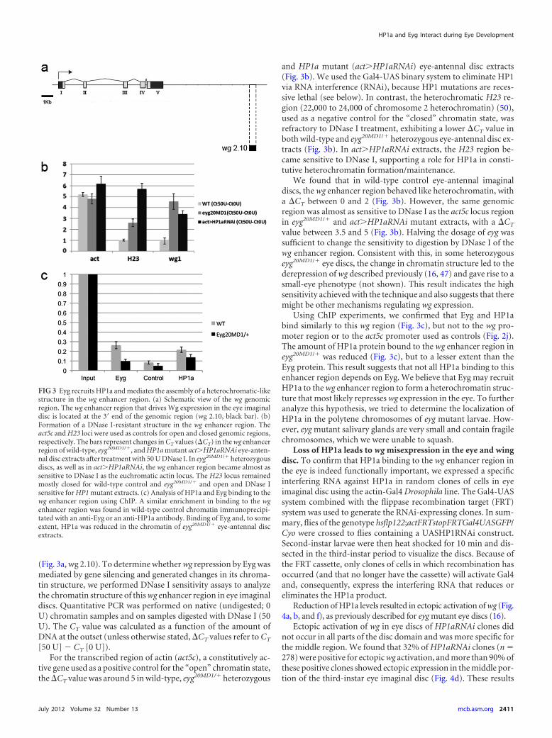
FIG 3 Eyg recruits HP1a and mediates the assembly of a heterochromatic-likestructure in the wg enhancer region. (a) Schematic view of the wg genomicregion. The wg enhancer region that drives Wg expression in the eye imaginaldisc is located at the 3= end of the genomic region (wg 2.10, black bar). (b)Formation of a DNase I-resistant structure in the wg enhancer region. Theact5c and H23 loci were used as controls for open and closed genomic regions,respectively. The bars represent changes in CT values (CT) in the wg enhancerregion of wild-type, eyg20MD1/�, and HP1a mutant act�HP1aRNAi eye-anten-nal disc extracts after treatment with 50 U DNase I. In eyg20MD1/� heterozygousdiscs, as well as in act�HP1aRNAi, the wg enhancer region became almost assensitive to DNase I as the euchromatic actin locus. The H23 locus remainedmostly closed for wild-type control and eyg20MD1/� and open and DNase Isensitive for HP1 mutant extracts. (c) Analysis of HP1a and Eyg binding to thewg enhancer region using ChIP. A similar enrichment in binding to the wgenhancer region was found in wild-type control chromatin immunoprecipi-tated with an anti-Eyg or an anti-HP1a antibody. Binding of Eyg and, to someextent, HP1a was reduced in the chromatin of eyg20MD1/� eye-antennal discextracts.
HP1a and Eyg Interact during Eye Development
July 2012 Volume 32 Number 13 mcb.asm.org 2411
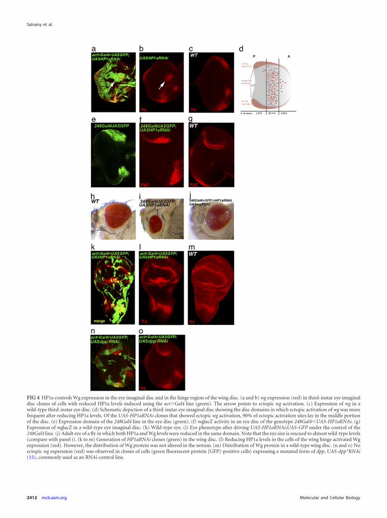
FIG 4 HP1a controls Wg expression in the eye imaginal disc and in the hinge region of the wing disc. (a and b) wg expression (red) in third-instar eye imaginaldisc clones of cells with reduced HP1a levels induced using the act�Gal4 line (green). The arrow points to ectopic wg activation. (c) Expression of wg in awild-type third-instar eye disc. (d) Schematic depiction of a third-instar eye imaginal disc showing the disc domains in which ectopic activation of wg was morefrequent after reducing HP1a levels. Of the UAS-HP1aRNAi clones that showed ectopic wg activation, 90% of ectopic activation sites lay in the middle portionof the disc. (e) Expression domain of the 248Gal4 line in the eye disc (green). (f) wglacZ activity in an eye disc of the genotype 248Gal4�UAS-HP1aRNAi. (g)Expression of wglacZ in a wild-type eye imaginal disc. (h) Wild-type eye. (i) Eye phenotype after driving UAS-HP1aRNAi;UAS-GFP under the control of the248Gal4 line. (j) Adult eye of a fly in which both HP1a and Wg levels were reduced in the same domain. Note that the eye size is rescued to almost wild-type levels(compare with panel i). (k to m) Generation of HP1aRNAi clones (green) in the wing disc. (l) Reducing HP1a levels in the cells of the wing hinge activated Wgexpression (red). However, the distribution of Wg protein was not altered in the notum. (m) Distribution of Wg protein in a wild-type wing disc. (n and o) Noectopic wg expression (red) was observed in clones of cells (green fluorescent protein [GFP]-positive cells) expressing a mutated form of dpp, UAS-dpp*RNAi(15), commonly used as an RNAi control line.
Salvany et al.
2412 mcb.asm.org Molecular and Cellular Biology
suggest a positional restriction for ectopic wg activation in the eyedisc, as has also been described for eyg mutants (16).
Interestingly, the HP1RNAi clones generated in the wing ima-ginal disc also exhibited derepression of wg in the correspondinghinge region, where eyg has been shown to regulate wg (20) (Fig.4k and l), but not in the notum, where wg expression does notdepend on eyg.
HP1a has been shown to control gene expression at the post-transcriptional level (34). To determine if the observed regulationof wg expression occurs at the transcriptional level, we expressedHP1a RNAi under the control of the 248Gal4 driver (which ex-presses Gal4 in a discrete domain of the eye disc) by crossing theUAS-HP1aRNAi line with the 248Gal4 line in a wgLacZ back-ground. Reducing HP1a levels resulted in ectopic wgLacZ expres-sion (Fig. 4f), suggesting that wg activation actually occurs at thetranscriptional level.
To rule out the possibility that derepression of wg by HP1aRNAi is a more general effect of the RNAi machinery, we per-formed a series of experiments in which shmiR-dpp2HBmutant (15)was expressed in clones of cells in the eye imaginal disc. In contrastto the effects of expressing HP1aRNAi, wg was not derepressed byexpressing shmiR-dpp2HBmutant (Fig. 4n and o), indicating thatderepression of wg in the eye disc is not a general effect of the RNAimachinery on heterochromatin assembly, but rather a specific ef-fect of reducing HP1a levels.
On the basis of these data, we conclude that HP1a is involved inwg repression in the eye imaginal disc and in the hinge region ofthe wing disc, where wg is a target of Eyg. We did not detect anyalteration of wg expression in the notum region of the wing disc(Fig. 4l), where Eyg is also expressed and was shown to be func-tional (3) but does not control wg expression (20).
We also sought to analyze the phenotypic outcome in adultflies with reduced HP1a levels in the eye disc. To this end, weexpressed UAS-HP1aRNAi together with UAS-GFP (see below)under the control of the above-mentioned 248Gal4 driver, whichallows flies to develop to adulthood (Fig. 4e) (see Materials andMethods). Adult flies carrying this genetic combination showed areduction in eye size (Fig. 4i). To further demonstrate that this wasdue, at least in part, to ectopic activation of wg in the eye field, weperformed an experiment in which we coexpressed UAS-HP1aRNAi and UAS-wgRNAi under the control of the 248Gal4driver. The flies were allowed to develop at 29°C until adulthood.The transgenes were balanced on different chromosomes, so thatwe were able to distinguish flies that carried either or both trans-genes in the same cross. Flies of the genotype 248Gal4;UAS-wgRNAi;UAS-HP1aRNAi showed some head defects, but eye sizewas rescued to almost normal (compare Fig. 4i to j). This resultstrongly suggests that the reduction in eye size that we observedfollowing reduction of HP1a levels depends on ectopic wg activa-tion. Introducing a second UAS transgene (UAS-GFP) when ex-pressing UAS-HP1aRNAi allowed us to eliminate the possibility ofa Gal4 protein titration effect in the experiment.
DISCUSSION
The Pax protein Eyg, a product of the eyg gene, is a transcriptionalrepressor known to function during all developmental stages inDrosophila. One known target of Eyg is wg. It has been previouslyreported that Eyg represses wg in the eye and that this repression isa critical function of Eyg (16). In this work, we investigated themolecular mechanism underlying the repressive function of Eyg,
examining repression of wg using the 3= cis-regulatory regionknown to control wg expression in the Drosophila eye as an assayto study Eyg’s mode of action. On the basis of our findings, weconclude that Eyg acts through heterochromatic gene silencing.
PEV experiments demonstrated that Eyg is a suppressor of var-iegation that is required for heterochromatic gene silencing andsuggested that Eyg represses its target genes through this mecha-nism. We also demonstrated a genetic interaction between eyg andHP1a in the modification of PEV, suggesting that the products ofthese two genes act together in the gene-silencing process. Consis-tent with this interpretation, we found that Eyg colocalized withHP1a protein on polytene chromosomes in the chromocenter, onchromosome 4, in telomeres, and in some discrete bands along thechromosomes. The localization of Eyg on polytene chromosomessuggests that the two other ubiquitously expressed HP1-like pro-teins, HP1b and HP1c, are not Eyg partners because we did notdetect Eyg protein on the many euchromatic bands where HP1band HP1c localize on polytene chromosomes (13, 49a) (Fig. 2c).
Eyg contains a PSVLL motif at residues 472 to 476 located inthe C-terminal domain of the protein. This is similar to the con-served pentapeptide motif present in the HP1 chromodomain andin other known HP1-interacting proteins (6, 38). However, whenthis domain was mutated in the Eyg protein, the interaction withHP1a was not affected, suggesting that the PSVLL motif in Eyg isdispensable for interaction with HP1a. This consensus sequence isabsent in other well-known HP1 interactors, like INCENP (2),Su(var)3-9 (1), and the lamin B receptor (49; for a review, seereference 39). Eyg could therefore be another example of an HP1interactor that engages HP1 differently from proteins that containthe consensus; alternatively, it could interact with HP1a indi-rectly. This result is consistent with a previous report by Yao andSun, who showed that the C-terminal domain of Eyg is not essen-tial for the repression of wg in the eye disc (47). The Eyg proteindomain responsible for HP1a binding and the identity of the scaf-fold protein(s) that complexes Eyg and HP1a are still to be deter-mined.
The results of ChIP experiments designed to determine if Eygand HP1a bind to the same DNA sequences in vivo showed thatboth proteins bound to the same terminal inverted-repeat 1360element, which initiates heterochromatic domains (9, 42); to theH23 locus, a region of heterochromatin of chromosome 2 (Fig.2j); and to the wg enhancer region known to be responsible fortrans-activation of the wg gene in the eye (and where binding sitesfor Pax-type paired-domain transcription factors have been pre-viously reported [33]). However, both chromatin domains be-haved differently in the absence of Eyg. Using DNase I sensitivityassays, we showed that, in eyg mutants, the 1360 element remainedconsiderably more refractory to DNase I treatment and thus moreclosed (although not as closed as in the wild type) (data notshown). It should also be noted that, for all the “closed” genomicregions tested in our DNase I sensitivity assays, none remained asclosed in eyg mutants after DNase I treatment as they did in thewild type; even the H23 locus was more open in the mutant back-ground than in the wild type (Fig. 3b). This result points to a moregeneral role for Eyg in heterochromatin formation and/or main-tenance that warrants further analysis. In this context, Eyg func-tion does not seem to depend on HP1a, because eliminating HP1aresults in a CT value of 5 to 6 in the DNase I sensitivity assay onthe H23 locus (Fig. 3b).
The wg enhancer region behaves differently. In eyg mutants, it
HP1a and Eyg Interact during Eye Development
July 2012 Volume 32 Number 13 mcb.asm.org 2413

adopted an open chromatin state (with CT values between 4 and5), as it does in the absence of HP1a (Fig. 3b). In this region, Eygand HP1a cooperate to silence gene expression. Binding of Eygand HP1a is not a general feature of the wg locus, because we didnot observe any binding to the wg promoter.
HP1a binding to the wg enhancer region was greatly reduced ineyg heterozygous mutant extracts, but to a lesser extent than it wasthe Eyg protein, suggesting that not all of the HP1a binding to thatregion depends on Eyg. However, the derepression of wg was ev-ident in the eye imaginal disc of heterozygous eyg mutants, result-ing in a small-eye phenotype in the adult fly (not shown). Onepossible explanation for this observation is that the HP1a thatremains bound to the wg locus is insufficient to repress wg expres-sion or that it has another, as yet undetermined role.
In yeast, it has been demonstrated that HP1 proteins formdistinct complexes with different heterochromatin-related func-tions (27). In Drosophila, HP1-mediated silencing has been re-ported to operate in both a Su(Var)3-9-dependent and -indepen-dent manner (8). HP1 silencing of up to 1.9 kb has been shown tobe Su(Var)3-9 independent, whereas long-range silencing de-pends upon the presence of Su(var)3-9. We propose that HP1-dependent wg silencing is Eyg dependent but Su(Var)3-9 inde-pendent, whereas for the heterochromatinization of other regions(e.g., H23 or the 1360 element), the presence of Eyg is not crucial,but H3K9 methylation is. Indeed, HP1a and 3mH3K9 colocalizeto constitutive heterochromatin regions in salivary gland nuclei ofwild-type larvae (12) and still do so in eyg mutant larvae (notshown). However, 3mH3K9 is never detected on the chromo-somal bands where HP1a and Eyg colocalize (reference 12 and ourobservations).
HP1a has recently been shown to have a role in the posttran-scriptional upregulation of many euchromatic genes (34) owingto the association of its chromodomain with different transcripts.This unexpected role of HP1a indicates that HP1a is a multifunc-tional protein whose versatility lies in its capacity to interact withdifferent partners in distinct contexts. It has been proposed thatthe main function of HP1a is nucleic acid compaction, by inter-acting either with modified histones or with specific hnRNP pro-teins (34). Our results point to the same conclusion in that theinteraction of HP1a with Eyg leads to compaction of the DNA atthe wg enhancer region and subsequent silencing of the gene. Inthis way, the HP1a protein could fulfill at least two different func-tions: assembly of the constitutive heterochromatin presentin telomeres and pericentromeres and on chromosome 4 by bind-ing to methylated histone H3K9 and (once targeted to regulatoryregions) specific euchromatic gene repression by interacting withtranscription factors to form a repressive chromatin structure. Inthe latter case, the resulting chromatin structure has a heterochro-matin appearance and exerts heterochromatin-like repression.
In conclusion, we describe a physical and functional interac-tion between Eyg and HP1a that regulates wg expression in the eye.Although HP1 has been shown to interact with other transcriptionfactors, like those encoded by retinoblastoma (Rb) or Krab (30,36), mediating the repression of their targets, a direct role of HP1ain this process has not previously been described. Our experi-ments show that HP1a is necessary for the repression of wg in theeye imaginal disc, endowing HP1a with a restricted function indevelopment. In the same way that Eyg represses wg in the eye disc,other repressors could also interact with HP1a or other hetero-chromatin proteins to mediate spatial and/or temporal repression
of their targets through heterochromatic gene silencing. This pos-sibility opens new avenues that link specific transcription factorswith general modifiers of chromatin.
ACKNOWLEDGMENTS
We thank Ginés Morata, Ernesto Sánchez-Herrero, Marco Milán, andHéctor Herranz for helpful discussions in the course of this work. We alsothank all the Morata and Sánchez-Herrero laboratory members for theircontinuous support, as well as Elise Corsetti and Sara Mederos for helpwith the experiments.
This work was supported by grants from the Ministerio Españolde Ciencia y Tecnología (BFU2009-12152), the Consolider program(CSD2007-00008), and the Fundación Ramón Areces.
REFERENCES1. Aagaard L, et al. 1999. Functional mammalian homologues of the Dro-
sophila PEV-modifier Su(var)3-9 encode centromere-associated proteinswhich complex with the heterochromatin component M31. EMBO J. 18:1923–1938.
2. Ainsztein AM, Kandels-Lewis SE, Mackay AM, Earnshaw WC. 1998.INCENP centromere and spindle targeting: identification of essential con-served motifs and involvement of heterochromatin protein HP1. J. CellBiol. 143:1763–1774.
3. Aldaz S, Morata G, Azpiazu N. 2003. The Pax-homeobox gene eyegoneis involved in the subdivision of the thorax of Drosophila. Development130:4473– 4482.
4. Chi N, Epstein JA. 2002. Getting your Pax straight: Pax proteins indevelopment and disease. Trends Genet. 18:41– 47.
5. Comet I, et al. 2006. PRE-mediated bypass of two Su(Hw) insulatorstargets PcG proteins to a downstream promoter. Dev. Cell 11:117–124.
6. Cowieson NP, Partridge JF, Allshire RC, McLaughlin PJ. 2000. Dimeri-sation of a chromo shadow domain and distinctions from the chromodo-main as revealed by structural analysis. Curr. Biol. 10:517–525.
7. Csink AK, Linsk R, Birchler JA. 1994. The Lighten up (Lip) gene ofDrosophila melanogaster, a modifier of retroelement expression, positioneffect variegation and white locus insertion alleles. Genetics 138:153–163.
8. Danzer JR, Wallrath LL. 2004. Mechanisms of HP1-mediated gene si-lencing in Drosophila. Development 131:3571–3580.
9. De Lucia F, Ni JQ, Vaillant C, Sun FL. 2005. HP1 modulates thetranscription of cell-cycle regulators in Drosophila melanogaster. NucleicAcids Res. 33:2852–2858.
10. Dominguez M, Ferres-Marco D, Gutierrez-Avino FJ, Speicher SA,Beneyto M. 2004. Growth and specification of the eye are controlledindependently by Eyegone and Eyeless in Drosophila melanogaster. Nat.Genet. 36:31–39.
11. Dorer DR, Henikoff S. 1994. Expansions of transgene repeats cause het-erochromatin formation and gene silencing in Drosophila. Cell 77:993–1002.
11a.Easow G, Teleman AA, Cohen SM. 2007. Isolation of microRNA targetsby miRNP immunopurification. RNA 13:1198 –1204.
12. Ebert A, Lein S, Schotta G, Reuter G. 2006. Histone modification and thecontrol of heterochromatic gene silencing in Drosophila. ChromosomeRes. 14:377–392.
13. Font-Burgada J, Rossell D, Auer H, Azorin F. 2008. Drosophila HP1cisoform interacts with the zinc-finger proteins WOC and Relative-of-WOC to regulate gene expression. Genes Dev. 22:3007–3023.
14. Grewal SI, Moazed D. 2003. Heterochromatin and epigenetic control ofgene expression. Science 301:798 – 802.
15. Haley B, Hendrix D, Trang V, Levine M. 2008. A simplified miRNA-based gene silencing method for Drosophila melanogaster. Dev. Biol. 321:482– 490.
16. Hazelett DJ, Bourouis M, Walldorf U, Treisman JE. 1998. decapentaple-gic and wingless are regulated by eyes absent and eyegone and interact todirect the pattern of retinal differentiation in the eye disc. Development125:3741–3751.
17. Hunt DM. 1970. Lethal interactions of the eye-gone and eyeless mutantsin Drosophila melanogaster. Genet. Res. 15:29 –34.
18. Inoue Y. 1980. Report of new mutants-Drosophila melanogaster. Dros-oph. Inf. Serv. 55:206.
19. James TC, et al. 1989. Distribution patterns of HP1, a heterochromatin-
Salvany et al.
2414 mcb.asm.org Molecular and Cellular Biology
associated nonhistone chromosomal protein of Drosophila. Eur. J. CellBiol. 50:170 –180.
20. Jang CC, et al. 2003. Two Pax genes, eye gone and eyeless, act coopera-tively in promoting Drosophila eye development. Development 130:2939 –2951.
21. Jones NA, Kuo YM, Sun YH, Beckendorf SK. 1998. The Drosophila Paxgene eye gone is required for embryonic salivary duct development. Devel-opment 125:4163– 4174.
22. Jun S, Wallen RV, Goriely A, Kalionis B, Desplan C. 1998. Lune/eyegone, a Pax-like protein, uses a partial paired domain and a homeodomainfor DNA recognition. Proc. Natl. Acad. Sci. U. S. A. 95:13720 –13725.
23. Kwon SH, Workman JL. 2008. The Heterochromatin Protein 1 (HP1)family: put away a bias toward HP1. Mol. Cells 26:217–227.
24. Lachner M, O’Carroll D, Rea S, Mechtler K, Jenuwein T. 2001. Meth-ylation of histone H3 lysine 9 creates a binding site for HP1 proteins.Nature 410:116 –120.
25. Lavrov S, Dejardin J, Cavalli G. 2004. Combined immunostaining andFISH analysis of polytene chromosomes. Methods Mol. Biol. 247:289 –303.
26. Lu BY, Ma J, Eissenberg JC. 1998. Developmental regulation of hetero-chromatin-mediated gene silencing in Drosophila. Development 125:2223–2234.
27. Motamedi MR, et al. 2008. HP1 proteins form distinct complexes andmediate heterochromatic gene silencing by nonoverlapping mechanisms.Mol. Cell 32:778 –790.
28. Murzina N, Verreault A, Laue E, Stillman B. 1999. Heterochromatindynamics in mouse cells: interaction between chromatin assembly factor 1and HP1 proteins. Mol. Cell 4:529 –540.
29. Negre N, Lavrov S, Hennetin J, Bellis M, Cavalli G. 2006. Mapping thedistribution of chromatin proteins by ChIP on chip. Methods Enzymol.410:316 –341.
30. Nielsen SJ, et al. 2001. Rb targets histone H3 methylation and HP1 topromoters. Nature 412:561–565.
31. Noll M. 1993. Evolution and role of Pax genes. Curr. Opin. Genet. Dev.3:595– 605.
32. Paro R. 1993. Mechanisms of heritable gene repression during develop-ment of Drosophila. Curr. Opin. Cell Biol. 5:999 –1005.
33. Pereira PS, Pinho S, Johnson K, Couso JP, Casares F. 2006. A 3=cis-regulatory region controls wingless expression in the Drosophila eyeand leg primordia. Dev. Dyn. 235:225–234.
34. Piacentini L, et al. 2009. Heterochromatin protein 1 (HP1a) positivelyregulates euchromatic gene expression through RNA transcript associa-tion and interaction with hnRNPs in Drosophila. PLoS Genet.5:e1000670. doi:10.1371/journal.pgen.1000670.
35. Rehwinkel J, et al. 2006. Genome-wide analysis of mRNAs regulated byDrosha and Argonaute proteins in Drosophila melanogaster. Mol. Cell.Biol. 26:2965–2975.
35a.Reuter G, Wolff Y. 1981. Isolation of dominant suppressor mutations for
position-effect variegation in Dorosophila melanogaster. Mol. Gen.Genet. 182:516 –519.
36. Ryan RF, et al. 1999. KAP-1 corepressor protein interacts and colocalizeswith heterochromatic and euchromatic HP1 proteins: a potential role forKruppel-associated box-zinc finger proteins in heterochromatin-mediated gene silencing. Mol. Cell. Biol. 19:4366 – 4378.
37. Sanchez L, Casares F, Gorfinkiel N, Guerrero I. 1997. The genital disc ofDrosophila melanogaster. Dev. Genes Evol. 207:219 –241.
38. Smothers JF, Henikoff S. 2001. The hinge and chromo shadow domainimpart distinct targeting of HP1-like proteins. Mol. Cell. Biol. 21:2555–2569.
39. Smothers JF, Henikoff S. 2000. The HP1 chromo shadow domain bindsa consensus peptide pentamer. Curr. Biol. 10:27–30.
40. Stephens GE, Craig CA, Li Y, Wallrath LL, Elgin SC. 2004. Immuno-fluorescent staining of polytene chromosomes: exploiting genetic tools.Methods Enzymol. 376:372–393.
41. Stewart MD, Li J, Wong J. 2005. Relationship between histone H3 lysine9 methylation, transcription repression, and heterochromatin protein 1recruitment. Mol. Cell. Biol. 25:2525–2538.
42. Sun FL, et al. 2004. cis-Acting determinants of heterochromatin forma-tion on Drosophila melanogaster chromosome four. Mol. Cell. Biol. 24:8210 – 8220.
43. Swaminathan J, Baxter EM, Corces VG. 2005. The role of histone H2Avvariant replacement and histone H4 acetylation in the establishment ofDrosophila heterochromatin. Genes Dev. 19:11.
44. Treisman J, Harris E, Desplan C. 1991. The paired box encodes a secondDNA-binding domain in the paired homeo domain protein. Genes Dev.5:594 – 604.
45. Vermaak D, Henikoff S, Malik HS. 2005. Positive selection drives theevolution of rhino, a member of the heterochromatin protein 1 family inDrosophila. PLoS Genet. 1:96 –108.
46. Wang LH, Chiu SJ, Sun YH. 2008. Temporal switching of regulation andfunction of eye gone (eyg) in Drosophila eye development. Dev. Biol.321:515–527.
47. Yao JG, Sun YH. 2005. Eyg and Ey Pax proteins act by distinct transcrip-tional mechanisms in Drosophila development. EMBO J. 24:2602–2612.
48. Yao JG, et al. 2008. Differential requirements for the Pax6(5a) geneseyegone and twin of eyegone during eye development in Drosophila. Dev.Biol. 315:535–551.
49. Ye Q, Worman HJ. 1996. Interaction between an integral protein of thenuclear envelope inner membrane and human chromodomain proteinshomologous to Drosophila HP1. J. Biol. Chem. 271:14653–14656.
49a.Zhang D, Wan D, Sun F. 2011. Drosophila melanogaster heterochromatinprotein HP1b plays important roles in transcriptional activation and de-velopment. Chromosoma 120:97–108.
50. Zhang Y, et al. 2008. Epigenetic blocking of an enhancer region controlsirradiation-induced proapoptotic gene expression in Drosophila em-bryos. Dev. Cell 14:481– 493.
HP1a and Eyg Interact during Eye Development
July 2012 Volume 32 Number 13 mcb.asm.org 2415