
Function, Pharmacology, Evolution and Anatomical Localization
50
Transcript of Function, Pharmacology, Evolution and Anatomical Localization

������������������� ���������� �
����
���������� �������������� ������������������� ������� ����������������������������
������� ������������� ���������� �������� ��������� �� � ������ ��!��� "���!��# ��� $���� �����#
%�%&�'� (�)%)'�
)$$' *+,*�+-.+)$/' 0�1�0*�,,����++�0���2�3�2#�2��2����00�*�

����������������������� ������� ��������������������������������������������� ���������������������� ��� !!"����!"#�$�%���&����'����%������%�(&����&��)��������%�������*+�,&�����������-�����������������.'���&+
��������
/�������,+� !!"+��������(&������'���.������������������0����1����%�2�(����3�������������������4�������������+������ ������������ ���������+����������� � ���� ������ ���������������� ������������� ������������ ����� ��56+��"���+ ������+������"673"�3$$�36�883"+
,&��2������3����������������)2(���*�������������������)40�*������-����'��%��������%�������3����������+�,&������%��&���������-������&��������1���&�����-�%���������������%�����������%����+�,&�����������)��*�������������'����&����������%������%2(�������-��������&�����������$����������%����&������-���������5������$�%���&��������'%��&���������������%����&���������������+�(&������'������&��������1���%� �&�� ����� ��� ��������� ����������� &�'&��� �%%����� %�� ��������������� &���)��,/*� �������� �� ��������� ���������'� &���� )�4/*� ��������� )���&�3�� ����3� ��'����3�4/*+� 9�� ���%����� ��������� �������� -��&� �������� ����������� (���� -&��&�&-����&����&�����������$�������������&������-��������������������������������-������ �������&����� �������+�,&���'%��&���5������$� ���������-�������������� �� �&�� �����-&���� �&�� ������������ ������� ���������-���� ���������� �� �&�������&���+��� ��������������������1������������-������%�����%���&��������%������%���� ����2(�������&������������+� ��'�:���������������3�����(���):�,3(��*�-�������������&����&����;�����%2(����-�����������������&��������%����������&���<4������:�����������&���<4���������&�����������+�9�������%��������3�%������2(�������&���'����������%�����&��������� ���������� ���������� �������������� �� ����%�������+�,&���'�2(���������������������������������������&��&���������������&�����&��������������������+�4�������������%������ $������������&������������������������+� ��'����%�����������&�:����-�������%������������������%��&��40� $�%�������-&��&�-�&����8��������+�9�������%������&�'�%��&������40� $�%����������������������������%����������������������%�"�%��&��-��&�:�,3(����������������'�5!����������������&�������������%����&�����+�,���������&���������&��������������=-���'��%��&������������%�'�������'�%���������3����������������������%������������&����%�����������+
�!���"���,/����&������'���'%��&������������2(�����������������������������4/���&������'���(>�������������
#��$����%������&�� ���� �����' ����� �� &�()�*+,&������������� �����&��-./*012�������&��! � �
?�,��;���/������ !!"
@44<��8$�38 !8@4�<�"673"�3$$�36�883"��#�#��#��#����3""��5�)&���#AA��+=�+��A������B��C��#�#��#��#����3""��5*

"My eyes already touch the sunny hill. going far ahead of the road I have begun.
So we are grasped by what we cannot grasp; it has inner light, even from a distance-
and charges us, even if we do not reach it,
into something else, which, hardly sensing it, we already are; a gesture waves us on
answering our own wave... but what we feel is the wind in our faces."
Rainer Maria Rilke
(March 1924)
To my family
���� �����


List of publications
This thesis is based on the following papers, which are referred to in the text by their Roman numerals.
I Haitina T, Klovins J, Andersson J, Fredriksson R, Lagerström MC, Larhammar D, Larson ET, Schiöth HB. (2004) Cloning, tissue distribution, pharmacology and three-dimensional modelling of melanocortin receptors 4 and 5 in rainbow trout suggest close evolutionary relationship of these subtypes. Biochem J, 380(Pt2):475-486.
II Klovins J*, Haitina T*, Ringholm A, Löwgren M, Fridmanis D, Slaidina M, Stier S, Schiöth HB. (2004) Cloning of two melanocortin (MC) receptors in spiny dogfish: MC3 receptor in cartilaginous fish shows high affinity to ACTH-derived peptides while it has lower preference to gamma-MSH. Eur J Biochem, 271(21):4320-4331. * contributed equally to this work
III Haitina T, Takahashi A, Holmén L, Enberg J, Schiöth HB. (2007) Further evidence for ancient role of ACTH peptides at melanocortin (MC) receptors; pharmacology of dogfish and lamprey peptides at dogfish MC receptors. Pep-tides, 28(4):798-805.
IV Haitina T, Klovins J, Takahashi A, Löwgren M, Ringholm A, Enberg J, Kawauchi H, Larson ET, Fredriksson R, Schiöth HB. (2007) Functional charac-terization of two melanocortin (MC) receptors in lamprey showing orthology to the MC1 and MC4 receptor subtypes. BMC Evol Biol, 7:101.
V Haitina T, Lindblom J, Renström T, Fredriksson R. (2006) Fourteen novel human members of mitochondrial solute carrier family 25 (SLC25) widely ex-pressed in the central nervous system. Genomics, 88(6):779-790.
VI Haitina T, Olsson F, Stephansson O, Alsiö J, Roman E, Ebendal T, Schiöth HB, Fredriksson R. (2008) Expression profile of the entire family of Adhesion G protein-coupled receptors in mouse and rat. BMC Neurosci, 9:43.
VII Haitina T, Fredriksson R, Foord SM, Schiöth HB, Gloriam DE. (2009) The G protein-coupled receptor subset of the dog genome is more similar to that in humans than rodents. BMC Genomics, 10(1):24.
Reprints were made with permission from the respective publishers.


Contents
G protein-coupled receptors..........................................................................11 The Glutamate family of GPCRs .............................................................14 The Rhodopsin family of GPCRs.............................................................14 The Adhesion family of GPCRs ...............................................................15 The Frizzled family of GPCRs.................................................................19 The Secretin family of GPCRs.................................................................19
The melanocortin system ..............................................................................20 The melanocortin 1 receptor.....................................................................21 The melanocortin 2 receptor.....................................................................23 The melanocortin 3 receptor.....................................................................24 The melanocortin 4 receptor.....................................................................26 The melanocortin 5 receptor.....................................................................28
Solute carriers ...............................................................................................30 Mitochondrial membrane transporters (SLC25 family) ...........................30
Conclusions...................................................................................................34
Perspectives ..................................................................................................37
Acknowledgements.......................................................................................39
References.....................................................................................................42


Abbreviations
AC Adenylate cyclase ACTH Adrenocorticotropic hormone ADORA Adenosine binding receptor AGRP Agouti-related protein ASIP Agouti signaling peptide BAI Brain-specific angiogenesis inhibitor BLAST Basic Local Alignment Search Tool BLAT BLAST-like alignment tool BMCP1 Brain mitochondrial carrier protein 1, SLC25A14 CA Cadherin repeats cAMP Cyclic adenosine monophosphate CELSR Cadherin EGF LAG seven-pass G-type receptor CNR Cannabinoid receptor CNS Central nervous system CUB C1r/C1 s urinary EGF and bone morphogenetic domain EDG Endothelial differentiation GPCR EGF Epidermal growth factor EGF_CA EGF, calcium binding domain EGF_Lam Laminin type epidermal growth factor domain EMR EGF-like module containing mucin-like receptor EST Expressed sequence tag ETL EGF-TM7-latrophilin-related protein FPR Formyl peptide receptor FZD Frizzled receptor GABABR Gamma-aminobutyric acid B receptor GBL Galactose-binding lectin domain GPCR G protein-coupled receptor GPS GPCR proteolytic site GRK GPCR kinase HBD Hormone binding domain HDMCP Hepatocellular carcinoma down-regulated mitochondrial car-
rier protein HFRW Histidine-Phenylalanine-Arginine-Tryptophan motif HMM Hidden Markov Model HS024 Cyclic MSH analog, MC4 receptor antagonist

HUGO Human Genome Organisation LEC Lectomedin receptor LNB-TM7 Long N-terminal seven transmembrane receptor related to
family B of GPCRs LRR Leucine-rich repeat MAPK Mitogen-activated protein kinase MC Melanocortin MCART Mitochondrial carrier triple repeat protein MRAP Melanocortin 2 receptor accessory protein MRGPR Mas-related GPCR MSH Melanocyte-stimulating hormone MTII Cyclic MSH analog, MC receptor agonist MTCH Mitochondrial carrier homolog NDP-MSH [Nle(4), D-Phe(7)]-alpha-MSH NPBWR2 Neuropeptide B/W receptor 2 OLF Olfactomedin domain OPN1LW Red opsin, long-wave-sensitive GPCR PDE Phosphodiesterase PI3K Phosphoinositide 3-kinase PLC Phospholipase POC Proopiocortin POM Proopiomelanotropin POMC Proopiomelanocortin PTHR1 Parathyroid hormone 1 receptor PTX Pentraxin domain RAMP Receptor activity-modifying protein RefSeq Reference Sequence collection S1P Sphingosine 1-phosphate SEA Sea urchin sperm protein domain SHU9119 Cyclic MSH analog, MC4 receptor antagonist SLC Solute Carrier family protein SLC25 Solute Carrier family 25 SMO Smoothened receptor SSTR4 Somatostatin receptor 4 TAAR Trace amine-associated receptor TAS1R Taste 1 receptor TG2 Tissue transglutaminase TM Transmembrane TSP1 Thrombospondin repeats, type 1 UCP Uncoupling protein VLGR1 Very large G protein-coupled receptor VPAC1R Vasoactive intestinal polypeptide type-1 receptor

11
G protein-coupled receptors
G protein-coupled receptors (GPCRs) belong to one of the largest and most widely studied families of membrane proteins. Bioinformatical analysis of the human genome sequence estimated 799 members in the GPCR super-family (Bjarnadottir et al 2006). GPCRs play a prominent role in major physiological processes in the organism such as neurotransmission, immu-nological responses, hormone secretion, cell metabolism, cell differentiation
and growth (Gao & Wang 2006). Around 30-45 % of medications available on the market are selectively targeted at GPCRs (Drews 2000; Hopkins & Groom 2002). But there is still a tremendous potential of possible drug tar-gets because existing pharmaceuticals only aim at less than 8 % of around 400 non-olfactory GPCRs (Tyndall & Sandilya 2005). Many different as-pects of GPCRs are explored today, such as structure, pharmacology, func-tion and evolution with one of the main goals to discover drugs that target these receptors.
The main structural components of the GPCRs are the extracellular amino-terminal domain, seven transmembrane (7TM) �-helices that span the cell membrane and the intracellular carboxy-terminal domain (Figure 1). These receptors are activated by a great variety of structurally diverse ligands, like photons, odorants, ions, amino acids, biogenic amines, peptides, proteins, lipids, hormones, neurotransmitters, nucleic acids and proteases (Jacoby et al 2006; Kristiansen 2004). Around 80 % of all known neuro-transmitters and hormones activate the intracellular signal transduction pathways via GPCRs (Birnbaumer et al 1990). The majority of the small molecule ligands bind within the ligand pocket between non-contiguous transmembrane helices, whereas large ligands bind the amino terminal of the GPCR (Kristiansen 2004).
The majority of GPCRs are assumed to lead signal transduction cascade into the cell via heterotrimeric G proteins, composed of �, � and subunits (Figure 1). Based on sequence similarities heterotrimeric G proteins can be divided into four families: Gs, Gi, Gq and G12 (Cabrera-Vera et al 2003). Stimulation of the Gs subfamily activates adenylate cyclase, AC1–9 through G�s, whereas activation of the Gi subfamily results in the inhibition of AC5 and AC6 through G�i. Stimulation of Gq subfamily members activates PLC�1–4 through G�q class, and the G12 family is implicated in the regula-tion of Rho family GTPase signaling through RhoGEFs (Kristiansen 2004). In its inactive state the G protein forms heterotrimer, where GDP is bound to

12
the � subunit and this complex is associated with the � functional unit. Upon agonist binding, the receptor changes its conformation, resulting in activation of the heterotrimeric G protein. This results in rapid release of GDP from its binding site on the � subunit and GDP replacement by GTP. This exchange leads to reduced affinity between the � subunit and the � complex and their functional dissociation from each other. The dissociated subunits can lead the intracellular signal transduction further through activa-tion or inhibition of several effector proteins such as AC1–9, PLC�1–4, phospholipase A2, tyrosine kinases, phosphodiesterase (PDE), phosphoinosi-tide 3-kinase (PI3K), GPCR kinases (GRKs), ion channels, and phosducin and molecules of the mitogen-activated protein kinase (MAPK) pathway, resulting in a wide range of cellular responses (Kristiansen 2004).
Figure 1. Schematic presentation of the general structure of G protein-coupled re-ceptors, heterotrimeric G proteins and mitochondrial membrane transporters.

13
It has been previously reported that not only do GPCRs couple to hetero-trimeric G proteins, but they can also interact with different intracellular proteins that regulate receptor trafficking, subcellular localization, signaling and desensitization. The carboxy-terminal and third intracellular loop of GPCRs have been considered the key domains responsible for interaction with intracellular proteins (Milligan & White 2001).
GPCRs can also form homo- and heterodimers. There is increasing evi-dence that homo- and heterodimerization may lead to formation of different binding sites for new ligands or altered ligand binding affinities. These re-ceptor complexes can acquire ligand binding and signaling properties that differ from the properties of individual receptors (George et al 2002). Many GPCRs are able to form and function as heterodimers of two GPCR mono-mers (for example, GABABR1-GABABR2 and TAS1R3-TAS1R2) or even as heterodimers of a GPCR monomer and a receptor activity-modifying pro-tein (RAMP) (for example, PTHR1-RAMP2 and VPAC1R-RAMP2) (Foord 2003).
The analysis of the sequenced human genome using recently advanced bionformatical methods has lead to the discovery of many novel GPCRs and therefore potential drug targets (Fredriksson et al 2003a; Gloriam et al 2005). Orphan GPCRs have no known ligand, no function and have not been demonstrated to couple to heterotrimeric G proteins. Classification of orphan GPCRs is thus based on structure similarity to other GPCRs and the pres-ence of a 7TM domain and it can be dubious until G protein coupling is con-firmed. Some of the orphan GPCRs have been proposed to possess ligand-independent functions. This idea is based on possible heterodimerization between the orphan GPCR and another GPCR with an already known ligand (Levoye et al 2006).
Classification of GPCRs in humans based on phylogenetic analysis led to recognition of five different families: Glutamate (class C), Rhodopsin (Class A), Adhesion (Class B), Frizzled/Taste 2 and Secretin (class B), making up the GRAFS classification system. (Fredriksson et al 2003b). The GRAFS families are found in all bilateral species (Fredriksson & Schioth 2005). Ver-tebrate GPCRs are also receptors for exogenous stimuli, such as odors, pheromones and taste. This group is characterized as sensory GPCRs and comprises olfactory receptors, sweet/umami taste 1 receptors, bitter taste 2 receptors and vomeronasal type 1 and type 2 receptor families (Liman 2006). The sensory receptors are of major importance for organism’s adaptation to different environmental conditions and this is most likely the reason for the highly divergent number of sensory GPCRs between different vertebrate species (Grus et al 2007). The following is a more detailed characterization of non-sensory GPCR families.

14
The Glutamate family of GPCRs The Glutamate receptors are recognized by relatively long amino-termini containing a probable ligand binding domain (Schioth & Fredriksson 2005). The human Glutamate family contains eight metabotropic receptors acti-vated by glutamate, one calcium-sensing receptor, two GABA receptors and three sweet and umami taste receptors (TAS1Rs). Based on structural simi-larity and the phylogenetic analysis, seven orphan GPCRs were also in-cluded in the Glutamate family (Gloriam et al 2007). In Paper VII we per-formed BLAST and BLAT searches for orthologs of human Glutamate re-ceptors in the dog genome assembly. We used the manual curation, align-ment and phylogenetic analysis and showed that orthologs of 22 Glutamate GPCRs from human, mouse and rat are all present and conserved in the dog. We determined that the average amino acid sequence identity between dog and human Glutamate receptor orthologs is 89 %, and is higher than between human and mouse orthologs, estimated at 86.5 %.
The Rhodopsin family of GPCRs The Rhodopsin family is largest within the GPCR superfamily and includes around 700 receptors (Gloriam et al 2007). Despite the fact that more than half of the Rhodopsin GPCRs represent olfactory receptors, this family still contains most of the GPCR drug targets, mainly amine and peptide receptors (Attwood & Findlay 1994). In contrast to other GPCR families, members of the Rhodopsin family have short amino-termini that in most cases are not involved in ligand binding. The ligands have been suggested to bind into a pocket between the transmembrane regions. These receptors share conserved sequence motifs within the 7TM segments (Lagerstrom & Schioth 2008). Two members of the Rhodopsin family are the only GPCRs that have been crystallized so far. The first crystallized structure of bovine rhodopsin was published in 2000 by Palszewski and colleagues (Palczewski et al 2000). Seven years later the high resolution crystal structure of human �2 adrenergic GPCR was presented (Cherezov et al 2007) making the comparison of these two models possible.
Based on the phylogenetic analysis, the Rhodopsin family can be divided into four groups: �, �, and (Fredriksson et al 2003b). The α-subfamily includes the well studied amine-, prostaglandin- and peptide binding recep-tors. The β-group contains mainly peptide binding receptors. The γ-subfamily receptors bind peptide and lipid-like substances. This group in-cludes receptors for important signaling molecules such as somatostatin, angiotensin and opioids. The last δ-subgroup contains purinergic, MAS-related and glycoprotein-binding receptors as well as olfactory receptors (Fredriksson et al 2003b). The phylogenetic analysis of the α-subfamily

15
makes it possible to distinguish the MECA cluster. This group contains melanocortin (MC) receptors, endothelial differentiation GPCRs (EDGs), sphingosine 1-phosphate (S1P) receptors, cannabinoid receptors (CNRs) and adenosine binding receptors (ADORAs). In Papers I, II and IV we cloned and characterized several MC receptors from the rainbow trout, spiny dog-fish and river lamprey.
In Paper VII we characterized the overall repertoire of Rhodopsin GPCRs in the dog genome. In the Rhodopsin family, we discovered the largest varia-tion between human and dog receptor repertoires compared to other GPCR families. The average amino acid sequence identities between dog and hu-man orthologs were estimated at 86.3 % which was higher than between the dog and mouse (81.8 %). We were unable to identify 24 human receptor orthologs in the dog genome, 20 of them were missing and 4 were pseu-dogenes. Conversely, the human genome was lacking 7 dog receptor orthologs, with 4 of those present as pseudogenes. The majority of the dif-ferences observed in our study were found in only three subgroups: MRGPRs (Mas-related GPCRs), TAAR (trace amine-associated receptors) and FPR (formyl peptide receptors). It was not surprising to find differences between MRGPRs and TAARs, since these groups have highly variable numbers of receptors among mammalian species (Hashiguchi & Nishida 2007). Due to this high variation it is reasonable to assume that these recep-tor families are under strong selection pressure that is species-specific. It was interesting that in the formyl peptide receptor cluster only FPRL1 was identi-fied in the dog genome, while FPR and FPRL2 were not present. These re-ceptors are thought to participate in immune responses (Migeotte et al 2006) and, therefore it is intriguing to speculate that these two receptors are not essential for the dog immune system or that FRPL1 has taken over their physiological functions. Our analysis also failed to find additional Rhodop-sin receptors in the dog genome, such as the neuropeptide B/W receptor 2 (NPBWR2), also missing in rodents and chickens, and the somatostatin re-ceptor 4 (SSTR4), which is present in human, mouse and rat.
The Adhesion family of GPCRs The Adhesion GPCRs are also called long N-terminal seven transmembrane receptors related to family B of GPCRs (LNB-TM7). The relation to the Secretin family or Class B of GPCRs is based on the amino acid sequence similarity of 7TM domains between these two families. However, in contrast to the Secretin family, Adhesion GPCRs are distinguished by very large ex-tracellular N-termini and GPCR proteolytic (GPS) domains (Fredriksson et al 2003b). The N-termini contain a large number of predicted glycosylation sites and a wide range of structural domains such as epidermal growth factor (EGF), hormone binding (HBD), thrombospondin, pentraxin, immunoglobu-

16
lin, olfactomedin and cadherin domains (Bjarnadottir et al 2007). Several of the N-terminal domains have been demonstrated to play a role in cell adhe-sion and cell-cell and cell-matrix interactions (Krasnoperov et al 1999; Obermann et al 2003). Another key structural component, the GPS domain is located just before the first TM domain and considered to be a cleavage mo-tif crucial for transport of the receptor from the endoplasmic reticulum to the cell membrane (Krasnoperov et al 2002). When Adhesion receptors are cleaved at the GPS domain, the N-terminal is separated from the 7TM do-main. Nevertheless the N-terminal is involved in ligand binding and, there-fore, thought to be able to re-associate with the rest of the receptor at the membrane (Volynski et al 2004).
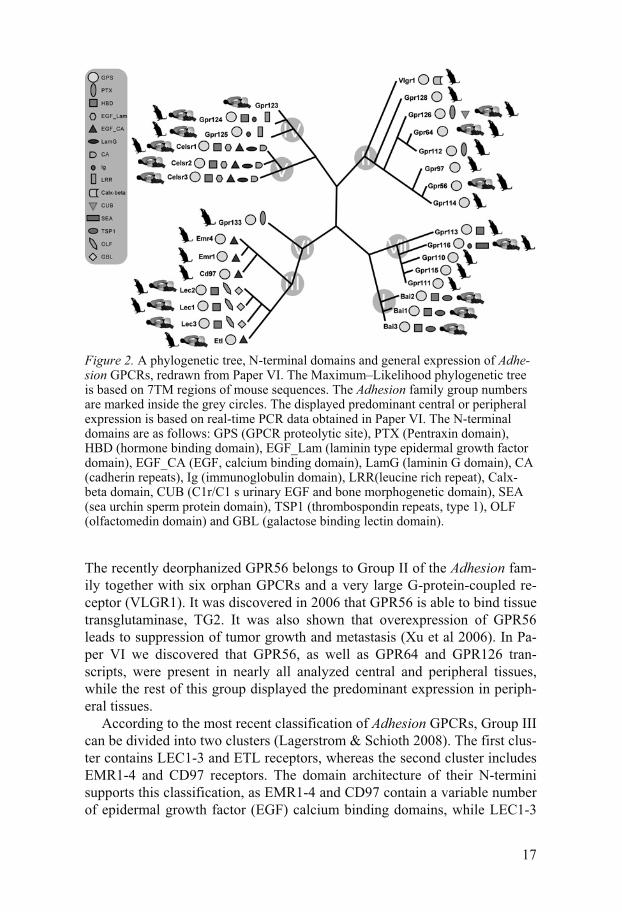
Comprising of 33 members, human Adhesion receptors form the second largest family of GPCRs. This family attracts a lot of attention from scien-tists today due to proposed essential roles in cell adhesion and signaling, even though the genes coding for Adhesion GPCRs are difficult to study because of their complex genomic structure and a large number of exons. The endogenous ligands have been discovered for only four members of the Adhesion family: BAI1, EMR2, CD97 and GPR56, whereas the rest of the Adhesion GPCRs remain orphans. Nearly all Adhesion GPCRs are lacking known physiological functions and have been poorly studied. The available expression data is limited to few tissues and has been gathered with diverse methods making it difficult to summarize and compare. Therefore, we sought to perform the overall expression analysis of the Adhesion family in two rodent species. In Paper VI we used real-time PCR approach and charac-terized expression profiles of all 30 Adhesion family members in the mouse and rat. We presented expression charts for a large number of brain regions and peripheral tissues and compared them to in situ data available from the Allen Mouse Brain Atlas (Lein et al 2007) and from human and mouse ESTs (Boguski et al 1993).
The Adhesion receptors can be divided into seven groups, I-VII, accord-ing to the phylogenetic analysis of 7TM domains (Figure 2); at the same time, the composition of functional domains in the N-termini also supports this grouping (Bjarnadottir et al 2007). Group I of the Adhesion family in-cludes the brain-specific angiogenesis inhibitors 1-3 (BAI1-3). The extracel-lular domains of BAI receptors contain hormone binding domains and thrombospondin type 1 repeats. The thrombospondin type 1 repeats in the N-terminal of BAI1 have been shown to mediate binding of phosphatidylserine to this receptor, where phosphatidylserine, located on the surface of apop-totic cells functions as an "eat me" signal (Park et al 2007). In our study (Pa-per VI), we confirmed that BAI1, BAI2 and BAI3 are expressed specifically in brain regions in both mouse and rat.
17
Figure 2. A phylogenetic tree, N-terminal domains and general expression of Adhe-sion GPCRs, redrawn from Paper VI. The Maximum–Likelihood phylogenetic tree is based on 7TM regions of mouse sequences. The Adhesion family group numbers are marked inside the grey circles. The displayed predominant central or peripheral expression is based on real-time PCR data obtained in Paper VI. The N-terminal domains are as follows: GPS (GPCR proteolytic site), PTX (Pentraxin domain), HBD (hormone binding domain), EGF_Lam (laminin type epidermal growth factor domain), EGF_CA (EGF, calcium binding domain), LamG (laminin G domain), CA (cadherin repeats), Ig (immunoglobulin domain), LRR(leucine rich repeat), Calx-beta domain, CUB (C1r/C1 s urinary EGF and bone morphogenetic domain), SEA (sea urchin sperm protein domain), TSP1 (thrombospondin repeats, type 1), OLF (olfactomedin domain) and GBL (galactose binding lectin domain).
The recently deorphanized GPR56 belongs to Group II of the Adhesion fam-ily together with six orphan GPCRs and a very large G-protein-coupled re-ceptor (VLGR1). It was discovered in 2006 that GPR56 is able to bind tissue transglutaminase, TG2. It was also shown that overexpression of GPR56 leads to suppression of tumor growth and metastasis (Xu et al 2006). In Pa-per VI we discovered that GPR56, as well as GPR64 and GPR126 tran-scripts, were present in nearly all analyzed central and peripheral tissues, while the rest of this group displayed the predominant expression in periph-eral tissues.
According to the most recent classification of Adhesion GPCRs, Group III can be divided into two clusters (Lagerstrom & Schioth 2008). The first clus-ter contains LEC1-3 and ETL receptors, whereas the second cluster includes EMR1-4 and CD97 receptors. The domain architecture of their N-termini supports this classification, as EMR1-4 and CD97 contain a variable number of epidermal growth factor (EGF) calcium binding domains, while LEC1-3

18
have more complex structures with several types of domains such as hor-mone binding, olfactomedin and galactose binding lectin domains (Bjarnadottir et al 2007). Our expression analysis in Paper VI presented fur-ther evidence for this clustering. All members of the LEC cluster were de-tected in nearly all analyzed brain regions and also - at lower levels - in pe-ripheral tissues. In contrast, members of the EMR cluster were detected at higher levels in the periphery. It has been previously shown that both mouse and rat lack EMR2 and EMR3 receptors (Bjarnadottir et al 2006; Gloriam et al 2007), therefore we were unable to include them in our expression analy-sis. Interestingly, our analysis of Adhesion receptors in the dog genome (Pa-per VII) revealed that the only variation between the human and dog Adhe-sion repertoires was found in the EMR cluster. It is evident that the dog ge-nome contains the expansions of EMR2 and EMR4 genes as we identified four different EMR2 receptors and three EMR4 receptors. Two of the EMR2-like GPCRs have been reported previously (Kwakkenbos et al 2006). In contrast to rodents, the dog does not have only the EMR2 receptor, but also the EMR3 receptor. Several studies on EMR receptors revealed their interactions with a variety of immune system cells, most probably indicating participation of EMR receptors in the immunological responses. EMR2 has been shown to bind chondroitin sulfate, in this case the mediator of cell at-tachment (Stacey et al 2003). EMR3 is interacting with ligands on the sur-face of macrophages and is able to activate neutrophils (Stacey et al 2001), while EMR4 was demonstrated to bind a ligand on B lymphoma cells (Stacey et al 2002). It is therefore intriguing to examine why several EMR receptors are not seemingly essential for the performance of the rodent im-mune system. Furthermore, explanation is needed as to why the dog genome has en expansion in the EMR cluster and what benefits, if any, this expan-sion provides to the immune system of the canine species. The dog serves as a model organism for testing toxicity of candidate drugs and it is therefore important to be aware of possible differences between human and dog im-mune systems.
Groups IV and V include GPR123-125 and CELSR1-3 receptors, respec-tively. A recent report has characterized GPR125 as a novel stem cell marker that is likely to be used therapeutically in the future (Seandel et al 2007). CELSR receptors have a combination of many different domains in their N-terminals. These receptors play an essential role in neurite growth and, gen-erally, in the CNS development (Curtin et al 2003; Shima et al 2007). The members of both groups are expressed in the CNS, whereas CELSR1, GPR124 and GPR125 are also abundant in peripheral tissues (Paper VI).
The members of the last two Groups VI and VII of Adhesion GPCRs do not have any known function and, in our study (Paper VI), they were de-tected predominantly in peripheral tissues. GPR144 is an orphan receptor missing in rodents, but present in the human and dog genomes (Paper VII).

19
The Frizzled family of GPCRs The Frizzled receptors are activated by the palmitoylated glycoprotein ligands Wnts and play an essential role in embryonic development. The amino-terminal domain of this receptor is typically N-glycosylated (Wang et al 2006). There are 11 Frizzled receptors described in the human and mouse, FZD1-10 and one Smoothened (SMO) receptor, whereas the rat has only 10 orthologs of Frizzled receptors, and FZD10 is a pseudogene (Gloriam et al 2007). The Taste2 or bitter taste receptor family was originally grouped with the Frizzled family, but they form two very diverse clusters (Fredriksson et al 2003b). It is not evident if the Frizzled and Taste2 groups have a common evolutionary origin and, therefore, they are now described as two different families. In Paper VII we investigated the Frizzled receptor repertoire in the dog genome. We discovered that all 11 human Frizzled receptor orthologs are present in the dog genome and their average amino acid sequence iden-tity is around 97 % between these two species.
The Secretin family of GPCRs The Secretin family is represented by 15 members in human and rodents. An extracellular hormone binding domain is located on the amino-termini of Secretin receptors and is involved in ligand binding. The receptor ligands are large peptides such as secretin, calcitonin, parathyroid hormone, glucagon, vasoactive intestinal peptide and pituitary adenylate cyclase activating pro-tein. Calcitonin, glucagon and parathyroid hormone are used in the clinic for the treatment of hypercalcaemia, hypoglycaemia and osteoporosis, respec-tively (Lagerstrom & Schioth 2008). In Paper VII we established that Se-cretin family repertoire in the dog is identical to that in humans and rodents and has an average amino acid sequence identity of 88.5 % and 83.4 % com-pared to the human and mouse, respectively.

20
The melanocortin system
The main components of the melanocortin (MC) system in mammals are five subtypes of MC receptors named MC1-MC5, four melanocortin peptides and two large peptides Agouti-related protein (AGRP) and Agouti signaling pep-tide (ASIP) (Barsh et al 1999; Schioth 2001). The melanocortin receptors belong to the Rhodopsin family of GPCRs (Fredriksson et al 2003b). The polypeptide termed proopiomelanocortin (POMC) is a precursor that under-goes posttranslational processing and it is cleaved into several different pep-tides, among others the melanocortin peptides α-, β- and γ-melanocyte stimulating hormones (MSH) and adrenocorticotropic hormone (ACTH). The common structural feature of all melanocortin peptides is a four amino acid-long core motif, HFRW, which is essential for activation of MC recep-tors (Figure 3). The evolution and transcriptional regulation of POMC has been widely studied in many vertebrate species (Takahashi & Kawauchi 2006). The melanocortin peptides function as endogenous agonists at MC receptors, whereas AGRP and ASIP are able to block MC receptors and antagonize the effect of the melanocortin receptor agonist, �-MSH (Jackson et al 2006). In addition to their roles as endogenous antagonists, these large peptides are also able to suppress the constitutive activity of MC receptors. ASIP has been shown to lower the basal level of constitutively active MC1 receptor (Siracusa 1994), whereas AGRP has been reported to suppress the constitutive activity of the MC3 and MC4 receptors (Adan & Kas 2003), which classifies both peptides as inverse agonists.
Figure 3. Amino acid sequence alignment of human, dogfish and lamprey melano-cortin peptides adapted from Paper III. These peptide sequences were used in phar-macological studies in Papers I-IV. The identical residues are marked with dark grey boxes; the conserved residues are marked with light grey boxes.

21
The melanocortin receptors have a typical structure characteristic for other Rhodopsin GPCRs with short N-termini and they are coupled to hetero-trimeric G�s protein. The binding of agonists to MC receptors results in dis-sociation of the G�s protein and activation of AC leading to production of intracellular cAMP. Similarly to other GPCRs, MC1, MC3 and MC4 recep-tors have been recently reported to be able to form homo- and heterodimers (Mandrika et al 2005; Nickolls & Maki 2006). Furthermore, it has been re-ported that the MC4 receptor dimer has two tandemly arranged binding sites with heterogeneous properties, that can affect ligand potencies in radioligand competition studies (Kopanchuk et al 2006). The melanocortin system modulates and controls many important physiological functions in the brain and in peripheral tissues, such as skin and hair pigmentation, adrenal func-tion, response to stress, energy homeostasis, feeding behavior, sexual func-tion, pain, immune responses and many others (Gantz & Fong 2003).
The melanocortin 1 receptor The mammalian melanocortin 1 receptor is expressed on melanocytes where it plays a major role in skin and hair pigmentation (Rana et al 1999). The binding of �-MSH to the MC1 receptor causes synthesis of the black pig-ment, eumelanin. In contrast, the endogenous antagonist, ASIP, is able to compete with �-MSH and block the MC1 receptor which results in interrup-tion of the eumelanin synthesis and, instead, switches on the red/yellow pig-ment, pheomelanin production (Rees 2000). A novel genetic locus responsi-ble for the black coat color in dogs has been reported recently (Candille et al 2007). It was named K locus and identified as beta-defensin 103. Beta-defensin is able to competitively inhibit ASIP in melanocytes, enabling the production of eumelanin. Moreover beta-defensin is also competing with �-MSH for binding to the MC1 receptor, but it does not induce the intracellular production of cAMP (Dorin & Jackson 2007). Loss of function mutations in the MC1 receptor in humans are associated with a red hair phenotype (Ringholm et al 2004) and higher risk for developing skin cancer (Mumm & Draznin 2006).
The MC1 receptor mutations affecting coat pigmentation have been pre-viously reported in a large number of laboratory, domestic and wild mam-mals and birds. It is apparent that gain-of-function mutations induce eumela-nin synthesis, whereas loss-of-function mutations result in pheomelanin pro-duction (Klungland et al 1995; Newton et al 2000; Robbins et al 1993; Rosenblum et al 2004; Takeuchi et al 1996). The role of the MC1 receptor in pigmentation has also been shown in other vertebrates, such as amphibians, reptiles and fishes (Castrucci et al 1997) where it seems to affect the distri-bution of the pigment rather than just synthesis of eumelanin/pheomelanin. For example, it has been reported that the MC1—�-MSH—ASIP system

22
plays a role in the dorsal-ventral pigment pattern in goldfish (Cerda-Reverter et al 2005) and can also affect the rapid distribution of the pigment during the background adaption in zebrafish. (Richardson et al 2008). The regula-tion of the MC1 receptor does not function only as a simple switch-on/ switch-off mechanism but it can also lead to regressive phenotypes. Genetic alterations in the MC1 receptor sequence are, for example, responsible for the degenerative brown phenotype of the blind Mexican cave tetra (Gross et al 2009).
Figure 4. Phylogeny of the chordate phylum displaying the relative timings of two genome duplications, as proposed by (Furlong & Holland 2002). The species used in our studies are stated in parenthesis.
The first cloning of the melanocortin receptors was reported in 1992, when human and mouse MC1 receptors and human MC2 receptor were cloned (Mountjoy et al 1992). Besides melanocytes, the expression of the MC1 receptor was described in keratinocytes, dermal fibroblasts as well as in im-mune system cells such as monocytes and macrophages (Bohm & Luger 2004; Luger et al 2003), where this receptor is thought to mediate anti-inflammatory actions (Chhajlani 1996). The great interest in the role of the MC1 receptor in adaptive pigmentation in non-mammalian vertebrates has led to cloning and characterization of this receptor in a number of teleosts also called bony fishes. The MC1 receptors have been described in the fugu (Klovins et al 2004), tilapia (van der Salm et al 2005), as well as in zebraf-ish, platyfish and medaka (Selz et al 2007). It seems that fish MC1 receptors share high amino acid sequence homology between teleosts and other verte-brates. In all above-mentioned species, except the fugu, the expression of the MC1 receptor was detected in the skin. Additionally, in the medaka, platy-fish and zebrafish MC1 transcripts were reported in a wide number of other

23
peripheral tissues as well as in the eye and brain (Selz et al 2007). In the tilapia, MC1 receptor expression in the skin was not altered in fish adapted to different backgrounds indicating that pigmentation in fish is most likely regulated at the protein signaling level rather than at the transcript level (van der Salm et al 2005).
In Paper IV we identified ancestral MC receptors in agnathan, the river lamprey (Figure 4). By applying a touch-down PCR and a genomic library screening methods, we were able to isolate two full-length genes, named MCa and MCb. Phylogenetic analysis revealed that the lamprey MCa recep-tor is most probably an ortholog of MC1 receptors. The presence of the MCa receptor in the skin of the river lamprey further supports this classification. It was previously reported that the melanocortin system is present in agnathans when POMC was cloned in the sea lamprey (Takahashi et al 1995a; Takaha-shi et al 1995b). The sea lamprey has two POMCs, one is named proopio-cortin (POC) and encodes ACTH, while another is named proopiomelano-tropin (POM) and encodes MSH-A and MSH-B that correspond to �-MSH and �-MSH, respectively. It is evident that the HFRW motif is conserved in lamprey melanocortin peptides (Figure 3). Since the MCa and MCb recep-tors in agnathans are the only ancestral MC receptors reported so far, it was essential to show whether they are able to respond to melanocortin peptides. We performed a comprehensive pharmacological characterization of the lamprey MCa receptor expressed in the eukaryotic cells using human and lamprey endogenous melanocortin ligands and several synthetic analogues. We were able to demonstrate the binding of all tested substances to the MCa receptor (Table 1). Moreover it is evident that MCa receptor is also coupled to G�s protein since agonist binding resulted in cAMP production. The lam-prey MCa receptor had a relatively high affinity for human ACTH derived peptides similarly to the fugu MC1 receptors (Klovins et al 2004). However, overall affinities to the MCa receptor were much lower than for the human MC1 receptor and interestingly the lowest affinities of all tested ligands had lamprey melanocortin peptides (Table 1).
The melanocortin 2 receptor The human melanocortin 2 receptor was cloned in 1992 (Mountjoy et al 1992). The mammalian MC2 receptor is also called ACTH receptor since it is the only MC receptor that does not bind MSH peptides and is activated only by ACTH (Schioth et al 1996). MC2R is expressed in the cortex of the adrenal gland, where it mediates the effects of ACTH on steroid secretion. Apart from steroidogenesis, it has been demonstrated that the MC2 receptor is also required for adrenal gland development and neonatal gluconeogenesis (Chida et al 2007). The functional characterization of the ACTH receptor is problematic since the cell surface expression of this receptor is only possible

24
in cells of adrenocortical origin. Recently it has been shown that the MC2R accessory protein named MRAP is essential for trafficking of MC2 receptor to the cell membrane (Cooray et al 2008). The unique quality of the MC2 receptor to respond only to ACTH, but not to MSH peptides, was also dem-onstrated in non-mammalian vertebrates such as the fugu (Klovins et al 2004) and trout (Aluru & Vijayan 2008). It is well-known that the homolog of the adrenal gland in teleosts is localized in the head kidney and it consists of interrenal and chromaffin cells. It is notable that the characteristic MC2 receptor expression is preserved during vertebrate evolution since this recep-tor is predominantly expressed within the head-kidney of fugu, trout and carp (Aluru & Vijayan 2008; Klovins et al 2004; Metz et al 2005). Surpris-ingly, in the fugu and trout, expression was also detected in several brain regions. In the aforementioned studies the differential expression of the MC2 receptor was investigated in the trout and carp. Acute ACTH stimulation was shown to result in the up-regulation of MC2 receptor transcripts in the inter-renal tissue of the trout (Aluru & Vijayan 2008). In another report, carps restrained for 7 days displayed down-regulation of the MC2 receptor tran-script in the head kidney (Metz et al 2005). In summary, these results from teleost fishes indicate the conservation of the MC2 receptor function during the vertebrate evolution; however the specific details of steroidogenesis in fish are yet to be elucidated.
The melanocortin 3 receptor The mammalian melanocortin 3 receptor is expressed in the brain and par-ticularly in the hypothalamus (Roselli-Rehfuss et al 1993). The MC3 recep-tor has a potential role in energy homeostasis since its inactivation results in increased fat mass, reduced lean mass and higher feeding efficiency (Chen et al 2000). The MC3 receptor knock-out mice also exhibit reduction in loco-motor behavior suggesting reduced energy expenditure. A recent study sug-gested that the MC3 receptor is required for the entrainment to a meal shed-ule. In contrast to wild-type mice, mice lacking the MC3 receptor showed significantly impaired activity-based response to food restriction and failed to increase wakefulness 2 hours before meal time (Sutton et al 2008). Each of the mammalian MC receptors has a specific binding profile to the melano-cortin peptides (Table 1). The human MC3 receptor has a unique preference to γ-MSH among different subtypes of MC receptors. Recently it has been demonstrated that the porcine MC3 receptor also has a higher binding affin-ity for γ-MSH compared to other melanocortin peptides (Fan et al 2008). It is not completely clear what physiological role γ-MSH exerts through the MC3 receptor, nevertheless, there are some novel studies suggesting that γ-MSH peptides induce anxiogenic effects in mice (Jansone et al 2009).

25
Table 1. The overview of the binding affinities of melanocortin ligands and their synthetic analogs to the MC receptors from different species. The data are retrieved from Papers I-IV, (Schioth et al 2005) and (Sanchez et al 2009). The dogfish and lamprey melanocortin peptides are marked with D and L, respectively. 0.01 — 1 (nmol/l) 1 — 10 (nmol/l) 10 — 100 (nmol/l) 100 — 1000 (nmol/l) � 1000 (nmol/l)
MC1
Human NDP-MSH, [125I] NDP-MSH, ACTH(1-24), �-MSH, ACTH(1-17), MTII
�-MSH, 1-MSH, ACTH(1-39)
HS024
Fugu [125I] NDP-MSH, NDP-MSH, ACTH(1-24)
MTII �-MSH, �-MSH, HS024 1-MSH
Lamprey Mca
[125I] NDP-MSH NDP-MSH, ACTH(1-17), �-MSH, ACTH(1-24), MTII, HS024
�-MSH, ACTH(1-39), L MSH-B, L ACTH(1-31), 1-MSH
L MSH-A
MC3
Human NDP-MSH, [125I] NDP-MSH 1-MSH ACTH(1-17), HS024, �-MSH, �-MSH, ACTH(1-24), MTII,
Dogfish [125I] NDP-MSH, NDP-MSH ACTH(1-24), ACTH(1-17), D �-MSH, D ACTH(1-25), �-MSH, L ACTH(1-31), HS024
�-MSH, MTII, D -MSH, 1-MSH, D -MSH, L MSH-B
L MSH-A D �-MSH(1-16)
MC4
Human HS024, SHU91119 [125I] NDP-MSH, NDP-MSH, MTII
�-MSH, �-MSH, ACTH(1-24)
ZnCl2, 1-MSH
Trout SHU91119, NDP-MSH, [125I] NDP-MSH
MTII, HS024 ACTH(1-24) �-MSH, �-MSH, 1-MSH, ZnCl2
Fugu MTII, ACTH(1-24), NDP-MSH, [125I] NDP-MSH, HS024
�-MSH, �-MSH 1-MSH
Goldfish [125I] NDP-MSH, NDP-MSH, HS024, MTII
�-MSH , �-MSH 1-MSH
Zebrafish [125I] NDP-MSH, NDP-MSH
�-MSH , �-MSH, HS014 1-MSH
Sea bass Diacetyl-MSH, MTII, [125I] NDP-MSH, �-MSH
AGRP, M �-MSH, Desacetyl-MSH, ACTH
Dogfish [125I] NDP-MSH, NDP-MSH
D ACTH(1-25), L ACTH(1-31) �-MSH, D -MSH, L MSH-B, D �-MSH, �-MSH, D -MSH
D �-MSH(1-16), L MSH-A, 1-MSH
MC5
Human SHU91119, NDP-MSH, HS024, [125I] NDP-MSH
MTII ACTH(1-24), �-MSH, �-MSH, 1-MSH
Trout [125I] NDP-MSH, NDP-MSH SHU91119, ACTH(1-24), MTII, HS024, �-MSH
�-MSH, 1-MSH
Fugu [125I] NDP-MSH, NDP-MSH
MTII, HS024 �-MSH �-MSH, 1-MSH
Goldfish [125I] NDP-MSH, NDP-MSH
HS024, MTII �-MSH , �-MSH, 1-MSH
Zebrafish MC5a
[125I] NDP-MSH NDP-MSH �-MSH, �-MSH, 1-MSH, HS014
Zebrafish MC5b
[125I] NDP-MSH, NDP-MSH
�-MSH, �-MSH, 1-MSH, HS014
The structure of POMC has been studied in a wide number of non-mammalian vertebrates and it is evident that the γ-MSH region in ray- finned fishes is quite variable and degenerated, either missing the γ-MSH core motif (Amemiya et al 1997; Dores et al 1997) or even the complete γ-MSH se-quence (Kitahara et al 1988; Takahashi & Kawauchi 2006). Despite the nu-merous efforts to clone the MC3 receptors in ray-finned fishes, the only MC3 receptor described so far is in zebrafish (Logan et al 2003). Even though the MC3 receptor is present in zebrafish, these species still lack the -MSH peptide (Gonzalez-Nunez et al 2003). Based on these findings, it has been suggested that -MSH—MC3 receptor system is either not present or impaired in this class of fishes. Interestingly, -MSH is present in the POMC of lobe-finned and cartilaginous fishes such as the lungfish and dogfish, re-spectively. Therefore, it can be speculated that MC3 receptors are also pre-sent in these fishes. In Paper II we describe cloning and characterization of MC3 and MC5 receptors in the spiny dogfish (Figure 4). The expression of the dogfish MC3 receptor was detected in the hypothalamus, similarly to

26
mammals, as well as in other brain regions. In this study we were interested to test whether the dogfish MC3 receptor has a preference to -MSH. In ad-dition, since cartilaginous fishes have a unique melanocortin peptide, -MSH, we also aimed to test its ability to bind the dogfish receptor. For that reason, in Paper II and III were performed an extensive pharmacological characterization of the dogfish MC3 receptors using human, dogfish and lamprey endogenous melanocortin peptides and their synthetic analogs (Ta-ble 1). We were able to demonstrate the binding of -MSH and its ability to induce intracellular synthesis of cAMP. Surprisingly, our analysis indicates that human, dogfish and lamprey ACTH peptides have a clear preference before human and dogfish -MSH peptides to the dogfish MC3 receptor in both binding and intracellular cAMP measurement experiments.
The melanocortin 4 receptor The melanocortin 4 receptor very rapidly captured the attention of scientific world after its cloning. The expression of the MC4 receptor was described in a number of brain regions including the paraventricular nucleus of the hypo-thalamus (Mountjoy et al 1994). The inactivation of the MC4 receptor in mice induced overeating and obesity (Huszar et al 1997). It was later shown that centrally expressed melanocortin peptides are mediating inhibition of food intake through the MC4 receptor (Fan et al 1997). Conversely, AGRP, which is expressed in the hypothalamus, acts as an endogenous antago-nist/inverse agonist at the MC3 and MC4 receptors and its overexpression leads to obesity (Ollmann et al 1997). To summarize, agonists of the MC4 receptor are highly anorexic, while antagonists generate orexigenic effects (Fan et al 1997). Furthermore, the MC4 receptor is one of the best-characterized monogenic factors of obesity (Farooqi 2008). A number of mutations in the MC4 receptor are linked to obese phenotypes in humans (Farooqi et al 2000; Miraglia Del Giudice et al 2002; Vaisse et al 1998). The MC4 receptor is also believed to have a role in antipyretic and hyperthermic control (Sinha et al 2004), sexual functions (Andersson & Hedlund 2002), cardiovascular functions (Pavia et al 2003) and prolactin secretion (Watanobe et al 2003).
The MC4 receptors have also been extensively studied in non-mammalian vertebrates. Remarkably, like in mammals, the expression of the MC4 recep-tors was described in brain regions of the zebrafish (Ringholm et al 2002), fugu (Klovins et al 2004) and dogfish (Ringholm et al 2003). In the goldfish and sea bass, detailed mapping of the MC4 receptor expression demonstrated the presence of the transcripts in key brain areas controlling food intake (Cerda-Reverter et al 2003b; Sanchez et al 2009). In Paper I we used the rainbow trout (Figure 4), a well-known model for physiological research, and demonstrated the presence of at least two melanocortin receptors, MC4

27
and MC5, in this species. We detected the expression of the trout MC4 re-ceptors in the hypothalamus, telencephalon and optic tectum. Altogether, this indicates that the central localization of the MC4 receptor is conserved in mammals and non-mammalian vertebrates, in this case, ray-finned and cartilaginous fishes. Interestingly, in contrast to mammals, we detected MC4 receptor transcripts in the head kidney of the rainbow trout. The presence of the MC4 receptor in the periphery has been demonstrated before in the gas-trointestinal tract, eye and ovary of the zebrafish; head kidney and gut of the fugu; eye, gill, spleen, dorsal skin and ovary of the goldfish as well as in the retina, liver, fat tissue and testis of the sea bass.
In Paper I and Paper III we presented a broad pharmacological characteri-zation of the trout and dogfish MC4 receptors, respectively. These results together with previously published pharmacological data are summarized in Table 1. Notably, all characterized MC4 receptors display highest affinities for synthetic melanocortin peptide analogs; antagonists SHU9119 and HS024, and agonists MTII and NDP-MSH. Noteworthy, all fish (except the sea bass) MC4 receptors preferred ACTH derived ligands over MSH ligands. This is in contrast to the human MC4 receptor binding profile, but in line with previous results from fugu MC1, lamprey MCa and dogfish MC3 re-ceptors. Using an intracellular cAMP assay in Paper I, we were able to dem-onstrate that, like in mammals, SHU9119 acts as an antagonist. It was able to compete with the agonist, MTII, and selectively blocked trout MC4, but not trout MC5 receptors. These findings indicated that these two substances are suitable for physiological in vivo experiments in the rainbow trout and al-most certainly in other teleost species as well. Supporting this, a recent re-port has demonstrated that these particular synthetic analogs affect food in-take in the trout (Schjolden et al 2009). In that study, MTII significantly decreased food intake, while HS024 and SHU9119 had an opposite effect, and HS024 was more potent in increasing food intake than SHU9119.
AGRP is an important key element of the food intake system in mam-mals. Therefore, upon the cloning of AGRP and MC4 in the goldfish, it was crucial to determine whether the AGRP–MC4 system is present in fishes (Cerda-Reverter & Peter 2003; Cerda-Reverter et al 2003b). The expression of AGRP was shown in the ventrobasal hypothalamic lateral tuberal nucleus also know as the teleostean homolog of the arcuate nucleus. Moreover, the fasting up-regulated AGRP transcript levels in this brain region of the gold-fish. In contrast to the effects on AGRP, food deprivation does not seem to affect the levels of centrally expressed MC4 receptors or POMC in fishes (Kobayashi et al 2008; Sanchez et al 2009). The expression of goldfish AGRP was also detected in several peripheral tissues, such as the ovary, muscle and ventral skin. It is very interesting since MC4 receptor expression in the goldfish and in other teleosts is also described in peripheral tissues (Metz et al 2006). Therefore, it can be suggested that AGRP might have some regulative action in the periphery, which is most likely mediated

28
through peripherally expressed MC4 receptors. The involvement of AGRP in food intake control in fish was also demonstrated in the zebrafish, where overexpression of AGRP resulted in obesity, increased linear growth and adipocyte hypertrophy (Song & Cone 2007). Moreover, it has been recently demonstrated in the sea bass that AGRP’s actions as a competitive antago-nist and inverse agonist are operated through different signaling pathways, where the inverse agonism, but not the competitive antagonism, depends on the presence of a phosphodiesterase inhibitor (Sanchez et al 2009). Alto-gether, these findings are indicative of a physiological role for the melano-cortin system in the control of the energy balance in teleost fish.
In Paper IV we were able to clone the ancestral MCb receptor from the river lamprey. The phylogenetic analysis indicated its probable orthology to the MC4 receptor subtype. We discovered that unlike other MC4 receptors, this ancestral protein does not display any expression in the lamprey brain and it is present only in peripheral tissues such as the skin, liver, heart and muscle. The POM and POC precursors encoding lamprey melanocortin pep-tides have been extensively described before (Takahashi et al 1995b), whereas there are no reports about the presence of AGRP in lampreys. Therefore, it is possible that the central food intake control system, mediated by the melanocortin system in other vertebrates, is most probably not yet developed in agnathans.
The melanocortin 5 receptor Melanocortin 5 receptors are expressed in the brain, but also widely distrib-uted in the human peripheral tissues, including the adrenal gland, adipocytes, leukocytes and others (Chhajlani 1996). The functional properties of the MC5 receptor are still not very well understood. Inactivation of the MC5 receptor resulted in exocrine gland dysfunction in mice (Chen et al 1997). Other studies described that mice with an inactivated MC5 receptor dis-played reduced pheromone signal for aggression (Morgan & Cone 2006) and that the MC5 receptor is essential for protection of the retina from the in-flammatory damage of experimental autoimmune uveoretinitis (Taylor et al 2006).
The MC5 receptors have been described in a number of fish species such as the zebrafish that has two MC5 receptors, named MC5a and MC5b (Ringholm et al 2002), goldfish (Cerda-Reverter et al 2003a), fugu (Klovins et al 2004) and common carp (Metz et al 2005). In Papers I and II we pre-sented two additional MC5 receptors, one from the rainbow trout and one from the spiny dogfish. It is interesting that all described MC5 receptors in fish are expressed in the brain. Furthermore, it is remarkable, that there is a great similarity in expression patterns between fugu MC4 and MC5 recep-tors; zebrafish MC4, MC5a and MC5b receptors; goldfish MC4 and MC5

29
receptors; trout MC4 and MC5 receptors; and between dogfish MC3, MC4 and MC5 receptors. The presence of MC transcripts in a similar range of tissues between the receptor subtypes might be an indicator of shared physiological roles these receptors play in fish. Another possible explanation is a common evolutionary origin of the MC3, MC4 and MC5 receptor sub-types. In order to support this hypothesis, we performed a number of phar-macological experiments and also analyzed the previously published data (Table 1). The detailed analysis of pharmacological data revealed that trout and dogfish MC5 receptors have a preference for ACTH derived peptides over MSH peptides. These results are in agreement with pharmacological preferences for ACTH of other MC receptor subtypes in fish described above. Together these studies gave raise to the hypothesis that MC receptors in early vertebrates had preference for ACTH peptides while the high sensi-tivity for the shorter melanocortin peptides, such as �-, �- and -MSH has appeared later when the MC receptor subtypes obtained more specialized functions (Schioth et al 2005).

30
Solute carriers
The membrane transport proteins can be divided into ion channels, water channels, ATP pumps, ABC transporters and solute carriers (SLCs) (Hediger et al 2004). Solute carriers constitute the largest group of membrane trans-porters in the human genome (Hediger et al 2004; Kusuhara & Sugiyama 2005). This group controls transport of amino acids, inorganic ions, sugars, nucleotides and fatty acids, among others. All of these transporters are asso-ciated with the membrane, but their cellular localization varies from the plasma membrane to mitochondria, synaptic vesicles (Wallen-Mackenzie et al 2006) or peroxisomes (Visser et al 2002). There are currently 46 families of solute carriers described (Fredriksson et al 2008). The SLC genes have a very complex genomic structure with a high number of introns making it difficult to bioinformatically mine and study these proteins. There have been only a small number of reports showing a phylogenetic relationship between different SLC families. A recent study demonstrated that 15 of the SLC families form four distinct phylogenetic clusters, most probably indicating a common evolutionary origin (Fredriksson et al 2008).
Mitochondrial membrane transporters (SLC25 family) The solute carrier family 25 (SLC25) is a group of mitochondrial proteins transporting a large variety of compounds, such as citrate, ornithine, phos-phate, adenine nucleotides, dicarboxylate, oxoglutarate, glutamate and others (Palmieri 2003). The common structural characteristics of this family of proteins are six �-helices spanning the mitochondrial membrane connected by hydrophobic loops both on the cytosolic and matrix side. Both the amino- and carboxy- terminals of the protein are located on the cytosolic side of the inner mitochondrial membrane (Palmieri 2003; Pebay-Peyroula et al 2003). Not so long ago the number of mitochondrial carriers was estimated to 23 members (Palmieri 2003). More recent report placed 27 members into this group (Hediger et al 2004). Since then 9 additional proteins were annotated through different human genome annotation projects. In Paper V we re-trieved all predicted protein sequences from the RefSeq database and applied a custom HMM model in order to identify additional SLC25 proteins. As a result of this analysis we discovered additional 14 transporters in the SLC25 family, making a total number of 46 members. The novel transporters were

31
annotated by the HUGO gene nomenclature committee and were named SLC25A32-A35 and SLC25A37-SLC255A46. Interestingly, according to the Pfam domain database (http://pfam.sanger.ac.uk) all mitochondrial carri-ers share a Mito_carr domain (accession number PF00153). Remarkably, in the UNIPROT Protein Knowledgebase (http://www.uniprot.org) there are 53 unique human proteins with the Mito_carr domain. It implies that there are, most probably, the additional 7 members that can be classified into the SLC25 family. These are currently annotated as mitochondrial carrier ho-molog 1 or presenilin-associated protein (MTCH1), mitochondrial carrier homolog 2 or Met-induced mitochondrial protein (MTCH2), mitochondrial carrier triple repeat protein 1, 2 and 6 (MCART1, MCART2 and MCART6), hepatocellular carcinoma down-regulated mitochondrial carrier protein (HDMCP) and putative mitochondrial carrier protein FLJ44862. Altogether, this makes the SLC25 family by far the largest of all solute carrier families.
The uncoupling proteins UCP1-3, also known as SLC25A7-A9, are among the most studied members of SLC25 family and are characterized as proton transporters (Kim-Han & Dugan 2005). The uncoupling proteins par-ticipate in various physiological processes and they are thought to be the genetic cause of several diseases. It has been demonstrated that UCP1 has a role in thermogenesis in mammalian brown adipose tissues, UCP2 nega-tively regulates insulin secretion, and overexpression of UCP3 causes in-creased fatty acid oxidation in mice (Echtay 2007). There are additional un-coupling proteins described, UCP4 (SLC25A27) (Mao et al 1999) and UCP5 or BMCP1 (SLC25A14) (Sanchis et al 1998). Mitochondrial uncoupling proteins have also been suggested to play a role in neurodegenerative dis-eases like Alzheimer’s disease, Parkinson’s disease, and Amyotrophic Lat-eral Sclerosis as well as normal aging of the brain (Andrews et al 2005). The UCPs are conserved during the evolution of eukaryotes since there has been a number of reports describing uncoupling mitochondrial proteins in plants (Vercesi et al 2006) and in slime mold (Satre et al 2007). Although the UCPs have been relatively extensively studied, the role of other SLC25 members is fairly unknown.

32
Figure 5. Phylogenetic analysis of full-length amino acid sequences of human SLC25A1-46 and 10 yeast SLC25 family members. This figure is modified from Paper V. The consensus tree was generated by the Neighbor-Joining analysis, the numbers at the nodes indicate bootstrap replicates. The novel sequences are marked with orange, the yeast transporters are marked with yellow. The preferred substrate, if known, is stated after the slash. Human proteins with recently identified substrate are circled in red.
In Paper V we retrieved 14 rat and 14 mouse orthologs of novel human SLC25 family members and confirmed their orthology with a help of the phylogenetic analysis. We also performed an extensive tissue localization analysis of nine of these novel genes (SLC25A33, SLC25A37, SLC25A40-A46) on a wide number of central and peripheral tissues from the rat with the quantitative real-time PCR technique. Our results indicate that these sol-

33
ute carriers are very widely distributed in the brain as well as in peripheral tissues of the rat. These findings are in agreement with expression profiles from other members of the SLC25 family, such as uncoupling proteins, that also have a very wide distribution (Palmieri 2003). There have been previous reports describing the mitochondrial transporters in yeast, but their relation-ship to the mammalian SLC25 family members was not clarified (Palmieri et al 2000). We performed en extensive phylogenetic analysis and included yeast transporters together with 46 members of SLC25 family SLC25SA1-A46 (Paper V). As a result of this analysis, we were able to identify 10 yeast proteins displaying the orthologous relationship to human SLC25 proteins (Figure 5). Our phylogenetic analysis showed that, remarkably, many solute carriers clustered together in the phylogenetic tree, shared identical or simi-lar substrates. Based on these findings, we proposed possible substrates for several members of SLC25 family with unknown functions. Notably, there have been recent reports characterizing several of the new solute carriers we identified, confirming our postulations about proposed substrates. For exam-ple, in our analysis the yeast proteins Mrs3p and Mrs4p, known to transport iron, formed a phylogenetic cluster together with SLC25A37 and SLC25A28. Recent study identified SLC25A37 as an iron transporter and it received the name mitoferrin (Shaw et al 2006) Later, the SLC25A28 was also characterized as an iron transporter, and it was shown that mitoferrin 1 (SLC25A37) and mitoferrin 2 (SLC25A28) regulate mitochondrial iron lev-els through a differential turnover (Paradkar et al 2009). Furthermore, there are three isoforms of ATP-Mg/P(i) transporters called SLC25A23-25. In our analysis, they formed a phylogenetic cluster with SLC25A41, which later was characterized as a calcium-independent ATP-Mg/P(i) carrier (Traba et al 2009). The mitochondrial carrier family was also described in slime mold (Satre et al 2007), where several proteins were classified as orthologs of human SLC25 proteins. In summary, the mitochondrial transporters form a surprisingly well conserved family within the eukaryote evolution, where clear orthologs can be identified between human and very basal organisms such as fungi and the social amoebae. Moreover, many of these orthologs seem to have conserved residues providing the contact points for the sub-strate, since they share identical or very similar substrates.

34
Conclusions
Paper I In this study we cloned two melanocortin receptors from the teleost rainbow trout. Phylogenetic analysis indicates that these receptors are orthologs of the human MC4 and MC5 receptors. We constructed 3D protein models of the trout and human MC4 and MC5 receptors. These models suggest that two human receptors are more diverged from each other than the trout receptors. The pharmacological analysis demonstrated that trout MC4 and MC5 recep-tors shared similar binding affinities for the endogenous human melanocortin peptides, where ACTH had higher potency than �-, �- or - MSH. Interest-ingly, we identified a natural high-affinity binding site for zinc ions at the rainbow trout MC4 receptor. This finding is in line with previous reports about zinc binding to the human MC4 receptor and indicates that zinc may play an evolutionary conserved role at this receptor subtype. In an intracellu-lar cAMP assay, the cyclic MSH analogues MTII and SHU9119, acted as a potent agonist and an antagonist, respectively, at the rainbow trout MC4 receptor. Thus, these ligands are suitable for physiological studies in the rainbow trout. We showed that the trout MC4 and MC5 receptors shared quite similar tissue localization in the brain and peripheral tissues. In sum-mary, our analysis indicates that the rainbow trout MC4 and MC5 receptors have more in common than their mammalian counterparts do, which may suggest that these two receptors have a closer evolutionary relationship than the other MC receptor subtypes.
Paper II We describe the cloning and characterization of two melanocortin receptors from the chondrichtyan spiny dogfish. The phylogenetic analysis shows that the cloned receptors are orthologs of the MC3 and MC5 receptor subtypes. The anatomical localization analysis revealed that both receptor transcripts are expressed in the hypothalamus, telencephalon, brain stem, optic tectum and olfactory bulb of the spiny dogfish. We expressed the cloned MC3 and MC5 receptors in the eukaryotic cells and visualized their localization using receptor–enhanced green fluorescence protein (EGFP) fusion proteins. We showed the presences of the dogfish MC3 receptors in the plasma mem-brane, whereas the dogfish MC5 receptors were retained in the intracellular compartments of the cells making it impossible to pharmacologically charac-terize the MC5 receptors. We performed the pharmacological characteriza-

35
tion of the dogfish MC3 receptors and discovered that ACTH derived pep-tides had higher affinity for these receptors than �-, �- or -MSH. In the in-tracellular assay we showed that the dogfish MC3 receptors are G�s-coupled and the agonist binding leads to the production of cAMP. Altogether, our results indicate that the important elements of the MC system appeared early in the vertebrate evolution and, most certainly, before the radiation of gna-thostomes.
Paper III We performed an extensive pharmacological characterization of the dogfish MC3 and MC4 receptors using the endogenous human, spiny dogfish and sea lamprey melanocortin peptides and their synthetic analogs. We dis-covered that, similarly to other fish melanocortin receptors, dogfish MC3 and MC4 had a preference for ACTH derived peptides. In addition, we pro-vide the first evidence that native dogfish ACTH ligand has a preference above native dogfish �-, �- and -MSH peptides to the MC3 and MC4 recep-tors in cartilaginous fishes. This further strengthens the hypothesis that the ligand contributing to the ancestral melanocortin ligand–receptor system resembled ACTH. In our study we were also able to show that -MSH, which is unique for cartilaginous fishes, is able to bind to the dogfish MC receptors and it has a higher affinity than its proposed precursor, dogfish �-MSH. Our results provide better insight into how new ligand-receptor inter-actions may form, which are essential for acquiring new physiological func-tions. Paper IV In this study we cloned two ancestral melanocortin receptors in the agnathan river lamprey. The lamprey receptors, named MCa and MCb, showed orthology to the MC1 and MC4 receptor subtypes, respectively. The molecu-lar clock analysis suggested that lamprey MC receptor genes diverged from each other more than 400 MYR ago. We performed expression analysis and found that both of the lamprey MC receptor transcripts were expressed in the skin, and the MCb receptor was also detected in the heart, liver, and skeletal muscle. Pharmacological characterization showed that the lamprey MCa receptor could bind and be activated by endogenous lamprey and human MSH peptides and synthetic MSH analogs. The lamprey MCa receptor had relatively high affinity for ACTH-derived peptides similarly to the fish MC receptors. In summary, this study shows the presence of MC receptors in agnathans indicating an early specialization of melanocortin receptor sub-types.
Paper V We identified 14 novel human transporters from the SLC25 family of mito-chondrial carriers. We also found the orthologs for these genes in the rat and

36
mouse. The novel transporters were annotated by the HGNC and were given names SLC25A32–SLC25A35 and SLC25A37–SLC25A46. We performed an extensive phylogenetic analysis for the entire SLC25 family with 46 hu-man transporters. We included recently published yeast mitochondrial carri-ers into our analysis and discovered that 10 of them were orthologs of human transporters. We compared the sequences of orthologous pairs and were able to show that the residues providing the contact points for the substrate are conserved between human and yeast proteins. The expression of nine novel transporters was determined with quantitative real-time PCR on the rat panel containing a large number of brain regions and peripheral tissues. In sum-mary, this study has provided new information about the repertoire of genes coding for mitochondrial transporter proteins and provided insights into their possible functional roles.
Paper VI We performed an extensive tissue localization analysis for the entire family of Adhesion GPCRs in the rat and mouse. We used quantitative real-time PCR technique and showed that the majority of GPCRs were expressed ei-ther specifically in the brain or ubiquitously in the brain and peripheral tis-sues. The results were compared with data from the Allen Brain Atlas pro-ject, ESTs and the literature. We performed the phylogenetic analysis of the mouse Adhesion GPCRs and combined it with obtained general expression data. Remarkably, we discovered that receptors within the same phyloge-netic clusters had similar expression profiles. This could be an indicator of shared physiological roles for members within the phylogenetically related clusters.
Paper VII We searched for the non-olfactory GPCRs in the dog genome and obtained 353 full-length and 18 incomplete GPCR gene sequences. All identified proteins were classified into Adhesion, Frizzled, Glutamate, Rhodopsin and Secretin families. We established orthologous relationships between human, dog, rat and mouse GPCRs and identified species-specific duplicates. The largest variation in a number of GPCRs between human and dog was found in the Rhodopsin and Adhesion families. We found that 12 dog GPCR genes are missing in humans while 24 human GPCR genes are not part of the dog GPCR repertoire. The comparison of the dog, human and rodent repertoires revealed several examples of species-specific gene duplications and dele-tions. Our results show that the dog GPCR repertoire is more similar to that in human than in rodents both with respect to the number of receptors in each family and to the number of orthologous pairs between the species. This information can be used for the selection of model organisms for phar-macological experiments and the interpretation of their results.

37
Perspectives
The number of available gene sequences has grown tremendously in the past few years as a result of several genome sequencing projects. The whole ge-nome sequences are very useful for studying repertoires of gene families, because they are essential for making conclusions about gene losses. Exist-ing genome projects have focused on species that are widely used as model organisms in physiological studies or/and located on the border of evolution-ary transitions, for example, located on either side of prokaryote–eukaryote, invertebrate–vertebrate and non-mammalian-mammalian transitions. Con-sidering these scientific advances, there are opportunities to perform much more detailed analysis of the gene sequences. A new interdisciplinary field of scientific research was described in 2003 as intersection of evolution and genomics, and was named phylogenomics (Eisen & Fraser 2003). Phyloge-nomics focuses on analyzing genes in a comparative perspective, for exam-ple, the analysis of gene repertoires between species, the gene order across species, and the identification of orthologous pairs, species specific expan-sions and deletions.
There is a great number of genes available in the genome databases that are automatically predicted, but not yet annotated. HMM model is an excel-lent tool for protein annotation through identification of homologous protein domains. This makes it possible to look at the domain conservation during the evolution of the organisms. In the case of evolutionary highly conserved families like the solute carriers, which are present in all eukaryotes as well as in prokaryotic species, phylogenetic analysis of the protein family reper-toires in different species is essential for classification and predictions of physiological function. It is likely that many transporters have substrate and physiological functions conserved during the evolution. Furthermore, it is experimentally much easier to study transporters in basal organisms, like yeast, than in mammals and from these results draw parallels to the physio-logical function of human proteins. There is a great potential in studying transporter families with approaches similar to that we used in our SLC25 study. The basal function of solute carriers indicates that they are essential for keeping physiological balance in the cell and, therefore, it is of great importance to understand the functional mechanisms of their action.
The melanocortin family is present only in vertebrates which makes it a relatively fast evolving family comparing to the solute carriers. Since there are five distinct types of melanocortin receptors, it indicates that these sub-

38
types have acquired their specialization very fast during the early evolution of vertebrates. Therefore, the melanocortin family represents a great model for studying how new physiological functions evolve. The MC4 receptor’s role in food intake is well studied today. It is evident now that ancestral MC4 receptor is present in the lamprey; already in teleosts the MC4 receptor has a distinct physiological role in food intake, which stays conserved through the evolution of vertebrates until modern animals, including human. However, none of the ancestral lamprey receptors show similarity to the MC2 receptor subtype. This makes the MC2 receptor the fastest evolving subtype. More-over, it is not clear if the MC2 receptor is present in cartilaginous fishes. It is known that MRAP is essential for transport of the MC2 receptor to the cell membrane. Therefore it is important to know when MRAP appeared in ver-tebrate evolution, since it was probably the key factor for specialization of the MC2 receptor. Altogether, in order to get a complete picture about the evolution of the specific protein family, it is important to analyze the evolu-tion of the whole signaling system, which involves these proteins. In the case of the melanocortin system, it means that we have to study the evolution of not only MC receptors, but also POMC, AGRP, ASIP and interacting pro-teins like MRAP. Furthermore, it is of crucial importance to combine the analysis of basic protein structure and orthologous relationships between species with functional studies like anatomical localization, pharmacological properties and intracellular signaling.
In order to understand how new proteins families evolve and how they acquire new ligands it is interesting to study the evolution and functional properties of phylogenetically related families. The MECA cluster of Rhodopsin GPCRs, which also includes the MC receptors, is a good model for this type of studies, since the families within this cluster bind very differ-ent ligands, such as peptides, lipids and adenosine. It is interesting to show if these families have a common evolutionary origin and may be to identify the ancestral proteins in invertebrates. Furthermore, the pharmacologically char-acterization of these ancestral proteins and 3D modeling of their binding sites would be of great significance, for understanding how ligand specificity evolves.

39
Acknowledgements
I would like to express my sincere gratitude and appreciation to my col-leagues, friends and family members whose presence, support and encour-agement helped me to reach this stage of my life. First, I would like to thank my supervisor Dr. Robert Fredriksson, for all the help, expert knowledge in the GPCR field, great ideas and good scientific discussions. Thank you for respecting my independence and being so pa-tient. Next, I would like to express my deepest gratitude to Professor Helgi Schiöth, our group leader and my friend, for believing in me and for con-tinuous encouragement. Your expert knowledge in pharmacology was of significant importance. I really appreciate that you taught me how to find the bottleneck of my projects and that you significantly improved my scientific writing skills. Thank you for participating with me in a great number of mul-tidisciplinary conferences all over the world and for introducing me to the traditional Icelandic cuisine, I am a great admirer. My first supervisor in Latvia, J�nis Klovi š, thank you for introducing me to the field of molecular biology, for teaching me my first PCR in Riga, back in 1999, for inspirational conversations about science. I have nice memories about long evenings in the lab; doing experiments, listening to Nick Cave songs and playing darts. I would like to thank the Department of Neuroscience for inspirational at-mosphere and good teaching opportunities and also staff members: Marita, Birgitta, Ulla, Else-Marie, Maria, Lena and Leena; thank you for all your help. All my co-authors, thank you for successful collaboration. I would like to send special thanks to all my project students, in chronologi-cal order, Maja Löwgren, Marie Svensson, Marie Karlsson, Filip Yström, Johan Enberg, Linnéa Holmén, Thomas Renström, Anela Hukanovic, Joanna Hammer and Fredrik Olsson, for hard work in the lab, genuine interest in the projects and great productivity.

40
Thanks to all members of J�nis Klovi š group in Riga, especially, D�vids Fridmanis, Ivo K�pa, Maija Slaidina, Ineta Kalni a, Ramona Petrovska and Ilona Mandrika for all great time we spent together in Riga and in Uppsala. Next, I would like to thank all past and present members of the following research groups:
Åsa Fex Svenningsen group: especially, Grzegorz, Katarzyna and Mikael, for interesting discussions.
Åsa Mackenzie group: especially, Åsa and Carolina, thank you for your support.
Dan Larhammar group: especially, Earl, for being my first Swedish-American supervisor, for teaching me library screening; Dan, for giving inspiration in the field of evolution and providing valuable literature refer-ences; Tomas, for keeping me nice company during long evenings in the lab and swimming on Saturdays; Ingrid, for great help in organizing teaching; Torun, Paula, Daniel, Görel and also Christina, Hille and Helena, thank your for your help in teaching and your support.
Klas Kullander group: especially, Malin, for teaching me binding and cell culture; Katarzyna, for moral support; Nadine, for always smiling; Anders, Henrik and Anna.
Lars Oreland group: especially, Camilla, Niklas and Hanna-Linn. Thanks to all past and present members of Finn Hallböök, Håkan Aldsko-gius, Elena Kozlova, Ted Ebendal and Manfred Kilimann groups and espe-cially: Elena, thank you for keeping Russian Easter traditions; Håkan, for nice discussions; Robert O, for introducing me to the special effects of the Japanese green tea; Nicole, for sharing ice-skating experiences; and Zvonimir, for long discussions about work matters in Café Linné. Special thanks to all past and present members of Helgi Schiöth group, espe-cially to: Aneta, for taking care of me in the lab; Anica, for all funny stories you told us; David, for great collaboration; Josefin, for nice talks about work and for understanding; Johan, for all paper discussions and nice choir con-certs; Kalle, for keeping me updated in the field of GPCR evolution and all the help with my computer; Markus, for great ideas and information about most advanced bioinformatic tools; Maria H, for keeping the lab organized; Smitha, for delicious Indian food; Ali, Björn S, Tobias, Maria L, Pär, Shahr-zad, Madeleine, Jon, Jonathan, Mathias, Ingrid, Maria N and Anna K. Thank you all for a warm atmosphere and inspirational discussions during fika and lunches.

41
I would like to thank my closest friends (in alphabetical order)
Åsa F S, you have always been so nice to me, thank you for fantastic Thanksgiving parties, your support and compliments, books and flowers for my garden.
Agnieszka, thank you for all activities we have done together: shopping, cooking, feeding babies, ice-skating, swimming...
Anna K-H, thank you for being a good friend, for all discussions about movies, photography, science, for all fun we had together and for giving me so much support during writing this thesis
Björn, thank you for nice music, great book suggestions and inspirational scientific discussions
Olga, thank you for taking care of me, for your help with my projects, for enjoyable home parties with Russian food, sushi and Wii.
Malena, thank you for being so positive, for great Box and Sculp train-ings, for our sushi lunches and sushi dinners and for being such a good lis-tener and always taking my side.
Marina, thank you for staying my true friend during all these years and for believing in me. �� ����������� ������-����� ���!�� ���# !� �����!
Pawel, you have a great sense of humor, thank you for understanding me so well, for all inspirational poetry and photography discussions. I really appreciate that you put such a tremendous effort into proofreading this the-sis.
Thora, for being a good friend, for all fun we had together in Uppsala and Reykjavik. Finally, I would like to send a big warm hug to my family: my mom, my grandmother, my brother Arsenij and Sanita. Thank you for always being there for me, for you love and understanding. I also want to thank my sister Olga. It is a true miracle that we found each other; I do not understand how I could live without you for 27 years. '�+ ����#�+ ���<+: ����, �=���>� ��>� �� =�����?��, �@>��<, =�!���!�� � �������; >�>�Q��, �=���>� �� ���@ ��>��� � ���=���!��; \���!��, �� ����� ��� �@>���� >��� !� �����, �=���>� ��>� �� ��>���; Sanita, paldies tev par atbalstu un par r^p`m; � \�{!�, �� �!��Q<, |�� }���, }�� �� ���# ���#� !�Q��, c=���>� ��>� �� =�!���!��, =�����?�� � ��>����.

42
References
Adan RA, Kas MJ. 2003. Inverse agonism gains weight. Trends in pharmacological sciences 24:315-21
Aluru N, Vijayan MM. 2008. Molecular characterization, tissue-specific expression, and regulation of melanocortin 2 receptor in rainbow trout. Endocrinology 149:4577-88
Amemiya Y, Takahashi A, Dores RM, Kawauchi H. 1997. Sturgeon proopiome-lanocortin has a remnant of gamma-melanotropin. Biochem Biophys Res Com-mun 230:452-6
Andersson KE, Hedlund P. 2002. New directions for erectile dysfunction therapies. International journal of impotence research 14 Suppl 1:S82-92
Andrews ZB, Diano S, Horvath TL. 2005. Mitochondrial uncoupling proteins in the CNS: in support of function and survival. Nat Rev Neurosci 6:829-40
Attwood TK, Findlay JB. 1994. Fingerprinting G-protein-coupled receptors. Protein Eng 7:195-203
Barsh GS, Ollmann MM, Wilson BD, Miller KA, Gunn TM. 1999. Molecular phar-macology of Agouti protein in vitro and in vivo. Ann N Y Acad Sci 885:143-52
Birnbaumer L, Abramowitz J, Brown AM. 1990. Receptor-effector coupling by G proteins. Biochimica et biophysica acta 1031:163-224
Bjarnadottir TK, Fredriksson R, Schioth HB. 2007. The adhesion GPCRs: a unique family of G protein-coupled receptors with important roles in both central and peripheral tissues. Cell Mol Life Sci 64:2104-19
Bjarnadottir TK, Gloriam DE, Hellstrand SH, Kristiansson H, Fredriksson R, Schioth HB. 2006. Comprehensive repertoire and phylogenetic analysis of the G protein-coupled receptors in human and mouse. Genomics 88:263-73
Boguski MS, Lowe TM, Tolstoshev CM. 1993. dbEST--database for "expressed sequence tags". Nat Genet 4:332-3
Bohm M, Luger TA. 2004. Melanocortins in fibroblast biology--current update and future perspective for dermatology. Experimental dermatology 13 Suppl 4:16-21
Cabrera-Vera TM, Vanhauwe J, Thomas TO, Medkova M, Preininger A, et al. 2003. Insights into G protein structure, function, and regulation. Endocrine reviews 24:765-81
Candille SI, Kaelin CB, Cattanach BM, Yu B, Thompson DA, et al. 2007. A -defensin mutation causes black coat color in domestic dogs. Science (New York, N.Y 318:1418-23
Castrucci AM, Almeida AL, al-Obeidi FA, Hadley ME, Hruby VJ, et al. 1997. Comparative biological activities of alpha-MSH antagonists in vertebrate pig-ment cells. General and comparative endocrinology 105:410-6
Cerda-Reverter JM, Haitina T, Schioth HB, Peter RE. 2005. Gene structure of the goldfish agouti-signaling protein: a putative role in the dorsal-ventral pigment pattern of fish. Endocrinology 146:1597-610

43
Cerda-Reverter JM, Ling MK, Schioth HB, Peter RE. 2003a. Molecular cloning, characterization and brain mapping of the melanocortin 5 receptor in the gold-fish. Journal of neurochemistry 87:1354-67
Cerda-Reverter JM, Peter RE. 2003. Endogenous melanocortin antagonist in fish: structure, brain mapping, and regulation by fasting of the goldfish agouti-related protein gene. Endocrinology 144:4552-61
Cerda-Reverter JM, Ringholm A, Schioth HB, Peter RE. 2003b. Molecular cloning, pharmacological characterization, and brain mapping of the melanocortin 4 re-ceptor in the goldfish: involvement in the control of food intake. Endocrinology 144:2336-49
Chen AS, Marsh DJ, Trumbauer ME, Frazier EG, Guan XM, et al. 2000. Inactiva-tion of the mouse melanocortin-3 receptor results in increased fat mass and re-duced lean body mass. Nature genetics 26:97-102
Chen W, Kelly MA, Opitz-Araya X, Thomas RE, Low MJ, Cone RD. 1997. Exo-crine gland dysfunction in MC5-R-deficient mice: evidence for coordinated regulation of exocrine gland function by melanocortin peptides. Cell 91:789-98
Cherezov V, Rosenbaum DM, Hanson MA, Rasmussen SG, Thian FS, et al. 2007. High-resolution crystal structure of an engineered human beta2-adrenergic G protein-coupled receptor. Science (New York, N.Y 318:1258-65
Chhajlani V. 1996. Distribution of cDNA for melanocortin receptor subtypes in human tissues. Biochem Mol Biol Int 38:73-80
Chida D, Nakagawa S, Nagai S, Sagara H, Katsumata H, et al. 2007. Melanocortin 2 receptor is required for adrenal gland development, steroidogenesis, and neona-tal gluconeogenesis. Proceedings of the National Academy of Sciences of the United States of America 104:18205-10
Cooray SN, Almiro Do Vale I, Leung KY, Webb TR, Chapple JP, et al. 2008. The melanocortin 2 receptor accessory protein exists as a homodimer and is essential for the function of the melanocortin 2 receptor in the mouse y1 cell line. Endo-crinology 149:1935-41
Curtin JA, Quint E, Tsipouri V, Arkell RM, Cattanach B, et al. 2003. Mutation of Celsr1 disrupts planar polarity of inner ear hair cells and causes severe neural tube defects in the mouse. Curr Biol 13:1129-33
Dores RM, Smith TR, Rubin DA, Danielson P, Marra LE, Youson JH. 1997. Deci-phering posttranslational processing events in the pituitary of a neopterygian fish: cloning of a gar proopiomelanocortin cDNA. General and comparative en-docrinology 107:401-13
Dorin JR, Jackson IJ. 2007. Genetics. Beta-defensin repertoire expands. Science (New York, N.Y 318:1395
Drews J. 2000. Drug discovery: a historical perspective. Science (New York, N.Y 287:1960-4
Echtay KS. 2007. Mitochondrial uncoupling proteins--what is their physiological role? Free radical biology & medicine 43:1351-71
Eisen JA, Fraser CM. 2003. Phylogenomics: intersection of evolution and genomics. Science (New York, N.Y 300:1706-7
Fan W, Boston BA, Kesterson RA, Hruby VJ, Cone RD. 1997. Role of melano-cortinergic neurons in feeding and the agouti obesity syndrome. Nature 385:165-8
Fan ZC, Sartin JL, Tao YX. 2008. Molecular cloning and pharmacological charac-terization of porcine melanocortin-3 receptor. The Journal of endocrinology 196:139-48
Farooqi IS. 2008. Monogenic human obesity. Frontiers of hormone research 36:1-11

44
Farooqi IS, Yeo GS, Keogh JM, Aminian S, Jebb SA, et al. 2000. Dominant and recessive inheritance of morbid obesity associated with melanocortin 4 receptor deficiency. J Clin Invest 106:271-9
Foord SM. 2003. Matching accessories. Sci STKE 2003:pe25 Fredriksson R, Hoglund PJ, Gloriam DE, Lagerstrom MC, Schioth HB. 2003a.
Seven evolutionarily conserved human rhodopsin G protein-coupled receptors lacking close relatives. FEBS letters 554:381-8
Fredriksson R, Lagerstrom MC, Lundin LG, Schioth HB. 2003b. The G-protein-coupled receptors in the human genome form five main families. Phylogenetic analysis, paralogon groups, and fingerprints. Mol Pharmacol 63:1256-72
Fredriksson R, Nordstrom KJ, Stephansson O, Hagglund MG, Schioth HB. 2008. The solute carrier (SLC) complement of the human genome: phylogenetic clas-sification reveals four major families. FEBS letters 582:3811-6
Fredriksson R, Schioth HB. 2005. The repertoire of G-protein-coupled receptors in fully sequenced genomes. Mol Pharmacol 67:1414-25
Furlong RF, Holland PW. 2002. Were vertebrates octoploid? Philosophical transac-tions of the Royal Society of London 357:531-44
Gantz I, Fong TM. 2003. The melanocortin system. Am J Physiol Endocrinol Metab 284:E468-74
Gao QB, Wang ZZ. 2006. Classification of G-protein coupled receptors at four lev-els. Protein Eng Des Sel 19:511-6
George SR, O'Dowd BF, Lee SP. 2002. G-protein-coupled receptor oligomerization and its potential for drug discovery. Nature reviews 1:808-20
Gloriam DE, Fredriksson R, Schioth HB. 2007. The G protein-coupled receptor subset of the rat genome. BMC genomics 8:338
Gloriam DE, Schioth HB, Fredriksson R. 2005. Nine new human Rhodopsin family G-protein coupled receptors: identification, sequence characterisation and evolu-tionary relationship. Biochim Biophys Acta 1722:235-46
Gonzalez-Nunez V, Gonzalez-Sarmiento R, Rodriguez RE. 2003. Identification of two proopiomelanocortin genes in zebrafish (Danio rerio). Brain Res Mol Brain Res 120:1-8
Gross JB, Borowsky R, Tabin CJ. 2009. A novel role for Mc1r in the parallel evolu-tion of depigmentation in independent populations of the cavefish Astyanax mexicanus. PLoS genetics 5:e1000326
Grus WE, Shi P, Zhang J. 2007. Largest vertebrate vomeronasal type 1 receptor gene repertoire in the semiaquatic platypus. Molecular biology and evolution 24:2153-7
Hashiguchi Y, Nishida M. 2007. Evolution of trace amine associated receptor (TAAR) gene family in vertebrates: lineage-specific expansions and degrada-tions of a second class of vertebrate chemosensory receptors expressed in the ol-factory epithelium. Mol Biol Evol 24:2099-107
Hediger MA, Romero MF, Peng JB, Rolfs A, Takanaga H, Bruford EA. 2004. The ABCs of solute carriers: physiological, pathological and therapeutic implica-tions of human membrane transport proteinsIntroduction. Pflugers Arch 447:465-8
Hopkins AL, Groom CR. 2002. The druggable genome. Nature reviews 1:727-30 Huszar D, Lynch CA, Fairchild-Huntress V, Dunmore JH, Fang Q, et al. 1997. Tar-
geted disruption of the melanocortin-4 receptor results in obesity in mice. Cell 88:131-41
Jackson PJ, Douglas NR, Chai B, Binkley J, Sidow A, et al. 2006. Structural and molecular evolutionary analysis of Agouti and Agouti-related proteins. Chemis-try & biology 13:1297-305

45
Jacoby E, Bouhelal R, Gerspacher M, Seuwen K. 2006. The 7 TM G-protein-coupled receptor target family. ChemMedChem 1:761-82
Jansone B, Rumaks J, Dzirkale Z, Pupure J, Svirskis S, et al. 2009. gamma1- and gamma2-melanocyte stimulating hormones induce central anxiogenic effects and potentiate ethanol withdrawal responses in the elevated plus-maze test in mice. Pharmacology, biochemistry, and behavior 92:267-71
Kim-Han JS, Dugan LL. 2005. Mitochondrial uncoupling proteins in the central nervous system. Antioxid Redox Signal 7:1173-81
Kitahara N, Nishizawa T, Iida K, Okazaki H, Andoh T, Soma GI. 1988. Absence of a gamma-melanocyte-stimulating hormone sequence in proopiomelanocortin mRNA of chum salmon Oncorhynchus keta. Comp Biochem Physiol B 91:365-70
Klovins J, Haitina T, Fridmanis D, Kilianova Z, Kapa I, et al. 2004. The melano-cortin system in Fugu: determination of POMC/AGRP/MCR gene repertoire and synteny, as well as pharmacology and anatomical distribution of the MCRs. Molecular biology and evolution 21:563-79
Klungland H, Vage DI, Gomez-Raya L, Adalsteinsson S, Lien S. 1995. The role of melanocyte-stimulating hormone (MSH) receptor in bovine coat color determi-nation. Mamm Genome 6:636-9
Kobayashi Y, Tsuchiya K, Yamanome T, Schioth HB, Kawauchi H, Takahashi A. 2008. Food deprivation increases the expression of melanocortin-4 receptor in the liver of barfin flounder, Verasper moseri. General and comparative endo-crinology 155:280-7
Kopanchuk S, Veiksina S, Mutulis F, Mutule I, Yahorava S, et al. 2006. Kinetic evidence for tandemly arranged ligand binding sites in melanocortin 4 receptor complexes. Neurochemistry international 49:533-42
Krasnoperov V, Bittner MA, Holz RW, Chepurny O, Petrenko AG. 1999. Structural requirements for alpha-latrotoxin binding and alpha-latrotoxin-stimulated secre-tion. A study with calcium-independent receptor of alpha-latrotoxin (CIRL) de-letion mutants. J Biol Chem 274:3590-6
Krasnoperov V, Lu Y, Buryanovsky L, Neubert TA, Ichtchenko K, Petrenko AG. 2002. Post-translational proteolytic processing of the calcium-independent re-ceptor of alpha-latrotoxin (CIRL), a natural chimera of the cell adhesion protein and the G protein-coupled receptor. Role of the G protein-coupled receptor pro-teolysis site (GPS) motif. J Biol Chem 277:46518-26
Kristiansen K. 2004. Molecular mechanisms of ligand binding, signaling, and regu-lation within the superfamily of G-protein-coupled receptors: molecular model-ing and mutagenesis approaches to receptor structure and function. Pharmacol-ogy & therapeutics 103:21-80
Kusuhara H, Sugiyama Y. 2005. Active efflux across the blood-brain barrier: Role of the solute carrier family. NeuroRx 2:73-85
Kwakkenbos MJ, Matmati M, Madsen O, Pouwels W, Wang Y, et al. 2006. An unusual mode of concerted evolution of the EGF-TM7 receptor chimera EMR2. Faseb J 20:2582-4
Lagerstrom MC, Schioth HB. 2008. Structural diversity of G protein-coupled recep-tors and significance for drug discovery. Nature reviews 7:339-57
Lein ES, Hawrylycz MJ, Ao N, Ayres M, Bensinger A, et al. 2007. Genome-wide atlas of gene expression in the adult mouse brain. Nature 445:168-76
Levoye A, Dam J, Ayoub MA, Guillaume JL, Jockers R. 2006. Do orphan G-protein-coupled receptors have ligand-independent functions? New insights from receptor heterodimers. EMBO reports 7:1094-8

46
Liman ER. 2006. Use it or lose it: molecular evolution of sensory signaling in pri-mates. Pflugers Arch 453:125-31
Logan DW, Bryson-Richardson RJ, Pagan KE, Taylor MS, Currie PD, Jackson IJ. 2003. The structure and evolution of the melanocortin and MCH receptors in fish and mammals. Genomics 81:184-91
Luger TA, Scholzen TE, Brzoska T, Bohm M. 2003. New insights into the functions of alpha-MSH and related peptides in the immune system. Ann N Y Acad Sci 994:133-40
Mandrika I, Petrovska R, Wikberg J. 2005. Melanocortin receptors form constitutive homo- and heterodimers. Biochem Biophys Res Commun 326:349-54
Mao W, Yu XX, Zhong A, Li W, Brush J, et al. 1999. UCP4, a novel brain-specific mitochondrial protein that reduces membrane potential in mammalian cells. FEBS letters 443:326-30
Metz JR, Geven EJ, van den Burg EH, Flik G. 2005. ACTH, alpha-MSH, and con-trol of cortisol release: cloning, sequencing, and functional expression of the melanocortin-2 and melanocortin-5 receptor in Cyprinus carpio. American jour-nal of physiology 289:R814-26
Metz JR, Peters JJ, Flik G. 2006. Molecular biology and physiology of the melano-cortin system in fish: a review. General and comparative endocrinology 148:150-62
Migeotte I, Communi D, Parmentier M. 2006. Formyl peptide receptors: a promis-cuous subfamily of G protein-coupled receptors controlling immune responses. Cytokine & growth factor reviews 17:501-19
Milligan G, White JH. 2001. Protein-protein interactions at G-protein-coupled re-ceptors. Trends in pharmacological sciences 22:513-8
Miraglia Del Giudice E, Cirillo G, Nigro V, Santoro N, D'Urso L, et al. 2002. Low frequency of melanocortin-4 receptor (MC4R) mutations in a Mediterranean population with early-onset obesity. Int J Obes Relat Metab Disord 26:647-51
Morgan C, Cone RD. 2006. Melanocortin-5 receptor deficiency in mice blocks a novel pathway influencing pheromone-induced aggression. Behavior genetics 36:291-300
Mountjoy KG, Mortrud MT, Low MJ, Simerly RB, Cone RD. 1994. Localization of the melanocortin-4 receptor (MC4-R) in neuroendocrine and autonomic control circuits in the brain. Molecular endocrinology (Baltimore, Md 8:1298-308
Mountjoy KG, Robbins LS, Mortrud MT, Cone RD. 1992. The cloning of a family of genes that encode the melanocortin receptors. Science (New York, N.Y 257:1248-51
Mumm CD, Draznin M. 2006. Melanocortin-1 receptor: loss of function mutations and skin cancer. Dermatology online journal 12:13
Newton JM, Wilkie AL, He L, Jordan SA, Metallinos DL, et al. 2000. Melanocortin 1 receptor variation in the domestic dog. Mamm Genome 11:24-30
Nickolls SA, Maki RA. 2006. Dimerization of the melanocortin 4 receptor: a study using bioluminescence resonance energy transfer. Peptides 27:380-7
Obermann H, Samalecos A, Osterhoff C, Schroder B, Heller R, Kirchhoff C. 2003. HE6, a two-subunit heptahelical receptor associated with apical membranes of efferent and epididymal duct epithelia. Molecular reproduction and develop-ment 64:13-26
Ollmann MM, Wilson BD, Yang YK, Kerns JA, Chen Y, et al. 1997. Antagonism of central melanocortin receptors in vitro and in vivo by agouti-related protein. Science (New York, N.Y 278:135-8

47
Palczewski K, Kumasaka T, Hori T, Behnke CA, Motoshima H, et al. 2000. Crystal structure of rhodopsin: A G protein-coupled receptor. Science (New York, N.Y 289:739-45
Palmieri F. 2003. The mitochondrial transporter family (SLC25): physiological and pathological implications Pflugers Arch-Eur J Physiol 447:689-709
Palmieri L, Runswick MJ, Fiermonte G, Walker JE, Palmieri F. 2000. Yeast mito-chondrial carriers: bacterial expression, biochemical identification and meta-bolic significance. Journal of bioenergetics and biomembranes 32:67-77
Paradkar PN, Zumbrennen KB, Paw BH, Ward DM, Kaplan J. 2009. Regulation of mitochondrial iron import through differential turnover of mitoferrin 1 and mi-toferrin 2. Molecular and cellular biology 29:1007-16
Park D, Tosello-Trampont AC, Elliott MR, Lu M, Haney LB, et al. 2007. BAI1 is an engulfment receptor for apoptotic cells upstream of the ELMO/Dock180/Rac module. Nature 450:430-4
Pavia JM, Schioth HB, Morris MJ. 2003. Role of MC4 receptors in the depressor and bradycardic effects of alpha-MSH in the nucleus tractus solitarii of the rat. Neuroreport 14:703-7
Pebay-Peyroula E, Dahout-Gonzalez C, Kahn R, Trezeguet V, Lauquin GJ, Bran-dolin G. 2003. Structure of mitochondrial ADP/ATP carrier in complex with carboxyatractyloside. Nature 426:39-44
Rana BK, Hewett-Emmett D, Jin L, Chang BH, Sambuughin N, et al. 1999. High polymorphism at the human melanocortin 1 receptor locus. Genetics 151:1547-57
Rees JL. 2000. The melanocortin 1 receptor (MC1R): more than just red hair. Pig-ment cell research / sponsored by the European Society for Pigment Cell Re-search and the International Pigment Cell Society 13:135-40
Richardson J, Lundegaard PR, Reynolds NL, Dorin JR, Porteous DJ, et al. 2008. mc1r Pathway regulation of zebrafish melanosome dispersion. Zebrafish 5:289-95
Ringholm A, Fredriksson R, Poliakova N, Yan YL, Postlethwait JH, et al. 2002. One melanocortin 4 and two melanocortin 5 receptors from zebrafish show remark-able conservation in structure and pharmacology. Journal of neurochemistry 82:6-18
Ringholm A, Klovins J, Fredriksson R, Poliakova N, Larson ET, et al. 2003. Pres-ence of melanocortin (MC4) receptor in spiny dogfish suggests an ancient ver-tebrate origin of central melanocortin system. Eur J Biochem 270:213-21
Ringholm A, Klovins J, Rudzish R, Phillips S, Rees JL, Schioth HB. 2004. Pharma-cological characterization of loss of function mutations of the human melano-cortin 1 receptor that are associated with red hair. The Journal of investigative dermatology 123:917-23
Robbins LS, Nadeau JH, Johnson KR, Kelly MA, Roselli-Rehfuss L, et al. 1993. Pigmentation phenotypes of variant extension locus alleles result from point mutations that alter MSH receptor function. Cell 72:827-34
Roselli-Rehfuss L, Mountjoy KG, Robbins LS, Mortrud MT, Low MJ, et al. 1993. Identification of a receptor for gamma melanotropin and other proopiomelano-cortin peptides in the hypothalamus and limbic system. Proceedings of the Na-tional Academy of Sciences of the United States of America 90:8856-60
Rosenblum EB, Hoekstra HE, Nachman MW. 2004. Adaptive reptile color variation and the evolution of the Mc1r gene. Evolution; international journal of organic evolution 58:1794-808
Sanchez E, Rubio VC, Thompson DA, Metz J, Flik G, et al. 2009. PHOSPHODI-ESTERASE INHIBITOR-DEPENDENT INVERSE AGONISM OF AGOUTI-

48
RELATED PROTEIN (AGRP) ON MELANOCORTIN 4 RECEPTOR IN SEA BASS (Dicentrarchus labrax). American journal of physiology
Sanchis D, Fleury C, Chomiki N, Goubern M, Huang Q, et al. 1998. BMCP1, a novel mitochondrial carrier with high expression in the central nervous system of humans and rodents, and respiration uncoupling activity in recombinant yeast. The Journal of biological chemistry 273:34611-5
Satre M, Mattei S, Aubry L, Gaudet P, Pelosi L, et al. 2007. Mitochondrial carrier family: repertoire and peculiarities of the cellular slime mould Dictyostelium discoideum. Biochimie 89:1058-69
Schioth HB. 2001. The physiological role of melanocortin receptors. Vitam Horm 63:195-232
Schioth HB, Chhajlani V, Muceniece R, Klusa V, Wikberg JE. 1996. Major phar-macological distinction of the ACTH receptor from other melanocortin recep-tors. Life Sci 59:797-801
Schioth HB, Fredriksson R. 2005. The GRAFS classification system of G-protein coupled receptors in comparative perspective. General and comparative endo-crinology 142:94-101
Schioth HB, Haitina T, Ling MK, Ringholm A, Fredriksson R, et al. 2005. Evolu-tionary conservation of the structural, pharmacological, and genomic character-istics of the melanocortin receptor subtypes. Peptides 26:1886-900
Schjolden J, Schioth HB, Larhammar D, Winberg S, Larson ET. 2009. Melanocortin peptides affect the motivation to feed in rainbow trout (Oncorhynchus mykiss). General and comparative endocrinology 160:134-8
Seandel M, James D, Shmelkov SV, Falciatori I, Kim J, et al. 2007. Generation of functional multipotent adult stem cells from GPR125+ germline progenitors. Nature 449:346-50
Selz Y, Braasch I, Hoffmann C, Schmidt C, Schultheis C, et al. 2007. Evolution of melanocortin receptors in teleost fish: the melanocortin type 1 receptor. Gene 401:114-22
Shaw GC, Cope JJ, Li L, Corson K, Hersey C, et al. 2006. Mitoferrin is essential for erythroid iron assimilation. Nature 440:96-100
Shima Y, Kawaguchi SY, Kosaka K, Nakayama M, Hoshino M, et al. 2007. Oppos-ing roles in neurite growth control by two seven-pass transmembrane cadherins. Nature neuroscience 10:963-9
Sinha PS, Schioth HB, Tatro JB. 2004. Roles of the melanocortin-4 receptor in anti-pyretic and hyperthermic actions of centrally administered alpha-MSH. Brain Res 1001:150-8
Siracusa LD. 1994. The agouti gene: turned on to yellow. Trends Genet 10:423-8 Song Y, Cone RD. 2007. Creation of a genetic model of obesity in a teleost. Faseb J
21:2042-9 Stacey M, Chang GW, Davies JQ, Kwakkenbos MJ, Sanderson RD, et al. 2003. The
epidermal growth factor-like domains of the human EMR2 receptor mediate cell attachment through chondroitin sulfate glycosaminoglycans. Blood 102:2916-24
Stacey M, Chang GW, Sanos SL, Chittenden LR, Stubbs L, et al. 2002. EMR4, a novel epidermal growth factor (EGF)-TM7 molecule up-regulated in activated mouse macrophages, binds to a putative cellular ligand on B lymphoma cell line A20. The Journal of biological chemistry 277:29283-93
Stacey M, Lin HH, Hilyard KL, Gordon S, McKnight AJ. 2001. Human epidermal growth factor (EGF) module-containing mucin-like hormone receptor 3 is a new member of the EGF-TM7 family that recognizes a ligand on human macro-phages and activated neutrophils. The Journal of biological chemistry 276:18863-70

49
Sutton GM, Perez-Tilve D, Nogueiras R, Fang J, Kim JK, et al. 2008. The melano-cortin-3 receptor is required for entrainment to meal intake. J Neurosci 28:12946-55
Takahashi A, Amemiya Y, Nozaki M, Sower SA, Joss J, et al. 1995a. Isolation and characterization of melanotropins from lamprey pituitary glands. Int J Pept Pro-tein Res 46:197-204
Takahashi A, Amemiya Y, Sarashi M, Sower SA, Kawauchi H. 1995b. Melanotro-pin and corticotropin are encoded on two distinct genes in the lamprey, the ear-liest evolved extant vertebrate. Biochem Biophys Res Commun 213:490-8
Takahashi A, Kawauchi H. 2006. Evolution of melanocortin systems in fish. Gen Comp Endocrinol 148:85-94
Takeuchi S, Suzuki H, Yabuuchi M, Takahashi S. 1996. A possible involvement of melanocortin 1-receptor in regulating feather color pigmentation in the chicken. Biochim Biophys Acta 1308:164-8
Taylor AW, Kitaichi N, Biros D. 2006. Melanocortin 5 receptor and ocular immu-nity. Cellular and molecular biology (Noisy-le-Grand, France) 52:53-9
Traba J, Satrustegui J, del Arco A. 2009. Characterization of SCaMC-3-like/slc25a41, a novel calcium-independent mitochondrial ATP-Mg/Pi carrier. Biochem J 418:125-33
Tyndall JD, Sandilya R. 2005. GPCR agonists and antagonists in the clinic. Medici-nal chemistry (Shariqah (United Arab Emirates)) 1:405-21
Vaisse C, Clement K, Guy-Grand B, Froguel P. 1998. A frameshift mutation in human MC4R is associated with a dominant form of obesity. Nat Genet 20:113-4
van der Salm AL, Metz JR, Bonga SE, Flik G. 2005. Alpha-MSH, the melanocortin-1 receptor and background adaptation in the Mozambique tilapia, Oreochromis mossambicus. General and comparative endocrinology 144:140-9
Vercesi AE, Borecky J, Maia Ide G, Arruda P, Cuccovia IM, Chaimovich H. 2006. Plant uncoupling mitochondrial proteins. Annual review of plant biology 57:383-404
Visser WF, van Roermund CW, Waterham HR, Wanders RJ. 2002. Identification of human PMP34 as a peroxisomal ATP transporter. Biochem Biophys Res Com-mun 299:494-7
Volynski KE, Silva JP, Lelianova VG, Atiqur Rahman M, Hopkins C, Ushkaryov YA. 2004. Latrophilin fragments behave as independent proteins that associate and signal on binding of LTX(N4C). The EMBO journal 23:4423-33
Wallen-Mackenzie A, Gezelius H, Thoby-Brisson M, Nygard A, Enjin A, et al. 2006. Vesicular glutamate transporter 2 is required for central respiratory rhythm generation but not for locomotor central pattern generation. J Neurosci 26:12294-307
Wang HY, Liu T, Malbon CC. 2006. Structure-function analysis of Frizzleds. Cellu-lar signalling 18:934-41
Watanobe H, Schioth HB, Izumi J. 2003. Pivotal roles of alpha-melanocyte-stimulating hormone and the melanocortin 4 receptor in leptin stimulation of prolactin secretion in rats. Journal of neurochemistry 85:338-47
Xu L, Begum S, Hearn JD, Hynes RO. 2006. GPR56, an atypical G protein-coupled receptor, binds tissue transglutaminase, TG2, and inhibits melanoma tumor growth and metastasis. Proceedings of the National Academy of Sciences of the United States of America 103:9023-8

��� 4����#�# 4!#����##���������� �������������� ������������������� ������� ����������������������������
����2 %�� 5��� �� �� ������ �� 6�����
� ������� �##����� ���� �� ������ �� 6������ 4!!#���4����#�� # �#����� � #������ �� � ���3�� �� !�!��#7 � ��8��!�# �� �� ���!��� �##����� ��� 9�! � ��:�� $8��#���#����� �3����#� 8��� �� #������ ����� # �#�3�������������� ������ �� #���# 5��� ��!�����#��$������# �� 4!!#��� 5##�����# ���� �� ������ ��6�����7 ;���� � &������� -..,� �� #���# 8�# !�3�#�������� �� �� < ��!�����#�� $������# �� 4!!#���5##�����# ���� �� ������ �� 6�����=7>
5#�3���2 !�3�����#7��7#����2�3�2#�2��2����00�*�
������������������� ���������� �
����