
Function ofthe Dimorphic Eyes in the Midwater Squid H ... · Pacific Science (1975), Vol. 29, No.2,...
8
Pacific Science (1975), Vol. 29, No.2, p. Printed in Great Britain Function of the Dimorphic Eyes in the Midwater Squid H istioteuthis dofl,eini I RICHARD EDWARD YOUNG 2 ABSTRACT: The squid Histioteuthis dofleini, like other members of the family Histioteuthidae, has a large left eye and a small right eye. The large eye points in a dorsal posterior direction while the squid typically orients at an oblique angle with the arms downward. The large eye, as a lesult, points vertically upward. The small eye appears to be directed ventrolaterally. This squid occurs primarily at depths of 500 to 700 m during the day where it is exposed to low levels of downwelling light. Presumably the large eye utilizes this faint downwelling light while the smaller eye utilizes bioluminescent light. SQUIDS of the family Histioteuthidae exhibit a peculiar modification of the visual system. During the larval stage, the eyes are normal in size and shape. At the termination of the larval period, the left eye becomes atypical in shape and rapidly enlarges relative to the right eye, the diameter becoming neatly twice that of the right eye in juveniles and adults. Two theories attempt to explain this peculiar devel- opment. Voss (1967; Lane 1960: 110) suggested that the large eye functions when the animal is in the dimly lit waters of the deep sea, whereas the normal eye functions in near- surface waters. Denton and Warren (1968) suggested the exact opposite; that the large eye is adapted for vision in near-surface waters, and the small eye for vision in deep waters. This idea, which is based on the presence of in the lens of the large eye which absorb ultraviolet radiation, will be examined at the conclusion of this paper. Voss's sugges- tion that the large eye is an adaptation to the deep-sea habitat seems quite possible. The value of a large eye to a deep-sea squid could be to increase visual sensitivity and/or visual acuity. Compared to a small eye, a large eye with a large retinal area may possess a greater number of visual cells and thereby I This study was supported by National Science Foundation grant no. GA-33659. Hawaii Institute of Geophysics contribution no. 644. Manuscript received . 21 January 1974. 2 University of Hawaii, Department of Oceanography, Honolulu, Hawaii 96822. produce a less "grainy" image. Walls (1942: 210) stated, however, that in nocturnal verte- brates an enlarged eye is designed for greater sensitivity rather than for resolution. Indeed, one of the primary means of attaining high sensitivity (retinal summation) is achieved at the sacrifice of acuity. The large eye may provide a compromise between acuity and sensitivity as apparently happens in geckos, Sphenodon, and possibly owls (Walls 1942: 206), but little can be said on this subject at present. . Eye size in fishes and squids has only a marginal effect on the intensity of the retinal image due to the fixed relationship between the size of the lens and the focal length of the eye. The spherical lens is the only refractive structure in the eye (the cornea, when present, plays no role in focusing), and the lens shape cannot be altered. The refractive index of the lens, which is graded from the COle to the periphery, is constantly readjusted with growth such that the size of the lens remains the only factor affecting focal length (Pumphrey 1961). This fixed relationship of lens size to focal length (retinal distance) is known as Matthies- sen's ratio (the distance from the center of the lens to the retina is 2.55 times the radius of the lens). Walls (1942: 211) pointed out that, in such an eye, doubling the eye diameter would double the diameter of the retinal image. Thus, while more light is admitted in a large eye, it is spread over a larger retina so that illumination of the retina per unit area remains the same. However, Denton and 211
Transcript of Function ofthe Dimorphic Eyes in the Midwater Squid H ... · Pacific Science (1975), Vol. 29, No.2,...

Pacific Science (1975), Vol. 29, No.2, p. 211-21~Printed in Great Britain
Function of the Dimorphic Eyes in the Midwater SquidH istioteuthis dofl,eini I
RICHARD EDWARD YOUNG2
ABSTRACT: The squid Histioteuthis dofleini, like other members of the familyHistioteuthidae, has a large left eye and a small right eye. The large eye points in adorsal posterior direction while the squid typically orients at an oblique angle withthe arms downward. The large eye, as a lesult, points vertically upward. The smalleye appears to be directed ventrolaterally. This squid occurs primarily at depths of500 to 700 m during the day where it is exposed to low levels of downwelling light.Presumably the large eye utilizes this faint downwelling light while the smaller eyeutilizes bioluminescent light.
SQUIDS of the family Histioteuthidae exhibit apeculiar modification of the visual system.During the larval stage, the eyes are normal insize and shape. At the termination of the larvalperiod, the left eye becomes atypical in shapeand rapidly enlarges relative to the right eye,the diameter becoming neatly twice that ofthe right eye in juveniles and adults. Twotheories attempt to explain this peculiar development. Voss (1967; Lane 1960: 110) suggestedthat the large eye functions when the animalis in the dimly lit waters of the deep sea,whereas the normal eye functions in nearsurface waters. Denton and Warren (1968)suggested the exact opposite; that the largeeye is adapted for vision in near-surface waters,and the small eye for vision in deep waters.This idea, which is based on the presence ofpigment~ in the lens of the large eye whichabsorb ultraviolet radiation, will be examinedat the conclusion of this paper. Voss's suggestion that the large eye is an adaptation to thedeep-sea habitat seems quite possible.
The value of a large eye to a deep-sea squidcould be to increase visual sensitivity and/orvisual acuity. Compared to a small eye, alarge eye with a large retinal area may possessa greater number of visual cells and thereby
I This study was supported by National ScienceFoundation grant no. GA-33659. Hawaii Institute ofGeophysics contribution no. 644. Manuscript received
. 21 January 1974.2 University ofHawaii, Department ofOceanography,
Honolulu, Hawaii 96822.
produce a less "grainy" image. Walls (1942:210) stated, however, that in nocturnal vertebrates an enlarged eye is designed for greatersensitivity rather than for resolution. Indeed,one of the primary means of attaining highsensitivity (retinal summation) is achieved atthe sacrifice of acuity. The large eye mayprovide a compromise between acuity andsensitivity as apparently happens in geckos,Sphenodon, and possibly owls (Walls 1942: 206),but little can be said on this subject at present.. Eye size in fishes and squids has only amarginal effect on the intensity of the retinalimage due to the fixed relationship betweenthe size of the lens and the focal length of theeye. The spherical lens is the only refractivestructure in the eye (the cornea, when present,plays no role in focusing), and the lens shapecannot be altered. The refractive index of thelens, which is graded from the COle to theperiphery, is constantly readjusted with growthsuch that the size of the lens remains the onlyfactor affecting focal length (Pumphrey 1961).This fixed relationship of lens size to focallength (retinal distance) is known as Matthiessen's ratio (the distance from the center of thelens to the retina is 2.55 times the radius ofthe lens). Walls (1942: 211) pointed out that,in such an eye, doubling the eye diameterwould double the diameter of the retinalimage. Thus, while more light is admitted in alarge eye, it is spread over a larger retina sothat illumination of the retina per unit arearemains the same. However, Denton and
211
212
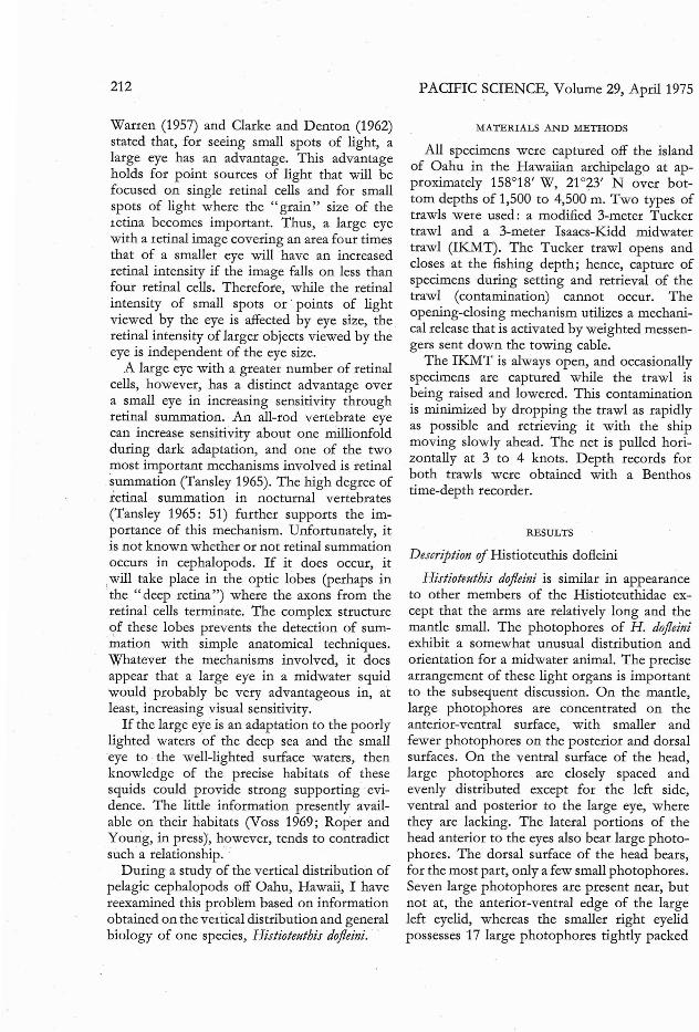
Wanen (1957) and Clarke and Denton (1962)stated that, for seeing small spots of light, alarge eye has an advantage. This advantageholds for point sources of light that will befocused on single retinal cells and for smallspots of light where the "grain" size of theretina becomes important. Thus, a large eyewith a retinal image covering an area four timesthat of a smaller eye will have an increasedretinal intensity if the image falls on less thanfour retinal cells. Therefore, while the retinalintensity of small spots or· points of lightviewed by the eye is affected by eye size, theretinal intensity of larger objects viewed by theeye is independent of the eye size.
A large eye with a greater number of retinalcells, however, has a distinct advantage overa small eye in increasing sensitivity throughretinal summation. An all-rod vertebrate eyecan increase sensitivity about one millionfoldduring dark adaptation, and one of the twomost important mechanisms involved is retinalsummation (Tansley 1965). The high degree ofretinal summation in nocturnal vertebrates(Tansley 1965: 51) further supports the importance of this mechanism. Unfortunately, itis not known whether or not retinal summationoccurs in cephalopods. If it does occur, it
,will take place in the optic lobes (perhaps in·the "deep retina") where the axons from therc·tinal cells terminate. The complex structure9f these lobes prevents the detection of summation with simple anatomical techniques.Whatever the mechanisms involved, it doesappear that a large eye in a midwater squidwould probably be very advantageous in, atleast, increasing visual sensitivity.
If the large eye is an adaptation to the poorlylighted waters of the deep sea and the smalleye to the well-lighted surface waters, thenknowledge of the precise habitats of thesesquids could provide strong supporting· evidence. The little information presently available on their habitats (Voss 1969; Roper andYoung, in press), however, tends to contradictsuch a relationship..
During a study of the vertical distribution ofpelagic cephalopods off Oahu, Hawaii, I havereexamined this problem based on informationobtained on the vertical distribution and generalbiology of one species, Histioteuthis dojleini. ..
PACIFIC SCIENCE, Volume 29, April 1975
MATERIALS AND METHODS
All specimens were captured off the islandof Oahu in the Hawaiian archipelago at approximately 158°18' W, 21°23' N over bottom depths of 1,500 to 4,500 m. Two types oftrawls were used: a modified 3-meter Tuckertrawl and a 3-meter Isaacs-Kidd midwatertrawl (IKMT). The Tucker trawl opens andcloses at the fishing depth; hence, capture ofspecimens during setting and retrieval of thetrawl (contamination) cannot occur. Theopening-closing mechanism utilizes a mechanical release that is activated by weighted messengers sent down the towing cable.
The IKMT is always open, and occasionallyspecimens are captured while the trawl isbeing raised and lowered. This contaminationis minimized by dropping the trawl as rapidlyas possible and retrieving it with the shipmoving slowly ahead. The net is pulled horizontally at 3 to 4 knots. Depth records forboth trawls were obtained with a Benthostime-depth recorder.
RESULTS
Description of Histioteuthis dofleini
Histioteuthis dojleini is similar in appeara~ceto other members of the Histioteuthidae except that the arms are relatively long and themantle small. The photophores of H. dojleiniexhibit a somewhat unusual distribution andorientation for a midwater animal. The precisearrangement of these light organs is importantto the subsequent discussion. On the mantle,large photophores are concentrated on theanterior-ventral surface, with smaller andfewer photophores on the posterior and dorsalsurfaces. On the ventral surface of the head,large photophores are closely spaced andevenly distributed except for the left side,ventral and posterior to the large eye, wherethey are lacking. The lateral portions of thehead anterior to the eyes also bear large photophores. The dorsal surface of the head bears,for the most part, only a few small photophores.Seven large photophores are present near, butnot at, the anterior-ventral edge of the largeleft eyelid, whereas the smaller right eyelidpossesses 17 large photophores tightly packed

Histioteuthis dofleini-YOUNG
Lt. Eye Ace. RetMain Ret.
Opt. l. Supraes. Mass........~
Tub. Eye
213
FIGURE 1. Histioteuthis dofleini. A, oblique section of the head of H. dofleini passing through the visual axes of botheyes; B, outline of the drawing in A but with the outline of a tubular eye superimposed on the large left eye; C,dorsal-posterior view of H. dofleini in an aquarium; this view is presumably what one would see if one were lookingvertically downward at a specimen floating in the water; D, lateral view showing the small right eye; E, lateralview showing the large left eye. The object in the photographs with the squid is a pair of 12-inch forceps.
ABBREVIATIONS: Lt. eye, left eye; Ace. Ret., accessory retina; Main Ret., main retina; Opt. L., optic lobe;Supraes. Mass, supraesophageal mass; Rt. Eye, right eye; and Tub. Eye, tubular eye.

214
along the entire circular edge of the eyelid.The arrangement of reflectors and pigment onthese latter photophores indicates that lightfrom them does not enter the eye but passesanteriorly and somewhat laterally. Largephotophores are found on the aboral surfacesof all four pairs of arms, although they aremost numerous on the fourth (ventral) armsand least numerous on the first (dorsal) arms.All of the photophores face anteriorly. Theskin of H. dofleini contains many reddish brownchromatophores that can greatly alter thecolor of the animal. When the chromatophorescontract, the animal looks silvery due to underlying iridophores; and when the chromatophores expand the animal becomes deep brownish red. Chromatophores can also expand overthe silvery tissue of the photophores. The iridescent layer is most prominent on the ventraland lateral surfaces of the head, the anteriorsurface of the mantle, and the aboral surfacesof the arms. A weaker layer is present on thedorsal surfaces of the head. Very little, if any,iridescent tissue exists on the dorsal andposterior surfaces of the mantle.
The right and left eyes differ greatly fromone another in size and shape (Figure lA).
,The right eye has a typical hemispherical shape.and a spherical lens. It differs from a typicalsquid eye primarily in the structure of theretina. The dorsal-posterior portion of theretina is noticeably thicker than the anteriorventral portion. The change in thickness isgradual, with the thickest portion being in thedorsal-posterior third and the thinnest in theanterior-ventral third.
The spherical lens of the left eye has twicethe diameter of the right one. In absoluteterms, the size of this eye is equally imp~essive.
In a specimen of only 75 mm mantle length,the lens diameter is 15 mm. The eye does nothave a hemispherical shape but has more theshape of a truncated cone with a curved base.The retina is divided into two portions ofdifferent thicknesses. The main retina (thickportion) is circular, nearly all of it being confined to the curved base of the cone and leavingmost of the converging region of the eye freepf any retina. The accessory retina (thin1?ortion) is continuous with the main retina onl\ll sides, but covers a much broader area
PACIFIC SCIENCE, Volume 29, April 1975
posterior-dorsally to the main retina thananterior-ventrally (Figure lA). Because theeye is easily distorted by contraction of thehead muscles during capture and fixation, it isnot certain whether the accessory retina liesat the same distance from the lens as the mainretina or whether it is closer to the lens, asillustrated in Figure lA, B.
The orientation of the large left eye isatypical. Instead of the usual lateral orientation,the eye faces in a posterior-dorsal direction.It is probably capable of limited movement.In a living specimen of H. heteropsis off California I have observed the large eye movefrom a posterior-dorsal direction to a dorsaldirection. Undistorted dead specimens of H.dofleini invariably have this eye directed posterior-dorsally, which is undoubtedly its moretypical position. Because of the eye's orientation and large size, the head bulges laterallyand the margin of the eyelid is elliptical andvery large, passing around the lens and thelateral wall of the bulbus of the eye. Thislateral portion of the eye that is not coveredby the eyelid contains a layer of iridophores.
Vertical Distribution
The vertical distribution of H. dofleini ispresented in Figure 2. The symbols in thefigure require some explanation. Tucker trawlcaptures are represented by a vertical bar thatindicates the total range fished by the net whileit was open. Within this range the net usuallyfishes predominately within a narrow zone,the midpoint of which is indicated by a dot.The IKMT also fishes primarily within anarrow vertical range. The total range, ofcourse, extends to the surface and, therefore,is not represented in Figure 2. The probabledepth of capture is determined from the horizontal phase of the tow in the same manner asfor captures from the Tucker trawl. EveryIKMT tow below 700 m during the day andbelow about 300 m at night passes throughthe habitat of most of the population while thenet is being set and retrieved. In such circumstances, some contamination is expected. Ihave assumed that five specimens (representedby the small dots in the figure) were capturedin this fashion.

i
Histioteuthis dofleini-YOlJNG 215
0
•200 :.'''''.. •• • • ••
,E 400 00
I 0+= • +I- 0
0Q..
,,~ 0w 600 ,
~a !O 000 0 0, 6 b 0I, If,
:800
0
1000
10 20 30 40 50 60MANTLE LENGTH, mm
FIGURE 2. Vertical distribution of Histioteuthis dofteini off Hawaii. Each symbol represents a single capture. Largesolid circle = depth of night captures; large open circles = depth of day captures; broken bars = depth range ofopening-closing day tows; solid bars = depth range of opening-closing night tows; small solid circles =
presumed contaminants.
The figure indicates that this species exhibitsa diel vertical migration, moving upwardseveral hundred meters at night. During boththe day and night, the larger animals occupyprogressively greater depths.
DISCUSSION
The vertical distribution of H. dofleini clearlyindicates that its dimorphic eyes are notadaptations to habitats with greatly differinglight intensities. Although this animal occursin different day and night habitats, bothhabitats are characterized by very low lightlevels.
The daytime habitat of most H. dofleini is azone of low light intensity, yet a zone wherelight plays a critical role in the ecology ofmany of the inhabitants. This twilight zonefrom about 400- to 700-m depth off Hawaiicorresponds to the habitat of most half-redshrimp (Foxton 1970; ]. Walters, personalcommunication), to the habitat of mostanimals bearing complex ventral photophores(Foxton 1970, Young 1973) and to the habitatof most fish (T. Clarke, personal communication; S. Amesbury, personal communication)
and squid (Young 1975) with tubular eyes.Although larvae of H. dofleini live well abovethe twilight zone in near-surface waters andlarger adults are found in the lower reaches ofthe twilight zone or occasionally below it,most juveniles and young adults occur withinit and exhibit characteristics typical of manysquid living there, i.e., a layer of silveryiridophores overlain by functional chromatophores, and complex ventral photophores.
Within this zone the intensity of downwelling light is over one hundred timesgreater than that passing upward (see Tylerand Preisendorfer 1962: 423). Determiningthe typical orientation of H. dofleini withinthis strongly directional radiance pattern iscritical to understanding the functions of theeyes. Clarke, Denton, and Gilpin-Brown (1969)have shown that a closely related squid, H.reversa, is neutrally buoyant. H. dofleini alsoappears to be nearly neutrally buoyant in anaquarium. Although the animal tends torotate to a position with the mantle downwardwhen motionless, it is probably capable oforienting in almost any direction with only aslight assist from the fins and funnel. Thenormal orientation of this species can be

216
deduced from the distribution of photophoreson the head, mantle, and arms. These photophores point in an anterior-ventral directionrelative to the longitudinal axis of the body.If the photophores are used in ventral countershading (i.e., elimination of the silhouettewhen viewed against the downwelling surfacelight), as are similar photophores in manyother midwater animals (Clarke 1963; Foxton1970; Denton, Gilpin-Brown, and Wtight 1972;Young 1973), they then must be directed downward. In order to direct the photophores downward the squid must be positioned with thebody axis at an angle of about 45° from thehorizontal with the mantle uppermost. In thisorientation the large eye looks upward in thedirection of maximum light intensity.
The brge eye of H. dofleini approaches atubular eye in shape and has certain functionalrelationships to tubular eyes. In nearly alltubular-eyed species, the eyes seem to bedirected either dorsally on a horizontallypositioned animal (e.g., Opisthoproctus) oranteriorly on a presumably vertically orientedanimal (most species with anteriorly directedeyes may orient vertically, as has been indicated for Gigantura and Srylephorus [Bruun 1957].However, the fish Winteria may be an exception. Unfortunately the evidence concerningorientation in midwater animals is meagre.)The large histioteuthid eye is also directedupward and its visual field completely includesthe vertical visual field of a tubular eye; yet,unlike the tubular eye, it maintains a broadlateral field of view (Figure 1E).
The probable function of a tubular eye hasbeen examined by Munk (1966) and others.Munk demonstrated that the tubular eye isequivalent to the central core of a hemisphericaleye. Fishes and cephalopods with tubular eyeshave the optical axes of their eyes parallel ornearly parallel. The compact configuration ofthe tubular eyes facilitates the parallel orientation and thus binocular vision. Brauer (1908)suggested that binocular vision results inbetter judgment of distances, whereas Weale(1955) suggested that it results in lowering ofthe visual threshold. (Pirenne [1967] statedthat binocular vision lowers the visual threshold in humans by 20 percent.) Fremlin (1972)suggested that binocular vision may increase
PACIFIC SCIENCE, Volume 29, April 1975
the ability to see details above the visualthreshold by increasing the signal-to-noiseratio; actual retinal stimulation (" signal") couldbe distinguished from fluctuations in cellactivity (noise) by analyzing retinal stimulationcoincident on the two retinas.
Although most authors (e.g., Walls 1942,Tansley 1965) have suggested that tubulareyes represent large eyes, i.e., they correspondto the central cores of large eyes, this has notbeen rigorously demonstrated. The tubulareyes in fishes and cephalopods could representthe reduction of normal-sized eyes into compact spaces for parallel alignment and binocularvision. However, the semitubular eye of H.dofleini is clearly not just a normal-sized eyein a somewhat compact form. Rather it isnearly twice the size of its right counterpart.The arrangement in H. dofleini demonstratesthe problem of having large upward-directedeyes. Even though the eye does not have thefull normal shape in this species, it still grosslydistorts a very large head. By analogy, thisarrangement in Histioteuthis suggests that tubular eyes of fish are indeed large eyes in a compact form designed for viewing the verticallydownwelling light, and that binocularity,therefore, is not the only factor involved.
The small eye of H. d?fleini has an anteriortilt. An additional ventral tilt results from thetilt of the head imposed by the large right eye.This latter tilt also explains the asymmetricalarrangement of the photophores on the ventralsurface of the head. When the animal is in itspresumed typical orientation, the small eyetilts slightly downward. Therefore, while thelarge and presumably more sensitive eye pointsupward in the direction of maximum lightintensity, the smaller eye, directed laterallyand ventrally, points in a direction of verylow light intensity. The latter eye probablyreceives less than 5 percent of the downwellingillumination that the upward-looking eye receives (see Tyler and Preisendorfer 1962: 423).These circumstances suggest that, whereas thelarge upward-looking eye detects downwellingsurface light, the small eye does not; rather,it detects bioluminescent light. Further, thecompact arrangement of photophores aroundthe smaller eye, combined with the modifications of the retina, suggests that counter-

Histioteuthis dofleini-YOUNG
shading is not the only function of these organs.These ocular photophores are ideally locatedto produce a strong beam of light that wouldilluminate the portion of the environment thatis surveyed by the thicker portions of theretina. In other words, these photophores mayfunction as a searchlight.
Lens Pigments
Denton and Warren (1968) suggested thatthe large eye of Histioteuthis functions in nearsurface waters because of the light-absorbingcharacteristics of the lens. They found that inH. meleagroteuthis the lens of the small eye istransparent to light down to about 310 nm,whereas the lens of the large eye always absorbs the near ultraviolet and sometimes bluelight. These authors pointed out that suchfeatures are characteristic of surface-dwellingfishes and squids. However, catch recordsdemonstrate that Histioteuthis spp. do not normally occur in near-surface waters during theday-time and the previous discussion hasattempted to demonstrate that the large eye isadapted for vision under conditions of lowlight intensity. In near-surface species, anultraviolet-absorbing lens may protect theretina from damage (Denton and Warren1968) or may improve visual acuity by reducing chromatic aberration (Wald and Griffin1947). The reason for the ultraviolet-absorbingpigments in the large lens of Histiotheuthisremains a mystery.
SUMMARY
1. Histioteuthis dofleini lives primarily in thetwilight zone (approximately 400 to 700 mdepth) during the day and migrates upwardseveral hundred meters at night.
2. This squid has a large left eye with a semitubular shape and a small right eye with atypical hemispherical shape.
3. H. dofleini probably orients at an obliqueangle in the water so that the large eye isdirected vertically upward while the smalleye is directed ventral-laterally.
4. The disparity in the size of the two eyessuggests that tubular eyes in other midwater animals are indeed enlarged eyes, as
217
has been previously suggested by severalauthors.
5. The small right eye may function primarilyin the detection of bioluminescent lightand the photophores that surround the lensmay function as a searchlight.
6. The large left eye functions primarily duringthe day in detecting downwelling light.
ACKNOWLEDGMENTS
I wish to thank Andrew Packard, UniversityMedical School, Edinburgh, and John Walters,University of Hawaii, for reading and commenting on the manuscript. I also thank JohnWalters for taking the photographs in Figure2, and Thomas Clarke, University of Hawaii,for supplying most of the specimens taken inopen nets.
LITERATURE CITED
BRAUER, A. 1908. Die Tiefsee-Fische. 2. Anat.Teil. Wiss. Ergebn. 'Valdivia' 15: 1-266.
BRUUN, A. F. 1957. Deep sea and abyssaldepths. Mem. geol. Soc. Amer. 67: 641-672.
CLARKE, G. L. 1971. Light conditions in thesea in relation to the diurnal vertical migrations of animals. Pages 41-50 in G. B.Farquhar, ed. Proceedings of an internationalsymposium on biological sound scatteringin the ocean. Maury Center for OceanScience, Washington.
CLARKE, G. L., and E. J. DENTON. 1962. Lightand animal life. Pages 456--468 in M. N. Hill,ed. The sea. Vol. 1. John Wiley & Sons,Interscience, New York.
CLARKE, M. R., E. ]. DENTON, and ]. B.GILPIN-BROWN. 1969. On the buoyancy ofsquid of the families Histioteuthidae, Octopoteuthidae and Chiroteuthidae. J. Physiol.203: 49-50.
CLARKE, W. D. 1963. Function of bioluminescence in mesopelagic organisms. Nature,Lond. 198: 1244-1246.
DENTON, E. ]., ]. B. GILPIN-BROWN, and P. G.WRIGHT. 1972. The angular distribution ofthe light produced by some mesopelagicfish in relation to their camouflage. Proc.roy. Soc., B, 182: 145-158.
DENTON, E. ]., and F. ]. WARREN. 1957. Thephotosensitive pigments in the retinae of

218
deep-sea fish. J. Mar. bioI. Ass. U.K. 36:651-662.
---. 1968. Eyes of the Histioteuthidae.Nature, Lond. 219: 400-401.
FOXTON, P. 1970. The vertical distribution ofpelagic decapods (Crustacea: Natantia)collected on the SOND cruise 1965. J. Mar.bioI. Ass. U.K. 50: 939-960.
FREMLIN, J. 1972. How stereoscopic visionevolved. New Sci. 56: 26-28.
KAMPA, E. M. 1971. Photoenvironment andsonic scattering. Pages 41-50 in G. B.Farquhar, ed. Proceedings of an internationalsymposium on biological sound scatteringin the ocean. Maury Center for OceanScience, Washington.
LANE, F. W. 1960. Kingdom of the octopus.Sheridan House, New YOlk. 300 pp.
MUNK, O. 1966. Ocular anatomy of some deepsea teleosts. Dana Rep. 70: 1-62.
PIRENNE, M. H. 1967. Vision and the eye.Chapman & Hall, London.
PUMPHREY, R. J. 1961. Concerning vision.Pages 193-208 in J. A. Ramsey and V. V.Wigglesworth, eds. The cell and the organism. At the University Press, Cambridge.
ROPER, C. F. E., and R. E. YOUNG. In press.The vertical distribution of pelagic cephalopods. Smithson. Contr. ZooI.
PACIFIC SCIENCE, Volume 29, April 1975
TANSLEY, K. 1965. Vision in vertebrates.Chapman & Hall, London.
TYLER, J. E., and R. W. PREISENDORFER. 1962.Transmission of energy within the sea.Pages 397-451 in M. N. Hill, ed. The sea.Vol. 1. John Wiley & Sons, Interscience,New York.
Voss, G. L. 1967. The biology and bathymetricdistribution of deep-sea cephalopods. Stud.trap. Oceanogr. 5: 511-535.
Voss, N. A. 1969. A monograph of the Cephalopoda of the North Atlantic: the familyHistioteuthidae. Bull. mar. Sci. 19(4): 713867.
WALD, G., and D. R. GRIFFIN. 1947. Thechange in refractive power of the humaneye in dim and bright light. J. opt. Soc.Amer. 37: 321-336.
WALLS, G. L. 1942. The:vertebrate eye. Bull.Cranbraok Inst. Sci. 19. 785 pp.
WEALE, R. A. 1955. Binocular vision and deepsea fish. Nature, Lond. 175: 996.
YOUNG, R. E. 1973. Information feedbackfrom photophores and ventral countershading in mid-water squid. Pacif. Sci.27(1): 1-7.
---. 1975. Transitory eye shapes and thevertical distribution of two midwater squids.Pacif. Sci. 29(3): 243-255.


