Function Human Heart
13
The human heart is an organ that pumps blood throughout the body via the circulatory system. Human heart anatomy In humans, the heart is roughly the s ize of a large fist and weighs between 9 and 12 ounces (250 and 350 grams). It has four chambers: two upper chambers (the atria) and two lower ones (the ventricles). The right atrium and right ventricle together make up the "right heart," and the left atrium and left ventricle make up the "left heart." A wall of muscle called the septum separates the two sides of the heart. The human heart is about the size of a fist. Credit: tlorna |Shutterstock View full size image A double-walled sac called the pericardium encases the heart, which serves to protect the heart and anchor it inside the chest. Between the outer layer, the parietal pericardium, and the inner layer, the serous pericardium, runs pericardial fluid, which lubricates the heart during contractions and movements of the lungs and diaphragm. The heart's outer wall consists of three layers. The outermost wall layer, or epicardium, is the inner wall of the pericardium. The middle layer, or myocardium, contains the muscle that contracts. The inner layer, or endocardium, is the lining that contacts the blood. The tricuspid valve and the mitral valve make up the atrioventricular (AV) valves, which connect the atria and the ventricles. The pulmonary semi-lunar valve separates the left ventricle from t he pulmonary artery, and the aortic valve s eparates the right ventricle from the aorta. The heartstrings, or chordae tendinae, anchor the valves to heart muscles. The sinoatrial node produces the electrical pulses that driv e heart contractions. Human heart function The heart circulates blood through two pathways: the pulmonary circ uit and the systemic circuit.
Transcript of Function Human Heart
7/27/2019 Function Human Heart
http://slidepdf.com/reader/full/function-human-heart 1/13
The human heart is an organ that pumps blood throughout the body via the
circulatory system.
Human heart anatomy
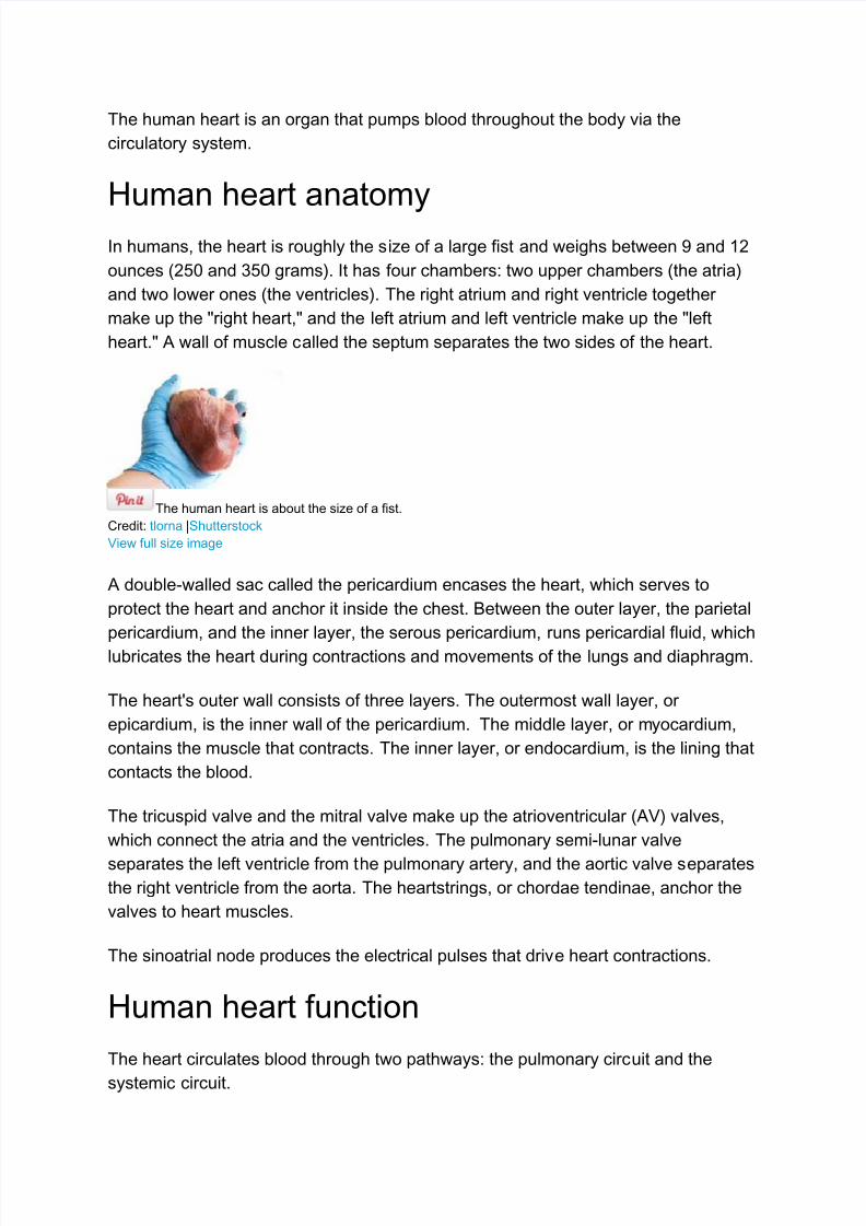
In humans, the heart is roughly the size of a large fist and weighs between 9 and 12
ounces (250 and 350 grams). It has four chambers: two upper chambers (the atria)
and two lower ones (the ventricles). The right atrium and right ventricle together
make up the "right heart," and the left atrium and left ventricle make up the "left
heart." A wall of muscle called the septum separates the two sides of the heart.
The human heart is about the size of a fist.
Credit: tlorna |Shutterstock
View full size image
A double-walled sac called the pericardium encases the heart, which serves to
protect the heart and anchor it inside the chest. Between the outer layer, the parietal
pericardium, and the inner layer, the serous pericardium, runs pericardial fluid, which
lubricates the heart during contractions and movements of the lungs and diaphragm.
The heart's outer wall consists of three layers. The outermost wall layer, or
epicardium, is the inner wall of the pericardium. The middle layer, or myocardium,
contains the muscle that contracts. The inner layer, or endocardium, is the lining that
contacts the blood.
The tricuspid valve and the mitral valve make up the atrioventricular (AV) valves,
which connect the atria and the ventricles. The pulmonary semi-lunar valve
separates the left ventricle from the pulmonary artery, and the aortic valve separatesthe right ventricle from the aorta. The heartstrings, or chordae tendinae, anchor the
valves to heart muscles.
The sinoatrial node produces the electrical pulses that drive heart contractions.
Human heart function
The heart circulates blood through two pathways: the pulmonary circuit and the
systemic circuit.
7/27/2019 Function Human Heart
http://slidepdf.com/reader/full/function-human-heart 2/13
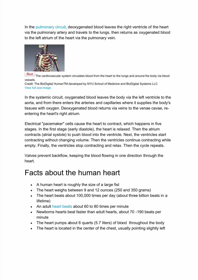
In the pulmonary circuit, deoxygenated blood leaves the right ventricle of the heart
via the pulmonary artery and travels to the lungs, then returns as oxygenated blood
to the left atrium of the heart via the pulmonary vein.
The cardiovascular system circulates blood from the heart to the lungs and around the body via blood
vessels.
Credit: The BioDigital HumanTM developed by NYU School of Medicine and BioDigital Systems LLC
View full size image
In the systemic circuit, oxygenated blood leaves the body via the left ventricle to theaorta, and from there enters the arteries and capillaries where it supplies the body's
tissues with oxygen. Deoxygenated blood returns via veins to the venae cavae, re-
entering the heart's right atrium.
Electrical "pacemaker" cells cause the heart to contract, which happens in five
stages. In the first stage (early diastole), the heart is relaxed. Then the atrium
contracts (atrial systole) to push blood into the ventricle. Next, the ventricles start
contracting without changing volume. Then the ventricles continue contracting while
empty. Finally, the ventricles stop contracting and relax. Then the cycle repeats.
Valves prevent backflow, keeping the blood flowing in one direction through the
heart.
Facts about the human heart
A human heart is roughly the size of a large fist
The heart weighs between 9 and 12 ounces (250 and 350 grams)
The heart beats about 100,000 times per day (about three billion beats in alifetime)
An adult heart beats about 60 to 80 times per minute
Newborns hearts beat faster than adult hearts, about 70 -190 beats per
minute
The heart pumps about 6 quarts (5.7 liters) of blood throughout the body
The heart is located in the center of the chest, usually pointing slightly left
7/27/2019 Function Human Heart
http://slidepdf.com/reader/full/function-human-heart 3/13
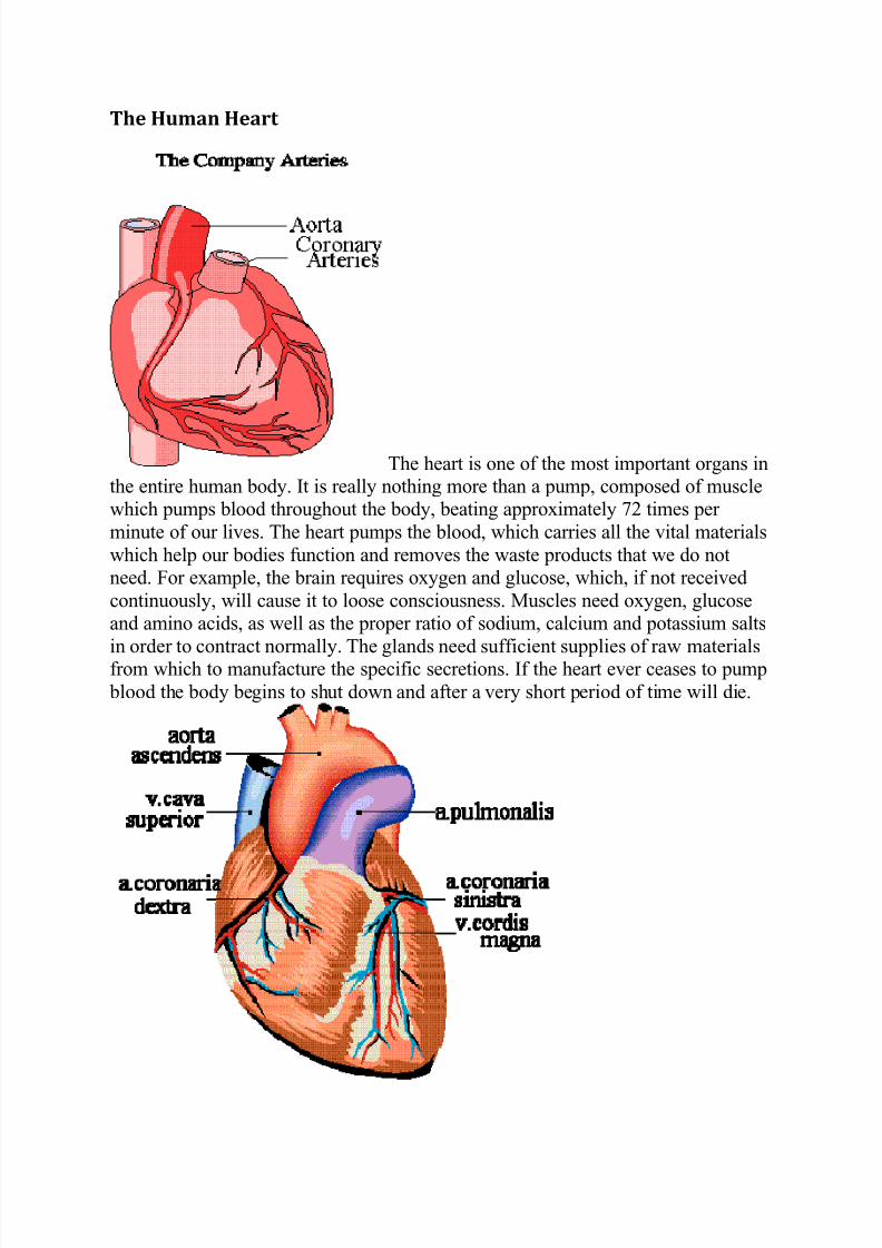
The Human Heart
The heart is one of the most important organs inthe entire human body. It is really nothing more than a pump, composed of musclewhich pumps blood throughout the body, beating approximately 72 times per
minute of our lives. The heart pumps the blood, which carries all the vital materials
which help our bodies function and removes the waste products that we do notneed. For example, the brain requires oxygen and glucose, which, if not received
continuously, will cause it to loose consciousness. Muscles need oxygen, glucoseand amino acids, as well as the proper ratio of sodium, calcium and potassium salts
in order to contract normally. The glands need sufficient supplies of raw materialsfrom which to manufacture the specific secretions. If the heart ever ceases to pump
blood the body begins to shut down and after a very short period of time will die.
7/27/2019 Function Human Heart
http://slidepdf.com/reader/full/function-human-heart 4/13
The heart is essentially a muscle(a little larger than the fist). Like any other muscle
in the human body, it contracts and expands. Unlike skeletal muscles, however, theheart works on the "All -or-Nothing Law". That is, each time the heart contracts it
does so with all its force. In skeletal muscles, the principle of "gradation" is
present. The pumping of the heart is called the Cardiac Cycle, which occurs about72 times per minute. This means that each cycle lasts about eight-tenths of a
second. During this cycle the entire heart actually rests for about four-tenths of a
second.
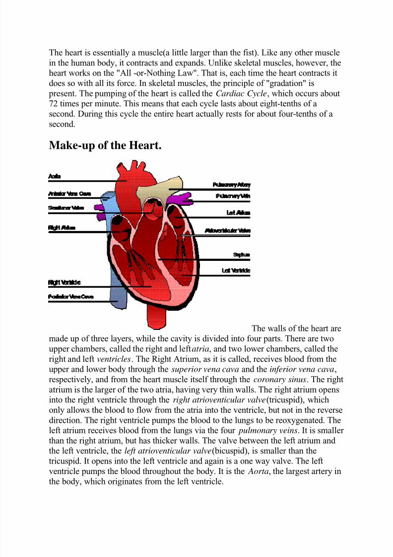
Make-up of the Heart.
The walls of the heart aremade up of three layers, while the cavity is divided into four parts. There are two
upper chambers, called the right and leftatria, and two lower chambers, called the
right and left ventricles. The Right Atrium, as it is called, receives blood from theupper and lower body through the superior vena cava and the inferior vena cava,
respectively, and from the heart muscle itself through the coronary sinus. The right
atrium is the larger of the two atria, having very thin walls. The right atrium opensinto the right ventricle through the right atrioventicular valve(tricuspid), which
only allows the blood to flow from the atria into the ventricle, but not in the reverse
direction. The right ventricle pumps the blood to the lungs to be reoxygenated. Theleft atrium receives blood from the lungs via the four pulmonary veins. It is smaller
than the right atrium, but has thicker walls. The valve between the left atrium andthe left ventricle, the left atrioventicular valve(bicuspid), is smaller than the
tricuspid. It opens into the left ventricle and again is a one way valve. The left
ventricle pumps the blood throughout the body. It is the Aorta, the largest artery in
the body, which originates from the left ventricle.
7/27/2019 Function Human Heart
http://slidepdf.com/reader/full/function-human-heart 5/13
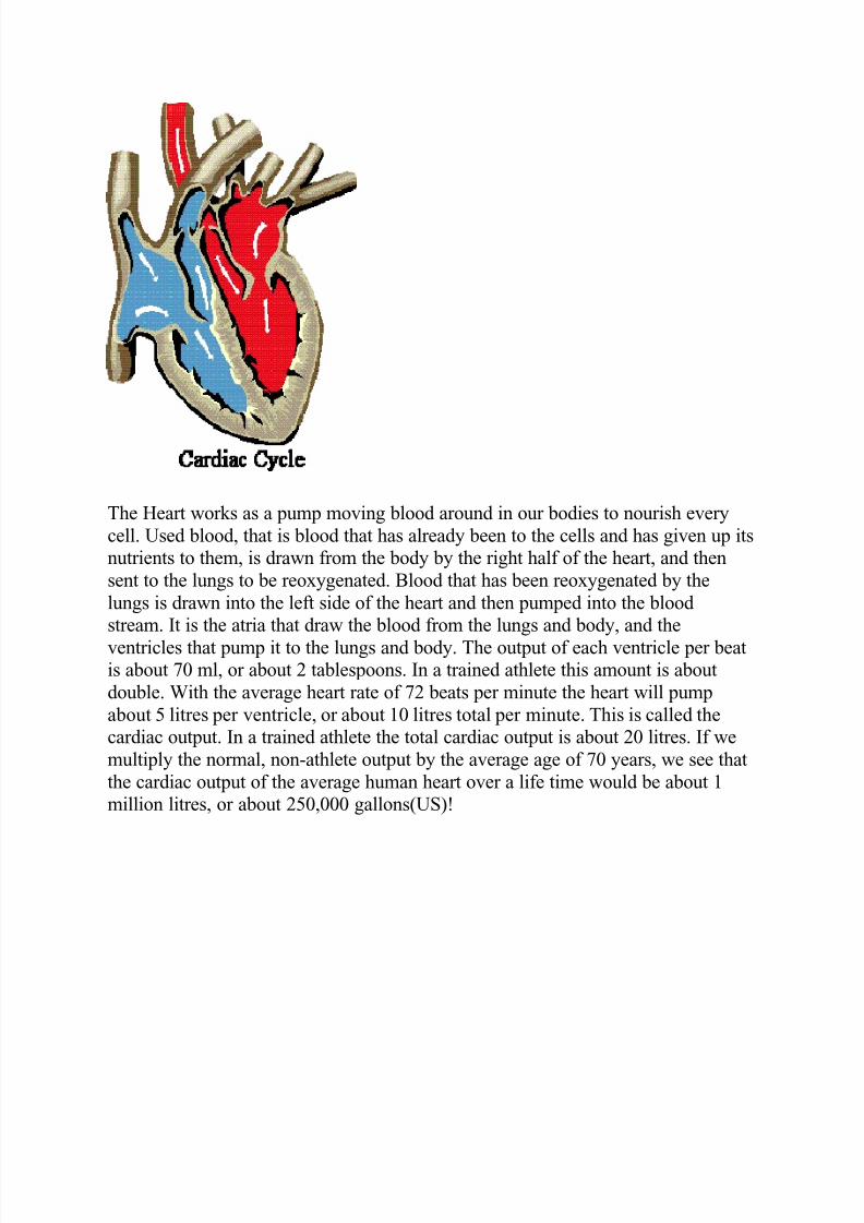
The Heart works as a pump moving blood around in our bodies to nourish every
cell. Used blood, that is blood that has already been to the cells and has given up itsnutrients to them, is drawn from the body by the right half of the heart, and then
sent to the lungs to be reoxygenated. Blood that has been reoxygenated by the
lungs is drawn into the left side of the heart and then pumped into the bloodstream. It is the atria that draw the blood from the lungs and body, and the
ventricles that pump it to the lungs and body. The output of each ventricle per beatis about 70 ml, or about 2 tablespoons. In a trained athlete this amount is about
double. With the average heart rate of 72 beats per minute the heart will pump
about 5 litres per ventricle, or about 10 litres total per minute. This is called thecardiac output. In a trained athlete the total cardiac output is about 20 litres. If we
multiply the normal, non-athlete output by the average age of 70 years, we see that
the cardiac output of the average human heart over a life time would be about 1million litres, or about 250,000 gallons(US)!
7/27/2019 Function Human Heart
http://slidepdf.com/reader/full/function-human-heart 6/13
Heart Basics
Function of the Heart
Introduction
The role of the heart is to pump oxygen-rich blood to every living cell in the body. Inorder to achieve its goal, it must continuously beat for a person’s entire lifespan. Becauseof its vital role, a non-beating heart always results in death. The human heart beatsapproximately 80,000 to 100,000 a day and pumps almost 2,000 gallons of blood. Thismeans that in a person’s life lasting 70 to 90 years, the heart beats approximately two tothree billion times and pumps 50 to 65 million gallons of blood. Because the heart is soessential for human sustenance, it is made up of a muscle different from skeletal musclethat allows it to constantly beat.
In order for the heart to deliver oxygenated blood to all cells, blood is pumped througharteries. Veins bring deoxygenated blood cells to the lungs, which then are oxygenated,and then sent back to heart. In this way, a continuous cycle is formed of the heartpumping oxygenated blood and deoxygenated blood out to their designated destinations,
and therefore the heart maintains the circulatory system.
Top
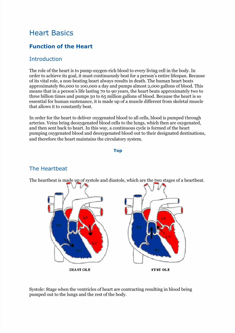
The Heartbeat
The heartbeat is made up of systole and diastole, which are the two stages of a heartbeat.
Systole: Stage when the ventricles of heart are contracting resulting in blood beingpumped out to the lungs and the rest of the body.
7/27/2019 Function Human Heart
http://slidepdf.com/reader/full/function-human-heart 7/13
Thick, muscular walls of both ventricles contract. Pressure rises in both ventricles, causing the bicuspid and tricuspid valves
to close. Therefore, blood is forced up the aorta and the pulmonary artery. The atria relax during this time. The left atrium receives blood from the
pulmonary vein, and the right atrium from the vena cava.
Diastole: Stage when the ventricles of the heart are relaxed and not contracting. Duringthis stage, the atria are filled with blood and pump blood into the ventricles.
Thick, muscular walls of both ventricles relax. Pressure in both ventricles falls low enough for bicuspid valves to open. The atria contract, and blood is forced into the ventricles, expanding them.
The blood pressure in the aorta is decreased, therefore the semi-lunar valves close.
How the heart worksWritten by Dr Roger Henderson, GP
127
What is the heart?The heart is a hollow, cone-shaped muscle that's about the size of an adult fist and usually found to the left of our
breastbone.
The heart is the most important organ in our body. It is basically a complex pump, responsible for
circulating blood, oxygen and nutrients around the body.
Parts of the heartDid you know? The average weight of a healthy female human heart is 9oz (255g).
A man's heart is usually slightly bigger at around 10.5oz (300g).
Every heart is made up of three layers:
an inner lining called the endocardium
a middle layer of muscle called the myocardium
an outer fluid-filled sac known as the pericardium.
The heart is divided into four chambers:
the right atrium and left atrium are the upper chambers of the heart
the right ventricle and left ventricle are the lower chambers.
A muscular wall called the septum separates the right and left sides of the heart.
Each of the chambers has valves. The valves have different names:
the tricuspid valve is at the exit of the right atrium the mitral valve is for the left atrium
the pulmonary valve is at the exit of the right ventricle
the aortic valve is at the exit of the left ventricle.
Their purpose is to allow blood to move forwards through the heart and to prevent it flowing backwards into the
previous chamber.
How does the heart work?The heart muscle contracts in two stages to squeeze blood out of the heart. This is known as systole.
In the first stage, the upper chambers (atria) contract at the same time, pushing blood down into the lower
chambers (ventricles).
Blood is pumped from the right atrium down into the right ventricle and from the left atrium down into the left
ventricle.
In the second stage, the lower chambers contract to push this blood out of the heart to either the body via your
main artery (aorta) or to the lungs to pick up oxygen.
7/27/2019 Function Human Heart
http://slidepdf.com/reader/full/function-human-heart 8/13
The heart then relaxes – known as diastole. Blood fills up the heart again, and the whole process, which takes a
fraction of a second, is repeated.
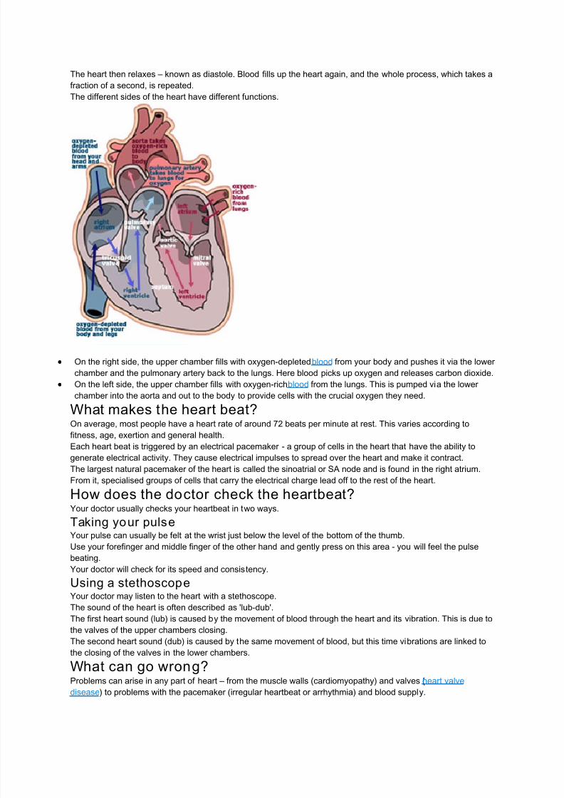
The different sides of the heart have different functions.
On the right side, the upper chamber fills with oxygen-depleted blood from your body and pushes it via the lower
chamber and the pulmonary artery back to the lungs. Here blood picks up oxygen and releases carbon dioxide.
On the left side, the upper chamber fills with oxygen-rich blood from the lungs. This is pumped via the lower
chamber into the aorta and out to the body to provide cells with the crucial oxygen they need.
What makes the heart beat?On average, most people have a heart rate of around 72 beats per minute at rest. This varies according to
fitness, age, exertion and general health.
Each heart beat is triggered by an electrical pacemaker - a group of cells in the heart that have the ability to
generate electrical activity. They cause electrical impulses to spread over the heart and make it contract.
The largest natural pacemaker of the heart is called the sinoatrial or SA node and is found in the right atrium.
From it, specialised groups of cells that carry the electrical charge lead off to the rest of the heart.
How does the doctor check the heartbeat?Your doctor usually checks your heartbeat in two ways.
Taking your pulseYour pulse can usually be felt at the wrist just below the level of the bottom of the thumb.
Use your forefinger and middle finger of the other hand and gently press on this area - you will feel the pulse
beating.Your doctor will check for its speed and consistency.
Using a stethoscopeYour doctor may listen to the heart with a stethoscope.
The sound of the heart is often described as 'lub-dub'.
The first heart sound (lub) is caused by the movement of blood through the heart and its vibration. This is due to
the valves of the upper chambers closing.
The second heart sound (dub) is caused by the same movement of blood, but this time vibrations are linked to
the closing of the valves in the lower chambers.
What can go wrong?Problems can arise in any part of heart – from the muscle walls (cardiomyopathy) and valves (heart valve
disease) to problems with the pacemaker (irregular heartbeat or arrhythmia) and blood supply.
7/27/2019 Function Human Heart
http://slidepdf.com/reader/full/function-human-heart 9/13
If a coronary artery becomes furred up or partially blocked with fatty material called atheroma, that artery cannot
then supply enough blood to the heart muscle to meet its needs during exertion or activity. The muscle ‘cramps’,
causing chest pain. This is known as angina.
If the poor blood supply to the heart worsens, so that chest pains start to happen more easily and with less
exertion, it's known as unstable angina. This requires increasing levels of heart medication or active surgical
intervention such as an angioplasty or heart bypass surgery.
When a coronary artery is completely blocked and no blood or oxygen reaches the heart muscle served by that
artery, it causes a heart attack. This also causes chest pain as the heart muscle served by that artery dies.
Depending on which part of the heart muscle is affected and the severity of damage to the heart muscle, the
effects of a heart attack can range from a good recovery to instant death. Heart disease is the biggest killer in the
UK, often causing death before the person reaches hospital.
Heart
The heart is a muscular organ about the size of a closed fist that functions as the
body’s circulatory pump. It takes in deoxygenated blood through the veins and
delivers it to the lungs for oxygenation before pumping it into the various arteries
(which provide oxygen and nutrients to body tissues by transporting the bloodthroughout the body). The heart is located in the thoracic cavity medial to the lungs
and posterior to the sternum.
On its superior end, the base of the heart is attached to the aorta,...
[Continued from above] . . . pulmonary arteries and veins, and the vena cava. The
inferior tip of the heart, known as the apex, rests just superior to the diaphragm.
The base of the heart is located along the body’s midline with the apex pointing
toward the left side. Because the heart points to the left, about 2/3 of the heart’s
mass is found on the left side of the body and the other 1/3 is on the right.
Anatomy of the HeartPericardium
The heart sits within a fluid-filled cavity called the pericardial cavity. The walls and
lining of the pericardial cavity are a special membrane known as the pericardium.
Pericardium is a type of serous membrane that produces serous fluid to lubricate the
heart and prevent friction between the ever beating heart and its surrounding
organs. Besides lubrication, the pericardium serves to hold the heart in position and
maintain a hollow space for the heart to expand into when it is full. The pericardium
has 2 layers—a visceral layer that covers the outside of the heart and a parietal
layer that forms a sac around the outside of the pericardial cavity.
Structure of the Heart Wall The heart wall is made of 3 layers: epicardium, myocardium and endocardium.
Epicardium. The epicardium is the outermost layer of the heart wall and is just
another name for the visceral layer of the pericardium. Thus, the epicardium is a
thin layer of serous membrane that helps to lubricate and protect the outside of the
heart. Below the epicardium is the second, thicker layer of the heart wall: the
myocardium.
Myocardium. The myocardium is the muscular middle layer of the heart wall that
contains the cardiac muscle tissue. Myocardium makes up the majority of the
thickness and mass of the heart wall and is the part of the heart responsible for
pumping blood. Below the myocardium is the thin endocardium layer.
7/27/2019 Function Human Heart
http://slidepdf.com/reader/full/function-human-heart 10/13
Endocardium. Endocardium is the simple squamous endothelium layer that lines the
inside of the heart. The endocardium is very smooth and is responsible for keeping
blood from sticking to the inside of the heart and forming potentially deadly blood
clots.
The thickness of the heart wall varies in different parts of the heart. The atria of the
heart have a very thin myocardium because they do not need to pump blood veryfar—only to the nearby ventricles. The ventricles, on the other hand, have a very
thick myocardium to pump blood to the lungs or throughout the entire body. The
right side of the heart has less myocardium in its walls than the left side because the
left side has to pump blood through the entire body while the right side only has to
pump to the lungs.
Chambers of the Heart
The heart contains 4 chambers: the right atrium, left atrium, right ventricle,
and left ventricle. The atria are smaller than the ventricles and have thinner, less
muscular walls than the ventricles. The atria act as receiving chambers for blood, sothey are connected to the veins that carry blood to the heart. The ventricles are the
larger, stronger pumping chambers that send blood out of the heart. The ventricles
are connected to the arteries that carry blood away from the heart.
The chambers on the right side of the heart are smaller and have less myocardium
in their heart wall when compared to the left side of the heart. This difference in size
between the sides of the heart is related to their functions and the size of the 2
circulatory loops. The right side of the heart maintains pulmonary circulation to the
nearby lungs while the left side of the heart pumps blood all the way to the
extremities of the body in the systemic circulatory loop.
Valves of the Heart
The heart functions by pumping blood both to the lungs and to the systems of the
body. To prevent blood from flowing backwards or “regurgitating” back into the
heart, a system of one-way valves are present in the heart. The heart valves can be
broken down into two types: atrioventricular and semilunar valves.
Atrioventricular valves. The atrioventricular (AV) valves are located in the middle of
the heart between the atria and ventricles and only allow blood to flow from the
atria into the ventricles. The AV valve on the right side of the heart is called
the tricuspid valve because it is made of three cusps (flaps) that separate to allow
blood to pass through and connect to block regurgitation of blood. The AV valve on
the left side of the heart is called the mitral valve or the bicuspid valve because it
has two cusps. The AV valves are attached on the ventricular side to tough strings
called chordae tendineae. The chordae tendineae pull on the AV valves to keep
them from folding backwards and allowing blood to regurgitate past them. During
the contraction of the ventricles, the AV valves look like domed parachutes with the
chordae tendineae acting as the ropes holding the parachutes taut.
Semilunar valves. The semilunar valves, so named for the crescent moon shape of
their cusps, are located between the ventricles and the arteries that carry blood
away from the heart. The semilunar valve on the right side of the heart is
the pulmonary valve, so named because it prevents the backflow of blood from the
pulmonary trunk into the right ventricle. The semilunar valve on the left side of the
heart is theaortic valve, named for the fact that it prevents the aorta from
regurgitating blood back into the left ventricle. The semilunar valves are smaller
than the AV valves and do not have chordae tendineae to hold them in place.
7/27/2019 Function Human Heart
http://slidepdf.com/reader/full/function-human-heart 11/13
Instead, the cusps of the semilunar valves are cup shaped to catch regurgitating
blood and use the blood’s pressure to snap shut.
Conduction System of the Heart
The heart is able to both set its own rhythm and to conduct the signals necessary to
maintain and coordinate this rhythm throughout its structures. About 1% of the
cardiac muscle cells in the heart are responsible for forming the conduction systemthat sets the pace for the rest of the cardiac muscle cells.
The conduction system starts with the pacemaker of the heart—a small bundle of
cells known as the sinoatrial (SA) node. The SA node is located in the wall of the
right atrium inferior to the superior vena cava. The SA node is responsible for
setting the pace of the heart as a whole and directly signals the atria to contract.
The signal from the SA node is picked up by another mass of conductive tissue
known as the atrioventricular (AV) node.
The AV node is located in the right atrium in the inferior portion of the interatrial
septum. The AV node picks up the signal sent by the SA node and transmits it
through the atrioventricular (AV) bundle. The AV bundle is a strand of conductive
tissue that runs through the interatrial septum and into the interventricular septum.The AV bundle splits into left and right branches in the interventricular septum and
continues running through the septum until they reach the apex of the heart.
Branching off from the left and right bundle branches are many Purkinje
fibers that carry the signal to the walls of the ventricles, stimulating the cardiac
muscle cells to contract in a coordinated manner to efficiently pump blood out of the
heart.
Physiology of the Heart
Coronary Systole and Diastole
At any given time the chambers of the heart may found in one of two states:
Systole. During systole, cardiac muscle tissue is contracting to push blood out of thechamber.
Diastole. During diastole, the cardiac muscle cells relax to allow the chamber to fill
with blood. Blood pressure increases in the major arteries during ventricular systole
and decreases during ventricular diastole. This leads to the 2 numbers associated
with blood pressure—systolic blood pressure is the higher number and diastolic
blood pressure is the lower number. For example, a blood pressure of 120/80
describes the systolic pressure (120) and the diastolic pressure (80).
The Cardiac Cycle
The cardiac cycle includes all of the events that take place during one heartbeat.
There are 3 phases to the cardiac cycle: atrial systole, ventricular systole, andrelaxation.
Atrial systole: During the atrial systole phase of the cardiac cycle, the atria contract
and push blood into the ventricles. To facilitate this filling, the AV valves stay open
and the semilunar valves stay closed to keep arterial blood from re-entering the
heart. The atria are much smaller than the ventricles, so they only fill about 25% of
the ventricles during this phase. The ventricles remain in diastole during this phase.
Ventricular systole: During ventricular systole, the ventricles contract to push blood
into the aorta and pulmonary trunk. The pressure of the ventricles forces the
semilunar valves to open and the AV valves to close. This arrangement of valves
allows for blood flow from the ventricles into the arteries. The cardiac muscles of the
7/27/2019 Function Human Heart
http://slidepdf.com/reader/full/function-human-heart 12/13
atria repolarize and enter the state of diastole during this phase.
Relaxation phase: During the relaxation phase, all 4 chambers of the heart are in
diastole as blood pours into the heart from the veins. The ventricles fill to about
75% capacity during this phase and will be completely filled only after the atria
enter systole. The cardiac muscle cells of the ventricles repolarize during this phaseto prepare for the next round of depolarization and contraction. During this phase,
the AV valves open to allow blood to flow freely into the ventricles while the
semilunar valves close to prevent the regurgitation of blood from the great arteries
into the ventricles.
Blood Flow through the Heart
Deoxygenated blood returning from the body first enters the heart from the superior
and inferior vena cava. The blood enters the right atrium and is pumped through
the tricuspid valve into the right ventricle. From the right ventricle, the blood is
pumped through the pulmonary semilunar valve into the pulmonary trunk.
The pulmonary trunk carries blood to the lungs where it releases carbon dioxide and
absorbs oxygen. The blood in the lungs returns to the heart through the pulmonaryveins. From the pulmonary veins, blood enters the heart again in the left atrium.
The left atrium contracts to pump blood through the bicuspid (mitral) valve into the
left ventricle. The left ventricle pumps blood through the aortic semilunar valve into
the aorta. From the aorta, blood enters into systemic circulation throughout the
body tissues until it returns to the heart via the vena cava and the cycle repeats.
The Electrocardiogram
The electrocardiogram (also known as an EKG or ECG) is a non-invasive device that
measures and monitors the electrical activity of the heart through the skin. The EKG
produces a distinctive waveform in response to the electrical changes taking place
within the heart.The first part of the wave, called the P wave, is a small increase in voltage of about
0.1 mV that corresponds to the depolarization of the atria during atrial systole. The
next part of the EKG wave is the QRS complex which features a small drop in
voltage (Q) a large voltage peak (R) and another small drop in voltage (S). The QRS
complex corresponds to the depolarization of the ventricles during ventricular
systole. The atria also repolarize during the QRS complex, but have almost no effect
on the EKG because they are so much smaller than the ventricles.
The final part of the EKG wave is the T wave, a small peak that follows the QRS
complex. The T wave represents the ventricular repolarization during the relaxation
phase of the cardiac cycle. Variations in the waveform and distance between thewaves of the EKG can be used clinically to diagnose the effects of heart attacks,
congenital heart problems, and electrolyte imbalances.
Heart Sounds
The sounds of a normal heartbeat are known as “lubb” and “dupp” and are caused
by blood pushing on the valves of the heart. The “lubb” sound comes first in the
heartbeat and is the longer of the two heart sounds. The “lubb” sound is produced
by the closing of the AV valves at the beginning of ventricular systole. The shorter,
sharper “dupp” sound is similarly caused by the closing of the semilunar valves at
the end of ventricular systole. During a normal heartbeat, these sounds repeat in a
regular pattern of lubb-dupp-pause. Any additional sounds such as liquid rushing or
gurgling indicate a structure problem in the heart. The most likely causes of these
7/27/2019 Function Human Heart
http://slidepdf.com/reader/full/function-human-heart 13/13
extraneous sounds are defects in the atrial or ventricular septum or leakage in the
valves.
Cardiac Output
Cardiac output (CO) is the volume of blood being pumped by the heart in one
minute. The equation used to find cardiac output is: CO = Stroke Volume x Heart
RateStroke volume is the amount of blood pumped into the aorta during each ventricular
systole, usually measured in milliliters. Heart rate is the number of heartbeats per
minute. The average heart can push around 5 to 5.5 liters per minute at rest.
Prepared by Tim Taylor, Anatomy and Physiology Instructor
Reference video clippings:
http://www.youtube.com/watch?v=gn6QmETEm8s
http://www.youtube.com/watch?v=yGlFBzaTuoI
http://www.youtube.com/watch?feature=player_detailpage&v=9M959pyzatc
http://www.youtube.com/watch?v=NF68qhyfcoM