
Function and regulation of microRNA-31 in development and ... · Function and Regulation of...
21
REVIEW ARTICLE Molecular Reproduction & Development (2016) Function and Regulation of MicroRNA-31 in Development and Disease NADEZDA A. STEPICHEVA AND JIA L. SONG* Department of Biological Sciences, University of Delaware, Newark, Delaware SUMMARY MicroRNAs (miRNAs) are small noncoding RNAs that orchestrate numerous cellular processes both under normal physiological conditions as well as in diseases. This review summarizes the functional roles and transcriptional regulation of the highly evolutionarily conserved miRNA, microRNA-31 (miR-31). miR-31 is an important regulator of embryonic implantation, development, bone and muscle homeostasis, and immune system function. Its own regulation is disrupted during the onset and progression of cancer and autoimmune disorders such as psoriasis and systemic lupus erythematosus. Limited studies suggest that miR-31 is transcriptionally regu- lated by epigenetics, such as methylation and acetylation, as well as by a number of transcription factors. Overall, miR-31 regulates diverse cellular and developmental processes by targeting genes involved in cell proliferation, apoptosis, cell differenti- ation, and cell motility. Mol. Reprod. Dev. 2016. ß 2016 Wiley Periodicals, Inc. Received 17 April 2016; Accepted 29 June 2016 [T]he function of miR-31 is context-dependent. Corresponding author: 323 Wolf Hall Newark, DE 19716. E-mail: [email protected] Grant sponsor: NSF IOS; Grant number: 1553338; Grant sponsor: NIH NIGMS; Grant number: 5P20GM103653 Published online in Wiley Online Library (wileyonlinelibrary.com). DOI 10.1002/mrd.22678 INTRODUCTION MicroRNAs (miRNAs) are conserved small non-coding regulatory RNAs that mediate translational repression and/ or induce decay of their target mRNAs in animal cells (Lewis et al., 2005; Lim et al., 2005; Iwama et al., 2007; Bartel, 2009; Hammond, 2015; Wilczynska and Bushell, 2015). Their transcripts form a hairpin secondary structure that is then subjected to a sequential processing by endoribonu- cleases DROSHA and DICER to yield short double- stranded RNAs (Bartel, 2009). One of the miRNA strands is then loaded onto an RNA-induced silencing complex (RISC). The miRNA in RISC binds to target mRNAs in a sequence-specific manner, primarily through the pairing of its seed sequence (28 bp of the miRNA’s 5 0 end) with the 3 0 untranslated region (UTR) of its target mRNA, although it can also bind to the 5 0 UTR or coding region of its target mRNA (Bartel, 2009). A single miRNA can have multiple gene targets, and a single gene can be regulated by multiple miRNAs (Lewis et al., 2005; Lim et al., 2005; Iwama et al., 2007; Hammond, 2015; Wilczynska and Bushell, 2015). miRNAs regulate numerous biological processes, and are present in all bilaterian animals or plants. According to the miRNA online database (miRBase.org), 466 mature miRNAs have been annotated in the fly Drosophila mela- nogaster; 70 in the sea urchin Strongylocentrotus purpur- atus; 1,915 in mice; and 2,588 in humans (Kozomara and Griffiths-Jones, 2014). Many of the miRNAs are evolution- arily conserved. Interestingly, analysis of the sequence divergence and miRNA expression among various species revealed that higher expression of miRNA directly Abbreviations: FIH1, factor inhibiting hypoxia inducible factor 1; FOXP3, forkhead box P3; lncRNA, long noncoding RNA; miRNA, microRNA; oncomiR, oncogenic microRNA; PKCe, protein kinase C epsilon; SATB2, special AT-rich sequence-binding protein 2; SP7/OSX, osterix; T reg , cells T regulatory cells; UTR, untranslated region; VEGF, vascular endothelial growth factor. ß 2016 WILEY PERIODICALS, INC.
Transcript of Function and regulation of microRNA-31 in development and ... · Function and Regulation of...

REVIEW ARTICLE
Molecular Reproduction & Development (2016)
Function and Regulation of MicroRNA-31 in Developmentand Disease
NADEZDA A. STEPICHEVA AND JIA L. SONG*
Department of Biological Sciences, University of Delaware, Newark, Delaware
SUMMARY
MicroRNAs (miRNAs) are small noncoding RNAs that orchestrate numerous cellularprocesses both under normal physiological conditions as well as in diseases. Thisreview summarizes the functional roles and transcriptional regulation of the highlyevolutionarily conserved miRNA, microRNA-31 (miR-31). miR-31 is an importantregulator of embryonic implantation, development, bone and muscle homeostasis,and immune system function. Its own regulation is disrupted during the onset andprogression of cancer and autoimmune disorders such as psoriasis and systemiclupus erythematosus. Limited studies suggest that miR-31 is transcriptionally regu-lated by epigenetics, such as methylation and acetylation, as well as by a number oftranscription factors. Overall, miR-31 regulates diverse cellular and developmentalprocesses by targeting genes involved in cell proliferation, apoptosis, cell differenti-ation, and cell motility.
Mol. Reprod. Dev. 2016. � 2016 Wiley Periodicals, Inc.
Received 17 April 2016; Accepted 29 June 2016
[T]he function of miR-31 iscontext-dependent.
�Corresponding author:323 Wolf HallNewark, DE 19716.E-mail: [email protected]
Grant sponsor: NSF IOS; Grant number:1553338; Grant sponsor: NIH NIGMS;Grant number: 5P20GM103653
Published online in Wiley Online Library(wileyonlinelibrary.com).DOI 10.1002/mrd.22678
INTRODUCTION
MicroRNAs (miRNAs) are conserved small non-codingregulatory RNAs that mediate translational repression and/or inducedecayof their targetmRNAs in animal cells (Lewiset al., 2005; Lim et al., 2005; Iwama et al., 2007; Bartel,2009; Hammond, 2015; Wilczynska and Bushell, 2015).Their transcripts form a hairpin secondary structure that isthen subjected to a sequential processing by endoribonu-cleases DROSHA and DICER to yield short double-stranded RNAs (Bartel, 2009). One of the miRNA strandsis then loaded onto an RNA-induced silencing complex(RISC). The miRNA in RISC binds to target mRNAs in asequence-specific manner, primarily through the pairing ofits seed sequence (2�8 bp of the miRNA’s 50 end) with the30 untranslated region (UTR) of its target mRNA, although itcan also bind to the 50UTR or coding region of its targetmRNA (Bartel, 2009). A single miRNA can have multiplegene targets, and a single gene can be regulated bymultiplemiRNAs (Lewis et al., 2005; Limet al., 2005; Iwama
et al., 2007; Hammond, 2015; Wilczynska and Bushell,2015).
miRNAs regulate numerous biological processes, andare present in all bilaterian animals or plants. According tothe miRNA online database (miRBase.org), 466 maturemiRNAs have been annotated in the fly Drosophila mela-nogaster; 70 in the sea urchin Strongylocentrotus purpur-atus; 1,915 in mice; and 2,588 in humans (Kozomara andGriffiths-Jones, 2014). Many of the miRNAs are evolution-arily conserved. Interestingly, analysis of the sequencedivergence andmiRNA expression among various speciesrevealed that higher expression of miRNA directly
Abbreviations: FIH1, factor inhibiting hypoxia inducible factor 1; FOXP3,forkhead box P3; lncRNA, long noncoding RNA; miRNA, microRNA; oncomiR,oncogenic microRNA; PKCe, protein kinase C epsilon; SATB2, special AT-richsequence-binding protein 2; SP7/OSX, osterix; Treg, cells T regulatory cells;UTR, untranslated region; VEGF, vascular endothelial growth factor.
� 2016 WILEY PERIODICALS, INC.

correlates with higher miRNA conservation (Liang and Li,2009). Approximately 15.6% of the humanmiRNAs appearto behuman-specific, and38.7%arenot conservedbeyondprimates��although only 453 of 834 humanmiRNAs anno-tated by 2012, were analyzed in this study (Kiezun et al.,2012; Mor and Shomron, 2013).
Here we review the function and regulation of the highlyconserved miR-31 (Fig. 1), which is involved in diversebiological processes including fertility, embryonic develop-ment, bone formation, and myogenesis (Table 1). miR-31has also been shown to be mis-regulated in a number ofdiseases, including cancer (Table 2) and autoimmune dis-eases, suchaspsoriasisand systemic lupuserythematosus.
miR-31 PROMOTES SPERMATOGENESIS ANDFACILITATES EMBRYONIC IMPLANTATION
Spermatogenesis is a complex process in which themale germ cells develop into spermatozoa in sequential,well-regulated phases of mitosis, meiosis, and spermio-genesis (Luo et al., 2016). The testes of patients sufferingfrom infertility may contain germ cells arrested at specificstages during development, with the most severe pheno-type being the absenceof any germcells (germcell aplasia)(Mu~noz et al., 2015).
Spermatogenesis is regulated at the post-transcriptionallevel by piRNAs (piwi-interacting RNAs) and miRNAs (re-viewed in Luteijn and Ketting, 2013; Luo et al., 2016). miR-31 is expressed in the testis but not in mature spermatozoa(Krawetz et al., 2011;Mu~noz et al., 2015), and was found to
be among the least-abundant miRNAs in the testes ofpatients suffering from the germ cell aplasia compared tothe testes of healthy men, suggesting its potential role inpromoting early sperm development (Mu~noz et al., 2015).Yet the molecular mechanism of how miR-31 regulatessperm development is unknown.
In contrast to the testes, human ovaries do not expressmiR-31, although the level of miR-31 expression was sig-nificantly elevated in both the endometrium and serumduring the time of embryonic implantation (Kuokkanenet al., 2010; Kresowik et al., 2014). Uterine endometriumreceptivity is critical for the success of embryo implantation(Kresowik et al., 2014). The exact role of miR-31 in endo-metrium receptivity is not yet known; however, the ob-served increased miR-31 expression during implantationsuggest that it participates in the creation of an immune-tolerant maternal environment.
miR-31 may mediate maternal immunotolerance bysuppressing FOXP3 (Forkhead Box P3) and CXCL12(C-X-C Motif Chemokine Ligand 12). FOXP3, which isdirectly suppressed bymiR-31, encodes amaster regulatorof the differentiation and activity of T regulatory (Treg) cells(Rouas et al., 2009). Treg cells are a subset of immune Tcells that are critical for the maintenance of immune cellhomeostasis as well as maternal immunotolerance to thedeveloping fetus (reviewed in Vignali et al., 2008; La Roccaet al., 2014; Li and Zheng, 2015). Down-regulation ofmiR-31 in the Treg cells is required for the accumulationof FOXP3 to regulate the differentiation and function of Treg
cells (Rouas et al., 2009). Interestingly, chromatin immu-noprecipitation assays revealed that FOXP3 may bind to
Figure 1. Theevolutionary conservation ofmiR-31.Alignment ofmaturemiR-31sequences inmetazoanspecies. miR-31 sequences were obtained from miRBase (Kozomara and Griffiths-Jones, 2014) andaligned using clustal multiple alignment (Sievers et al., 2011). Red nucleotides indicate miR-31 seedsequences. Blue indicates conserved nucleotides.
Molecular Reproduction & Development STEPICHEVA AND SONG
2 Mol. Reprod. Dev. (2016)

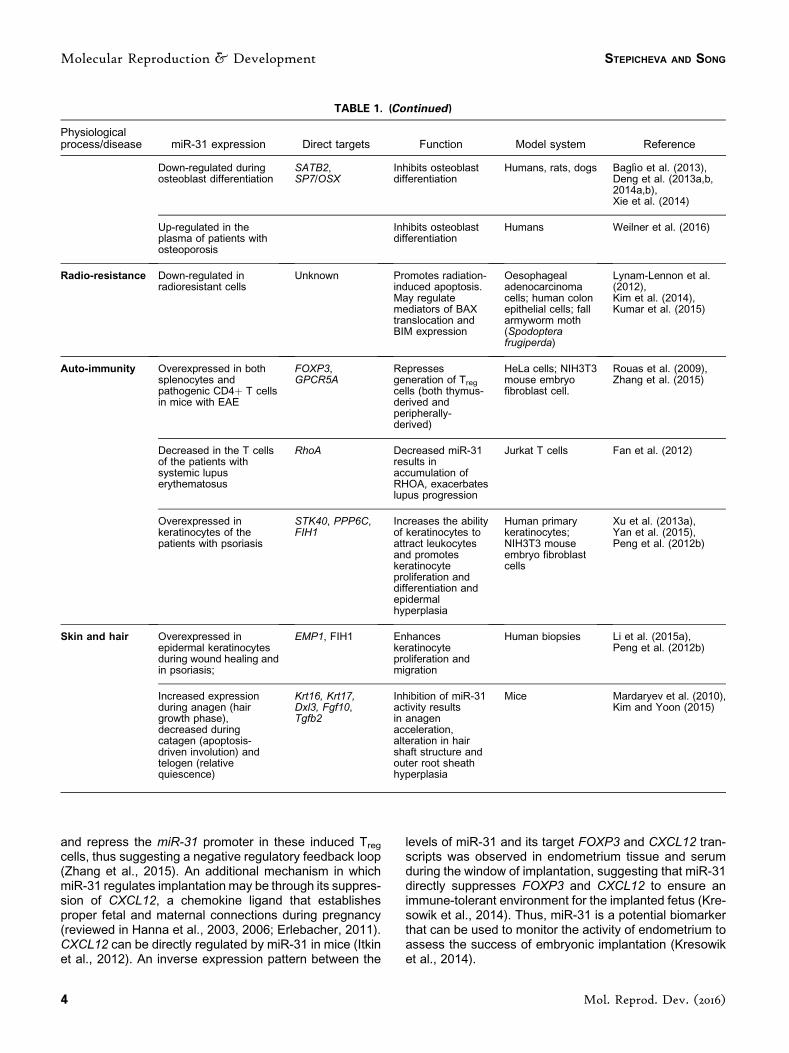
TABLE 1. Summary of miR-31 in Development and Disease
Physiologicalprocess/disease miR-31 expression Direct targets Function Model system Reference
Spermatogenesis Down-regulated in testisof infertile patients
Unknown Potential role inpromoting earlysperm development
Human testis Munoz et al. (2015)
Embryoimplantation
Elevated in bothendometrium and serumduring the time ofimplantation
Possibly throughFOXP3 andCXCL12
Provides animmune-tolerantmaternalenvironment
Human (embryoimplantation); HeLacells (FOXP3regulation);HEK293T cells(CXCL12regulation)
Kresowik et al. (2014),Rouas et al. (2009),Itkin et al. (2012)
Embryonicdevelopment
Expressed in a pair-rulepattern in the foregut,anterior endoderm andhindgut
Unknown Promotes propersegmentation
Fruitfly (Drosophila)embryos
Aboobaker et al. (2005),Leaman et al. (2005)
Expressed ubiquitouslyduring early development
Pmar1, Alx1,Snail, VegfR7
Promotesskeletogenesis
Sea urchin(S. purpuratus)embryos
Stepicheva andSong (2015)
Expressed ubiquitously inzebrafish embryos
PROX1 Suppressesvasculardevelopment
Frog (Xenopuslaevis) andzebrafish (Daniorerio) embryos;primary humanlymphatic andblood vascularendothelial cellsisolated fromneonatal humanforeskins
Pedrioli et al. (2010),Wienholds et al. (2005)
Expressed in prosomeresp1/p4 of the brain and inthe extremities of thesomites, notably thehypaxial dermomyotome
Myf5 Preventsinappropriateaccumulation of thekey myogenic factorMYF5 in the brain
Mouse embryo Daubas et al. (2009)
Vasculardevelopment
Down-regulated indifferentiated vascularsmooth muscle cells andup-regulated in de-differentiated andproliferative vascularsmooth muscle cells
LATS1, CREG Increased miR-31levels results inincreasedproliferation ofvascular smoothmuscle cells
Rat vascularsmooth musclecells from the aorticmedia; humanvascular smoothmuscle cells fromsegments ofinternal thoracicarteries retrievedduring coronarybypass surgery
Liu et al. (2011),Wang et al. (2013)
Myogenesis Highly expressed at theearly stages of differentia-tion of muscle satellitecells; progressivelydecreases at later differ-entiation stages.
MYF5, DMD Maintainsquiescence ofmuscle satellitecells
Muscle satellitecells isolated frommice and humans
Crist et al. (2012),Cacchiarelli et al. (2011)
Increased in thecardiomyocytes afterischemia/reperfusioninjury
Pkce Downregulation ofmiR-31 increasesheart resistance toischemia/reperfusion injury
Mice Wang et al. (2015b)
Bonehomeostasis
Highly abundant inosteoclasts
RhoA Promotesosteoclastpolarization andbone resorption
Mouse bonemarrow-derivedmacrophages
Mizoguchi et al. (2013)
(Continued )
miR-31 IN DEVELOPMENT AND DISEASE
Mol. Reprod. Dev. (2016) 3
and repress the miR-31 promoter in these induced Treg
cells, thus suggesting a negative regulatory feedback loop(Zhang et al., 2015). An additional mechanism in whichmiR-31 regulates implantation may be through its suppres-sion of CXCL12, a chemokine ligand that establishesproper fetal and maternal connections during pregnancy(reviewed in Hanna et al., 2003, 2006; Erlebacher, 2011).CXCL12 can be directly regulated by miR-31 in mice (Itkinet al., 2012). An inverse expression pattern between the
levels of miR-31 and its target FOXP3 and CXCL12 tran-scripts was observed in endometrium tissue and serumduring the window of implantation, suggesting that miR-31directly suppresses FOXP3 and CXCL12 to ensure animmune-tolerant environment for the implanted fetus (Kre-sowik et al., 2014). Thus, miR-31 is a potential biomarkerthat can be used to monitor the activity of endometrium toassess the success of embryonic implantation (Kresowiket al., 2014).
TABLE 1. (Continued)
Physiologicalprocess/disease miR-31 expression Direct targets Function Model system Reference
Down-regulated duringosteoblast differentiation
SATB2,SP7/OSX
Inhibits osteoblastdifferentiation
Humans, rats, dogs Bagl�ıo et al. (2013),Deng et al. (2013a,b,2014a,b),Xie et al. (2014)
Up-regulated in theplasma of patients withosteoporosis
Inhibits osteoblastdifferentiation
Humans Weilner et al. (2016)
Radio-resistance Down-regulated inradioresistant cells
Unknown Promotes radiation-induced apoptosis.May regulatemediators of BAXtranslocation andBIM expression
Oesophagealadenocarcinomacells; human colonepithelial cells; fallarmyworm moth(Spodopterafrugiperda)
Lynam-Lennon et al.(2012),Kim et al. (2014),Kumar et al. (2015)
Auto-immunity Overexpressed in bothsplenocytes andpathogenic CD4þ T cellsin mice with EAE
FOXP3,GPCR5A
Repressesgeneration of Treg
cells (both thymus-derived andperipherally-derived)
HeLa cells; NIH3T3mouse embryofibroblast cell.
Rouas et al. (2009),Zhang et al. (2015)
Decreased in the T cellsof the patients withsystemic lupuserythematosus
RhoA Decreased miR-31results inaccumulation ofRHOA, exacerbateslupus progression
Jurkat T cells Fan et al. (2012)
Overexpressed inkeratinocytes of thepatients with psoriasis
STK40, PPP6C,FIH1
Increases the abilityof keratinocytes toattract leukocytesand promoteskeratinocyteproliferation anddifferentiation andepidermalhyperplasia
Human primarykeratinocytes;NIH3T3 mouseembryo fibroblastcells
Xu et al. (2013a),Yan et al. (2015),Peng et al. (2012b)
Skin and hair Overexpressed inepidermal keratinocytesduring wound healing andin psoriasis;
EMP1, FIH1 Enhanceskeratinocyteproliferation andmigration
Human biopsies Li et al. (2015a),Peng et al. (2012b)
Increased expressionduring anagen (hairgrowth phase),decreased duringcatagen (apoptosis-driven involution) andtelogen (relativequiescence)
Krt16, Krt17,Dxl3, Fgf10,Tgfb2
Inhibition of miR-31activity resultsin anagenacceleration,alteration in hairshaft structure andouter root sheathhyperplasia
Mice Mardaryev et al. (2010),Kim and Yoon (2015)
Molecular Reproduction & Development STEPICHEVA AND SONG
4 Mol. Reprod. Dev. (2016)

TABLE 2. Role of miR-31 in Various Cancers�
Cancer type Validated target Function of the target References
Tumor suppressor(downregulated)
Breast cancer GNA13 GNA-13 promotes cell invasion mainly throughactivation of RHOA.
Rasheed et al. (2015)
Liver cancer HDAC2, CDK2 HDAC2 and CDK2 are cell cycle regulators(acceleration of cell cycle if overexpressed).
Kim et al. (2015)
CDH1 & 2,VMN, FN1
CDH1/2, VMN, and FN1 are regulators of theepithelial-to-mesenchymal transition.
Ovariancancer
STMN1 STMN1 destabilized microtubules. Hassan et al. (2015)MET MET is a membrane receptor that mediates
apoptotic resistance to therapeutic drugs ifoverexpressed
Mitamura et al. (2013)
Brain Tumors DOCK1 Dock1 promotes epithelial-to-mesenchymaltransition through NF-kB/SNAIL signaling.
Zhang et al. (2016)
TRADD TRADD is an upstream activator of NF-kB. Rajbhandari et al. (2015)FIH1 FIH1 inhibits HIF1a and NOTCH. Down-
regulation of FIH1 promotes angiogenesis.Wong et al. (2015)
Follicularlymphoma
E2F2 E2F2 regulates pro-proliferation genes. Thompson et al. (2016)PIK3C2A PIK3C2A is an oncogene, and its suppression
results in apoptosis.Naso-pharyngealcarcinoma
FIH1 FIH1 inhibits HIF1a, which is a master regulatorof oxygen homeostasis.
Cheung et al. (2014)
MCM2 MCM2 is important in the initiation of DNAreplication.
Medullo-blastoma
MCM2 MCM2 is important in the initiation of DNAreplication.
Jin et al. (2014)
OncomiR(up-regulated)
Lung cancer BAP1 BAP1 is a tumor suppressor in lung cancer(nuclear-localized deubiquitinating enzyme).
Yu et al. (2016)
MET MET is a proto-oncogene and a hepatocytegrowth factor receptor.
Hou et al. (2016)
ABCB9 ABCB9 is a transporter involved in cellulartrafficking and chemotherapy-related multidrugresistance.
Dong et al. (2014)
RASA1,SPRED1 & 2,SPRY1, 3, & 4
RASA1, SPRED1, SPRED2, SPRY1, SPRY3,and SPRY4 are negative regulators of RAS/MAPK signaling.
Edmonds et al. (2016)
Cervicalcancer
ARID1A ARID1A is a tumor suppressor that remodelschromatin to regulate cell cycle progression.
Wang et al. (2014)
Colon/colorectalcancer
E2F2 E2F2 acts as a tumor suppressor in coloncancer by inhibiting cell cycle.
Li et al. (2015c)
SATB2 SATB2 is a tumor suppressor; its down-regulation is associated with metastasis.
Yang et al. (2013)
FIH1 FIH1 inhibits HIF1a, which is a master regulatorof oxygen homeostasis. Down-regulation ofFIH1 promotes tumor angiogenesis, cellproliferation and cell invasion.
Chen et al. (2014)
CDKN2B CDKN2B is a cell growth regulator that controlscell cycle G1 progression.
Lei et al. (2014)
Pancreaticcancer
RASA1 RASA1 is a suppressor of RAS function. Down-regulation of RASA1 increases cell proliferation.
Kent et al. (2016)
Intra-hepaticcholangio-carcinoma
Hu et al. (2013)
Eosopha-gealneoplasia
STK40 STK40 is a negative regulator of NF-kB-mediated transcription.
Taccioli et al. (2015)
CPM CPM is a cancer biomarker, but the mechanismfor its carcinogenesis not known.
ABCB9, ATP-Binding Cassette, sub-family B (MDR/TAP), member 9;ARID1A, AT-rich Interactive Domain 1A (SWI-like);BAP1, BRCA1-associated Protein-1;CDH1,E-Cadherin;CDH2, N-Cadherin; CDK2, Cyclin-dependent Kinase 2;CDKN2B, Cyclin-dependent Kinase Inhibitor 2B; CPM, Carboxypeptidase M;DOCK1, Dedicatorof Cytokinesis 1;E2F2, E2F Transcription Factor 2; FIH1, Factor Inhibiting Hypoxia-inducible Transcription Factor 1 alpha; FN1, Fibronectin;GNA13, G Protein Alpha-13;HDAC2, Histone Deacetylase 2;MCM2, MinichromosomeMaintenance Complex Component 2;MET, receptor tyrosine kinase;PIK3C2A, Phosphatidylinositol-4-Phosphate 3 Kinase, catalytic subunit type 2 alpha;RASA1, RAS P21 Protein Activator 1; SATB2, Special AT-rich Sequence-binding Protein 2; SPRED1/ 2, Sprouty-related, EVH1 domain containing 1/2; SPRY1/3/4, Sprouty RTK Signaling Antagonist 1/3/4; STK40, Serine/Threonine Kinase 40; STMN1, Stathmin 1; TRADD, TNFReceptor-associated Death Domain; VMN, Vimentin.�This table does not include references in the review by Laurila and Kallioniemi (2013).
miR-31 IN DEVELOPMENT AND DISEASE
Mol. Reprod. Dev. (2016) 5
miR-31 REGULATES DIVERSE PROCESSESDURING EMBRYONIC DEVELOPMENT
miRNAsareknownregulatorsofembryonicdevelopment(reviewed in Pauli et al., 2011). Several studies reported theexpression pattern and importance of miR-31 in regulatingembryogenesisofvariousorganisms.DepletionofmiR-31 inDrosophila embryos results in severe segmentation defects(Leaman et al., 2005). The segmented body plan in theDrosophila embryo is determined by transcription factors,encoded by gap genes, that control the expression of pair-rule genes. Pair-rule genes, in turn, activate segment-polar-ity genes that regulate theWNT andHH (Hedgehog) signal-ing pathways in determining the polarity of the embryonicparasegments. Mutation of pair-rule genes results in loss ofthe normal developmental pattern of the segmented insectembryos. The direct targets of miR-31 in the Drosophilaembryo have not been identified; however, miR-31 knock-down induced the mis-expression of pair-rule genes eve(even skipped), ftz (fushi tarazu), and hairy, as detected byRNA in situ hybridization, indicating pattern-formation de-fects (Leaman et al., 2005). This observation suggests thatmiR-31may indirectly regulate these pair-rule genes, whichis consistent with the spatial expression of miR-31 in a pair-rule pattern in the foregut, anterior endoderm, andhindgut ofDrosophila embryos (Aboobaker et al., 2005).
miR-31maybeoneof themost highly abundantmiRNAsthat is ubiquitously expressed during Strongylocentrotuspurpuratus (sea urchin) early development (Song et al.,2012; Stepicheva and Song, 2015). The knockdown ofmiR-31 in the sea urchin embryos resulted in a range ofdose-dependent phenotypes, including the formation ofextra cells and cell clumps in the blastocoel of the embryo,gut widening and reduction of the embryo size, as well asskeletogenesis defects (discussed below) (Stepicheva andSong, 2015).
In Danio rerio (zebrafish) embryos, the knockdown ofmiR-31 with loss-of-function morpholinos did not result insignificant phenotypes, whereas the overexpression ofmiR-31 resulted in internal lymphatic vascular defects(Pedrioli et al., 2010). Similarly, overexpression of miR-31 in Xenopus laevis (frog) embryos resulted in dose-dependent reduction in venous sprouting, but no otherdevelopmental defects (Pedrioli et al., 2010). While miR-31 is ubiquitously expressed in the zebrafish embryos(Wienholds et al., 2005), it has a specific function in vascu-lar development.
The regulation of vascular development by miR-31 mayinvolve its direct repression of PROX1 (Prospero Homeo-box 1), which encodes a well-characterized lymphatic tran-scription factor. Bioinformatics analysis of potential miR-31targets revealed a high conservation of the binding site ofmiR-31 in the 30UTR of PROX1 in humans, frogs, andzebrafish (Pedrioli et al., 2010). Moreover, PROX1 canbe directly suppressed by miR-31 in primary human lym-phatic and blood vascular endothelial cells isolated fromneonatal human foreskins (Pedrioli et al., 2010), indicatingthat the function of miR-31 may be conserved in regulatinglymphatic vasculature via PROX1 in vertebrates. In
addition, miR-31 also inhibits vasculature development inthe adult by directly suppressing LATS2 (Large TumorSuppressor Homolog 2) in rats and CREG (Cellular Re-pressor of E1A-stimulated Genes) in humans, which bothpromote cell proliferation (Liu et al., 2011; Wang et al.,2013).
miR-31 REGULATES MYOGENESIS
No data are currently available on the effect of systemicmiR-31 knockout or overexpression in the mouse. miR-31in the mouse brain may be involved in preventing inappro-priate accumulation of the key myogenic transcription fac-tor Myf5 (Daubas et al., 2009). Myf5 is transcribed at theonset of myogenesis in the somite and limb bud, as well asin some of the restricted domains of the ventral mesen-cephalon, prosencephalon, and neural tube of the mouseembryo (Daubaset al., 2009).miR-31 is highly expressed inareas whereMyf5 is transcribed but not translated, asmiR-31 directly suppresses the translation of Myf5 to preventmyogenesis in the developing brain (Daubas et al., 2009).
miR-31 also regulates myogenesis in adult murine tis-sues by suppressing the activation of muscle satellite cells(Crist et al., 2012). Skeletal muscle satellite cells are theequivalent of myogenic stem cells, and are usually main-tained in a quiescent state. They are activated in responseto injury, giving rise to regeneratedmuscle and newsatellitecells (Morgan and Partridge, 2003). The activation of mus-cle satellite cells requires MYF5. In the quiescent satellitecells,Myf5mRNA is transcribed, but not translated, due toits sequestration in the messenger ribonucleoprotein gran-ules along with miR-31 (Crist et al., 2012). These granulesare dynamic, self-assembling structures containing trans-lationally silent mRNAs bound by various proteins (re-viewed in Buchan, 2014). Upon activation of the satellitecells, messenger ribonucleoprotein granules dissociate,leading to a rapid release of translatable Myf5 mRNA.Importantly, exogenous overexpression of miR-31 sup-pressed the translation of Myf5 in activated satellite cells,resulting in the disruption of normal muscle satellite cellactivation (Crist et al., 2012). Thus, decreased abundanceof miR-31 or increased abundance of MYF5 is necessaryfor the activation of satellite cells in muscle regeneration.
Skeletal muscles are highly dynamic tissues that adaptto the level of exercise performed. Interestingly, acuteendurance exercise resulted in decreased abundance ofmiR-31 in human muscle biopsies, despite the increasedtranscript abundanceof the keymiRNAbiogenesis proteinsDROSHA,DICER1, andXPOT5 (exportin 5) (Russell et al.,2013). The exact mechanism or role of miR-31 down-regulation has not been elucidated; nevertheless, miR-31may be involved in regeneration and delaying skeletalmuscle atrophy after exercising.
Mis-regulation of miR-31 may lead to myopathies. Inwild-type mice, miR-31 expression was high at the earlystages of differentiation of muscle satellite cells, but pro-gressively decreased at later differentiation stages (Cac-chiarelli et al., 2011). Persistent accumulation of miR-31 in
Molecular Reproduction & Development STEPICHEVA AND SONG
6 Mol. Reprod. Dev. (2016)

muscle satellite cells was associated with severe myopa-thies, as in the case of Duchenne muscular dystrophy(DMD), a genetic disorder caused by the mutations inDYS (Dystrophin) (Cacchiarelli et al., 2011). DYSTRO-PHIN is critical for the proper muscle formation, linkingthe internal cytoskeleton to the transmembrane protein(sarcoglycan complex) at the plasma membrane that in-teractswith the extracellularmatrix (reviewed inNowakandDavies, 2004). miR-31 directly suppress DYS in mice andhumans, and its accumulation in regeneratingmyoblasts inthe patients with Duchenne muscular dystrophy resulted ina lower differentiation potential (Cacchiarelli et al., 2011).Thus, repression of miR-31 function in compromised adultmuscles may improve the efficiency of therapeutic treat-ments aimed at the accumulation of DYSTROPHIN inmuscles (Cacchiarelli et al., 2011).
miR-31 also plays a role in mouse cardiac myocytesduring injury (Wang et al., 2015b). Cardiac ischemia/reper-fusion injury results in the accumulation of miR-31 in themyocardium and the down-regulation of its direct targetPkce (Protein Kinase C epsilon) (Wang et al., 2015b).Decreased PKCe down-regulates NFkB, whose activationis important for ischemic late pre-conditioning (a delayedadaptive response to increase heart resistance to ische-mia/reperfusion injury) (Xuan et al., 1999; Wang et al.,2015b). Treatment of post-injury cardiac myocytes withmiR-31 inhibitor was cardioprotective and reduced myo-cardial infarct size (Wang et al., 2015b).
Thus, miR-31 has been shown to regulate myogenesisin adult tissues through suppression of Myf5 and Dystro-phin during the differentiation of muscle cells and to re-spond to cardiac myocyte ischemia/reperfusion injurythrough suppression of PKCe.
miR-31 MAINTAINS BONE HOMEOSTASIS IN THEADULT VERTEBRATES AND MODULATESSKELETOGENESIS IN THE INVERTEBRATE SEAURCHIN EMBRYOS
The vertebrate skeleton is a complex, metabolicallyactive tissue that is remodeled throughout life in order tomaintain the shape, quality, and size of the bone (Hadji-dakis and Androulakis, 2006; Fisher and Franz-Odendaal,2012; Long, 2012). A number of evolutionarily conservedpathways are reportedly involved in proper formation andmaintenance of bone in vertebrates. Recent studies alsoindicate that miRNAs control multiple targets of the verte-brate skeletogenic gene regulatory networks, from initialresponse of stem/progenitor cells to the structural andmetabolic activity of the mature tissue (Lian et al., 2012;Zhao et al., 2014).
Bone remodeling in vertebrates begins with the resorp-tion of mineralized bone by osteoclasts specialized multi-nucleated cells that differentiate from hematopoietic stemcells (Fig. 2) (Boyle et al., 2003; Hadjidakis and Androula-kis, 2006). Following adhesion to the fragment of the bonethat needs to be remodeled, osteoclasts undergo a signifi-cant polarization and cytoskeleton reorganization in order
to form a specialized extracellular compartment whereprotons and proteases are released to demineralize thebone (Boyle et al., 2003; Hadjidakis and Androulakis, 2006;Itzstein et al., 2011). miR-31 is highly abundant in osteo-clasts, and inhibition of miR-31 resulted in impaired osteo-clast function (Mizoguchi et al., 2013). One of the directmiR-31 targets involved in osteoclast function is RHOA(RAS Homolog Family Member A), which is essential forosteoclast cytoskeleton reorganization, and thus is criticalfor the ability of osteoclasts to resorb the bone (Destainget al., 2005; Itzstein et al., 2011;Mizoguchi et al., 2013). Theidentity of RHOA as a miR-31 target is supported by thefinding that inhibition of RHOA restores osteoclast matura-tion in bone marrow-derived macrophages treated withmiR-31 inhibitor (Mizoguchi et al., 2013).
Following the resorption of bone by osteoclasts, special-ized mononuclear cells removing demineralized undi-gested collagen from the bone surface (Raggatt andPartridge, 2010). These mononuclear cells also producesignals to attract osteoblasts, the specialized cells that areresponsible for the formation of the new bone matrix (Had-jidakis and Androulakis, 2006). Osteoblasts differentiatefrom multi-potent mesenchymal cells in response to twomain transcription factors, SATB2 (SATB Homeobox 2)and SP7/OSX (Osterix), whose transcripts are negativelyregulatedbymiR-31 (Bagl�ıoet al., 2013;Denget al., 2013a;Xie et al., 2014). miR-31 is down-regulated in bone mes-enchymal stem cells during osteogenic differentiation inhumans and rats (Bagl�ıo et al., 2013; Deng et al., 2013a).Down-regulation of miR-31 in bone marrow mesenchymalstem cells, bone marrow stromal stem cells, and adiposetissue-derived stem cells of the rats and dogs was alsonecessary for repairing critical-size bone defects,which arethe smallest size defects that would not heal without medi-cal intervention, indicating the potential importance ofmiR-31 in bone injury therapeutics (Deng et al., 2013b,2014a,b).
Several studies suggest a regulatory loop in the osteo-genic differentiation process (Deng et al., 2013a; Ge et al.,2015) (Fig. 3). RUNX2 (RUNT-related Transcription Factor2) transcriptionally activates SP7/OSX, resulting in anaccumulation of its downstream target SATB2 in differenti-ating osteoblasts (Nakashima et al., 2002; Tang et al.,2011). SATB2, in turn, physically interacts with RUNX2to enhance the transcriptional activation of SP7/OSX (Do-breva et al., 2006). In addition, RUNX2 suppressesmiR-31transcription through direct binding of its promoter, thusremoving the miR-31-mediated translational silencing ofSP7/OSX and SATB2 (Deng et al., 2013a).
This regulatory loop plays an important role not only inosteogenic differentiation but also in tooth eruption. Pa-tients with cleidocranial dysplasia (delayed tooth eruption)were found to have mutations in RUNX2, and thus higherlevels of miR-31 and down-regulated SATB2 compared tothehealthy individuals (Geet al., 2015). KnockdownofmiR-31 in dental follicle cells from patients with cleidocranialdysplasia led to increased levels of SATB2 and RUNX2, aswell as the rescue of osteoclast-inductive and matrix deg-radation capacities (Ge et al., 2015).
miR-31 IN DEVELOPMENT AND DISEASE
Mol. Reprod. Dev. (2016) 7

In addition to its role in the bone injury repair and teetheruption, miR-31 is a critical component of the age-relatedreduction of osteogenesis (Weilner et al., 2016). miR-31 issignificantly elevated in the plasma of elderly people orpatients with osteoporosis. Senescent endothelial cellssecrete miR-31 in microvesicles that are taken up bymesenchymal stem cells, where miR-31 may inhibit osteo-genic differentiation by suppressing FZD3 (Frizzled-3),which encodes a receptor for WNT5 signaling (Weilneret al., 2016). Previously increased WNT5A is associatedwith BMP2 (Bone Morphogenetic Protein 2)-mediated os-teoblast differentiation (Nemoto et al., 2012), and FZD3mRNA was up-regulated during osteogenesis (Chakra-vorty et al., 2014). Elevated FZD3 transcript abundancein age-related reduction of osteogenesis correlated withreduced miR-31 (Weilner et al., 2016).
Thus, miR-31 regulates bone maintenance in verte-brates by modulating both osteoclasts (through RHOA)and osteoblasts (through SP7/OSX, SATB2, and
Figure 2. Modulation of miR-31 during bone remodeling.A: Bone remodeling begins with the resorptionof mineralized bone by osteoclasts, which must undergo cytoskeletal reorganization, involving theformation of an actin-rich sealing zone followed by apico-basal polarization and formation of the ruffledborder (Boyle et al., 2003; Hadjidakis and Androulakis, 2006). Activated osteoclasts adhere to thefragment of the bone that needs to be remodeled / resorbed. One of the miR-31 targets involved inosteoclast function is RHOA, which is essential for osteoclast cytoskeleton reorganization (Mizoguchiet al., 2013). The second phase of bone remodeling involves specialized mononuclear cells that preparethe bone surface and attract osteoblasts (Raggatt and Partridge, 2010). Differentiation of osteoblasts isregulated mainly by SATB2 and SP7/OSX, whose transcripts are suppressed by miR-31 (Bagl�ıo et al.,2013; Deng et al., 2013a; Xie et al., 2014). Differentiated osteoblasts promote bone matrix formation. B:Down-regulation of miR-31 increases bone volume and mineralization due to the suppression ofosteoclast function (decrease in bone resorption) (Mizoguchi et al., 2013) and promotion of osteoblastdifferentiation (increase in matrix deposition) (Deng et al., 2013a; Xie et al., 2014).
Figure 3. Regulatory feedback of miR-31 during osteogenic differen-tiation. RUNX2 transcriptionally activates (solid arrow) SP7/OSX,resulting in the accumulation of SATB2 in differentiating osteoblasts(Nakashima et al., 2002; Tang et al., 2011). SATB2, in turn, physicallyinteracts (dashed arrow) with RUNX2 to auto-enhance RUNX2 ex-pression, thus positively increasing SP7/OSX production (Dobrevaet al., 2006). RUNX2 also suppresses expression of miR-31 by directlybinding to its promoter, thus removing the miR-31-mediated transla-tional silencing of SP7/OSX and SATB2 transcripts (Deng et al.,2013a).
Molecular Reproduction & Development STEPICHEVA AND SONG
8 Mol. Reprod. Dev. (2016)

potentially FZD3) (Fig. 2A). Further, knockdown of miR-31resulted in increased bone volume and mineralization dueto the suppression of osteoclast function and promotion ofosteoblast differentiation (Fig. 2B) (Bagl�ıo et al., 2013;Deng et al., 2013a,b; Mizoguchi et al., 2013).
The role of miR-31 in regulating skeletogenesis is notrestricted to vertebrates; indeed, this function seems to be aconservedmechanism in invertebrates.miR-31 is critical forregulation of skeletogenesis in the sea urchin embryo (Ste-picheva andSong, 2015), an echinodermand a sister groupto the chordates (McClay, 2011). Sea urchin embryos un-dergo less complex skeletogenesis than vertebrates. Thelarval skeleton comes from a single cell type, the primarymesenchyme cells (PMCs) (Oliveri et al., 2003). The larvalskeleton supports larval swimming, the shape of the larvae,aswell as larval feeding (PenningtonandStrathmann, 1990;Hart and Strathmann, 1994; Piacentino et al., 2015). Forproper skeletogenesis, the PMCs need to differentiate,undergo an epithelial-to-mesenchymal transition, fusewitheachother, and localize into thecorrectpattern (Sharmaand Ettensohn, 2010; Rafiq et al., 2012; Lyons et al., 2014;Saunders andMcClay, 2014; McClay, 2016). The complex-ity of the signals received by the PMCs is not known, but theprocess of PMC positioning or patterning is, in part, depen-dent on VEGF (Vascular Endothelial Growth Factor) signal-ing,ALK4/5/7 (TransformingGrowthFactorBeta receptors),SLC26A2/7 (Solute Carriers 26a2 and 7), LOX (Lipoxyge-nase), and BMP5/8 (BoneMorphogenetic Proteins 5 and 8)(Duloquin et al., 2007; Adomako-Ankomah and Ettensohn,2013,2014;Piacentinoetal., 2015,2016a,b).KnockdownofmiR-31 resulted in a significant decrease in the length ofdorsoventral connecting rods, formation of extra tri-radiates,aswell asPMCpatterningdefects in theseaurchingastrulae(StepichevaandSong,2015).miR-31directly suppressesatleast three transcription factors (SpPmar1, SpAlx1, andSpSnail) and one effector gene (SpVegfr7) within the seaurchin skeletogenic gene regulatory network. Inhibition ofSpAlx1 and/or SpVegfr7 by miR-31 in the developing em-bryo results in a less severe, but similar, PMC defect to thephenotype of miR-31 inhibition, suggesting that those tar-gets contribute to the same regulatory pathways in seaurchin embryo. In addition, miR-31 regulates expressionof SpVegf3, which encodes an ectodermal ligand that iscritical for the positioning of the PMCs, by a yet-to-be-identified mechanism (Stepicheva and Song, 2015). Thefact that miR-31 regulates both the signal receiving PMCsand signal-sending ectoderm suggests its ability to cross-regulatemultiplepathways toensureproperskeletogenesis.
ROLE OF miR-31 IN CANCER ISCONTEXT-DEPENDENT
miR-31 plays an important role in different types ofcancers, including breast (Sossey-Alaoui et al., 2011; Luet al., 2012; K€orner et al., 2013; Mulrane et al., 2014; Vir�eet al., 2014), ovarian (Anderson et al., 2010; Yu et al., 2010;Hassan et al., 2015), lung (Liu et al., 2010; Meng et al.,2013; Dong et al., 2014; Edmonds et al., 2016; Yu et al.,
2016), colon (Cottonham et al., 2010; Cekaite et al., 2012;Xu et al., 2013b; Kim et al., 2014; Li et al., 2015c; Kuriharaet al., 2016), and melanoma (Greenberg et al., 2011;Asangani et al., 2012). Intriguingly, even though the ex-pression of miR-31 is consistently altered in various can-cers, miR-31 can perform either tumor-suppressive oroncogenic functions, depending on the type of cancer(Table 2) (reviewed in Laurila and Kallioniemi, 2013).
The best-characterized example of differential miR-31expression in cancer cells is its down-regulation in breastcancer, where miR-31 has been shown to serve as a tumorsuppressor miRNA (Augoff et al., 2011). In breast cancercell lines, miR-31 suppresses translation of genes involvedin apoptosis (such as PKCe), cell motility (such as actinremodeling genesWASF3 [WASProtein FamilyMember 3]and RHOA, and ITGB1 [Integrin Beta 1]), and cell invasion(GNA13 [G protein alpha-13]), through activation of RHOA)(Augoff et al., 2011; Sossey-Alaoui et al., 2011; K€orneret al., 2013; Rasheed et al., 2015).
miR-31 is also down-regulated in patientswith leukemia,in which it suppresses MAP3K14 (NFkB-inducing Kinase)to repress NFkB signaling (Yamagishi et al., 2012). Theloss ofmiR-31 results in constitutive activation of NFkB thatcontributes to abnormal cell proliferation as well as inhibi-tion of apoptosis (Yamagishi et al., 2012). miR-31 also hasa tumor suppressor function in glioblastomas, which are afast-growing, aggressive tumor of the central nervous sys-tem that form on the supportive tissue of the brain (Bleekeret al., 2012; Hua et al., 2012; Zhou et al., 2015; Zhang et al.,2016). In glioma cells, miR-31 inhibits RADIXIN, whichencodes a cytoskeletal protein that is essential for cellmotility, adhesion, and proliferation, as well as DOCK1(dedicator of cytokinesis 1), which encodes a promoterof epithelial-to-mesenchymal transition through NFkB/SNAIL signaling (Hua et al., 2012; Zhang et al., 2016).The down-regulation of miR-31 during progression of glio-blastoma results in increased migration and invasion ofthese cancer cells (Hua et al., 2012; Zhang et al., 2016). Inaddition, miR-31 can promote angiogenesis in glioma tu-mors through the direct suppression of FIH1 (Factor In-hibiting Hypoxia-inducible Factor 1) (Wong et al., 2015).FIH1 is a multi-functional hydroxylase whose downstreamtargets include HIF1A (Hypoxia-inducible Factor 1 alpha)and NOTCH. Suppression of FIH1 by miR-31 up-regulatesHIF1A, resulting in the up-regulation of VEGF and promo-tion of angiogenesis (Wong et al., 2015).
RecentlymiR-31was found to be at aberrantly low levelsin the patients with hepatocellular carcinoma (Kim et al.,2015). The molecular targets of miR-31 in hepatocellularcarcinoma include HDAC2 (Histone Deacetylase 2) andCDK2 (Cyclin-dependent Kinase 2), which promote the cellcycle, as well as CDH1 and CDH2 (N- and E-Cadherins),VIM (Vimentin), and FN1 (Fibronectin), which are involvedin the epithelial-to-mesenchymal transition (Kim et al.,2015). Thus, in hepatocellular carcinoma,miR-31 functionsas a tumor suppressor that represses genes that promotecell proliferation and cell metastasis.
In ovarian cancer, loss of miR-31 increases chemore-sistance to taxane through the lack of suppression of
miR-31 IN DEVELOPMENT AND DISEASE
Mol. Reprod. Dev. (2016) 9
STMN1 (Stathamin1) andMET (a receptor tyrosine kinase)(Mitamura et al., 2013; Hassan et al., 2015). Taxane che-motherapy is based on its binding to beta-tubulin, resultingin stabilized microtubules. Microtubule stabilization causescell cycle arrest in G2/M phase, leading to apoptosis (Has-san et al., 2015). STMN1 is a cytosolic, tubulin-bindingprotein shown to stimulate microtubule depolymerization.Down-regulation ofmiR-31 in ovarian cancer cells results inaccumulation of STMN1, which counteracts taxane che-motherapy aimed at microtubule stabilization (Hassanet al., 2015). MET, on the other hand, is a transmembranereceptor that contributes toacquiredapoptotic resistance tochemotherapy (Tang et al., 2010). One MET-dependentmechanism that contributes to apoptotic resistance is theactivation ofPI3K (Phosphoinositol-3Kinase)/AKT (ProteinKinase B) signaling (Tang et al., 2010; Xiao et al., 2001).AKT promotes resistance to apoptosis via multiple mecha-nisms, including phosphorylation and activation of CHUK(Conserved Helix-Loop-Helix Ubiquitous Kinase/IkB ki-nase), which results in nuclear localization of NFkB toactivate transcription of anti-apoptotic genes, or phosphory-lation of BAD to prevent cell death (del Peso et al., 1997;Ozeset al., 1999;Xiaoet al., 2001).Down-regulation ofmiR-31 results in the accumulation of MET and development ofchemoresistance due to a block to apoptosis (Mitamuraet al., 2013). These findings suggest that miR-31 has aprotective effect on these cancers, and may be a potentialtherapeutics tool for improving the success of taxane-pre-scribed ovarian cancer treatment.
miR-31 is also up-regulated in some cancers, acting asan oncogenic miRNA (oncomiR). One well-documentedoncomiR role of miR-31 was described in colorectal can-cers (Cottonham et al., 2010; Cekaite et al., 2012; Xu et al.,2013b; Yang et al., 2013; Ito et al., 2014; Lei et al., 2014; Liet al., 2015c; Tateishi et al., 2015). Some of the miR-31targets of colon cancer include important tumor suppres-sors such as E2F2 (E2F Transcription Factor 2); SATB2,RASA1 (RAS p21GTPase activating protein 1), which wasrecently shown to be targeted by miR-31 in pancreaticcancer);RHOBTB1 (Rho-Related BTBDomain Containing1); and TIAM1 (T Lymphoma and Metastasis Gene 1)(Cottonham et al., 2010; Sun et al., 2013; Xu et al.,2013b; Yang et al., 2013; Li et al., 2015c; Kent et al.,2016). Importantly, suppression of miR-31 in colon cancercells resulted in the increased sensitivity to chemothera-peutic drug fluorouracil, suggesting the potential of usingmiR-31 as a therapeutic target to enhance the efficacy ofchemotherapy treatments (Wang et al., 2010).
miR-31 also acts as an oncomiR in the lung cancer,where it directly targets tumor-suppressing genes, such asLATS2 (Large Tumor Suppressor 2), PPP2R2A (ProteinPhosphatase 2 Regulatory Subunit B alpha), and BAP1(BRCA1-associated Protein 1) (Liu et al., 2010; Yu et al.,2016). Additional miR-31 targets involved in the progres-sion of lung cancer include negative regulators of RAS/MAPK signaling RASA1, SPRED1, SPRED2 (Sprouty-re-lated EVH1 Domain Containing 1/2), SPRY1, SPRY3, andSPRY4 (Sprouty RTK Signaling Antagonist 1/3/4) (Ed-monds et al., 2016). The overexpression of miR-31 is
proposed to be a predictor of lymph node metastasis,and results in a poor prognosis in patients with lung ade-nocarcinoma (Meng et al., 2013). Even though miR-31 isgenerally overexpressed in the lung cancers, some lungcancer tumors were reported to have a decreased miR-31expression (Okudela et al., 2014a,b).
miR-31 may additionally contribute to chemotherapy-related multi-drug resistance by targeting ABCB9 (ATP-Binding Cassette B9 Transporter) (Dong et al., 2014). Inhi-bition of ABCB9 expression leads to chemoresistance,presumably throughdecreaseddruguptake.miR-31directlyinhibits translation of ABCB9, thus contributing to poortreatment outcomes (Dong et al., 2014). Further, in cervicalcancer, miR-31 directly inhibits tumor suppressor ARID1A(AT-rich Interactive Domain 1A), which activates transcrip-tion of genes by chromatin remodeling (Wang et al., 2014).InhibitionofmiR-31expression in cervical cancer resulted ingrowth arrest and a decrease in cell migration. Importantly,the anti-tumor effects of miR-31 inhibitor could be reversedby the knockdown of ARID1A (Wang et al., 2014).
Taken together, the function of miR-31 in various can-cers depends on its local environment where its complexinteraction with other factors determine its role as a tumorsuppressor or an oncomiR.
miR-31 PROMOTES RADIATION-INDUCEDAPOPTOSIS
Several independent studies reported miR-31 to beinvolved in radioresistance, the ability of an organism towithstand ionizing radiation. For example, miR-31 wassignificantly down-regulated in radioresistant esophagealadenocarcinoma cells (Lynam-Lennon et al., 2012). Over-expression of miR-31 re-sensitized the cells to radiation-induced cell death and down-regulated thirteen DNA repairgenes. The exact mechanism of miR-31 in radiosensitivityhas not been elucidated (Lynam-Lennon et al., 2012). Asimilar study demonstrated that inhibition of miR-31-5pprotected human colon epithelial cells against ionizingradiation (Kim et al., 2014).
The role ofmiR-31 in radiation-inducedapoptosis seemsto be evolutionarily conserved. An interesting study con-ducted recently unraveled the mechanism behind the un-usual resistance to radiation-induced apoptosis of the fallarmyworm moth (Spodoptera frugiperda) (Kumar et al.,2015). Increased radiation led to increasedmiR-31 expres-sion, which induced caspase-3-dependent apoptosis. Ap-optosis is a complex process dependent on a number ofpro- and anti-apoptotic proteins (Elmore, 2007). Uponirradiation, the ratio between the pro-apoptotic proteinBax and the anti-apoptotic protein Bcl2 was not altered,yet Baxwas translocated to themitochondria, thus inducingapoptosis. Importantly, inhibition of miR-31 resulted indecreased translocation of Bax to mitochondria and de-creased levels of the pro-apoptotic Bim, thereby reducingapoptosis. Moreover, ectopic overexpression of miR-31 inunirradiated cells resulted in increased apoptosis throughmitochondrial Bax translocation and upregulation of Bim
Molecular Reproduction & Development STEPICHEVA AND SONG
10 Mol. Reprod. Dev. (2016)

expression, suggesting that miR-31 may regulate media-tors of Bax translocation and Bim expression by yet to beidentified mechanism (Kumar et al., 2015).
The evidence that miR-31 is important for mediatingradiation-induced apoptosis may be important in under-standing the underlying mechanism of increased radiationresistance of some cancer cells and contribute to improvedcancer therapeutics.
miR-31 IS MIS-REGULATED IN AUTOIMMUNEDISEASES AND ALLERGY
Immune system homeostasis is mediated by regulatoryT (Treg) cells (reviewed in Vignali et al., 2008; Li and Zheng,2015). miR-31 regulates Treg cells through several mecha-nisms, including the suppression of FOXP3 involved in Treg
cells differentiation (discussed earlier). In addition, miR-31can repress the generation of peripherally derived Treg cells(Dhamne et al., 2013), which differentiate in secondarylymphoid organs and tissues to control autoimmuneresponses under certain inflammatory conditions (Yadavet al., 2013). One of the mechanisms for the induction ofperipherally derived Treg cells is mediated by retinoic acid,which indirectly induces expression of Foxp3 (Hill et al.,2008; Zhang et al., 2015). miR-31 directly suppressesGprc5a (G Protein-Coupled Receptor Class C Group 5Member A/Retinoic Acid-inducible Protein 3), leading todecreased retinoic acid. This in turn indirectly decreasesFoxp3 expression, resulting in the suppression of periph-erally derived Treg cell differentiation (Zhang et al., 2015).Suppression of Gprc5a by miR-31 in peripherally derivedTreg cells is important in mice with experimental autoim-mune encephalomyelitis (EAE), an animal model for multi-ple sclerosis. miR-31 was significantly overexpressed inboth splenocytes and pathogenic CD4-positive T cells inmice with EAE (Zhang et al., 2015). Further, conditionaldeletion of miR-31 resulted in a significant decrease in theseverity of EAE (Zhang et al., 2015). Thus, the abundanceof miR-31 correlates with EAE severity.
miR-31 has also been shown to regulate the function of Tcells in patients with systemic lupus erythematosus, achronic autoimmune disease in the connective tissuethat affects various organs, such as skin, heart, lungs,kidneys, and the nervous system (Fan et al., 2012). Thelevel ofmiR-31 is significantly decreased in T cells obtainedfrom lupus patients compared to the healthy controls (Fanet al., 2012). The proposed mechanism of miR-31 in theprogression of lupus involves miR-31 directly repressingtranslation of RHOA, which is critical for the proper homingof lymphocytes to sites of infection (Helms et al., 2007; Fanet al., 2012;). RHOA indirectly inhibits IL2 (Interleukin 2)transcriptionby interferingwith theNFAT (NuclearFactor ofActivated T cells) activity that decreases the abundance ofacetylated histone 3 at the IL2 promoter (Helms et al.,2007). IL2 is a multi-functional cytokine that is crucial forT cell activation, proliferation, and contraction; its abun-dance is reduced in lupus, resulting in the systemic mis-regulation of immune responses in patients (reviewed in
Lieberman and Tsokos, 2010). Decreased levels ofmiR-31result in the accumulation of RHOA, leading to decreasedproduction of IL2 and thus progression of lupus. A separatestudy demonstrated that miR-31 regulates IL2 levels bydirectly suppressing KSR2 (Kinase Suppressor of RAS 2),which encodes an upstream kinase suppressor of IL2 (Xueet al., 2013). Activation of primary lymphocytes throughstimulation of T-cell receptor results in the up-regulation ofmiR-31, followed by increased expression of IL2 and thusactivation of an immune response (Xue et al., 2013).
miR-31 is also overexpressed in keratinocytes of thepatients suffering from psoriasis, a chronic autoimmunedisease that is characterized by the formation of itchy,silvery scales on the skin (Xu et al., 2013a; Yan et al.,2015). Overexpression of miR-31 has been linked to in-creased expression of chemokines and cytokines, and thusan increase in the ability of psoriatic keratinocytes to attractleukocytes, which result in chronic inflammation (Xu et al.,2013a). miR-31 can directly suppress translation of STK40(Serine/Threonine Kinase 40), which encodes a negativeregulator of the NFkB pathway; the NFkB pathway, in turn,activates the expression of cytokine and chemokines)(reviewed in Pasparakis, 2009). Thus, overexpression ofmiR-31 can indirectly and constitutively activate the NFkBpathway, contributing to increased inflammation in psoriaticepidermis (Xu et al., 2013a; Yan et al., 2015). Interestingly,activation of NFkB signaling induces miR-31 expression(Yan et al., 2015), creating a potential feed-forward loopthat enhances NFkB pathway activity.
In addition to regulating inflammation responses,miR-31can directly suppress PPP6C (Protein Phosphatase 6),which encodes a negative regulator of G1-to-S phase pro-gression, indicating that this miR can regulate keratinocyteproliferation (Yan et al., 2015).miR-31waspreviously foundto enhance keratinocyte proliferation and migration duringthe process of wound healing by suppressing EMP1(Epithelial Membrane Protein 1), which encodes a tumorsuppressor (Li et al., 2015a). The rate of wound healing isincreased inpsoriasis (Morhennetal., 2013), so it is possiblethat that miR-31 promotes keratinocyte proliferation in pso-riasis not only through suppression of PPP6C, but alsothrough suppression of EMP1. Up-regulation of miR-31 inthe psoriatic epithelium might also result in increased ker-atinocyte differentiation through NOTCH signaling as over-expression of miR-31 indirectly activates NOTCH throughdirect suppression of FIH1 (Peng et al., 2012b).
Overall, overexpression of miR-31 in psoriatic keratino-cytes results in: (i) the ability of keratinocytes to attractleukocytes through direct suppression of STK40; (ii) kera-tinocyte proliferation and epidermal hyperplasia throughreduction of PPP6C and EMP1 expression; and (iii) en-hanced keratinocyte differentiation through suppression ofFIH1 (Peng et al., 2012b; Xu et al., 2013a; Yan et al., 2015).InhibitionofmiR-31 led todecreasedepidermal hyperplasiaand reduced disease severity, suggesting that miR-31maybe a potential therapeutic target for the treatment of psori-asis (Yan et al., 2015).
Keratinocytes are present not only in the skin, but also inthe eyes, where they may contribute to the complications
miR-31 IN DEVELOPMENT AND DISEASE
Mol. Reprod. Dev. (2016) 11

arising fromautoimmune diabetes. ThemiR-31 target FIH1regulates corneal epithelial glycogen metabolism. In-creased levels of FIH1 result in decreased AKT signaling,activation of GSK3b (Glycogen Synthetase Kinase 3 beta),and inactivation of glycogen synthase (Peng et al., 2012a).The exact role of miR-31 in diabetes has not been eluci-dated, although it might be involved in regulation of cornealepithelial glycogen metabolism by suppressing FIH1.Moreover, miR-31 abundance is reported to be elevatedin the sera and skin of diabetic patients compared to healthyindividuals (Sebastiani et al., 2013; Ramirez et al., 2015).
miR-31 has also been linked to the progression of theallergic airway disease inmice (RutledGe et al., 2015). Theabundance of miR-31 in the lungs of mice sensitized withimmunodominant allergen positively correlated with thedegree of neutrophil recruitment to the airways and nega-tively correlatedwith the levels of OXRS1 (Oxidative StressResponsive 1) and NSF (N-ethylmaleimide Sensitive Fu-sion Protein). NSF is involved in vesicular trafficking (re-quired for the proper immune response) by facilitatingdisassembly of SNAREs (Stow et al., 2006), whereasOXSR1 protects against oxidative stress, which occursin many allergic and autoimmune diseases (Ingramet al., 2007). Both OXSR1 and NSF trancsripts containmiR-31 binding sites, and their regulation by miR-31 iscorrelative (Rutledge et al., 2015).
The level and effect of miR-31 thus vary by the type ofautoimmune disease. miR-31 is increased in the T cells ofmicewith EAE and keratinocytes of the patients with psoria-sis, where it exacerbates these disease conditions. On theother hand, miR-31 is decreased in T cells, and has aprotective effect in patients with lupus. miR-31 mayalso modulate immune response of the neutrophils. Thus,miR-31 emerges as an important regulator of autoimmuneresponses that acts through a number of mechanisms.
REGULATION OF miR-31 EXPRESSION
Most studies to date have focused on identifyingmiR-31targets, so relatively little is known of how miR-31 expres-sion itself is regulated. Several studies, mostly in thecontext of cancer, have shown that the transcriptionalregulation of miR-31 is repressed in part by hypermethy-lation in breast, prostate, liver, leukemia, and melanomacancer cells (Asangani et al., 2012; Augoff et al., 2012;Yamagishi et al., 2012; Lin et al., 2013; Vrba et al., 2013;Kim et al., 2015). Treatment of breast cancer cells express-ing low levels ofmiR-31with demethylating agents resultedin increased miR-31 expression (Augoff et al., 2012). In-terestingly, treatment of these cells with a demethylationagent in addition to the deacetylating agent resulted inhighermiR-31 expression compared to the level in the cellstreatedwith the demethylation agent alone, suggesting thatthe regulation of the miR-31 gene may involve both pro-moter methylation and acetylation (Augoff et al., 2012).Usually the abundance of miR-31 is increased in the lungcancer; however, one study found that in some of lungcancer cells, miR-31 expression was decreased with a
methylated promoter. Treatment of these cells with DNAmethylation inhibitors did not affect miR-31 expression,indicating that in these lung cancer tumors, methylationcannot be a source of the reduced miR-31 expression(Okudela et al., 2014b).
Histone modification was also demonstrated to be in-volved in the regulation ofmiR-31expression. For example,thePolycombgroup protein EZH2 (EnhancerOf Zeste 2), atranscriptional repressor that catalyzes histone H3K27trimethylation, suppressesmiR-31 expression in colorectalcancer, prostate cancer, melanoma, and leukemia (Asan-gani et al., 2012; Yamagishi et al., 2012; Zhang et al., 2014;Kurihara et al., 2016). Histone deacetylase inhibitors alsoregulate cell proliferation and senescence in breast cancercell lines via up-regulation ofmiR-31 expression (Cho et al.,2015). In esophageal cancer cells, EZH2 form a co-repres-sor complex with SOX4 (SRY-box 4) and HDAC3 (HistoneDeacetylase 3) to repress miR-31 transcription through anepigenetic silencing and by histone acetylation (Kouman-goye et al., 2015).
In addition to epigenetic silencing, miR-31 expressionwas directly silenced by transcription factors such as thebreast cancer oncogene EMSY, which is recruited to themiR-31 promoter by the transcription factor ETS1 and thehistone lysine demethylaseKDM5B to repress its transcrip-tion (Vir�e et al., 2014). In vitro findings further demonstratedthat expression of EMSY promoted oncogenic cell trans-formation as well as migration, whereas the re-expressionof miR-31 reversed those phenotypes, suggesting thatmiR-31 is an important antagonist of EMSY function inbreast cancer (Vir�e et al., 2014).
In colorectal cancer,miR-31 levelswere up-regulated bythe overexpression of AEG1 (Astrocyte Elevated Gene 1)(Huang et al., 2014), a multi-functional oncoprotein (re-viewed in Ying et al., 2011). Similarly, miR-31 expressionwas induced by the MAPK/ERK pathway during vascularsmoothmuscle cell proliferation (Liu et al., 2011). Theexactmechanism of how AEG1 or MAPK/ERK regulate miR-31has not been elucidated.
In oral carcinoma, up-regulation ofmiR-31was linked tothe activation of EGFR (Epidermal Growth Factor Recep-tor) (Lu et al., 2014). EGFR activation initiates AKT signal-ing, which, in turn, induces the expression of the basicleucine zipper transcription factor C/EBPb (CCAAT/en-hancer binding protein). C/EBPb directly binds to the pro-moter ofmiR-31 in a lung cancer cell line (Xi et al., 2010). Astrong positive correlation was also observed between thelevels of C/EBPb and miR-31 expression in an oral carci-noma cell lines (Lu et al., 2014). These data thus suggest amodel involving anEGFR-AKT-C/EBPb-miR-31 regulatoryaxis (Lu et al., 2014).
In Kaposi’s sarcoma,miR-31 was shown to be regulatedby the minor form of the K15 protein (K15M) expressed byKaposi’s sarcoma-associated herpesvirus (Tsai et al.,2009). Expression of K15M up-regulated the expressionof miR-31 and miR-21, which promoted cell migration andinvasion. Down-regulation of miR-31 and miR-21 in theinfected host cells expressing K15M disrupted K15M-in-duced cell migration, suggesting that K15M regulates cell
Molecular Reproduction & Development STEPICHEVA AND SONG
12 Mol. Reprod. Dev. (2016)

motility via these miRNAs (Tsai et al., 2009). miR-31 mayincrease cell motility in response to Kaposi’s sarcoma-associated herpesvirus infection by directly suppressingthe tumor suppressor FAT4 (FAT Atypical Cadherin 4),which can reduce cell motility and proliferation (Wu et al.,2011).
Regulation ofmiR-31 expression by transcription factorsand signaling pathways has been described in cancer, inbone homeostasis (as discussed earlier), and in macro-phages during microbial infections (Ghorpade et al., 2013).miR-31 expression is activated by SHH (Sonic Hedgehog)signaling that is induced in macrophages duringMycobac-teriumbovis infections. This pathogenactivatesTLR2 (Toll-like Receptor 2), which initiates the expression of the first-responder cytokine TNFa (Tumor Necrosis Factor alpha).TNFa, in turn, activates SHH signaling, which induces theexpression of miR-31. Interestingly, miR-31 regulates itsown transcription by directly suppressing MYD88 (MyeloidDifferentiation Primary Response 88), a key anchor adap-tor that is recruited to TLR2. Recruitment of TLR2 results inactivation of TNFA expression. During M. bovis infection,miR-31 turns off transcription of SHH and its own gene byinhibiting MyD88-medated activation of TNFA expressionand SHH signaling (Ghorpade et al., 2013). Of note, M.bovis is a known causative agent of tuberculosis, and miR-31 is down-regulated in patients with tuberculosis (Wanget al., 2015a; Zhou et al., 2016), suggesting an aberrantimmune response that failed to activate miR-31.
Lastly, a number of non-protein molecules (hormonesand indoles) were demonstrated to regulate miR-31 ex-pression (Kuokkanen et al., 2010; Busbee et al., 2015). Anumber of studies reported that miR-31 expression isactivated by RELA (p65 subunit of NF-kB) and SP1 (azinc-finger transcription factor) in esophageal cells underconditions that lack zinc (Alder et al., 2012; Fong et al.,2016; Taccioli et al., 2015). These data were correlative,and no molecular mechanism has been elucidated.
In summary, even though the regulation of miR-31expression is not well understood, these studies suggestmultiple regulatory mechanisms that control miR-31expression.
miR-31 AND lnc-31 MAY BE CO-REGULATED ANDSHARE SIMILAR FUNCTIONS
miRNAs are one of several types of non-coding regula-tory RNAs that modulate gene expression in the cells. Infact, a large proportion of eukaryotic genome is transcribedinto long non-coding RNAs (lncRNAs) (Ponting et al.,2009). The definition of the lncRNA is variable, but ingeneral this refers to RNAs that are >200 base pairslong and have low or no protein-coding potential (Pontinget al., 2009; Pauli et al., 2011; Rinn and Chang, 2012).lncRNAsplay a critical role in regulating anumber of cellularprocesses, such as differentiation, development, and dis-ease progression��yet lncRNAs are among the least un-derstood non-coding RNAs, and the exact mechanism oftheir regulation depends on the specific lncRNA (Ponting
et al., 2009; Pauli et al., 2011; Rinn and Chang, 2012; Deyet al., 2014).
A structural relationship between the lncRNAs andmiRNAs was recently identified. Some lncRNAs havemiRNA sequences embedded within them. In humansand mice, the primary transcript of lncRNA-31 (lnc-31)contains miR-31, and some evidence indicates that theymay be co-regulated and share similar functions (Augoffet al., 2012; Ballarino et al., 2015). For example, in triple-negative breast cancer cells, which lack HER2 (hormoneepidermal growth factor receptor 2), ER (estrogen recep-tor), and PR (progesterone receptors), both miR-31 andits host lnc-31 are silenced by an epigenetic mechanisminvolving promoter hypermethylation, suggesting thattheses genes might be co-transcribed (Augoff et al.,2012). In a different context, both lnc-31 and miR-31were down-regulated during myogenic differentiation inmice and humans (Ballarino et al., 2015). Interestingly,treatment with silencing RNAs against the exon sequen-ces of lnc-31 reduced the abundance of mature lnc-31,but not miR-31, suggesting that the cytoplasmic lnc-31transcript undergoes biogenesis along a pathway that isindependent from that of miR-31 biogenesis (Ballarinoet al., 2015). Further studies are required to determine theexact mechanism of lnc-31 biogenesis and its relation tomiR-31 biogenesis.
lnc-31 and miR-31 are induced upon activation of theoncogene-induced senescence program, using 4-hydrox-ytamoxifen, in human diploid fibroblasts (Montes et al.,2015). Both lnc-31 and miR-31 also promote oncogene-induced cellular senescence (Cho et al., 2015; Monteset al., 2015). Cellular senescence can be induced by theexpression of several tumor suppressor pathways, includ-ing the p16INK4A/RB (Retinoblastoma) pathway. The ex-pression of p16INK4A is dependent on the activation ofINK4B-ARF-INK4A locus, which is tightly repressed bythe Polycomb group proteins. lnc-31 directly interacts withPolycomb group proteins to de-repress the INK4 locusupon induction of senescence, leading to cell cyclearrest. Thus, lnc-31 expression up-regulates thep16INK4A-dependent senescence in human diploid fibro-blasts (Montes et al., 2015).
Similar to lnc-31 in the oncogene-induced senescentcells (Montes et al., 2015), miR-31 acts as a tumor sup-pressor in promoting senescence in breast cancer cells,albeit miR-31-mediated activation of cellular senescencemay be dependent on p21 rather than on p16 INK4A (Choet al., 2015). The overall evidence indicates that miR-31and lnc-31 may be co-transcribed (Ballarino et al., 2015;Montes et al., 2015), and that they may perform similarfunctions, as in the case of their activation of cellularsenescence (Cho et al., 2015; Montes et al., 2015).
CONCLUSION
The gene regulatory networks orchestrating gene ex-pression have been under a close investigation for deca-des, and are conventionally believed to be controlled by
miR-31 IN DEVELOPMENT AND DISEASE
Mol. Reprod. Dev. (2016) 13

transcription factors that work as on/off switches of geneexpression.Over the last 15 years, significant progress hasbeen made towards the understanding of the regulatoryrole of miRNAs in various biological processes. The ac-cepted model is that miRNAs can impact developmentaldecisions bymodulatingmorphogen gradients or transcrip-tion factor levels, and thus contributing to the precisenessof the gene expression programs inside the living cells (Inuiet al., 2010; Song et al., 2015).
miR-31 is a highly conserved miRNAs that has beenexamined in both normal physiological processes as wellas in the context of various diseases. Numerous studiesdemonstrate that the function of miR-31 is context-de-pendent. This complexity may be caused by its broadspectrum of molecular targets and specific expression invarious tissues and organs (Fig. 4). miR-31 regulatesgenes involved in cell differentiation, cell proliferation,migration, and apoptosis, so its function as a tumorsuppressor or oncomiR depends on the combination offactors involved in the onset and progression of a specificdisease. Despite the existing experimental data, we arestill far from understanding biological pathways thatmiR-31 cross-regulates and how it is regulated. Identifi-cation of miR-31 gene targets in the developing embryosusing systems biology approach may reveal a global viewof its function.
ACKNOWLEDGMENTS
We thank the two anonymous reviewers for their valu-able feedback. We also acknowledge miRTex, where weextracted many of the miR-31 literature (Li et al., 2015b).
REFERENCES
Aboobaker AA, Tomancak P, Patel N, Rubin GM, Lai EC. 2005.
Drosophila microRNAs exhibit diverse spatial expression pat-
terns during embryonic development. Proc Natl Acad Sci USA
102:18017�18022.
Adomako-Ankomah A, Ettensohn CA. 2013. Growth factor-medi-
ated mesodermal cell guidance and skeletogenesis during sea
urchin gastrulation. Development 140:4214�4225.
Adomako-Ankomah A, Ettensohn CA. 2014. Growth factors and
early mesoderm morphogenesis: Insights from the sea urchin
embryo. Genesis 52:158�172.
Alder H, Taccioli C, Chen H, Jiang Y, Smalley KJ, Fadda P, Ozer
HG, Huebner K, Farber JL, Croce CM, Fong LY. 2012. Dysre-
gulation of miR-31 and miR-21 induced by zinc deficiency
promotes esophageal cancer. Carcinogenesis 33:1736�1744.
Anderson M, Creighton C, Fountain M, Yu Z, Nagaraja A, Zhu H,
Khan M, Olokpa E, Gunaratne P, Matzuk M. 2010. Massively
parallelsequencingidentifiesmiR-31asakeytumorsuppressorin
papillary serous ovarian cancers. Gynecol Oncol 116:S11�S11.
Asangani IA, Harms PW, Dodson L, Pandhi M, Kunju LP, Maher
CA, Fullen DR, Johnson TM, Giordano TJ, Palanisamy N,
Chinnaiyan AM. 2012. Genetic and epigenetic loss of micro-
RNA-31 leads to feed-forward expression of EZH2 in mela-
noma. Oncotarget 3:1011�1025.
AugoffK,DasM,BialkowskaK,McCueB,PlowEF,Sossey-Alaoui
K. 2011. miR-31 is a broad regulator of beta 1-integrin expres-
sion and function in cancer cells.MolCancerRes 9:1500�1508.
Augoff K, McCue B, Plow EF, Sossey-Alaoui K. 2012. miR-31 and
its host gene lncRNA LOC554202 are regulated by promoter
hypermethylation in triple-negative breast cancer. Mol Cancer
11:5�17.
Bagl�ıo SR, Devescovi V, Granchi D, Baldini N. 2013. MicroRNA
expression profiling of human bonemarrowmesenchymal stem
cells during osteogenic differentiation revealsOsterix regulation
by miR-31. Gene 527:321�331.
Ballarino M, Cazzella V, D’Andrea D, Grassi L, Bisceglie L,
Cipriano A, Santini T, Pinnar�o C, Morlando M, Tramontano A,
Bozzoni I. 2015. Novel long noncoding RNAs (lncRNAs) in
myogenesis: A miR-31 overlapping lncRNA transcript controls
myoblast differentiation. Mol Cell Biol 35:728�736.
Bartel DP. 2009. MicroRNAs: Target recognition and regulatory
functions. Cell 136:215�233.
Bleeker FE, Molenaar RJ, Leenstra S. 2012. Recent advances in
the molecular understanding of glioblastoma. J Neurooncol
108:11�27.
Boyle WJ, Simonet WS, Lacey DL. 2003. Osteoclast differentia-
tion and activation. Nature 423:337�342.
Buchan JR. 2014. mRNP granules. Assembly, function, and con-
nections with disease. RNA Biol 11:1019�1030.
Busbee PB, Nagarkatti M, Nagarkatti PS. 2015. Natural indoles,
indole-3-carbinol (I3C) and 3,30-diindolylmethane (DIM),
Figure 4. The complexity of miR-31 regulation. miR-31 regulatesmany biological processes��including cell differentiation, proliferation,migration, and apoptosis��in various tissues and organs. Dependingon a combination of factors involved in the onset and progression of aspecific disease, miR-31 abundance can influence the prognosis.Arrows indicate a postiive regulatory relationship. CAD, coronaryartery disease; INF, inflammation. �Internal organs include the brain,esophagus, lungs, kidney, stomach, colon, and etc.
Molecular Reproduction & Development STEPICHEVA AND SONG
14 Mol. Reprod. Dev. (2016)

attenuate staphylococcal enterotoxin B-mediated liver injury by
downregulating miR-31 expression and promoting caspase-2-
mediated apoptosis. PLoS One 10:e0118506.
Cacchiarelli D, Incitti T, Martone J, Cesana M, Cazzella V, Santini
T, Sthandier O, Bozzoni I. 2011. miR-31 modulates dystrophin
expression: New implications for Duchennemuscular dystrophy
therapy. EMBO Rep 12:136�141.
CekaiteL,RantalaJK,BruunJ,GuribyM,AgesenTH,DanielsenSA,
Lind GE, Nesbakken A, Kallioniemi O, Lothe RA, Skotheim RI.
2012.MiR-9,-31, and-182 deregulation promote proliferation and
tumor cell survival in colon cancer. Neoplasia 14:868�879.
Chakravorty N, Hamlet S, Jaiprakash A, Crawford R, Oloyede A,
Alfarsi M, Xiao Y, Ivanovski S. 2014. Pro-osteogenic topograph-
ical cues promote early activation of osteoprogenitor differenti-
ation via enhanced TGFb, Wnt, and Notch signaling. Clin Oral
Implants Res 25:475�486.
ChenT, Yao LQ,ShiQ,RenZ, Ye LC,Xu JM, ZhouPH, ZhongYS.
2014. MicroRNA-31 contributes to colorectal cancer develop-
ment by targeting factor inhibiting HIF-1a (FIH-1). Cancer Biol
Ther 15:516�523.
Cheung CC, Chung GT, Lun SW, To KF, Choy KW, Lau KM, Siu
SP, Guan XY, Ngan RK, Yip TT, Busson P, Tsao SW, Lo KW.
2014. miR-31 is consistently inactivated in EBV-associated
nasopharyngeal carcinoma and contributes to its tumorigene-
sis. Mol Cancer 13:184.
Cho JH,DimriM,DimriGP. 2015.MicroRNA-31 is a transcriptional
target of histonedeacetylase inhibitors anda regulator of cellular
senescence. J Biol Chem 290:10555�10567.
Cottonham CL, Kaneko S, Xu L. 2010. miR-21 and miR-31
converge on TIAM1 to regulate migration and invasion of colon
carcinoma cells. J Biol Chem 285:35293�35302.
CristCG,MontarrasD,BuckinghamM.2012.Muscle satellite cells
are primed for myogenesis but maintain quiescence with se-
questration of Myf5 mRNA targeted by microRNA-31 in mRNP
granules. Cell Stem Cell 11:118�126.
Daubas P, Crist CG, Bajard L, Relaix F, Pecnard E, Rocancourt D,
BuckinghamM. 2009. The regulatorymechanisms that underlie
inappropriate transcription of the myogenic determination gene
Myf5 in the central nervous system. Dev Biol 327:71�82.
del Peso L, Gonz�alez-Garc�ıa M, Page C, Herrera R, Nu~nez G.
1997. Interleukin-3-inducedphosphorylationofBAD through the
protein kinase Akt. Science 278:687�689.
Deng Y, Wu S, Zhou H, Bi X, Wang Y, Hu Y, Gu P, Fan X. 2013a.
Effects of a miR-31, Runx2, and Satb2 regulatory loop on the
osteogenic differentiation of bone mesenchymal stem cells.
Stem Cells Dev 22:2278�2286.
Deng Y, Zhou HF, Zou DH, Xie Q, Bi XP, Gu P, Fan XQ. 2013b.
The role ofmiR-31-modified adipose tissue-derived stemcells in
repairing rat critical-sized calvarial defects. Biomaterials
34:6717�6728.
Deng Y, Bi X, Zhou H, You Z,WangY, Gu P, Fan X. 2014a. Repair
of critical-sized bone defects with anti-miR-31-expressing bone
marrow stromal stem cells and poly(glycerol sebacate) scaf-
folds. Eur Cell Mater 27:13�24; discussion 24�15.
Deng Y, Zhou H, Gu P, Fan X. 2014b. Repair of canine medial
orbital bone defects with miR-31-modified bone marrow mes-
enchymal stem cells. Invest Ophthalmol Vis Sci 55:6016�6023.
Destaing O, Saltel F, Gilquin B, Chabadel A, Khochbin S, Ory S,
Jurdic P. 2005. A novel Rho-mDia2-HDAC6 pathway controls
podosome patterning through microtubule acetylation in osteo-
clasts. J Cell Sci 118:2901�2911.
Dey BK, Mueller AC, Dutta A. 2014. Long non-coding RNAs as
emerging regulators of differentiation, development, and dis-
ease. Transcription 5:e944014.
Dhamne C, Chung Y, Alousi AM, Cooper LJ, Tran DQ. 2013.
Peripheral and thymic foxp3(þ) regulatory T cells in search of
origin, distinction, and function. Front Immunol 4:253.
Dobreva G, Chahrour M, Dautzenberg M, Chirivella L, Kanzler B,
Fari~nas I, Karsenty G, Grosschedl R. 2006. SATB2 is a multi-
functional determinant of craniofacial patterning and osteoblast
differentiation. Cell 125:971�986.
Dong Z, Zhong Z, Yang L, Wang S, Gong Z. 2014. MicroRNA-31
inhibits cisplatin-induced apoptosis in non-small cell lung cancer
cells by regulating the drug transporter ABCB9. Cancer Lett
343:249�257.
Duloquin L, Lhomond G, Gache C. 2007. Localized VEGF signal-
ing from ectoderm to mesenchyme cells controls morphogene-
sis of the sea urchin embryo skeleton. Development
134:2293�2302.
EdmondsMD, BoydKL,Moyo T,Mitra R, Duszynski R, ArrateMP,
Chen X, Zhao Z, Blackwell TS, Andl T, Eischen CM. 2016.
MicroRNA-31 initiates lung tumorigenesis and promotesmutant
KRAS-driven lung cancer. J Clin Invest 126:349�364.
Elmore S. 2007. Apoptosis: A review of programmed cell death.
Toxicol Pathol 35:495�516.
Erlebacher A. 2011. Strangers no more: Uterine NK cell recogni-
tion of the placenta in mice. Proc Natl Acad Sci USA
108:4267�4268.
Fan W, Liang D, Tang Y, Qu B, Cui H, Luo X, Huang X, Chen S,
Higgs BW, Jallal B, Yao Y, Harley JB, Shen N. 2012. Identifi-
cation of microRNA-31 as a novel regulator contributing to
impaired interleukin-2 production in T cells from patients
with systemic lupus erythematosus. Arthritis Rheum 64:
3715�3725.
Fisher S, Franz-Odendaal T. 2012. Evolution of the bone gene
regulatory network. Curr Opin Genet Dev 22:390�397.
FongLY,Taccioli C, JingR,SmalleyKJ,AlderH, JiangY,FaddaP,
Farber JL, Croce CM. 2016. MicroRNA dysregulation and
esophageal cancer development depend on the extent of zinc
dietary deficiency. Oncotarget 7:10723�10738.
Ge J, GuoS, Fu Y, Zhou P, ZhangP, DuY, Li M, Cheng J, JiangH.
2015. Dental follicle cells participate in tooth eruption via the
RUNX2-MiR-31-SATB2 loop. J Dent Res 94:936�944.
miR-31 IN DEVELOPMENT AND DISEASE
Mol. Reprod. Dev. (2016) 15

Ghorpade DS, Holla S, Kaveri SV, Bayry J, Patil SA, Balaji KN.
2013. Sonic hedgehog-dependent induction of microRNA 31
andmicroRNA 150 regulatesMycobacterium bovis BCG-driven
toll-like receptor 2 signaling. Mol Cell Biol 33:543�556.
Greenberg E, Hershkovitz L, Itzhaki O, Hajdu S, Nemlich Y,
Ortenberg R, Gefen N, Edry L, Modai S, Keisari Y, Besser
MJ, Schachter J, Shomron N, Markel G. 2011. Regulation of
cancer aggressive features in melanoma cells by microRNAs.
PLoS One 6:e18936.
Hadjidakis DJ, Androulakis II. 2006. Bone remodeling. Ann N Y
Acad Sci 1092:385�396.
Hammond SM. 2015. An overview of microRNAs. Adv Drug Deliv
Rev 87:3�14.
Hanna J, Wald O, Goldman-Wohl D, Prus D, Markel G, Gazit R,
Katz G, Haimov-Kochman R, Fujii N, Yagel S, Peled A, Man-
delboim O. 2003. CXCL12 expression by invasive trophoblasts
induces the specific migration of CD16- human natural killer
cells. Blood 102:1569�1577.
Hanna J, Goldman-Wohl D, Hamani Y, Avraham I, Greenfield C,
Natanson-YaronS,PrusD,Cohen-Daniel L,ArnonTI,Manaster
I, Gazit R, Yutkin V, BenharrochD, Porgador A, Keshet E, Yagel
S, Mandelboim O. 2006. Decidual NK cells regulate key devel-
opmental processes at the human fetal-maternal interface. Nat
Med 12:1065�1074.
Hart MW, Strathmann RR. 1994. Functional consequences of
phenotypic plasticity in echinoid larvae. Biol Bull 186:291�299.
Hassan MK, Watari H, Mitamura T, Mohamed Z, El-Khamisy SF,
Ohba Y, Sakuragi N. 2015. P18/Stathmin1 is regulated by miR-
31 in ovarian cancer in response to taxane. Oncoscience
2:294�308.
Helms WS, Jeffrey JL, Holmes DA, Townsend MB, Clipstone NA,
Su L. 2007.Modulation of NFAT-dependent gene expression by
the RhoA signaling pathway in T cells. J Leukoc Biol
82:361�369.
Hill JA,Hall JA,SunCM,CaiQ,GhyselinckN,ChambonP,Belkaid
Y, Mathis D, Benoist C. 2008. Retinoic acid enhances Foxp3
induction indirectly by relieving inhibition from CD4þCD44hi
Cells. Immunity 29:758�770.
Hou C, Sun B, Jiang Y, Zheng J, Yang N, Ji C, Liang Z, Shi J,
Zhang R, Liu Y, Ye C, Zuo P. 2016. MicroRNA-31 inhibits lung
adenocarcinoma stem-like cells via down-regulation of MET-
PI3K-Akt signaling pathway. Anticancer Agents Med Chem
16:501�518.
Hu C, Huang F, Deng G, Nie W, HuangW, Zeng X. 2013. miR-31
promotes oncogenesis in intrahepatic cholangiocarcinoma
cells via the direct suppression of RASA1. Exp Ther Med
6:1265�1270.
HuaD,DingD,HanX, ZhangW,ZhaoN, FoltzG, LanQ,HuangQ,
LinB. 2012.HumanmiR-31 targets radixin and inhibitsmigration
and invasion of glioma cells. Oncol Rep 27:700�706.
Huang S, Wu B, Li D, Zhou W, Deng G, Zhang K, Li Y. 2014.
Knockdown of astrocyte elevated gene-1 inhibits tumor growth
and modifies microRNAs expression profiles in human colorec-
tal cancer cells. Biochem Biophys Res Commun 444:338�345.
Ingram JL, Antao-Menezes A, Turpin EA, Wallace DG, Mangum
JB, Pluta LJ, Thomas RS, Bonner JC. 2007. Genomic analysis
of human lung fibroblasts exposed to vanadium pentoxide to
identify candidate genes for occupational bronchitis. Respir Res
8:34.
Inui M, Martello G, Piccolo S. 2010. MicroRNA control of signal
transduction. Nat Rev Mol Cell Biol 11:252�263.
Itkin T, Ludin A, Gradus B, Gur-Cohen S, Kalinkovich A, Schajno-
vitz A, Ovadya Y, Kollet O, Canaani J, Shezen E, Coffin DJ,
Enikolopov GN, Berg T, Piacibello W, Hornstein E, Lapidot T.
2012. FGF-2 expands murine hematopoietic stem and progeni-
tor cells via proliferation of stromal cells, c-Kit activation, and
CXCL12 down-regulation. Blood 120:1843�1855.
Ito M, Mitsuhashi K, Igarashi H, Nosho K, Naito T, Yoshii S,
Takahashi H, Fujita M, Sukawa Y, Yamamoto E, Takahashi
T, Adachi Y, Nojima M, Sasaki Y, Tokino T, Baba Y, Maruyama
R, Suzuki H, Imai K, Yamamoto H, Shinomura Y. 2014. Micro-
RNA-31 expression in relation to BRAF mutation, CpG island
methylation and colorectal continuum in serrated lesions. Int J
Cancer 135:2507�2515.
Itzstein C, Coxon FP, Rogers MJ. 2011. The regulation of osteo-
clast function and bone resorption by small GTPases. Small
GTPases 2:117�130.
Iwama H, Masaki T, Kuriyama S. 2007. Abundance of microRNA
target motifs in the 30-UTRs of 20527 human genes. Febs Lett
581:1805�1810.
Jin Y, Xiong A, Zhang Z, Li S, Huang H, Yu TT, Cao X, Cheng SY.
2014. MicroRNA-31 suppresses medulloblastoma cell growth
by inhibiting DNA replication through minichromosome mainte-
nance 2. Oncotarget 5:4821�4833.
Kent OA, Mendell JT, Rottapel R. 2016. Transcriptional regulation
ofmiR-31 by oncogenic KRASmediatesmetastatic phenotypes
by repressing RASA1. Mol Cancer Res 14:267�277.
Kiezun A, Artzi S, Modai S, Volk N, Isakov O, Shomron N. 2012.
miRviewer: AmultispeciesmicroRNAhomologous viewer. BMC
Res Notes 5:92.
Kim SB, Zhang L, Barron S, Shay JW. 2014. Inhibition of micro-
RNA-31-5p protects human colonic epithelial cells against ion-
izing radiation. Life Sci Space Res (Amst) 1:67�73.
Kim HS, Lee KS, Bae HJ, Eun JW, Shen Q, Park SJ, Shin WC,
Yang HD, Park M, Park WS, Kang YK, Nam SW. 2015. Micro-
RNA-31 functions as a tumor suppressor by regulating cell cycle
and epithelial-mesenchymal transition regulatory proteins in
liver cancer. Oncotarget 6:8089�8102.
K€orner C, Keklikoglou I, Bender C, W€orner A, M€unstermann E,
Wiemann S. 2013. MicroRNA-31 sensitizes human breast cells
to apoptosis by direct targeting of protein kinase C epsilon
(PKCepsilon). J Biol Chem 288:8750�8761.
Koumangoye RB, Andl T, Taubenslag KJ, Zilberman ST, Taylor
CJ, Loomans HA, Andl CD. 2015. SOX4 interacts with EZH2
Molecular Reproduction & Development STEPICHEVA AND SONG
16 Mol. Reprod. Dev. (2016)

and HDAC3 to suppress microRNA-31 in invasive esophageal
cancer cells. Mol Cancer 14:24.
Kozomara A, Griffiths-Jones S. 2014. miRBase: Annotating high
confidence microRNAs using deep sequencing data. Nucleic
Acids Res 42:D68�D73.
Krawetz SA, Kruger A, Lalancette C, Tagett R, Anton E, Draghici
S, DiamondMP. 2011. A survey of small RNAs in human sperm.
Hum Reprod 26:3401�3412.
Kresowik JD, Devor EJ, Van Voorhis BJ, Leslie KK. 2014. Micro-
RNA-31 is significantly elevated in both human endometrium
and serum during the window of implantation: A potential bio-
marker for optimum receptivity. Biol Reprod 91:17.
Kumar A, Ghosh S, Chandna S. 2015. Evidence for microRNA-31
dependent Bim-Bax interaction preceding mitochondrial Bax
translocation during radiation-induced apoptosis. Sci Rep
5:15923.
Kuokkanen S, Chen B, Ojalvo L, Benard L, Santoro N, Pollard JW.
2010. Genomic profiling of microRNAs and messenger RNAs
reveals hormonal regulation in microRNA expression in human
endometrium. Biol Reprod 82:791�801.
Kurihara H, Maruyama R, Ishiguro K, Kanno S, Yamamoto I,
Ishigami K, Mitsuhashi K, Igarashi H, Ito M, Tanuma T,
Sukawa Y, Okita K, Hasegawa T, Imai K, Yamamoto H,
Shinomura Y, Nosho K. 2016. The relationship between
EZH2 expression and microRNA-31 in colorectal cancer
and the role in evolution of the serrated pathway. Oncotarget
7:12704�12717.
La Rocca C, Carbone F, Longobardi S, Matarese G. 2014. The
immunology of pregnancy: Regulatory T cells control maternal
immune tolerance toward the fetus. Immunol Lett 162:41�48.
Laurila EM, Kallioniemi A. 2013. The diverse role of miR-31 in
regulating cancer associated phenotypes. Genes Chromo-
somes Cancer 52:1103�1113.
Leaman D, Chen PY, Fak J, Yalcin A, Pearce M, Unnerstall U,
Marks DS, Sander C, Tuschl T, Gaul U. 2005. Antisense-
mediated depletion reveals essential and specific functions
of microRNAs in Drosophila development. Cell 121:
1097�1108.
Lei SL, Zhao H, Yao HL, Chen Y, Lei ZD, Liu KJ, Yang Q. 2014.
Regulatory roles of microRNA-708 and microRNA-31 in prolif-
eration, apoptosis and invasion of colorectal cancer cells. Oncol
Lett 8:1768�1774.
Lewis BP, Burge CB, Bartel DP. 2005. Conserved seed pairing,
often flanked by adenosines, indicates that thousands of human
genes are microRNA targets. Cell 120:15�20.
Li D, Li X, Wang A, Meisgen F, Pivarcsi A, Sonkoly E, Ståhle M,
Land�enNX. 2015a.MicroRNA-31 promotes skin wound healing
by enhancing keratinocyte proliferation and migration. J Invest
Dermatol 135:1676�1685.
Li G, Ross KE, Arighi CN, Peng Y, Wu CH, Vijay-Shanker K.
2015b. miRTex: A text mining system for miRNA-gene relation
extraction. PLoS Comput Biol 11:e1004391.
Li T, LuoW, LiuK, LvX, Xi T. 2015c.miR-31 promotes proliferation
of colon cancer cells by targeting E2F2. Biotechnol Lett
37:523�532.
Li X, Zheng Y. 2015. Regulatory T cell identity: Formation and
maintenance. Trends Immunol 36:344�353.
Lian JB, Stein GS, van Wijnen AJ, Stein JL, Hassan MQ, Gaur T,
Zhang Y. 2012. MicroRNA control of bone formation and ho-
meostasis. Nat Rev Endocrinol 8:212�227.
Liang H, Li WH. 2009. Lowly expressed human microRNA genes
evolve rapidly. Mol Biol Evol 26:1195�1198.
Lieberman LA, Tsokos GC. 2010. The IL-2 defect in systemic
lupus erythematosus disease has an expansive effect on host
immunity. J Biomed Biotechnol 2010:740619.
LimLP, LauNC,Garrett-EngeleP,GrimsonA,Schelter JM,Castle
J, Bartel DP, LinsleyPS, JohnsonJM. 2005.Microarray analysis
shows that some microRNAs downregulate large numbers of
target mRNAs. Nature 433:769�773.
LinPC,ChiuYL, BanerjeeS, ParkK,Mosquera JM,Giannopoulou
E, Alves P, Tewari AK, Gerstein MB, Beltran H, Melnick AM,
Elemento O, Demichelis F, Rubin MA. 2013. Epigenetic repres-
sion of miR-31 disrupts androgen receptor homeostasis and
contributes to prostate cancer progression. Cancer Res
73:1232�1244.
Liu X, Sempere LF, Ouyang HX, Memoli VA, Andrew AS, Luo Y,
Demidenko E, Korc M, Shi W, Preis M, Dragnev KH, Li H,
DiRenzo J, Bak M, Freemantle SJ, Kauppinen S, Dmitrovsky
E. 2010. MicroRNA-31 functions as an oncogenic microRNA in
mouse and human lung cancer cells by repressing specific
tumor suppressors. J Clin Invest 120:1298�1309.
Liu X, Cheng Y, Chen X, Yang J, Xu L, Zhang C. 2011. MicroRNA-
31 regulated by the extracellular regulated kinase is involved in
vascular smooth muscle cell growth via large tumor suppressor
homolog 2. J Biol Chem 286:42371�42380.
Long F. 2012. Building strong bones: Molecular regulation of the
osteoblast lineage. Nat Rev Mol Cell Biol 13:27�38.
Lu ZD, Ye YP, Jiao DC, Qiao JH, Cui SD, Liu ZZ. 2012. miR-155
and miR-31 are differentially expressed in breast cancer pa-
tients and are correlated with the estrogen receptor and pro-
gesterone receptor status. Oncol Lett 4:1027�1032.
Lu WC, Kao SY, Yang CC, Tu HF, Wu CH, Chang KW, Lin SC.
2014. EGF up-regulates miR-31 through the C/EBPb signal
cascade in oral carcinoma. PLoS One 9:e108049.
LuoLF,HouCC,YangWX.2016.Small non-codingRNAsand their
associated proteins in spermatogenesis. Gene 578:141�157.
Luteijn MJ, Ketting RF. 2013. PIWI-interacting RNAs: From gen-
eration to transgenerational epigenetics. Nat Rev Genet
14:523�534.
Lynam-Lennon N, Reynolds JV, Marignol L, Sheils OM, Pidgeon
GP, Maher SG. 2012. MicroRNA-31 modulates tumour sensi-
tivity to radiation in oesophageal adenocarcinoma. J Mol Med-
Jmm 90:1449�1458.
miR-31 IN DEVELOPMENT AND DISEASE
Mol. Reprod. Dev. (2016) 17

Lyons DC, Martik ML, Saunders LR, McClay DR. 2014. Specifi-
cation to biomineralization: Following a single cell type as it
constructs a skeleton. Integr Comp Biol 54:723�733.
Mardaryev AN, Ahmed MI, Vlahov NV, Fessing MY, Gill JH,
Sharov AA, Botchkareva NV. 2010. Micro-RNA-31 controls
hair cycle-associated changes in gene expression programs
of the skin and hair follicle. FASEB J 24:3869�3881.
McClay DR. 2011. Evolutionary crossroads in developmental
biology: Sea urchins. Development 138:2639�2648.
McClay DR. 2016. Sea Urchin Morphogenesis. Curr Top Dev Biol
117:15�29.
Meng W, Ye Z, Cui R, Perry J, Dedousi-Huebner V, Huebner A,
Wang Y, Li B, Volinia S, Nakanishi H, Kim T, Suh SS, Ayers LW,
Ross P, Croce CM, Chakravarti A, Jin VX, Lautenschlaeger T.
2013. MicroRNA-31 predicts the presence of lymph node me-
tastases and survival in patientswith lung adenocarcinoma.Clin
Cancer Res 19:5423�5433.
Mitamura T,Watari H,Wang L, KannoH, HassanMK,Miyazaki M,
Katoh Y, Kimura T, Tanino M, Nishihara H, Tanaka S,
Sakuragi N. 2013. Downregulation ofmiRNA-31 induces taxane
resistance in ovarian cancer cells through increase of receptor
tyrosine kinase MET. Oncogenesis 2:e40.
Mizoguchi F, Murakami Y, Saito T, Miyasaka N, Kohsaka H. 2013.
miR-31 controls osteoclast formation and bone resorption by
targeting RhoA. Arthritis Res Ther 15:R102.
MontesM,NielsenMM,MaglieriG,JacobsenA,HøjfeldtJ,Agrawal-
SinghS,HansenK,HelinK,vandeWerkenHJ,PedersenJS,Lund
AH. 2015. The lncRNAMIR31HG regulates p16(INK4A) expres-
sion to modulate senescence. Nat Commun 6:6967.
Mor E, Shomron N. 2013. Species-specific microRNA regulation
influences phenotypic variability: Perspectives on species-spe-
cific microRNA regulation. Bioessays 35:881�888.
Morgan JE, Partridge TA. 2003. Muscle satellite cells. Int J Bio-
chem Cell Biol 35:1151�1156.
Morhenn VB, Nelson TE, Gruol DL. 2013. The rate of wound
healing is increased in psoriasis. J Dermatol Sci 72:87�92.
Mulrane L, Gallagher WM, O’Connor DP. 2014. A novel mecha-
nism of regulation of the anti-metastatic miR-31 by EMSY in
breast cancer. Breast Cancer Res 16:467.
Mu~noz X, Mata A, Bassas L, Larriba S. 2015. Altered miRNA
signature of developing germ-cells in infertile patients relates to
the severity of spermatogenic failure and persists in spermato-
zoa. Sci Rep 5:17991�18003.
Nakashima K, Zhou X, Kunkel G, Zhang Z, Deng JM, Behringer
RR, de Crombrugghe B. 2002. The novel zinc finger-containing
transcription factor osterix is required for osteoblast differentia-
tion and bone formation. Cell 108:17�29.
NemotoE,EbeY,KanayaS, TsuchiyaM,NakamuraT, TamuraM,
Shimauchi H. 2012.Wnt5a signaling is a substantial constituent
in bonemorphogenetic protein-2-mediated osteoblastogenesis.
Biochem Biophys Res Commun 422:627�632.
Nowak KJ, Davies KE. 2004. Duchenne muscular dystrophy and
dystrophin: Pathogenesis and opportunities for treatment.
EMBO Rep 5:872�876.
Okudela K, Suzuki T, Umeda S, Tateishi Y, Mitsui H, Miyagi Y,
Ohashi K. 2014a. A comprehensive search for microRNAs with
expression profiles modulated by oncogenic KRAS: Potential
involvement of miR-31 in lung carcinogenesis. Oncol Rep
32:1374�1384.
Okudela K, Tateishi Y, Umeda S, Mitsui H, Suzuki T, Saito Y,Woo
T, Tajiri M, Masuda M, Miyagi Y, Ohashi K. 2014b. Allelic
imbalance in the miR-31 host gene locus in lung cancer-its
potential role in carcinogenesis. PLoS One 9:e 100581.
Oliveri P, Davidson EH, McClay DR. 2003. Activation of pmar1
controls specification of micromeres in the sea urchin embryo.
Dev Biol 258:32�43.
Ozes ON, Mayo LD, Gustin JA, Pfeffer SR, Pfeffer LM,
Donner DB. 1999. NF-kappaB activation by tumour necrosis
factor requires the Akt serine-threonine kinase. Nature 401:
82�85.
Pasparakis M. 2009. Regulation of tissue homeostasis by NF-
kappaB signalling: Implications for inflammatory diseases. Nat
Rev Immunol 9:778�788.
Pauli A, Rinn JL, Schier AF. 2011. Non-codingRNAs as regulators
of embryogenesis. Nat Rev Genet 12:136�149.
Pedrioli DM, Karpanen T, Dabouras V, Jurisic G, van de Hoek G,
Shin JW, Marino D, K€alin RE, Leidel S, Cinelli P, Schulte-
Merker S, Br€andli AW, Detmar M. 2010. miR-31 functions as
a negative regulator of lymphatic vascular lineage-specific dif-
ferentiation in vitro and vascular development in vivo. Mol Cell
Biol 30:3620�3634.
Peng H, Hamanaka RB, Katsnelson J, Hao LL, Yang W,
Chandel NS, Lavker RM. 2012a. MicroRNA-31 targets FIH-1
to positively regulate corneal epithelial glycogen metabolism.
FASEB J 26:3140�3147.
Peng H, Kaplan N, Hamanaka RB, Katsnelson J, Blatt H, YangW,
Hao L, Bryar PJ, Johnson RS, Getsios S, Chandel NS,
Lavker RM. 2012b. microRNA-31/factor-inhibiting hypoxia-in-
ducible factor 1 nexus regulates keratinocyte differentiation.
Proc Natl Acad Sci USA 109:14030�14034.
Pennington JT, Strathmann RR. 1990. Consequences of the
calcite skeletons of planktonic echinoderm larvae for orienta-
tion, swimming, and shape. Biol Bull 121�133.
PiacentinoML,Ramachandran J, BradhamCA. 2015. LateAlk4/5/
7 signaling is required for anterior skeletal patterning in sea
urchin embryos. Development 142:943�952.
Piacentino ML, Chung O, Ramachandran J, Zuch DT, Yu J,
Conaway EA, Reyna AE, Bradham CA. 2016a. Zygotic
LvBMP5-8 is required for skeletal patterning and for left-right
but not dorsal-ventral specification in the sea urchin embryo.
Dev Biol 412:44�56.
Piacentino ML, Zuch DT, Fishman J, Rose S, Speranza EE, Li C,
Yu J, Chung O, Ramachandran J, Ferrell P, Patel V, Reyna A,
Molecular Reproduction & Development STEPICHEVA AND SONG
18 Mol. Reprod. Dev. (2016)

HameeduddinH,Chaves J,Hewitt FB,BardotE,LeeD,CoreAB,
Hogan JD, Keenan JL, Luo L, Coulombe-Huntington J, Blute TA,
Oleinik E, Ibn-Salem J, Poustka AJ, Bradham CA. 2016b. RNA-
Seq identifies SPGs as a ventral skeletal patterning cue in sea
urchins. Development 143:703�714.
Ponting CP, Oliver PL, Reik W. 2009. Evolution and functions of
long noncoding RNAs. Cell 136:629�641.
Rafiq K, CheersMS, EttensohnCA. 2012. The genomic regulatory
control of skeletal morphogenesis in the sea urchin. Develop-
ment 139:579�590.
Raggatt LJ, Partridge NC. 2010. Cellular and molecular mecha-
nisms of bone remodeling. J Biol Chem 285:25103�25108.
Rajbhandari R, McFarland BC, Patel A, Gerigk M, Gray GK,
Fehling SC, Bredel M, Berbari NF, Kim H, Marks MP, Meares
GP, Sinha T,Chuang J, BenvenisteEN,Nozell SE. 2015. Loss of
tumor suppressive microRNA-31 enhances TRADD/NF-kB sig-
naling in glioblastoma. Oncotarget 6:17805�17816.
Ramirez HA, Liang L, Pastar I, Rosa AM, Stojadinovic O,
Zwick TG, Kirsner RS, Maione AG, Garlick JA, Tomic-
Canic M. 2015. Comparative genomic, microRNA, and tissue
analyses reveal subtle differences between non-diabetic and
diabetic foot skin. PLoS One 10:e0137133.
Rasheed SA, Teo CR, Beillard EJ, Voorhoeve PM, Zhou W,
Ghosh S, Casey PJ. 2015. MicroRNA-31 controls G protein
alpha-13 (GNA13) expression and cell invasion in breast cancer
cells. Mol Cancer 14:67.
Rinn JL, Chang HY. 2012. Genome regulation by long noncoding
RNAs. Annu Rev Biochem 81:145�166.
Rouas R, Fayyad-Kazan H, El Zein N, Lewalle P, Rothe F,
Simion A, Akl H, Mourtada M, El Rifai M, Burny A, Romero P,
Martiat P, Badran B. 2009. Human natural Treg microRNA
signature: Role of microRNA-31 and microRNA-21 in FOXP3
expression. Eur J Immunol 39:1608�1618.
Russell AP, Lamon S, Boon H, Wada S, G€uller I, Brown EL,
Chibalin AV, Zierath JR, Snow RJ, Stepto N, Wadley GD,
Akimoto T. 2013. Regulation of miRNAs in human skeletal
muscle following acute endurance exercise and short-term
endurance training. J Physiol 591:4637�4653.
Rutledge H, Baran-Gale J, de Villena FP, Chesler EJ,
Churchill GA, Sethupathy P, Kelada SN. 2015. Identification
of microRNAs associated with allergic airway disease using a
genetically diverse mouse population. BMC Genomics 16:633.
Saunders LR, McClay DR. 2014. Sub-circuits of a gene regulatory
network control a developmental epithelial-mesenchymal tran-
sition. Development 141:1503�1513.
Sebastiani G, Nigi L, Spagnuolo I, Morganti E, Fondelli C,
Francesko D. 2013. MicroRNA profiling in sera of patients
with type 2 diabetes mellitus reveals an upregulation of miR-
31 expression in subjects with microvascular complications.
J Biomed Sci Eng 6:58�64.
Sharma T, Ettensohn CA. 2010. Activation of the skeletogenic
gene regulatory network in the early sea urchin embryo. Devel-
opment 137:1149�1157.
Sievers F, Wilm A, Dineen D, Gibson TJ, Karplus K, Li W,
Lopez R, McWilliam H, Remmert M, Soding J,
Thompson JD, Higgins DG. 2011. Fast, scalable generation
of high-quality protein multiple sequence alignments using
Clustal Omega. Mol Syst Biol 7:539.
Song JL, Stoeckius M, Maaskola J, Friedlander M, Stepicheva N,
JulianoC, LebedevaS, ThompsonW,RajewskyN,WesselGM.
2012. Select microRNAs are essential for early development in
the sea urchin. Dev Biol 362:104�113.
Song JL, Nigam P, Tektas SS, Selva E. 2015. microRNA regula-
tion ofWnt signaling pathways in development and disease.Cell
Signal 27:1380�1391.
Sossey-Alaoui K, Downs-Kelly E, Das M, Izem L, Tubbs R,
Plow EF. 2011. WAVE3, an actin remodeling protein, is regu-
lated by the metastasis suppressor microRNA, miR-31, during
the invasion-metastasis cascade. Int JCancer 129:1331�1343.
Stepicheva NA, Song JL. 2015. microRNA-31 modulates skeletal
patterning in the sea urchin embryo. Development
142:3769�3780.
Stow JL, Manderson AP, Murray RZ. 2006. SNAREing immunity:
The role of SNAREs in the immune system. Nat Rev Immunol
6:919�929.
Sun D, Yu F, Ma Y, Zhao R, Chen X, Zhu J, Zhang CY, Chen J,
Zhang J. 2013. MicroRNA-31 activates the RAS pathway and
functions as an oncogenic MicroRNA in human colorectal can-
cer by repressing RAS p21 GTPase activating protein 1
(RASA1). J Biol Chem 288:9508�9518.
Taccioli C, Garofalo M, Chen H, Jiang Y, Tagliazucchi GM, Di
Leva G, Alder H, Fadda P, Middleton J, Smalley KJ, Selmi T,
Naidu S, Farber JL, Croce CM, Fong LY. 2015. Repression of
esophageal neoplasia and inflammatory signaling by Anti-miR-
31 delivery in vivo. J Natl Cancer Inst 107:1�11.
Tang MK, Zhou HY, Yam JW,Wong AS. 2010. c-Met overexpres-
sion contributes to the acquired apoptotic resistance of non-
adherent ovarian cancer cells through a cross talk mediated by
phosphatidylinositol 3-kinase and extracellular signal-regulated
kinase 1/2. Neoplasia 12:128�138.
Tang W, Li Y, Osimiri L, Zhang C. 2011. Osteoblast-specific
transcription factor Osterix (Osx) is an upstream regulator of
Satb2 during bone formation. J Biol Chem 286:32995�33002.
Tateishi Y, Okudela K, Mitsui H, Umeda S, Suzuki T, Kojima Y,
Watanabe K, Kawano N, Endo I, Ohashi K. 2015. The potential
role of microRNA-31 expression in early colorectal cancer.
Pathol Int 65:513�518.
Thompson MA, Edmonds, MD, Liang S, McClintock-Treep,
Wantg, X, Li S, Eichen CM. 2016. miR-31 and miR-17-5p levels
change during transformation of follicular lymphoma. Hum
Pathol 50:118�126.
Tsai YH, Wu MF, Wu YH, Chang SJ, Lin SF, Sharp TV,
Wang HW. 2009. The M type K15 protein of Kaposi’s sar-
coma-associated herpesvirus regulates microRNA expression
via its SH2-binding motif to induce cell migration and invasion.
J Virol 83:622�632.
miR-31 IN DEVELOPMENT AND DISEASE
Mol. Reprod. Dev. (2016) 19

Vignali DA, Collison LW, Workman CJ. 2008. How regulatory T
cells work. Nat Rev Immunol 8:523�532.
Vir�eE,CurtisC, DavalosV,Git A, RobsonS, VillanuevaA, Vidal A,
Barbieri I, Aparicio S, Esteller M, Caldas C, Kouzarides T. 2014.
The breast cancer oncogene EMSY represses transcription of
antimetastatic microRNA miR-31. Mol Cell 53:806�818.
Vrba L, Mu~noz-Rodr�ıguez JL, Stampfer MR, Futscher BW. 2013.
miRNA gene promoters are frequent targets of aberrant DNA
methylation in human breast cancer. PLoS One 8:e 54398.
Wang CJ, Stratmann J, Zhou ZG, Sun XF. 2010. Suppression of
microRNA-31 increases sensitivity to 5-FU at an early stage,
and affects cell migration and invasion in HCT-116 colon cancer
cells. BMC Cancer 10:616.
Wang J, Yan CH, Li Y, Xu K, Tian XX, Peng CF, Tao J, Sun MY,
Han YL. 2013. MicroRNA-31 controls phenotypic modulation of
human vascular smooth muscle cells by regulating its target
gene cellular repressor of E1A-stimulated genes. Exp Cell Res
319:1165�1175.
Wang N, Zhou Y, Zheng L, Li H. 2014. MiR-31 is an independent
prognostic factor and functions as an oncomir in cervical cancer
via targeting ARID1A. Gynecol Oncol 134:129�137.
Wang JX, Xu J, Han YF, Zhu YB, Zhang WJ. 2015a. Diagnostic
values of microRNA-31 in peripheral blood mononuclear cells
for pediatric pulmonary tuberculosis in Chinese patients. Genet
Mol Res 14:17235�17243.
Wang Y, Men M, Yang W, Zheng H, Xue S. 2015b. MiR-31
downregulation protects against cardiac ischemia/reperfusion
injury by targeting protein kinase C epsilon (PKCe) directly. CellPhysiol Biochem 36:179�190.
Weilner S, Schraml E, Wieser M, Messner P, Schneider K,
Wassermann K, Micutkova L, Fortschegger K, Maier AB,
Westendorp R, Resch H, Wolbank S, Redl H, Jansen-D€urr P,
Pietschmann P, Grillari-Voglauer R, Grillari J. 2016. Secreted
microvesicular miR-31 inhibits osteogenic differentiation of
mesenchymal stem cells. Aging Cell 15:744�754.
Wienholds E, Kloosterman WP, Miska E, Alvarez-Saavedra E,
BerezikovE, deBruijnE,HorvitzHR,KauppinenS,PlasterkRH.
2005. MicroRNA expression in zebrafish embryonic develop-
ment. Science 309:310�311.
Wilczynska A, Bushell M. 2015. The complexity of miRNA-medi-
ated repression. Cell Death Differ 22:22�33.
Wong HK, Fatimy RE, Onodera C, Wei Z, Yi M, Mohan A,
Gowrisankaran S, Karmali P, Marcusson E, Wakimoto H,
Stephens R, Uhlmann EJ, Song JS, Tannous B,
Krichevsky AM. 2015. The cancer genome atlas analysis pre-
dicts microRNA for targeting cancer growth and vascularization
in glioblastoma. Mol Ther 23:1234�1247.
WuYH,HuTF,ChenYC,Tsai YN, Tsai YH,ChengCC,WangHW.
2011. The manipulation of miRNA-gene regulatory networks by
KSHV induces endothelial cell motility. Blood 118:2896�2905.
Xi S, Yang M, Tao Y, Xu H, Shan J, Inchauste S, Zhang M,
Mercedes L, Hong JA, Rao M, Schrump DS. 2010. Cigarette
smoke induces C/EBP-b-mediated activation of miR-31 in nor-
mal human respiratory epithelia and lung cancer cells. PLoS
One 5:e 13764.
Xiao GH, Jeffers M, Bellacosa A, Mitsuuchi Y, Vande Woude GF,
Testa JR. 2001. Anti-apoptotic signaling by hepatocyte growth
factor/Met via the phosphatidylinositol 3-kinase/Akt and mito-
gen-activated protein kinase pathways. ProcNatl AcadSci USA
98:247�252.
Xie Q, Wang Z, Bi X, Zhou H, Wang Y, Gu P, Fan X. 2014. Effects
of miR-31 on the osteogenesis of human mesenchymal stem
cells. Biochem Biophys Res Commun 446:98�104.
Xu N, Meisgen F, Butler LM, Han G, Wang XJ, S€oderberg-
Naucl�er C, Ståhle M, Pivarcsi A, Sonkoly E. 2013a. Micro-
RNA-31 is overexpressed in psoriasis and modulates inflam-
matory cytokine and chemokine production in keratinocytes
via targeting serine/threonine kinase 40. J Immunol 190:
678�688.
Xu RS, Wu XD, Zhang SQ, Li CF, Yang L, Li DD, Zhang BG,
Zhang Y, Jin JP, Zhang B. 2013b. The tumor suppressor gene
RhoBTB1 is a novel target of miR-31 in human colon cancer. Int
J Oncol 42:676�682.
Xuan YT, Tang XL, Banerjee S, Takano H, Li RC, Han H, Qiu Y,
Li JJ, Bolli R. 1999. Nuclear factor-kappaB plays an essential
role in the late phase of ischemic preconditioning in conscious
rabbits. Circ Res 84:1095�1109.
Xue F, Li H, Zhang J, Lu J, Xia Y, Xia Q. 2013. miR-31 regulates
interleukin 2 and kinase suppressor of ras 2 during T cell
activation. Genes Immun 14:127�131.
Yadav M, Stephan S, Bluestone JA. 2013. Peripherally induced
tregs��role in immune homeostasis and autoimmunity. Front
Immunol 4:232.
Yamagishi M, Nakano K, Miyake A, Yamochi T, Kagami Y,
Tsutsumi A, Matsuda Y, Sato-Otsubo A, Muto S,
Utsunomiya A, Yamaguchi K, Uchimaru K, Ogawa S,
Watanabe T. 2012. Polycomb-mediated loss of miR-31 acti-
vates NIK-dependent NF-kB pathway in adult T cell leukemia
and other cancers. Cancer Cell 21:121�135.
Yan S, Xu Z, Lou F, Zhang L, Ke F, Bai J, Liu Z, Liu J,
Wang H, Zhu H, Sun Y, Cai W, Gao Y, Su B, Li Q, Yang X,
Yu J, Lai Y, Yu XZ, Zheng Y, Shen N, Chin YE. 2015. NF-
kB-induced microRNA-31 promotes epidermal hyperplasia
by repressing protein phosphatase 6 in psoriasis. Nat Com-
mun 6:7652.
Yang MH, Yu J, Chen N, Wang XY, Liu XY, Wang S, Ding YQ.
2013. Elevated microRNA-31 expression regulates colorectal
cancer progression by repressing its target gene SATB2. PLoS
One 8:e85353.
Ying Z, Li J, Li M. 2011. Astrocyte elevated gene 1: biological
functions andmolecular mechanism in cancer and beyond. Cell
Biosci 1:36.
Yu ZF, Creighton C, Fountain MD, Nagaraja AK, Zhu HF,
Khan MF, Han DY, Olokpa E, Hawkins SM, Gunaratne P,
AndersonML, MatzukMM. 2010. MiR-31 Is a tumor suppressor
Molecular Reproduction & Development STEPICHEVA AND SONG
20 Mol. Reprod. Dev. (2016)

microRNA that functions in ovarian cancer. Biol Reprod
83:59�59.
Yu M, Liang H, Fu Z, Wang X, Liao Z, Zhou Y, Liu Y, Wang Y,
HongY, ZhouX,YanX,MaM,ZhangW,GuoB, Zhang J, ZenK,
Zhang CY,Wang T, Zhang Q, Chen X. 2016. BAP1 suppresses
lung cancer progression and is inhibited by miR-31. Oncotarget
7:13742�13753.
Zhang Q, Padi SK, Tindall DJ, Guo B. 2014. Polycomb protein
EZH2 suppresses apoptosis by silencing the proapoptotic miR-
31. Cell Death Dis 5:e 1486.
Zhang L, Ke F, Liu Z, Bai J, Liu J, Yan S, Xu Z, Lou F, Wang H,
ZhuH,SunY,CaiW,GaoY, LiQ, YuXZ,QianY,HuaZ,Deng J,
Li QJ. 2015. MicroRNA-31 negatively regulates peripherally
derived regulatory T-cell generation by repressing retinoic
acid-inducible protein 3. Nat Commun 6:7639.
Zhang B, Li H, Yin C, Sun X, Zheng S, Zhang C, Shi L, Liu Y, Lu S.
2016. Dock1 promotes the mesenchymal transition of glioma
and is modulated byMiR-31. Neuropathol Appl Neurobiol [Epub
ahead of print].
Zhao X, Xu D, Li Y, Zhang J, Liu T, Ji Y, Wang J, Zhou G, Xie X.
2014. MicroRNAs regulate bone metabolism. J Bone Miner
Metab 32:221�231.
Zhou RJ, Xu XY, Liu BX, Dai WZ, Cai MQ, Bai CF, Zhang XF,
Wang LM, Lin L, Jia SZ,WangWH. 2015. Growth-inhibitory and
chemosensitizing effects of microRNA-31 in human glioblas-
toma multiforme cells. Int J Mol Med 36:1159�1164.
Zhou M, Yu G, Yang X, Zhu C, Zhang Z, Zhan X. 2016.
Circulating microRNAs as biomarkers for the early diag-
nosis of childhood tuberculosis infection. Mol Med Rep 13:
4620�4626.
miR-31 IN DEVELOPMENT AND DISEASE
Mol. Reprod. Dev. (2016) 21