Full Text (PDF) - Drug Metabolism &...
9
1521-009X/43/7/1028–1036$25.00 http://dx.doi.org/10.1124/dmd.115.063388 DRUG METABOLISM AND DISPOSITION Drug Metab Dispos 43:1028–1036, July 2015 U.S. Government work not protected by U.S. copyright Hepatoselective Nitric Oxide (NO) Donors, V-PYRRO/NO and V-PROLI/NO, in Nonalcoholic Fatty Liver Disease: A Comparison of Antisteatotic Effects with the Biotransformation and Pharmacokinetics Kamil Kus, Maria Walczak, Edyta Maslak, Agnieszka Zakrzewska, Anna Gonciarz-Dytman, Piotr Zabielski, Barbara Sitek, Krystyna Wandzel, Agnieszka Kij, Adrian Chabowski, Ryan J. Holland, Joseph E. Saavedra, Larry K. Keefer, and Stefan Chlopicki Jagiellonian Centre for Experimental Therapeutics (K.K., M.W., E.M., A.Z., A.G.-D., B.S., K.W., A.K., S.Ch.), Department of Pharmacokinetics and Physical Pharmacy, Medical College (K.K., M.W., A.G.-D., A.K.), and Department of Experimental Pharmacology, Chair of Pharmacology, Medical College (S.Ch.), Jagiellonian University, Krakow, Poland; Department of Physiology, Medical University of Bialystok, Bialystok, Poland (P.Z., A.Ch.); Leidos Biomedical Research, Inc., Frederick National Laboratory for Cancer Research, Frederick, Maryland (J.E.S.); and Chemical Biology Laboratory, National Cancer Institute, Frederick, Maryland (R.J.H., L.K.K.) Received January 17, 2015; accepted April 9, 2015 ABSTRACT V-PYRRO/NO [O(2)-vinyl-1-(pyrrolidin-1-yl)diazen-1-ium-1,2-diolate] and V-PROLI/NO (O2-vinyl-[2-(carboxylato)pyrrolidin-1-yl]diazen-1- ium-1,2-diolate), two structurally similar diazeniumdiolate deriva- tives, were designed as liver-selective prodrugs that are metabolized by cytochrome P450 isoenzymes, with subsequent release of nitric oxide (NO). Yet, their efficacy in the treatment of nonalcoholic fatty liver disease (NAFLD) and their comparative pharmacokinetic and metabolic profiles have not been characterized. The aim of the present work was to compare the effects of V-PYRRO/NO and V-PROLI/NO on liver steatosis, glucose tolerance, and liver fatty acid composition in C57BL/6J mice fed a high-fat diet, as well as to comprehensively characterize the ADME (absorption, distribution, metabolism and excretion) profiles of both NO donors. Despite their similar structure, V-PYRRO/NO and V-PROLI/NO showed differences in pharmacological efficacy in the murine model of NAFLD. V-PYRRO/NO, but not V-PROLI/NO, attenuated liver steatosis, improved glucose tolerance, and favorably modified fatty acid composition in the liver. Both compounds were characterized by rapid absorption following i.p. administration, rapid elimination from the body, and incomplete bioavailability. However, V-PYRRO/NO was eliminated mainly by the liver, whereas V-PROLI/NO was excreted mostly in unchanged form by the kidney. V-PYRRO/NO was metabolized by CYP2E1, CYP2C9, CYP1A2, and CYP3A4, whereas V-PROLI/NO was metabolized mainly by CYP1A2. Importantly, V-PYRRO/NO was a better NO releaser in vivo and in the isolated, perfused liver than V-PROLI/NO, an effect compatible with the superior antisteatotic activity of V-PYRRO/NO. In conclusion, V-PYRRO/NO displayed a pronounced antisteatotic effect associated with liver-targeted NO release, whereas V-PROLI/NO showed low effectiveness, was not taken up by the liver, and was eliminated mostly in unchanged form by the kidney. Introduction Many commonly used drugs do not possess biologic activity per se, but are metabolized into active metabolites that exert a therapeutic effect. Increasing bioavailability, reducing toxicity, and achieving organ-selective delivery represent the major aims of prodrug development (Han and Amidon, 2000; Huttunen et al., 2008; Testa, 2009). One of the strategies for achieving hepatoselectivity in drugs is to develop prodrugs that are metabolized by specific enzymes in the liver (Han and Amidon, 2000; Erion et al., 2005; Zawilska et al., 2013). Since cytochrome P450– dependent enzymes represent a large family of enzymes responsible for metabolizing a vast number of xenobiotics, and are located mainly in the liver, they constitute an excellent target for liver-specific prodrugs (Huttunen et al., 2008; Ortiz de Montellano, 2013). V-PYRRO/NO [O(2)-vinyl-1-(pyrrolidin-1-yl)diazen-1-ium-1,2- diolate] and V-PROLI/NO (O2-vinyl-[2-(carboxylato)pyrrolidin-1- yl]diazen-1-ium-1,2-diolate) (Fig. 1) are both diazeniumdiolates that were designed to deliver nitric oxide (NO) directly to the liver via cytochrome P450–dependent metabolism. The structure of these prodrugs was designed to avoid spontaneous decomposition under physiological conditions and to facilitate cytochrome P450–related biotransformation into their respective epoxides. These unstable intermediates are formed in hepatocytes and hydrolyze, either spontaneously or via enzymatic reaction by hepatic epoxide hydrolase, to generate diazeniumdiolate ions, which spontaneously release This work was supported by the Polish National Science Center [DEC-2013/11/ N/NZ7/00749] and the European Union from the resources of the European Regional Development Fund under the Innovative Economy Programme, grant coordinated by JCET-UJ [POIG.01.01.02-00-069/09]. dx.doi.org/10.1124/dmd.115.063388. ABBREVIATIONS: AGP, a-acid glycoprotein; AUC, area under the curve; BSA, bovine serum albumin; HFD, high-fat diet; NAFLD, nonalcoholic fatty liver disease; NO, nitric oxide; PUFA, polyunsaturated fatty acids; SFA, saturated fatty acids; TAG, triacylglycerol; V-PROLI/NO, O2-vinyl-[2- (carboxylato)pyrrolidin-1-yl]diazen-1-ium-1,2-diolate; V-PYRRO/NO, O(2)-vinyl-1-(pyrrolidin-1-yl)diazen-1-ium-1,2-diolate. 1028 at ASPET Journals on May 7, 2018 dmd.aspetjournals.org Downloaded from
Transcript of Full Text (PDF) - Drug Metabolism &...

1521-009X/43/7/1028–1036$25.00 http://dx.doi.org/10.1124/dmd.115.063388DRUG METABOLISM AND DISPOSITION Drug Metab Dispos 43:1028–1036, July 2015U.S. Government work not protected by U.S. copyright
Hepatoselective Nitric Oxide (NO) Donors, V-PYRRO/NO andV-PROLI/NO, in Nonalcoholic Fatty Liver Disease: A Comparison of
Antisteatotic Effects with the Biotransformationand Pharmacokinetics
Kamil Kus, Maria Walczak, Edyta Maslak, Agnieszka Zakrzewska, Anna Gonciarz-Dytman,Piotr Zabielski, Barbara Sitek, Krystyna Wandzel, Agnieszka Kij, Adrian Chabowski,
Ryan J. Holland, Joseph E. Saavedra, Larry K. Keefer, and Stefan Chlopicki
Jagiellonian Centre for Experimental Therapeutics (K.K., M.W., E.M., A.Z., A.G.-D., B.S., K.W., A.K., S.Ch.), Department ofPharmacokinetics and Physical Pharmacy, Medical College (K.K., M.W., A.G.-D., A.K.), and Department of Experimental
Pharmacology, Chair of Pharmacology, Medical College (S.Ch.), Jagiellonian University, Krakow, Poland; Department of Physiology,Medical University of Bialystok, Bialystok, Poland (P.Z., A.Ch.); Leidos Biomedical Research, Inc., Frederick National Laboratory forCancer Research, Frederick, Maryland (J.E.S.); and Chemical Biology Laboratory, National Cancer Institute, Frederick, Maryland
(R.J.H., L.K.K.)
Received January 17, 2015; accepted April 9, 2015
ABSTRACT
V-PYRRO/NO [O(2)-vinyl-1-(pyrrolidin-1-yl)diazen-1-ium-1,2-diolate]and V-PROLI/NO (O2-vinyl-[2-(carboxylato)pyrrolidin-1-yl]diazen-1-ium-1,2-diolate), two structurally similar diazeniumdiolate deriva-tives, were designed as liver-selective prodrugs that are metabolizedby cytochrome P450 isoenzymes, with subsequent release of nitricoxide (NO). Yet, their efficacy in the treatment of nonalcoholic fattyliver disease (NAFLD) and their comparative pharmacokinetic andmetabolic profiles have not been characterized. The aim of thepresent work was to compare the effects of V-PYRRO/NO andV-PROLI/NO on liver steatosis, glucose tolerance, and liver fatty acidcomposition in C57BL/6J mice fed a high-fat diet, as well as tocomprehensively characterize the ADME (absorption, distribution,metabolism and excretion) profiles of both NO donors. Despite theirsimilar structure, V-PYRRO/NO and V-PROLI/NO showed differencesin pharmacological efficacy in themurinemodel of NAFLD. V-PYRRO/NO,
but not V-PROLI/NO, attenuated liver steatosis, improved glucosetolerance, and favorably modified fatty acid composition in the liver.Both compounds were characterized by rapid absorption followingi.p. administration, rapid elimination from the body, and incompletebioavailability. However, V-PYRRO/NO was eliminated mainly by theliver, whereas V-PROLI/NO was excreted mostly in unchanged formby the kidney. V-PYRRO/NO was metabolized by CYP2E1, CYP2C9,CYP1A2, and CYP3A4, whereas V-PROLI/NO was metabolized mainlyby CYP1A2. Importantly, V-PYRRO/NO was a better NO releaser invivo and in the isolated, perfused liver than V-PROLI/NO, an effectcompatible with the superior antisteatotic activity of V-PYRRO/NO.In conclusion, V-PYRRO/NO displayed a pronounced antisteatoticeffect associated with liver-targeted NO release, whereas V-PROLI/NOshowed low effectiveness, was not taken up by the liver, and waseliminated mostly in unchanged form by the kidney.
Introduction
Many commonly used drugs do not possess biologic activity per se,but are metabolized into active metabolites that exert a therapeutic effect.Increasing bioavailability, reducing toxicity, and achieving organ-selectivedelivery represent the major aims of prodrug development (Han andAmidon, 2000; Huttunen et al., 2008; Testa, 2009). One of the strategiesfor achieving hepatoselectivity in drugs is to develop prodrugs that aremetabolized by specific enzymes in the liver (Han and Amidon, 2000;
Erion et al., 2005; Zawilska et al., 2013). Since cytochrome P450–dependent enzymes represent a large family of enzymes responsible formetabolizing a vast number of xenobiotics, and are located mainly in theliver, they constitute an excellent target for liver-specific prodrugs(Huttunen et al., 2008; Ortiz de Montellano, 2013).V-PYRRO/NO [O(2)-vinyl-1-(pyrrolidin-1-yl)diazen-1-ium-1,2-
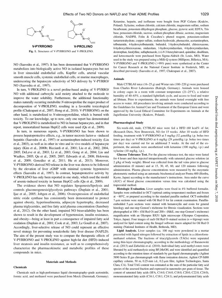
diolate] and V-PROLI/NO (O2-vinyl-[2-(carboxylato)pyrrolidin-1-yl]diazen-1-ium-1,2-diolate) (Fig. 1) are both diazeniumdiolates thatwere designed to deliver nitric oxide (NO) directly to the liver via cytochromeP450–dependent metabolism. The structure of these prodrugs was designedto avoid spontaneous decomposition under physiological conditions and tofacilitate cytochrome P450–related biotransformation into their respectiveepoxides. These unstable intermediates are formed in hepatocytes andhydrolyze, either spontaneously or via enzymatic reaction by hepatic epoxidehydrolase, to generate diazeniumdiolate ions, which spontaneously release
This work was supported by the Polish National Science Center [DEC-2013/11/N/NZ7/00749] and the European Union from the resources of the EuropeanRegional Development Fund under the Innovative Economy Programme, grantcoordinated by JCET-UJ [POIG.01.01.02-00-069/09].
dx.doi.org/10.1124/dmd.115.063388.
ABBREVIATIONS: AGP, a-acid glycoprotein; AUC, area under the curve; BSA, bovine serum albumin; HFD, high-fat diet; NAFLD, nonalcoholicfatty liver disease; NO, nitric oxide; PUFA, polyunsaturated fatty acids; SFA, saturated fatty acids; TAG, triacylglycerol; V-PROLI/NO, O2-vinyl-[2-(carboxylato)pyrrolidin-1-yl]diazen-1-ium-1,2-diolate; V-PYRRO/NO, O(2)-vinyl-1-(pyrrolidin-1-yl)diazen-1-ium-1,2-diolate.
1028
at ASPE
T Journals on M
ay 7, 2018dm
d.aspetjournals.orgD
ownloaded from
NO (Saavedra et al., 1997). It has been demonstrated that V-PYRRO/NOmetabolizes into biologically active NO in isolated hepatocytes but notin liver sinusoidal endothelial cells, Kupffer cells, arterial vascularsmooth muscle cells, systemic endothelial cells, or murine macrophages,underscoring the hepatocyte selectivity of NO delivery by V-PYRRO/NO (Saavedra et al., 1997).In turn, V-PROLI/NO is a novel proline-based analog of V-PYRRO/
NO with additional carboxylic acid moiety attached to the molecule toimprove the water solubility. Furthermore, the additional functionalitymakes naturally occurring metabolite N-nitrosoproline the major product ofdecomposition of V-PROLI/NO, resulting in a favorable toxicologicalprofile (Chakrapani et al., 2007; Hong et al., 2010). V-PYRRO/NO, on theother hand, is metabolized to N-nitrosopyrrolidine, which is burned withtoxicity. To our knowledge, up to now, only one report has demonstratedthat V-PROLI/NO is metabolized to NO in human hepatocytes, and in thatreport, immortalized human HepG2 cells were used (Qu et al., 2009).In turn, in numerous reports, V-PYRRO/NO has been shown to
possess hepatoprotective effects, e.g., in tumor necrosis factor-a –inducedhepatitis (Saavedra et al., 1997) or acetaminophen-induced toxicity (Liuet al., 2003), as well as in other in vitro and in vivo models of hepatocyteinjury (Kim et al., 2000b; Ricciardi et al., 2001; Liu et al., 2002, 2004,2005; DeLeve et al., 2003; Li et al., 2003; Gong et al., 2004; Liu andWaalkes, 2005; Qu et al., 2005, 2007; Edwards et al., 2008; Holowniaet al., 2009; González et al., 2011; Hu et al., 2013). Moreover,V-PYRRO/NO–derived NO release in the liver was shown to be followedby the elevation of liver cGMP with minimal systemic hypotensiveeffects (Saavedra et al., 1997). In contrast, hepatoprotective activity byV-PROLI/NO has only been reported in one study, which used the modelof arsenic-induced toxicity in human HepG2 cells (Qu et al., 2009).The evidence shows that NO regulates lipogenesis/lipolysis and
controls gluconeogenesis/glycolysis pathways (Duplain et al., 2001;Ijaz et al., 2005; Jobgen et al., 2006). Overexpression of endothelialnitric oxide synthase has consistently been demonstrated to protectagainst obesity, hyperinsulinemia, adipocyte hypertrophy, decreasedplasma triglycerides, and free fatty acid plasma concentration (Sansburyet al., 2012). On the other hand, impaired NO bioavailability has beenshown to result in the development of hypertension, insulin resistance,and obesity—being at least in part a consequence of impaired fatty acidoxidation (Duplain et al., 2001; Cook et al., 2003; Le Gouill et al., 2007).Accordingly, liver-selective release of NO could represent an effectivenovel strategy for preventing nonalcoholic fatty liver disease (NAFLD).The aim of the present study was to compare the therapeutic effects ofV-PYRRO/NO and V-PROLI/NO against high-fat diet (HFD)–inducedliver steatosis and insulin resistance, as well as to comprehensivelycharacterize the pharmacokinetic and metabolic profiles of thesecompounds in mice.
Materials and Methods
Chemicals
Chemicals such as high-performance liquid chromatography–grade acetonitrile,formic acid, and methanol were purchased from Merck (Darmstadt, Germany).
Ketamine, heparin, and isoflurane were bought from PGF Cefarm (Kraków,Poland). Xylazine, sodium chloride, calcium chloride, magnesium sulfate, sodiumbicarbonate, potassium dihydrogen phosphate, glucose, pyruvic acid, EDTA, Trisbase, potassium chloride, sucrose, sodium phosphate dibasic, acetone, magnesiumchloride, NADPH, Folin & Ciocalteu’s phenol reagent, potassium-sodiumtartaretetrahydrate, copper sulfate, sodium hydroxide, phenacetin, acetaminophen,tolbutamide, 4-hydroxytolbutamide, bufuralol, 1-hydroxybufuralol, chlorzoxazone,6-hydroxychlorzoxazone, midazolam, 1-hydroxymidazolam, 4-hydroxymidazolam,dextrorphan, furafylline, sulfaphenazole, (+)-N-3-benzylnirvanol, quinidine, disulfiram,and ketoconazole were purchased from Sigma-Aldrich (St. Louis, MO). Waterused in the study was prepared using a Milli-Q system (Millipore, Billerica, MA).V-PYRRO/NO and V-PROLI/NO (.99% pure) were synthetized at the Centerfor Cancer Research at the National Cancer Institute in Frederick, MD, asdescribed previously (Saavedra et al., 1997; Chakrapani et al., 2007).
Animals
Male C57BL/6J mice (16–25 g) and Wistar rats (180–220 g) were purchasedfrom Charles River Laboratories (Raleigh, Germany). Animals were housedin colony cages in a room with constant temperature (21–25�C), a relativehumidity of 40–65%, a standard light/dark cycle, and access to food and waterad libitum. Prior to experiments, the animals were fasted overnight with freeaccess to water. All procedures involving animals were conducted according tothe Guidelines for Animal Care and Treatment of the European Union and wereapproved by the Local Ethical Committee for Experiments on Animals at theJagiellonian University (Krakow, Poland).
Pharmacological Study
Six-week-old, male, C57BL/6J mice were fed a HFD (60 kcal% of fat;(Research Diets, New Brunswick, NJ) for 15 weeks. After 10 weeks of HFDfeeding, treatment with V-PYRRO/NO at 5 mg/kg (32 mmol/kg i.p. bolus twotimes per day) or V-PROLI/NO at 6 mg/kg (29 mmol/kg i.p. bolus two timesper day) was carried out for an additional 5 weeks. At the end of the ex-periment, the animals were anesthetized with ketamine (100 mg/kg, i.p.) andxylazine (10 mg/kg, i.p.).
Glucose Tolerance Test. For the glucose tolerance test, the mice were fastedfor 4 hours and then injected intraperitoneally with saturated glucose solution (at2 g/kg of body weight). Blood was collected from the tail veins prior to glucoseadministration (0 minute) and at 15, 30, 45, 60, and 120 minutes followingadministration. Plasma glucose concentrations were measured by the enzymaticphotometric method using an automatic biochemical analyzer Pentra 400 (Horiba,Kyoto, Japan) according to the manufacturer’s instructions. Area under the curve(AUC) of blood glucose concentration versus time was calculated using thetrapezoidal method.
Histologic Evaluation. Liver samples were fixed in 4% buffered formalin.Samples were embedded in OCT (optimal cutting temperature) medium and frozenat 280�C, or prepared according to the standard paraffin method. OCT-embedded7-mm sections were stained with Oil Red O for fat content examination. Paraffin-embedded 5-mm sections were stained with hematoxylin and eosin for generalhistology and one-step Gomori’s trichrome for fibrosis visualization. Sections werephotographed at 100� (Oil Red O) and 200� (H&E, one-step Gomori’s trichrome)magnification with an Olympus BX51 light microscope (Olympus Corporation,Tokyo, Japan). Four images of each Oil Red O–stained section (n = 6/group) wereanalyzed for lipid content using the ImageJ software macro adapted for Oil Red Ostaining (National Institutes of Health, Bethesda, MD).
Lipid Analysis. Liver samples (ca. 100 mg) were powdered in a mortarprecooled with liquid nitrogen followed by extraction of lipids in a chloroform-methanol solution. The fractions of triacylglycerols (TAGs) were separatedusing thin-layer chromatography, according to the methodology of Baranowskiet al. (2013) and Zabielski et al. (2010). Individual fatty acid methyl esters wereliberated by acid methanolysis using BF3MeOH, and were identified and quantifiedaccording to the retention times of standards by gas-liquid chromatography (Agilent5890 Series II gas chromatograph with flame ionization detector, Agilent CP-Sil88capillary column, 50 m, 0.25-mm i.d., 0.2-mm film; Agilent Technologies, SantaClara, CA). Total TAG content was estimated as the sum of the particular fatty acidspecies of the assessed fraction and expressed in nanomoles per gram of tissue. Thecontent of saturated fatty acids (SFA, C14:0, C16:0, C18:0, C20:0, C22:0, C24:0),monounsaturated fatty acids (C16:1, C18:1, C24:1), and polyunsaturated fatty acids
Fig. 1. Structures of V-PYRRO/NO and V-PROLI/NO.
Effects of Two NO Donors on NAFLD and Their ADME Profiles 1029
at ASPE
T Journals on M
ay 7, 2018dm
d.aspetjournals.orgD
ownloaded from

(PUFA, 18:2 n-6, 18:3 n-3, 20:4 n-6, 22:6 n-3) in the TAG fractions was analyzed,along with the PUFA/SFA ratio (Wierzbicki et al., 2009).
In Vivo Pharmacokinetics
The 6-week-old male C57BL/6J mice were injected with V-PYRRO/NOor V-PROLI/NO as a single i.v. and i.p. administration at a dose of 5 mg/kg(32 mmol/kg) or 6 mg/kg (29 mmol/kg) for V-PYRRO/NO and V-PROLI/NO,respectively. Animals were anesthetized with isoflurane at a concentration of3–4% in 100% oxygen and sacrificed at the following time intervals: 0 (beforedosing) and 2, 5, 7, 10, 15, 20, 25, 30, 45, 60, 90, 120, and 240 minutes aftercompound administration. Blood samples were collected into heparinizedmicrofuge tubes and centrifuged at 1000 rpm for 15 minutes. The plasma wasseparated into clean tubes and frozen at 220�C prior to analysis. The livertissues were collected and rinsed with phosphate-buffered saline (pH 7.4) andstored at 280�C until analysis.
All data in the pharmacokinetic experiments were processed using PhoenixWinNonlin 6.3 software (Certara, St. Louis, MO). The noncompartmental approachwas applied to calculate the basic pharmacokinetic parameters such as meanresidence time, AUC, systemic clearance (ClT), and volume of distribution atsteady state.
Renal Clearance
After a single i.v. injection of V-PYRRO/NO (5 mg/kg) or V-PROLI/NO(6 mg/kg), the 6-week-old mice (n = 7) were individually placed in stainlesssteel metabolic cages for collection of urine. The urine samples were collectedat 0–6, 6–12, and 12–24 hours post dosing. The volumes of the urine sampleswere recorded, and samples were stored at 220�C until analysis.
Renal elimination was assessed based on the fraction of administered doseexcreted in the urine in unchanged form:
fR ¼ Ae‘
Divð1Þ
where fR is the fraction of dose excreted by the kidney, Ae‘ is the total amountof studied substance in the urine, and Div is the administered dose.
Renal clearance (ClR) of V-PYRRO/NO and V-PROLI/NO was calculatedas follows:
ClR ¼ fR � ClT ð2ÞClR ¼ Ae‘
AUC‘0
ð3Þ
Hepatic Extraction Ratio
Hepatic elimination was determined using the ex vivo model of perfusedmouse liver. Following i.p. injection of ketamine (100 mg/kg), xylazine (10 mg/kg),and 0.8 mg/kg of heparin, the vena porta and the vena cava inferior were cannulatedand ligated, and the liver was perfused using the U-100 system for organ perfusion(Hugo Sachs Elektronik, Harvard Apparatus, March-Hugstetten, Germany) untileffluent was blood-free. The liver was than excised and moved to a moist chamber.Perfusion was carried out with Krebs-Hanseleit buffer of the following composition:118.0 mM NaCl, 2.52 mM CaCl2, 1.16 mM MgSO4, 24.88 mM NaHCO3, 1.18mM KH2PO4, 4.7 mM KCl, 10.0 mM glucose, 2.0 mM pyruvic acid, and 0.5 mMEDTA. After the initial stabilization period (15 minutes), either V-PYRRO/NO orV-PROLI/NO was added at a final concentration of 10 or 50 mM, respectively. Theexperiments were completed within 1 hour after starting perfusion. The perfusionflow rate was 4.3 ml/min, and samples were collected as follows: inlet samplesevery 20 minutes, and outlet effluents every 1 minute, between 5 and 20 minutes,and then every 10 minutes. After the experiments, the livers were excised, dried,and weighed.
To ensure the viability of the liver, alanine aminotransferase and lactatedehydrogenase activity in the effluents were measured every 15 minutes for theduration of the experiment by the enzymatic photometric method, using theautomatic biochemical analyzer Pentra 400 (Horiba, Kyoto, Japan), accordingto the manufacturer’s instructions.
A control experiment to exclude the binding of V-PYRRO/NO or V-PROLI/NOto the experimental setup was conducted. For this purpose, buffer containing
V-PYRRO/NO or V-PROLI/NO was perfused through the perfusion systemwithout mounting of the isolated liver. Buffer samples were collected thesame way as in the experiments with the isolated liver.
The hepatic extraction ratio was calculated based on the V-PYRRO/NO andV-PROLI/NO concentrations in the inlet and outlet effluents:
E ¼ Cin 2Cout
Cinð4Þ
where E is the hepatic extraction ratio and Cin and Cout are concentrations ofstudied compounds in inlet or outlet liver effluent.
Protein Binding
Binding of V-PYRRO/NO and V-PROLI/NO to bovine serum albumin (BSA)and a-acid glycoprotein (AGP) was determined using capillary electrophoresisin frontal analysis mode on a Beckman Coulter P/ACE MDQ CE system witha photodiode array detector fixed at 230 nm (Beckman Coulter, Brea, CA). Theworking conditions were as follows: uncoated fused silica capillary length60.2 cm (50 cm to the detector) with 50-mm i.d. and 360-mm o.d.; temperature ofthe capillary 37�C; applied voltage of 15 kV; observed currents of about 56 mA.Following the standard rinsing procedure, the samples were injected at 0.5 psifor 40 seconds (injected sample volume represented 5% of the total capillaryvolume). The unbound concentrations of V-PYRRO/NO and V-PROLI/NO weredetermined by comparing the plateau peak height of the equilibrated samples withthe peak height of V-PYRRO/NO or V-PROLI/NO in the absence of BSA or AGP.
The results were expressed as the saturation fraction (r), representing thenumber of moles of compound bound (Cb) per mole of protein (P):
r ¼ Cb
Pð5Þ
The percentage of binding was determined using eq. 6:
% bound ¼ Ct 2 Cu
Ct x 100 ð6Þ
where Ct is the total concentration of compound in the protein solution, and Cu
is the free analyte concentration.The equilibrium association constant (Ka) in the binding class (m) and the
number of binding sites (n) were determined by a nonlinear regression analysisusing Wolfram Mathematica 8.0 software (La Jolla, CA) to fit the data to eq. 7:
r ¼ +m
i¼1
ni × Cu
Kdi þ Cu ð7Þ
where r is the number of moles of drug bound per mole of protein (Cb/Pt,where Pt is the total protein concentration), m is the number of independentclasses of binding sites, Kdi is the dissociation constant for the ith class, andni is the number of binding sites in the ith class.
In Vitro Metabolism by Cytochrome P450
Rats were sacrificed by decapitation, and liver microsomes were prepared bydifferential centrifugation. In brief, liver fragments were washed with 20 mMTris/KCl buffer (pH 7.4) and homogenized (IKA-Werke GmbH & Co. KGStaufen, Janke, Germany). The homogenate was centrifuged (Sorvall WX UltraSeries; Thermo Scientific, Waltham, MA) at approximately 11,500 � g for 20minutes at 4�C. The supernatant (S9 fraction) was transferred to new tubes andcentrifuged at 100,000 � g for 1 hour at 4�C. The pellet was suspended in0.15 M KCl and centrifuged again at 100,000 � g for 1 hour at 4�C. Theobtained pellet was dispersed in Tris/sucrose buffer and stored at 280�C untiluse. Protein concentration in the microsomal fraction was determined by Lowryprotein assay (Lowry et al., 1951).
V-PYRRO/NO or V-PROLI/NO at a concentration range of 0.5–50 mM wasincubated with the rat liver microsomes (1 mg/ml) in 0.1 M phosphate buffer(pH 7.4) containing 10 mM MgCl2. After 5 minutes of preincubation at 37�C,the incubation reactions were initiated with the addition of NADPH to a finalconcentration of 1 mM and were stopped after 20 minutes by placing samples on iceand adding ice-cold acetonitrile containing internal standard (4-hydroxymephenytoin
1030 Kus et al.
at ASPE
T Journals on M
ay 7, 2018dm
d.aspetjournals.orgD
ownloaded from
at a concentration of 20 ng/ml). Basic kinetic parameters, Vmax and Km forV-PYRRO/NO and V-PROLI/NO, were calculated with GraphPad Prism6.02 software (La Jolla, CA) using nonlinear regression.
To test the involvement of cytochrome P450 isoenzymes in V-PYRRO/NOand V-PROLI/NOmetabolism, the following cytochrome P450–dependent isoenzymeinhibitors were used: furafylline (1.5 mg/ml) for CYP1A2, sulfaphenazole (15 mg/ml)for CYP2C9, (+)-N-3-benzylnirvanol (1 mg/ml) for CYP2C19, quinidine (15 mg/ml)for CYP2D6, disulfiram (15 mg/ml) for CYP2E1, and ketoconazole (5 mg/ml) forCYP3A4.
To study the effects of V-PYRRO/NO and V-PROLI/NO on cytochromeP450 isoenzyme activity, a “cocktail” method was used based on measurementof the concentration of metabolites derived from cytochrome P450 isoenzyme–specific substrates. In brief, V-PYRRO/NO or V-PROLI/NO, in the con-centration range of 0.1 mM to 1 mM, was incubated with rat liver microsomessuspended in 0.1 M phosphate buffer (pH 7.4), 10 mM MgCl2, and substratecocktail, phenacetin (7.5 mg/ml) for CYP1A2, tolbutamide (2.5 mg/ml) forCYP2C9, bufuralol (12.5 mg/ml) for CYP2D6, chlorzoxazone (12.5 mg/ml) forCYP2E1, and midazolam (7 mg/ml) for CYP3A4. After addition of V-PYRRO/NO or V-PROLI/NO, the reaction mixtures were preincubated for 5 minutes at+37�C in a shaking water bath (Grant Instruments, Royston, UK), and next thereaction was initiated by addition of 1 mM NADPH. Following a 10-minuteincubation, the reaction was terminated with an ice-cold mixture of acetonitrile:acetone (1:1; v/v) containing the internal standard dextrorphan (50 ng/ml). Sampleswere subsequently cooled on ice for 20 minutes to precipitate the protein, and thencentrifuged at approximately 15,000 � g for 15 minutes at 4�C. The supernatantwas analyzed immediately after incubation.
Chromatographic and Mass Spectrometric Analysis
V-PYRRO/NO and V-PROLI/NO concentrations in plasma, liver homo-genates, urine, liver effluents, and in the incubation mixture of micro-somes (50 ml) were measured after deproteinization using ice-coldacetonitrile (500 ml) containing internal standard (4-hydroxymephenytoin,20 ng/ml). Samples were subsequently cooled on ice for 20 minutes toprecipitate the protein and then centrifuged at approximately 15,000 � g for15 minutes at 4�C. The supernatant was transferred to a high-performanceliquid chromatography vial, and 5 ml of supernatant was injected into theanalytical column. The analytical system consisted of UFLC Nexera(Shimadzu, Kyoto, Japan) coupled with a QTrap 5500 mass spectrometer(AB Sciex, Framingham, MA). Chromatographic separation was achievedusing the Acquity UPLC BEH C18 (1.7 mm, 3.0 � 100 mm; Waters, Milford,MA) analytical column, with acetonitrile and water containing 0.1% formicacid in the isocratic elution (45:55 v/v), at a flow rate of 0.4 ml/min. Theanalyzed compounds were detected in positive ionization multiple reactionmonitoring mode, monitoring the transitions of protonated ions m/z 158→70for V-PYRRO/NO, m/z 202→100 for V-PROLI/NO, and m/z 235→150 forinternal standard. The operating conditions were as follows: curtain gas20 psi, ion spray voltage 5000 V, temperature 400�C, ion source gases 50 and15 psi. Nitrogen was used as the curtain and collision gas.
Chromatographic separation of the metabolites of model cytochrome P450substrates was performed on a Kinetex analytical column (2.6 mm PFP 100 Å,3� 100 mm; Phenomenex, Torrance, CA) using an Ultimate 3000 Ultra PerformanceLiquid Chromatography system (Dionex, Sunnyvale, CA). The mobile phaseconsisted of acetonitrile containing 0.1% formic acid (eluent A) and watercontaining 0.1% formic acid (eluent B). At a flow rate of 500 ml/min, the amountof eluent A was increased linearly from 20 to 95% over 2 minutes, maintained at95% for 6 minutes, returned to 20% over 3 minutes, and left to re-equilibrate for 6minutes. The total run time was 15 minutes. The analytes were detected usinga TSQ Quantum Ultra triple quadrupole mass spectrometer (Thermo Scientific).Heated electrospray ionization was used in both positive and negative mode (for6-hydroxychlorzoxazone). The selected reaction monitoring transitions for eachquantified substance and the internal standard were as follows: acetaminophen m/z152→110, 4-hydroxytolbutamide m/z 287→89, 4-hydroxymephenytoin m/z 235→150,1-hydroxybufuralol m/z 278→186, 6-hydroxychlorzoxazone m/z 184→120,1-hydroxymidazolam m/z 342→324, 4-hydroxymidazolam m/z 342→297,and dextrorphan (internal standard) m/z 258→157. The operating conditionswere as follows: needle voltage 4020 V, vaporizer temperature 250�C, sheathgas (nitrogen) pressure 30 psi, auxiliary gas (nitrogen) pressure 10 psi, and
capillary temperature 370�C. The argon gas pressure in the collision cell wasapproximately 2 mTorr.
Nitrite and Nitrate Measurements
The plasma samples were precipitated with methanol at a ratio of 1:1 (v/v).Liver samples were homogenized with methanol at a ratio of 1:2 (w/v).Following centrifugation at 10,000 � g for 10 minutes, 10 ml of supernatantwas injected into the separation column.
The concentrations of nitrite and nitrate were measured by ENO-20 system(Eicom, Kyoto, Japan), based on the liquid chromatography method withpostcolumn derivatization with Griess reagent. Nitrite and nitrate were separatedon a NO-PAK column (4.6 � 50 mm; Eicom). Nitrate was reduced to nitrite bya cadmium-copper column (NO-RED; Eicom). Nitrite was detected based on theGriess reaction, with sulfanilamide and naphthylethylenediamine forming a purplediazo compound with the absorbance of dye product measured at 540 nm. Theflow of the mobile phase (carrier solution; Eicom) was 0.33 ml/min. The Griessreagent was delivered at a flow rate of 0.11 ml/min.
Statistical Analysis
Data are expressed as the mean 6 S.E.M. The assessment of normalityand heterogeneity of variances was performed using the Shapiro-Wilk testand Fligner-Killeen test, respectively. To assess the statistical significance ofthe pharmacokinetics results, Student’s t test and the nonparametric Mann-Whitney test were used. For pharmacological experiments, the nonparametricKruskal-Wallis test or one-way analysis of variance and Tukey’s post-hoctest were used. The results were analyzed using Statistica 10.0 software(Statsoft, Tulsa, OK).
Results
Effects of V-PYRRO/NO and V-PROLI/NO Treatment onNAFLD. Treatment with V-PYRRO/NO, but not V-PROLI/NO, attenuatedliver steatosis as evidenced by histopathological analysis (Fig. 2, A–C)with semiautomatic quantitative analysis of liver fat content based onOil Red O–stained slides (Fig. 2D), as well as by liver triglyceride contentbased on gas chromatography–flame ionization detector chromatography(Fig. 2E). Furthermore, V-PYRRO/NO, but not V-PROLI/NO, favorablychanged fatty acid composition by increasing the PUFA/SFA ratio inmice fed a HFD (Fig. 2F). Finally, the overall glucose exposure,calculated on the basis of AUC of blood glucose concentration versustime on the tolerance chart, was significantly improved in V-PYRRO/NO–treated mice, whereas there was no improvement seen in V-PROLI/NO–treated mice (Fig. 2, G and H).Pharmacokinetic Profile of V-PYRRO/NO and V-PROLI/NO.
The mean plasma concentration versus time profiles of V-PYRRO/NOand V-PROLI/NO after i.v. and i.p. administration are depicted in Fig.3A, and the pharmacokinetic parameters are given in Table 1.V-PYRRO/NO was eliminated more rapidly than V-PROLI/NO, as
evidenced by a lower mean residence time and higher clearance values.V-PYRRO/NO was detectable in plasma for up to 60 minutes after i.v.administration and 30 minutes after i.p. administration, respectively,whereas V-PROLI/NO was measurable over the entire sampling period.Moreover, V-PYRRO/NO was widely distributed in the intra- andextracellular water (Vdss [steady -state volume of distribution] = 0.88l/kg), whereas V-PROLI/NO distribution was limited to the extracellularfluid (Vdss = 0.15 l/kg). In view of the fact that the pharmacologicaleffects were studied following intraperitoneal administration ofV-PYRRO/NO and V-PROLI/NO, pharmacokinetic studies follow-ing i.p. dosing were also conducted. Both compounds were rapidlyabsorbed after i.p. administration, with lower bioavailability forV-PYRRO/NO as compared with V-PROLI/NO (about 28 and 51%,respectively). Systemic NO release following i.v. administration ofV-PYRRO/NO was higher than with V-PROLI/NO, as evidenced bynitrate and nitrite plasma concentrations (Fig. 3, B and C).
Effects of Two NO Donors on NAFLD and Their ADME Profiles 1031
at ASPE
T Journals on M
ay 7, 2018dm
d.aspetjournals.orgD
ownloaded from

Hepatic Metabolism of V-PYRRO/NO and V-PROLI/NO. Toassess the liver disposition of V-PYRRO/NO and V-PROLI/NO, theconcentration of each compound in liver homogenates was measured
following i.v. or i.p. administration of the compounds. V-PYRRO/NOwas not detectable, whereas V-PROLI/NO was distributed in the liverwith maximum concentrations of 35.8 nmol/g (tmax [time to reach the
Fig. 2. Representative pictures of liver sections stained with H&E (A), one-step Gomori’s trichrome (B), and Oil Red O (C). Liver fat content calculated from Oil Red Opictures (D), liver triglyceride (TG) concentration (E), and PUFA/SFA ratio (F) in the liver of mice. Glucose tolerance curve (G) and AUC of blood glucose concentrationversus time (H). Values are the means6 S.E.M. (n = 6). Values with different superscript letters within each animal group are significantly different (P# 0.05). HF, high-fatgroup; HF+V-PYRRO/NO, high-fat group treated with V-PYRRO/NO; HF+V-PROLI/NO, high-fat group treated with V-PROLI/NO.
Fig. 3. Comparison of plasma pharmacokinetic profiles of V-PYRRO/NO and V-PROLI/NO after i.v. and i.p. administration in mice (A), and NO22, NO3
2 concentrationsin plasma after i.v. administration of each compound (B and C). Values are expressed as the mean 6 S.E.M. (n = 3 for i.p. and n = 4 for i.v. administration).
1032 Kus et al.
at ASPE
T Journals on M
ay 7, 2018dm
d.aspetjournals.orgD
ownloaded from

maximal concentration] = 2 minutes) and 23.9 nmol/g (tmax = 7 minutes)following i.v. and i.p. administration, respectively. Even after 60 minutes,the concentration of V-PROLI/NO remained elevated (5.3 and 2.5 nmol/gafter i.v. and i.p., respectively).To compare the decomposition of V-PYRRO/NO and V-PROLI/NO
in the liver in greater detail, the uptake and metabolism of these analogswere studied in the isolated, perfused mouse liver setup. V-PYRRO/NOexhibited nonspecific binding to the experimental setup, resulting ina 25% decrease in the dose introduced to the liver that was includedin the calculations of the hepatic extraction ratio, whereas binding ofV-PROLI/NO to the perfusion setup was not significant. As shown inFig. 4, A and B, the concentrations of V-PYRRO/NO in the livereffluents amounted to about 70% of the initial concentration, taking intoaccount the binding to experimental setup; therefore, the calculatedhepatic extraction ratio was about 0.3, and the hepatic clearance was0.058 l/min/kg. In contrast, V-PROLI/NO was hardly metabolized in theliver, with outflow concentrations of the compound near its inflowconcentration. The hepatic extraction ratio for V-PROLI/NO was below0.1 (about 0.05) and hepatic clearance was 0.0005 ml/min/kg, suggestingnegligible liver metabolism of V-PROLI/NO, in contrast with V-PYRRO/NO. These findings were confirmed by the measurements of concen-trations of nitrite and nitrate in the liver samples after a 1-hour perfusionof V-PYRRO/NO or V-PROLI/NO at a concentration of 50 mM.V-PYRRO/NO perfusion resulted in elevations in nitrite and nitrate livercontent by more than 2-fold (NO2
2: 4.8 nmol/g; NO32: 23.3 nmol/g),
whereas V-PROLI/NO perfusion did not increase nitrite or nitrateconcentration in the liver homogenate (NO2
2: 1.7 nmol/g; NO32: 13.4
nmol/g). Altogether, these results confirm considerable liver metabolismof V-PYRRO/NO, with subsequent NO release, but not of V-PROLI/NO.Renal Elimination of V-PYRRO/NO and V-PROLI/NO. As
shown in Fig. 4C, less than 0.1% of V-PYRRO/NO was eliminated in
the urine, whereas approximately 61% of V-PROLI/NO was excretedin the urine as unmodified compound. The calculated renal clearancefor V-PYRRO/NO was very low and without physiologic significance,whereas the renal clearance for V-PROLI/NO was significantly higherand amounted to 0.0032 l/min/kg.Protein Binding of V-PYRRO/NO and V-PROLI/NO. Both
analogs bound to BSA with one class of binding site, and the per-centage of binding amounted to 25.13 6 4.5% and 53.33 6 7.1% forV-PYRRO/NO and V-PROLI/NO, respectively. However, binding affinityto BSA was relatively low (Ka = 1.93 � 103 and 7.57 � 103 M21 forV-PYRRO/NO and V-PROLI/NO, respectively). Binding of studiedanalogs to AGP was also very low and not of physiologic significance(3.15 6 2.9 and 3.05 6 1.43 for V-PYRRO/NO and V-PROLI/NO,respectively), probably due to the acidic character of these molecules.Role of Cytochrome P450 in V-PYRRO/NO and V-PROLI/NO
Metabolism. The calculated kinetic parameters derived using theMichaelis-Menten transformation (Fig. 5A) for V-PYRRO/NO wereKm = 131.6 6 38.15 mM and Vmax = 6.35 6 1.41 mmol/min/mgprotein. Based on the kinetic plots, V-PROLI/NO seems to have a lowaffinity for the cytochrome P450 isoenzymes. The Eadie-Hofstee plotswere nonlinear, suggesting multiple-enzyme catalysis (data not shown).The effect of selective cytochrome P450 inhibitors on the bio-
transformation of V-PYRRO/NO and V-PROLI/NO using isoenzyme-selective chemical inhibitors is shown in Fig. 5B. As evidenced by theeffects of their respective inhibitors, V-PYRRO/NO was metabolizedmainly by CYP2E1, but also by CYP2C9, CYP1A2, and CYP3A4,whereas V-PROLI/NO was biotransformed mainly by CYP1A2 andCYP2C9.Neither V-PYRRO/NO nor V-PROLI/NO inhibited cytochrome
P450 isoenzymes as evidenced by the lack of inhibition of phenacetin-O-deethylation (CYP1A2), tolbutamide-4-hydroxylation (CYP2C9),bufuralol-1-hydroxylation (CYP2D6), chlorzoxazone-6-hydroxylation(CYP2E1), and midazolam-1- and -4-hydroxylation (CYP3A4), up toa concentration of 1 mM V-PYRRO/NO and V-PROLI/NO (data notshown).
Discussion
In the present study, we compared two NO donors (V-PYRRO/NOand V-PROLI/NO) that were designed to deliver NO to the liver viacytochrome P450–dependent metabolism by looking at their effects onliver steatosis, liver fatty acid composition, and insulin resistance, andcomprehensively analyzing their pharmacokinetics and metabolismprofiles. In the study, we demonstrated that, despite having similarchemical structures, only V-PYRRO/NO attenuated liver steatosis,increased the liver PUFA/SFA ratio, and improved postprandialglucose tolerance, whereas V-PROLI/NO was ineffective. Pharmaco-kinetic studies revealed rapid absorption following i.p. administration,
TABLE 1
Pharmacokinetic parameters of V-PYRRO/NO and V-PROLI/NO after i.v. and i.p.administration in control mice
ParameterV-PYRRO/NO V-PROLI/NO
Intravenous Intraperitoneal Intravenous Intraperitoneal
Co (mM) 41.5 — 222.48 —
AUC0→‘ (mM*min) 218.62 60.9 5669.26 2895.27MRT (min) 6 7.7 27.87 28.57Cmax (mM) — 7.93 — 79.57tmax (min) — 5 — 10Vss (l/kg) 0.88 — 0.149 —
Cl (l/min/kg) 0.146 — 0.0053 —
F (%) — 27.85 — 51.07
AUC0→‘, area under the curve extrapolated to infinity; Cl, clearance; Co, initial concentration;MRT, mean residence time; tmax, the time to reach the maximum plasma concentration;Vss, steady-state volume of distribution.
Fig. 4. Calculated differences for V-PYRRO/NO and V-PROLI/NO concentrations between inlet and outlet effluent samples following perfusion with 10 mM (A) and50 mM compound (B). (C) Fraction of dose that was eliminated unchanged by the kidney. Values are expressed as the mean 6 S.E.M. *P , 0.05; **P , 0.01 (n = 4 forisolated, perfused mouse liver experiments, and n = 7 for renal elimination evaluation).
Effects of Two NO Donors on NAFLD and Their ADME Profiles 1033
at ASPE
T Journals on M
ay 7, 2018dm
d.aspetjournals.orgD
ownloaded from

intense elimination, and incomplete bioavailability for both V-PYRRO/NOand V-PROLI/NO. However, it was noticed that V-PYRRO/NO wasmetabolized in the liver by CYP2E1, CYP2C9, and CYP3A4, whereasV-PROLI/NO was eliminated mostly in unchanged form by the kidney.Importantly, V-PYRRO/NO proved to be a better NO releaser in themouse in vivo and in the isolated liver as compared with V-PROLI/NO.These results were consistent with superior antisteatotic activity forV-PYRRO/NO. In conclusion, we provide clear-cut evidence thatV-PYRRO/NO displays a pronounced antisteatotic effect associatedwith liver-targeted NO release, whereas V-PROLI/NO is not effective,is not taken up by the liver, and is eliminated mostly unchanged by thekidney.In physiological conditions, NO generated mainly by endothelial
nitric oxide synthase (Förstermann and Sessa, 2012) plays a crucialrole in liver homeostasis. NO activates soluble guanyl cyclase, whichactivates cGMP-dependent protein kinase (Pfeifer et al., 2013), and itsrole is not limited to the regulation of hepatic arterial resistance(Rockey and Shah, 2004). In fact, the NO/cGMP–dependent signalingpathway exerts an anti-inflammatory and antifibrotic effect by limitingKupffer cell and hepatic stellate cell activation (Tateya et al., 2011).NO also inhibits caspase activation, and therefore apoptosis, in hepatocytes(Kim et al., 2000a). Furthermore, the remarkable regenerative ability of theliver is linked to NO activity (Carnovale and Ronco, 2012). NO alsoregulates glucose and lipid metabolism (Jobgen et al., 2006), decreases fattyacid storage by improving their catabolism, and attenuates de novo fattyacid synthesis. Moreover, NO regulates glucose metabolism by increasingglucose transport and metabolism as well as inhibiting gluconeogenesis andglycogen deposition (Jobgen et al., 2006).Clearly, inadequate NO production by liver sinusoidal endothelial
cells results in unopposed hepatic stellate cell activation and promotion ofliver inflammation, which may contribute to the progression of portalhypertension (Hu et al., 2013), sinusoidal obstruction syndrome (DeLeve,2008), ischemia-reperfusion injury (Siriussawakul et al., 2010), and liversteatosis (Maslak et al., 2015). In fact, impairment of NO signalingresulted in decreased glucose uptake, fasting hyperglycemia, and thedevelopment of insulin resistance (Lutz et al., 2011; An et al., 2012).In line with the importance of NO in the maintenance of metabolic
homeostasis, the beneficial effect of NO-based therapy has beendemonstrated in obesity (Jobgen et al., 2009), insulin resistance (Sadriand Lautt, 1999), and liver steatosis (de Oliveira et al., 2006).However, in contrast to the present paper, none of these reportsconcerned the direct delivery of NO to the liver (Ricardo et al., 2002).In the current study, we aimed at comparing the antisteatotic efficacy oftwo structurally related hepatocyte-specific NO donors, and revealeda hepatospecific and hepatoprotective action of V-PYRRO/NO, but notof V-PROLI/NO, against NAFLD. Moreover, the liver-specific effects
of V-PYRRO/NO–derived NO release not only attenuated liver steatosis,but were also associated with changes in fatty acid composition, inparticular the amelioration of the PUFA/SFA ratio, most likely due toinhibition of endogenous fatty acid synthesis. Our findings are of particularimportance because fatty acid saturation has been shown to be involved inthe pathogenesis of insulin resistance (van den Berg et al., 2010) andmetabolic syndrome (Warensjö et al., 2005). In fact, in our previous work,we demonstrated that the effect of V-PYRRO/NO on insulin resistance andsteatosis was mediated by NO-dependent Akt activation and inhibitionof de novo fatty acid synthesis by Acetyl-CoA carboxylase (ACC)phosphorylation (Maslak et al., 2015). Interestingly, despite previouslyreported data indicating NO release in vitro (Hong et al., 2010) andfavorable toxicity (Chakrapani et al., 2007), V-PROLI/NO did notinfluence insulin resistance and fatty acid composition in mice in thepresent study.The superior pharmacological activity of V-PYRRO/NO was not
associated with higher bioavailability than V-PROLI/NO followingi.p. administration. In fact, V-PROLI/NO showed greater bioavail-ability (51%) than V-PYRRO/NO (27%). Similar results for the bio-availability of V-PYRRO/NO were reported by Stinson et al. (2002).The incomplete bioavailability, besides the first-pass effect, might bea result of extrahepatic metabolism of V-PYRRO/NO and V-PROLI/NO, since the cytochrome P450 enzyme system is also located in otherextrahepatic tissues, including the lungs, kidneys, and small intestine(Guengerich, 1992; Nebert and Russell, 2002; Ding and Kaminsky,2003; Paine et al., 2006; Pavek and Dvorak, 2008). Partial de-composition of V-PYRRO/NO and V-PROLI/NO in other organs mayexplain the increased nitrite and nitrate plasma concentrations detectedfollowing i.v. administration of the compounds. However, we areconfident that, even though V-PYRRO/NO was partially metabolizedin extrahepatic tissues, its pharmacological effect came mostly, if notentirely, from liver-specific NO release.The results from the specific cytochrome P450 inhibitors demon-
strated that V-PYRRO/NO was mainly a substrate for CYP2E1, aswell as for CYP1A2, CYP2C9, and CYP3A4, whereas V-PROLI/NOwas shown to be metabolized mainly by CYP1A2 and CYP2C9 in theliver. These results are partially in line with the findings of Inami et al.(2006), who showed that CYP2E1, CYP2A6, and CYP2B6 areresponsible for V-PYRRO/NO metabolism in human liver micro-somes. On the other hand, V-PROLI/NO was metabolized mainlyby CYP1A2 and, to a lesser extent, by CYP2E1 and CYP3A4, asreported previously (Chakrapani et al., 2007), but with low enzymeaffinity.Increased metabolism and NO production in V-PROLI/NO–treated
HepG2 cells, as compared with V-PYRRO/NO treatment, was previouslydemonstrated by Hong et al. (2010). However, high concentrations of
Fig. 5. (A) Michaelis-Menten plots of the enzyme kinetics of V-PYRRO/NO and V-PROLI/NO in rat liver microsomes. (B) The effect of selective cytochrome P450inhibitors on V-PYRRO/NO and V-PROLI/NO (50 mM) metabolism in rat liver microsomes. Values are shown as the mean percentage 6 S.E.M. (n = 3 for furafylline,sulfaphenazole, N-benzylnirvanol, and ketoconazole; n = 6 for disulfiram).
1034 Kus et al.
at ASPE
T Journals on M
ay 7, 2018dm
d.aspetjournals.orgD
ownloaded from
V-PYRRO/NO and V-PROLI/NO were required to generate relativelylow concentrations of nitrite [reported by Hong et al., 2010 andconfirmed by us (unpublished results)], suggesting a relatively lowbiotransformation of the compounds in HepG2 cells. This notion seemsto be in line with recent studies showing that, in human hepatoma cells(e.g., HepG2), the expression of most of cytochrome P450 isoenzymesis substantially reduced as compared with primary human hepatocytes(about 100- to 1000-fold lower) (Westerink and Schoonen, 2007;Pawłowska and Augustin, 2011; Lin et al., 2012). Moreover, in HepG2cell lines, the activity of cytochrome P450 enzymes is much lower thanin ex vivo isolated primary hepatocytes (Westerink and Schoonen,2007; Lin et al., 2012). Additionally, since the expression and activity ofCYP1A2 are higher than those of CYP2E1 in HepG2 cells (Westerinkand Schoonen, 2007), Hong et al. (2010) found high release of NO fromV-PROLI/NO, as compared with V-PYRRO/NO. In our hands, bio-transformation of V-PROLI/NO was dependent on CYP1A2 andCYP2C9, whereas biotransformation of V-PYRRO/NO was mainlydependent on CYP2E1. It is not surprising that Gong et al. (2004)demonstrated that HepG2 cells failed to generate nitrite and nitratefrom V-PYRRO/NO. Accordingly, we believe that use of the HepG2cell line as a model system to study NO release from V-PYRRO/NOand V-PROLI/NO is not relevant to primary hepatocytes or the invivo setting, thus giving contradictory results from our presentstudy, which was performed in an in vivo and ex vivo isolated liversetup.In the present work, we also demonstrated that the differences in the
plasma pharmacokinetic profiles of V-PYRRO/NO and V-PROLI/NOmay be linked to differences in their lipophilicity caused by an ad-ditional carboxylic acid moiety in V-PROLI/NO’s structure. In silico–predicted log P values for both analogs differed from 21.63 to1.01 for V-PYRRO/NO and from 23.92 to 22.76 for V-PROLI/NO,depending on the pH of the environment. V-PYRRO/NO, being morelipophilic, was widely distributed in the total body water (Vdss = 0.88l/kg), whereas V-PROLI/NO distribution was limited only to theextracellular fluid (Vdss = 0.149 l/kg). Differences in pharmacokineticprofiles can also be related to the participation of proline transportersin the case of V-PROLI/NO, but not of V-PYRRO/NO.Moreover, the results showed that the affinity of V-PYRRO/NO and
V-PROLI/NO to BSA was rather low. V-PROLI/NO was bound toBSA to a greater extent, probably due to the stronger acidic character,which can partially explain its slower elimination and longer biologicalhalf-life. Furthermore, we provide evidence that neither compoundaffects the activity of cytochrome P450, as shown in the direct inhibitionstudy. In summary, both analogs display distinct cytochrome P450metabolism, distinct pharmacokinetic profile, and differences in renalexcretion.
Conclusions
V-PYRRO/NO, but not V-PROLI/NO, protected against high fatdiet-induced liver steatosis and improved insulin resistance in micefed a high-fat diet. The compounds’ distinct pharmacological effectscan be explained by their pharmacokinetic and metabolic profiles.V-PYRRO/NO displayed a pronounced antisteatotic effect associatedwith liver-targeted NO release, whereas V-PROLI/NO was ineffective,not taken up by the liver, and was eliminated mostly unchanged by thekidney. It is worth adding that therapy with liver-targeted NO donors,free of systemic hypotensive effects, represents a promising therapeuticstrategy not only in NAFLD but also in other liver disorders, such asliver cirrhosis, liver fibrosis, and postischemic injury (Ricciardi et al.,2001; Moal et al., 2002; Edwards et al., 2008) that warrants furtherstudies.
Authorship ContributionsParticipated in research design: Kus, Chlopicki.Conducted experiments: Kus, Walczak, Maslak, Zakrzewska, Gonciarz-
Dytman, Zabielski, Sitek, Wandzel, Kij, Chabowski.Contributed new reagents or analytic tools: Saavedra.Performed data analysis: Kus, Maslak, Holland, Keefer.Wrote or contributed to the writing of the manuscript: Kus, Chlopicki,
Maslak, Walczak, Holland, Keefer.
References
An Z, Winnick JJ, Moore MC, Farmer B, Smith M, Irimia JM, Roach PJ, and Cherrington AD(2012) A cyclic guanosine monophosphate-dependent pathway can regulate net hepatic glucoseuptake in vivo. Diabetes 61:2433–2441.
Baranowski M, Blachnio-Zabielska AU, Zabielski P, Harasim E, Harasiuk D, Chabowski A,and Gorski J (2013) Liver X receptor agonist T0901317 enhanced peroxisome proliferator-activated receptor-delta expression and fatty acid oxidation in rat skeletal muscle. J PhysiolPharmacol 64:289–297.
Carnovale CE and Ronco MT (2012) Role of nitric oxide in liver regeneration. Ann Hepatol 11:636–647.
Chakrapani H, Showalter BM, Kong L, Keefer LK, and Saavedra JE (2007) V-PROLI/NO,a prodrug of the nitric oxide donor, PROLI/NO. Org Lett 9:3409–3412.
Cook S, Hugli O, Egli M, Vollenweider P, Burcelin R, Nicod P, Thorens B, and Scherrer U(2003) Clustering of cardiovascular risk factors mimicking the human metabolic syndrome X ineNOS null mice. Swiss Med Wkly 133:360–363.
Deleve LD (2008) Sinusoidal obstruction syndrome. Gastroenterol Hepatol (N Y) 4:101–103.DeLeve LD, Wang X, Kanel GC, Ito Y, Bethea NW, McCuskey MK, Tokes ZA, Tsai J,and McCuskey RS (2003) Decreased hepatic nitric oxide production contributes to the de-velopment of rat sinusoidal obstruction syndrome. Hepatology 38:900–908.
de Oliveira CPMS, Simplicio FI, de Lima VMR, Yuahasi K, Lopasso FP, Alves VAF,Abdalla DSP, Carrilho FJ, Laurindo FRM, and de Oliveira MG (2006) Oral administrationof S-nitroso-N-acetylcysteine prevents the onset of non alcoholic fatty liver disease in rats.World J Gastroenterol 12:1905–1911.
Ding X and Kaminsky LS (2003) Human extrahepatic cytochromes P450: function in xenobioticmetabolism and tissue-selective chemical toxicity in the respiratory and gastrointestinal tracts.Annu Rev Pharmacol Toxicol 43:149–173.
Duplain H, Burcelin R, Sartori C, Cook S, Egli M, Lepori M, Vollenweider P, Pedrazzini T,Nicod P, and Thorens B, et al. (2001) Insulin resistance, hyperlipidemia, and hypertension inmice lacking endothelial nitric oxide synthase. Circulation 104:342–345.
Edwards C, Feng H-Q, Reynolds C, Mao L, and Rockey DC (2008) Effect of the nitric oxidedonor V-PYRRO/NO on portal pressure and sinusoidal dynamics in normal and cirrhotic mice.Am J Physiol Gastrointest Liver Physiol 294:G1311–G1317.
Erion MD, van Poelje PD, Mackenna DA, Colby TJ, Montag AC, Fujitaki JM, Linemeyer DL,and Bullough DA (2005) Liver-targeted drug delivery using HepDirect prodrugs. J PharmacolExp Ther 312:554–560.
Förstermann U and Sessa WC (2012) Nitric oxide synthases: regulation and function. Eur Heart J33:829–837, 837a–837d.
Gong P, Cederbaum AI, and Nieto N (2004) The liver-selective nitric oxide donor O2-vinyl1-(pyrrolidin-1-yl)diazen-1-ium-1,2-diolate (V-PYRRO/NO) protects HepG2 cells against cy-tochrome P450 2E1-dependent toxicity. Mol Pharmacol 65:130–138.
González R, Cruz A, Ferrín G, López-Cillero P, Fernández-Rodríguez R, Briceño J, Gómez MA,Rufián S, Mata MdeL, and Martínez-Ruiz A, et al. (2011) Nitric oxide mimics transcriptional andpost-translational regulation during a-tocopherol cytoprotection against glycochenodeoxycholate-induced cell death in hepatocytes. J Hepatol 55:133–144.
Guengerich FP (1992) Characterization of human cytochrome P450 enzymes. FASEB J 6:745–748.Han HK and Amidon GL (2000) Targeted prodrug design to optimize drug delivery. AAPSPharmSci 2:E6.
Holownia A, Jablonski J, Skiepko A, Mroz R, Sitko E, and Braszko JJ (2009) Ruthenium redprotects HepG2 cells overexpressing CYP2E1 against acetaminophen cytotoxicity. NaunynSchmiedebergs Arch Pharmacol 379:27–35.
Hong SY, Borchert GL, Maciag AE, Nandurdikar RS, Saavedra JE, Keefer LK, Phang JM,and Chakrapani H (2010) The Nitric Oxide Prodrug V-PROLI/NO Inhibits Cellular Uptake ofProline. ACS Med Chem Lett 1:386–389.
Hu LS, George J, and Wang JH (2013) Current concepts on the role of nitric oxide in portalhypertension. World J Gastroenterol 19:1707–1717.
Huttunen KM, Mähönen N, Raunio H, and Rautio J (2008) Cytochrome P450-activated prodrugs:targeted drug delivery. Curr Med Chem 15:2346–2365.
Ijaz S, Yang W, Winslet MC, and Seifalian AM (2005) The role of nitric oxide in the modu-lation of hepatic microcirculation and tissue oxygenation in an experimental model of hepaticsteatosis. Microvasc Res 70:129–136.
Inami K, Nims RW, Srinivasan A, Citro ML, Saavedra JE, Cederbaum AI, and Keefer LK (2006)Metabolism of a liver-selective nitric oxide-releasing agent, V-PYRRO/NO, by human mi-crosomal cytochromes P450. Nitric Oxide 14:309–315.
Jobgen W, Meininger CJ, Jobgen SC, Li P, Lee M-J, Smith SB, Spencer TE, Fried SK, and Wu G(2009) Dietary L-arginine supplementation reduces white fat gain and enhances skeletal muscleand brown fat masses in diet-induced obese rats. J Nutr 139:230–237.
Jobgen WS, Fried SK, Fu WJ, Meininger CJ, and Wu G (2006) Regulatory role for the arginine-nitric oxide pathway in metabolism of energy substrates. J Nutr Biochem 17:571–588.
Kim YM, Chung HT, Simmons RL, and Billiar TR (2000a) Cellular non-heme iron content isa determinant of nitric oxide-mediated apoptosis, necrosis, and caspase inhibition. J Biol Chem275:10954–10961.
Kim YM, Kim TH, Chung HT, Talanian RV, Yin XM, and Billiar TR (2000b) Nitric oxide preventstumor necrosis factor alpha-induced rat hepatocyte apoptosis by the interruption of mitochondrialapoptotic signaling through S-nitrosylation of caspase-8. Hepatology 32:770–778.
Le Gouill E, Jimenez M, Binnert C, Jayet PY, Thalmann S, Nicod P, Scherrer U,and Vollenweider P (2007) Endothelial nitric oxide synthase (eNOS) knockout mice havedefective mitochondrial beta-oxidation. Diabetes 56:2690–2696.
Effects of Two NO Donors on NAFLD and Their ADME Profiles 1035
at ASPE
T Journals on M
ay 7, 2018dm
d.aspetjournals.orgD
ownloaded from

Li C, Liu J, Saavedra JE, Keefer LK, and Waalkes MP (2003) The nitric oxide donor, V-PYRRO/NO, protects against acetaminophen-induced nephrotoxicity in mice. Toxicology 189:173–180.
Lin J, Schyschka L, Mühl-Benninghaus R, Neumann J, Hao L, Nussler N, Dooley S, Liu L,Stöckle U, and Nussler AK, et al. (2012) Comparative analysis of phase I and II enzymeactivities in 5 hepatic cell lines identifies Huh-7 and HCC-T cells with the highest potential tostudy drug metabolism. Arch Toxicol 86:87–95.
Liu J, He Y-Y, Chignell CF, Clark J, Myers P, Saavedra JE, and Waalkes MP (2005) Limitedprotective role of V-PYRRO/NO against cholestasis produced by alpha-naphthylisothiocyanatein mice. Biochem Pharmacol 70:144–151.
Liu J, Li C, Waalkes MP, Clark J, Myers P, Saavedra JE, and Keefer LK (2003) The nitric oxidedonor, V-PYRRO/NO, protects against acetaminophen-induced hepatotoxicity in mice.Hepatology 37:324–333.
Liu J, Qu W, Saavedra JE, and Waalkes MP (2004) The nitric oxide donor, O2-vinyl 1-(pyrrolidin-1-yl)diazen-1-ium-1,2-diolate (V-PYRRO/NO), protects against cadmium-induced hepatotoxicity inmice. J Pharmacol Exp Ther 310:18–24.
Liu J, Saavedra JE, Lu T, Song J-G, Clark J, Waalkes MP, and Keefer LK (2002) O(2)-Vinyl 1-(pyrrolidin-1-yl)diazen-1-ium-1,2-diolate protection against D-galactosamine/endotoxin-induced hepatotoxicity in mice: genomic analysis using microarrays. J Pharmacol Exp Ther300:18–25.
Liu J and Waalkes MP (2005) Nitric oxide and chemically induced hepatotoxicity: beneficialeffects of the liver-selective nitric oxide donor, V-PYRRO/NO. Toxicology 208:289–297.
Lowry OH, Rosebrough NJ, Farr AL, and Randall RJ (1951) Protein measurement with the Folinphenol reagent. J Biol Chem 193:265–275.
Lutz SZ, Hennige AM, Feil S, Peter A, Gerling A, Machann J, Kröber SM, Rath M, SchürmannA, and Weigert C, et al. (2011) Genetic ablation of cGMP-dependent protein kinase type Icauses liver inflammation and fasting hyperglycemia. Diabetes 60:1566–1576.
Maslak E, Zabielski P, Kochan K, Kus K, Jasztal A, Sitek B, Proniewski B, Wojcik T, Gula K,and Kij A, et al. (2015) The liver-selective NO donor, V-PYRRO/NO, protects against liversteatosis and improves postprandial glucose tolerance in mice fed high fat diet. BiochemPharmacol 93:389–400.
Moal F, Chappard D, Wang J, Vuillemin E, Michalak-Provost S, Rousselet MC, Oberti F, Calès P(2002) Fractal dimension can distinguish models and pharmacologic changes in liver fibrosis inrats. Hepatology 36(4 Pt 1), 840–9.
Nebert DW and Russell DW (2002) Clinical importance of the cytochromes P450. Lancet 360:1155–1162.
Ortiz de Montellano PR (2013) Cytochrome P450-activated prodrugs. Future Med Chem 5:213–228.
Paine MF, Hart HL, Ludington SS, Haining RL, Rettie AE, and Zeldin DC (2006) The humanintestinal cytochrome P450 “pie”. Drug Metab Dispos 34:880–886.
Pavek P and Dvorak Z (2008) Xenobiotic-induced transcriptional regulation of xenobiotic me-tabolizing enzymes of the cytochrome P450 superfamily in human extrahepatic tissues. CurrDrug Metab 9:129–143.
Pawłowska M and Augustin E (2011) Expression systems of cytochrome P450 proteins in studiesof drug metabolism in vitro. Postepy Hig Med Dosw 65:367–376.
Pfeifer A, Kili�c A, and Hoffmann LS (2013) Regulation of metabolism by cGMP. PharmacolTher 140:81–91.
Qu W, Liu J, Dill AL, Saavedra JE, Keefer LK, and Waalkes MP (2009) V-PROLI/NO, a nitricoxide donor prodrug, protects liver cells from arsenic-induced toxicity. Cancer Sci 100:382–388.
Qu W, Liu J, Fuquay R, Saavedra JE, Keefer LK, and Waalkes MP (2007) The nitric oxideprodrug, V-PYRRO/NO, mitigates arsenic-induced liver cell toxicity and apoptosis. CancerLett 256:238–245.
Qu W, Liu J, Fuquay R, Shimoda R, Sakurai T, Saavedra JE, Keefer LK, and Waalkes MP (2005)The nitric oxide prodrug, V-PYRRO/NO, protects against cadmium toxicity and apoptosis atthe cellular level. Nitric Oxide 12:114–120.
Ricardo KFS, Shishido SM, de Oliveira MG, and Krieger MH (2002) Characterization of thehypotensive effect of S-nitroso-N-acetylcysteine in normotensive and hypertensive consciousrats. Nitric Oxide 7:57–66.
Ricciardi R, Foley DP, Quarfordt SH, Saavedra JE, Keefer LK, Wheeler SM, Donohue SE, CalleryMP, and Meyers WC (2001) V-PYRRO/NO: an hepato-selective nitric oxide donor improvesporcine liver hemodynamics and function after ischemia reperfusion. Transplantation 71:193–198.
Rockey DC and Shah V (2004) Nitric oxide biology and the liver: report of an AASLD researchworkshop. Hepatology 39:250–257.
Saavedra JE, Billiar TR, Williams DL, Kim YM, Watkins SC, and Keefer LK (1997) Targeting nitricoxide (NO) delivery in vivo. Design of a liver-selective NO donor prodrug that blocks tumornecrosis factor-alpha-induced apoptosis and toxicity in the liver. J Med Chem 40:1947–1954.
Sadri P and Lautt WW (1999) Blockade of hepatic nitric oxide synthase causes insulin resistance.Am J Physiol 277:G101–G108.
Sansbury BE, Cummins TD, Tang Y, Hellmann J, Holden CR, Harbeson MA, Chen Y, Patel RP,Spite M, and Bhatnagar A, et al. (2012) Overexpression of endothelial nitric oxide synthaseprevents diet-induced obesity and regulates adipocyte phenotype. Circ Res 111:1176–1189.
Siriussawakul A, Zaky A, and Lang JD (2010) Role of nitric oxide in hepatic ischemia-reperfusion injury. World J Gastroenterol 16:6079–6086.
Stinson SF, House T, Bramhall C, Saavedra JE, Keefer LK, and Nims RW (2002) Plasmapharmacokinetics of a liver-selective nitric oxide-donating diazeniumdiolate in the maleC57BL/6 mouse. Xenobiotica 32:339–347.
Tateya S, Rizzo NO, Handa P, Cheng AM, Morgan-Stevenson V, Daum G, Clowes AW, MortonGJ, Schwartz MW, and Kim F (2011) Endothelial NO/cGMP/VASP signaling attenuates Kupffercell activation and hepatic insulin resistance induced by high-fat feeding. Diabetes 60:2792–2801.
Testa B (2009) Prodrugs: bridging pharmacodynamic/pharmacokinetic gaps. Curr Opin ChemBiol 13:338–344.
van den Berg SA, Guigas B, Bijland S, Ouwens M, Voshol PJ, Frants RR, Havekes LM, RomijnJA, and van Dijk KW (2010) High levels of dietary stearate promote adiposity and deterioratehepatic insulin sensitivity. Nutr Metab (Lond) 7:24.
Warensjö E, Risérus U, and Vessby B (2005) Fatty acid composition of serum lipids predicts thedevelopment of the metabolic syndrome in men. Diabetologia 48:1999–2005.
Westerink WMA and Schoonen WGEJ (2007) Cytochrome P450 enzyme levels in HepG2 cellsand cryopreserved primary human hepatocytes and their induction in HepG2 cells. Toxicol InVitro 21:1581–1591.
Wierzbicki M, Chabowski A, Zendzian-Piotrowska M, Harasim E, and Górski J (2009) Chronic,in vivo, PPARalpha activation prevents lipid overload in rat liver induced by high fat feeding.Adv Med Sci 54:59–65.
Zabielski P, Baranowski M, Błachnio-Zabielska A, Zendzian-Piotrowska M, and Górski J (2010)The effect of high-fat diet on the sphingolipid pathway of signal transduction in regenerating ratliver. Prostaglandins Other Lipid Mediat 93:75–83.
Zawilska JB, Wojcieszak J, and Olejniczak AB (2013) Prodrugs: a challenge for the drug de-velopment. Pharmacol Rep 65:1–14.
Address correspondence to: Stefan Chlopicki, Jagiellonian Centre for Experi-mental Therapeutics (JCET), Jagiellonian University. Bobrzynskiego 14, 30-348Krakow, Poland. E-mail: [email protected]
1036 Kus et al.
at ASPE
T Journals on M
ay 7, 2018dm
d.aspetjournals.orgD
ownloaded from