FULL PAPER Promising anti-inflammatory bio-efficacy of ...
18
*Corresponding author: Shalini Srivastava, Email: [email protected] Tel: 0098-84- 05622205/+918859159055 Asian Journal of Nanoscience and Materials, 2018, 1(4), 244-261. Promising anti-inflammatory bio-efficacy of saponin loaded silver nanoparticles prepared from the plant Madhuca longifolia Mukti Sharma a , Saurabh Yadav a , Man Mohan Srivastava a , Narayanan Ganesh b , Shalini Srivastava a, * a Department of Chemistry, Faculty of Science, Dayalbagh Educational Institute, Agra, 282005, India. b Jawaharlal Nehru Cancer Hospital & Research Centre, Bhopal, 462001, India. Received: 02 July 2018, Revised: 28 July 2018 and Accepted: 15 September 2018. ABSTRACT: Phyto-compounds facilitated synthesis of nanoparticles has created an exceptional impact in the formation of nanoparticles and is used for the synthesis of modern nano drugs. Ignorance about phytochemical composition particularly knowledge of the bio-active principle of medicinal plant restricts the demonstration of the real picture of the enhancement of any bio-efficacy. The present communication scientifically established anti-inflammatory bio-efficacy in seeds of the folk plant Madhuca longifolia and its significant enhancement by bio-active principle (saponin) loaded silver nanoparticles (S@AgNps). A family of four saponins has been explored quantified (3.59%) and characterized (Micro Mass ESI-TOF MS spectra). Synthesis of S@AgNps has been conducted in a green single step and thoroughly characterized. In- vivo assessment of anti- inflammatory bio-efficacy has been carried out using carrageenan induced hind paw edema in Swiss albino mice model. Anti-inflammation bio-efficacy of native seed extract (15 mg/kg/bw) was found 46.84% which was further elevated and further rose to 56.10% by saponin at considerable low optimized dose (1.5 mg/kg/bw). Anti-inflammatory bio-efficacy was further successfully enhanced to (70.99%) by S@AgNps, almost close to that of reference drug (Diclofenac sodium; 76.42%). Saponin loaded silver nanoparticles (S@AgNps) prepared from the seed extract of the plant M. longifolia seem to be an ideal candidate for the development of complimentary herbal nanomedicine for anti- inflammation. KEYWORDS: Madhuca longifolia, Green synthesis, Saponin loaded silver nanoparticles Enhanced anti-inflammatory activity. GRAPHICAL ABSTRACT: FULL PAPER
Transcript of FULL PAPER Promising anti-inflammatory bio-efficacy of ...

*Corresponding author: Shalini Srivastava, Email: [email protected] Tel: 0098-84- 05622205/+918859159055
Asian Journal of Nanoscience and Materials, 2018, 1(4), 244-261.
Promising anti-inflammatory bio-efficacy of saponin loaded silver
nanoparticles prepared from the plant Madhuca longifolia
Mukti Sharmaa, Saurabh Yadava, Man Mohan Srivastavaa, Narayanan Ganeshb,
Shalini Srivastavaa,*
aDepartment of Chemistry, Faculty of Science, Dayalbagh Educational Institute, Agra, 282005, India. bJawaharlal Nehru Cancer Hospital & Research Centre, Bhopal, 462001, India.
Received: 02 July 2018, Revised: 28 July 2018 and Accepted: 15 September 2018.
ABSTRACT: Phyto-compounds facilitated synthesis of nanoparticles has created an exceptional
impact in the formation of nanoparticles and is used for the synthesis of modern nano drugs.
Ignorance about phytochemical composition particularly knowledge of the bio-active principle of
medicinal plant restricts the demonstration of the real picture of the enhancement of any bio-efficacy.
The present communication scientifically established anti-inflammatory bio-efficacy in seeds of the
folk plant Madhuca longifolia and its significant enhancement by bio-active principle (saponin)
loaded silver nanoparticles (S@AgNps). A family of four saponins has been explored quantified
(3.59%) and characterized (Micro Mass ESI-TOF MS spectra). Synthesis of S@AgNps has been
conducted in a green single step and thoroughly characterized. In- vivo assessment of anti-
inflammatory bio-efficacy has been carried out using carrageenan induced hind paw edema in Swiss
albino mice model. Anti-inflammation bio-efficacy of native seed extract (15 mg/kg/bw) was found
46.84% which was further elevated and further rose to 56.10% by saponin at considerable low
optimized dose (1.5 mg/kg/bw). Anti-inflammatory bio-efficacy was further successfully enhanced to
(70.99%) by S@AgNps, almost close to that of reference drug (Diclofenac sodium; 76.42%). Saponin
loaded silver nanoparticles (S@AgNps) prepared from the seed extract of the plant M. longifolia seem
to be an ideal candidate for the development of complimentary herbal nanomedicine for anti-
inflammation.
KEYWORDS: Madhuca longifolia, Green synthesis, Saponin loaded silver nanoparticles Enhanced
anti-inflammatory activity.
GRAPHICAL ABSTRACT:
FULL PAPER
Almasi. 245
Asian Journal of
Nanoscience and
Materials
1. Introduction Several tactics are available for the
synthesis of metal nanoparticles viz
electrochemical, microwave, sonochemical,
phase transfer and photochemical synthesis
[1-3]. These methods are over-priced and
also require chemicals which ultimately
lead to health risks. Chemically synthesized
metal nanoparticles show an enormous
range of side effects like hepatic problems
and metabolic disorders which are quite
annoying [4]. Numerous variety of metal
nanomaterials are being acquired using
copper, zinc, titanium, magnesium, gold,
and silver [5-7]. Green approaches for the
synthesis of metal nanoparticles using
plants and microorganisms are becoming
highly popular [8,9]. However, multiple
purification steps and sophisticated
processes are required in the microbial-
based synthesis of nanoparticles [10].
Phyto-compounds facilitated synthesis of
nanoparticles has received an unprecedented
significance and becomes one of the thrust
areas of medicinal research [11-13]. Ample
reports are available on the use of medicinal
plants in the synthesis of silver and gold
nanoparticles [14,15]. Among the noble
metals, silver has been a focus of interest
particularly for anti-inflammatory drugs
[16]. Silver itself has potent antimicrobial
activity including antifungal, anti-oxidant
and anti-inflammatory [17]. Lack of
knowledge on the phytochemical
constituents of the plant extract, ruin the
fate of entire prospects of the study. Thus
characterization of a proper phytochemical
as the bio-active principle is essentially
required. In the current scenario, bio-active
principle loaded metal nanoparticles having
enhanced bio-efficacy have been found
highly attractive [18-20].
Inflammation, although appears as a simple
ailment but now seeking serious attention
all over the world. It is a perplexing process
which is affiliated with alternation in
vascular permeability, physiological

246 Almasi.
Asian Journal of
Nanoscience and
Materials
change, denaturation of proteins, an
imbalance in enzymes and hormones [21].
Several conventional anti-inflammatory
drugs are common in use but are assorted
with side effects like kidney, heart and liver
failure [22]. Little efforts have been made
towards synthesis and characterization of
metal nanoparticles, loaded with the bio-
active principle of medicinal plants having
anti-inflammatory properties.
Madhuca longifolia (Mahua) is an
indigenous plant belongs to the Sapotaceae
family and has been reported to have
several nutritional potentials [23]. It is
generally valued for its seed which has an
abundant amount of oil-bearing capacity for
which about 0.14 million ton of seeds are
produced in India [24]. Apart from its
nutritional values, seeds have not been
much investigated for medicinal uses and
remained unseen in the eyes of the
researchers. In the tribal’s life, the plant M.
longifolia is frequently used as folk
medicine for wound healing, inflammation
or swelling [25,26].
The present communication for the first
time reports the promising anti-
inflammatory bio-efficacy of S@AgNps,
prepared from the extracted saponin of the
seeds of the plant M.longifolia. In- vivo
experiments have been carried out in
carrageenan-induced hind paw edema in
Swiss albino mice model. Isolation of
saponin (bio-active principle) from aq. alc.
seed extract has been carried out and
characterized using high-resolution mass
spectrum (Micro Mass ESI-TOF MS
spectrometer). S@AgNps exhibited
excellent anti-inflammatory bio-efficacy
almost close to that of reference drug
(Diclofenac sodium).
2. Experimental
2.1 Collection and identification of seeds
Seeds of Madhuca longifolia were collected
from the local area of Rajaborari, Madhya
Pradesh, India and were identified by
Taxonomy Division of Department of

Almasi. 247
Asian Journal of
Nanoscience and
Materials
Botany, Dayalbagh Educational Institute
Agra, India.
2.2 Microwave-Ultrasound assisted
extraction of seeds
Defatted seed powder (100 g) was treated in
aqueous ethanol (300 ml) in a beaker and
placed in a microwave oven (3 min) with a
maximum output power of 1150 W and
frequency of 2.45 GHz and cooled. The
sample was transferred to an ultrasonic bath
and sonicated for 40 min at room
temperature. The suspension was cooled
and filtered.
2.3 Isolation, characterization and
quantitative estimation of saponin
Seed extract (20 ml) was treated with two-
fold fractions of diethyl ether (40 ml) and
finally transferred into a separating funnel.
The ethereal layer was discarded and the
aqueous layer was extracted twice with a
biphasic solvent mixture (n-butanol: 5%
NaCl; 6:1) in a separating funnel. The n-
butanol layer was heated in a water bath for
30 min, dried in a crucible and finally
weighed. A mixture of saponins was
characterized using high-resolution
spectrometer (Micro Mass ESI-TOF MS).
Recording of spectra involved ionization of
sample (2 µl) with ethanol through a small
heated capillary (flow rate of 1-10 µl min-1)
in negative mode [M-H-].
2.4 Synthesis of saponin loaded silver
nanoparticles (S@AgNps)
Synthesis of silver nanoparticles was carried
out at different pH, varying concentration of
silver nitrate solution and saponin.
Optimized experimental conditions for the
synthesis of S@AgNps were as follows:
saponins (1 ml; 1.5 mg /ml), silver nitrate
solution (10 ml; 1 mM) and sonication (45
min; 20 KHz) at pH 11.5. The formation of
S@AgNps was observed by the change in
color from pale yellow to dark brown.
2.5 Characterization of S@AgNps
S@AgNps were characterized using Ultra
Violet–visible spectroscopy (UV-vis3000+
Lab India), X-ray diffraction (Bruker AXS
D8 Advance, Germany), Field emission

248 Almasi.
Asian Journal of
Nanoscience and
Materials
scanning electron microscopy (Nova Nano
FE-SEM 450, Netherland), Transmission
electron microscopy (at magnification of
300,000X), Energy dispersive X-ray
spectroscopy (Tecnai G2 T 20 ST,
Germany) Dynamic light scattering (Nano
ZS90 model Malvern, Germany) and
Fourier transform Infrared spectroscopy
(Agilent Cary 630 FTIR, India).
2.6 Anti-inflammatory in-vivo bioassay
Male Swiss albino mice (weight: 25-30 g)
were obtained from animal house of
Jawaharlal Nehru Cancer & Research
Centre Bhopal, Madhya Pradesh and used
for the evaluation of in-vivo acute toxicity
(Ethical permission; CPCSEA Registration
No. 500/01/9/CPCSEA/2001). Animals
were kept at a temperature of 25-28°C in
clean polypropylene cages with 12 h light
and dark cycle with proper pellet diet and
water ad libitum. Mice were monitored at
the one-time exposure of dose ranging
(1000- 4500 mg/kg/bw) for the evaluation
of LD50 of S@AgNps.
In-vivo anti-inflammatory bioassay in Swiss
albino mice has been carried out.
Carrageenan acute hind paw edema was
produced by injecting carrageenan (1%
suspension in sterile normal saline; 0.1 ml)
locally into the planter aponeurosis of the
right hind paw of mice [27]. Mice were
divided into ten groups, each consisting of
six animals. Group I treated as a control
group. Group II was given reference drug
Diclofenac sodium (50 mg/kg/bw). Groups
III, IV, V and VI were given seed extract at
the doses of (5, 10, 15 and 20 mg/kg/bw).
Groups VII, VIII, IX were given saponin
(1.0, 1.5, 3.0 mg/kg/bw). Group X was
given S@AgNps (1.5 mg/kg/bw). After half
an hour, the carrageenan (0.1 ml; 1%) was
injected into the right hind paw of each
mouse. The volume of the paw edema was
measured by plythesmometre (at 0.5, 1, 2
and 4 h). Percent inhibition in each case was
calculated using the formula:
,
where, Vc = mean increase in paw edema
Almasi. 249
Asian Journal of
Nanoscience and
Materials
volume in control group; Vt = mean
increase in paw edema volume in the treated
group.
2.7 Statistical analysis
Results were expressed as the mean ±
standard deviation. Data were analyzed by
one way ANOVA (Tukey-Kramer) using
Graph pad Insat software. Significance of
the values was considered at P< 0.001.
3. Results and Discussion
It is evidenced that occurrence of the
saponin as plant secondary metabolites are
mainly responsible for anti-inflammatory
property [28,29]. Detailed phytochemical
studies of the plant M.longifolia is lacking.
The fact has motivated us to isolate and
characterize saponin in the seed extract of
M.longifolia plant. The comparison of anti-
inflammatory bio-efficacy of native seed
extract, saponin and saponin loaded silver
nanoparticles (S@AgNps) has not only
ascertained the important role of saponin as
a bio-active principle but also highlighted
the extent of green nanotechnological
enhancement in the anti-inflammatory bio-
efficacy.
Micro Mass ESI-TOF MS chromatogram
exhibited the presence of a family of four
saponins (3.59%). Micro Mass ESI-TOF
MS chromatogram (Figure 1) ascertained
the presence of Mi-saponin A (1); m/z
1221.5963, Mi-saponin B (2); m/z
1353.6344, Madhucoside A (3); m/z
1483.6742 and Madhucoside B (4); m/z
1515.6863 along with some unassigned
peaks of non- saponin contents.

250 Almasi.
Asian Journal of
Nanoscience and
Materials
Fig. 1. Micro Mass ESI-TOF MS chromatogram of saponin extract.
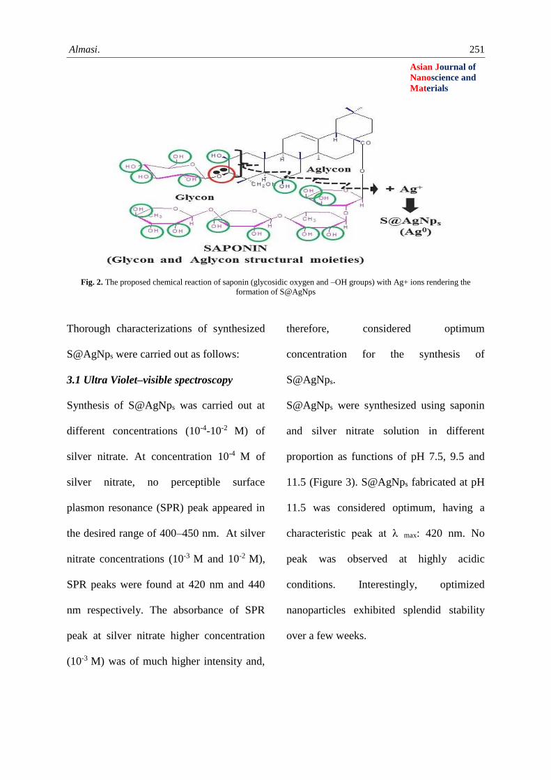
At pH, 11.5 saponin started reducing silver
nitrate to nano silver (S@AgNps). Saponin
consists of structurally-related steroid or
triterpenoid aglycone (sapogenin) linked to
sugar moieties through glycosidic linkage.
The oxygen atom of a glycosidic linkage
having a lone pair of electrons is likely to
reduce Ag+ ions into Ag0, forming
S@AgNps. Further, hydroxyl groups of the
sugar moieties and sapogenin might also
play a favorable role in the reduction of Ag+
ions [30]. The oxidized saponin being
electron deficient in nature may impart free
radical scavenging abilities, acting as an
anti-oxidant. Thus saponin, not only forms
silver nanoparticles but also get involved in
its simultaneous loading or capping on
silver nanoparticles. This medicated coating
on the freshly generated silver nanoparticles
and enhance pharmacological efficacy
provides robust shielding from
aggregations, keeping them in nano state
(stability). Hence, the overall strong
synergistic reduction potential of saponin
(Figure 2) is capable of forming silver
nanoparticles, acting as reducing and
capping agents.
Almasi. 251
Asian Journal of
Nanoscience and
Materials
Fig. 2. The proposed chemical reaction of saponin (glycosidic oxygen and –OH groups) with Ag+ ions rendering the
formation of S@AgNps
Thorough characterizations of synthesized
S@AgNps were carried out as follows:
3.1 Ultra Violet–visible spectroscopy
Synthesis of S@AgNps was carried out at
different concentrations (10-4-10-2 M) of
silver nitrate. At concentration 10-4 M of
silver nitrate, no perceptible surface
plasmon resonance (SPR) peak appeared in
the desired range of 400–450 nm. At silver
nitrate concentrations (10-3 M and 10-2 M),
SPR peaks were found at 420 nm and 440
nm respectively. The absorbance of SPR
peak at silver nitrate higher concentration
(10-3 M) was of much higher intensity and,
therefore, considered optimum
concentration for the synthesis of
S@AgNps.
S@AgNps were synthesized using saponin
and silver nitrate solution in different
proportion as functions of pH 7.5, 9.5 and
11.5 (Figure 3). S@AgNps fabricated at pH
11.5 was considered optimum, having a
characteristic peak at λ max: 420 nm. No
peak was observed at highly acidic
conditions. Interestingly, optimized
nanoparticles exhibited splendid stability
over a few weeks.

252 Almasi.
Asian Journal of
Nanoscience and
Materials
Fig. 3. UV–vis spectra of saponin loaded silver nanoparticles at different pH.
3.2 X-ray diffraction
Bragg reflection peaks for S@AgNps
appeared at the position of 2θ at 38.11° and
44.27°. The fact indexed to (111) and (200)
lattice planes of the face-centered cubic
structure of silver nanoparticles (JCPDS file
04-0783). The intensity of the diffraction
peak corresponding to (200)
crystallographic planes was found to lower
than (111). The fact established lattice plane
(111) as a transcendent crystallographic
plane (Figure 4). The (111) plane is known
to be more reactive because of its high atom
density [31]. Some unassigned peaks were
observed which were due to the
crystallization of bioorganic phase [32].
Fig. 4. XRD of saponin loaded silver nanoparticles.

Almasi. 253
Asian Journal of
Nanoscience and
Materials
3.3 FE-SEM, TEM, and EDX studies
FE-SEM image (Figure 5) acquired from
drop coated films of nanoparticles indicated
polydispersed spherical shaped surface
morphology of S@AgNps. TEM analysis
confirmed the formation of S@AgNps in the
size range 20-48 nm. (Figure 6). The
morphology of S@AgNps was almost
spherical in shape with a light-colored
coating. The desirable signals of silver
metal were found at 3KeV in EDX spectra
of the saponin loaded silver nanoparticles
(Figure 7). The presence of other peaks may
be ascribed to Cu and C which is an artifact
of the Cu-grid on which the sample was
coated. The peaks of O and N might have
initiated from the biomolecules that are
adhered to the surface nanoparticles.
Fig. 5. FE-SEM image of saponin loaded silver nanoparticles.
Fig. 6. TEM image of saponin loaded silver nanoparticles.

254 Almasi.
Asian Journal of
Nanoscience and
Materials
Fig. 7. EDX record of saponin loaded silver nanoparticles.
3.4 Dynamic light Scattering
Average hydrodynamic size of S@AgNps
was 52.40 nm in an asymmetric distribution
between of 13 to 200 nm (Figure 8). The
high intensity distribution of particle size
highlighted the fact that fabricated
nanoparticles are in quite lower range. Zeta
potential of the fabricated S@AgNps,
determined in water as a dispersant, was -
33.5 mV (Figure 9). The high negative
charge constitutes a repulsive barrier that
physically separates the nanoparticles,
avoiding aggregation [33]. The fact finds
support from TEM images of nanoparticles
exhibiting light-colored coating which
might be due to the presence of saponin
around the nanoparticles, acting as a
protective barrier against aggregation [34]
Fig. 8. Size distribution of saponin loaded silver nanoparticles.

Almasi. 255
Asian Journal of
Nanoscience and
Materials
Fig. 9. Zeta potential of saponin loaded silver nanoparticles.
3.5 FTIR analysis
FTIR spectra of native saponin and saponin
loaded silver nanoparticles were recorded
(Figure 10). Almost overlapping of the
peaks, depicting the structure of saponin (-
OH, -C-H-, -C=C- and -C-O-C-) indicates
the presence of residue saponin on the
surface of the S@AgNps. Consequently, the
occurrence of these peaks in the FTIR
spectra of the S@AgNps indicates the dual
role of saponin as reducing and stabilizing
agents.
Fig. 10. FTIR spectra of saponin and saponin loaded silver nanoparticles.
3.6 Anti-inflammatory in -vivo bioassay:
Carrageenan-induced hind paw edema
Toxicity of S@AgNps was monitored after
one-time exposure of the dose ranging
(1000- 45000 mg/kg/bw). LD50 (table 1)
was found (3000mg/kg bw).

256 Almasi.
Asian Journal of
Nanoscience and
Materials
Table1. Mortality percentage of S@AgNps in treatment groups.
S.No. Dose
(mg/kg/bw)
Time
Interval
Mortality
(%)
0-6h 6-24h 24-48 h 48-72 h
1. 1000 - - - - Nil
2. 1500 - - - - Nil
3. 2000 - - - - Nil
4. 2500 - - - - Nil
5. 3000 - 2 1 - 50.00
6. 3500 - 2 2 - 66.60
7. 4000 - 3 2 - 83.30
8. 4500 - 3 3 - 100.00
In- vivo anti-inflammatory bioassay was
carried out in carrageenan induced paw
edema volume of Swiss albino mice by
giving the oral administration of reference
drug and various treatments (Figure 11). A
perusal of figure exhibited statistically
(P<0.001) significant decreasing trend in
reduction in paw edema volume in all the
treated groups. However, in the control
group it increases initially then starts
decreasing. The trend may be ascribed to
the development of natural immunity with
time.
Fig. 11. Decreasing trend in reduction in paw edema volume in various treated groups
(mean ± standard deviation, n = 6).
Percentage inhibition of inflammation was
calculated from a reduction in paw edema
values in each case and presented in table 2.

Almasi. 257
Asian Journal of
Nanoscience and
Materials
Table 2. Percentage inhibition of inflammation in mice: Carrageenan induce hind paw edema
A time-dependent (0.5-4 h) increase in
percentage inhibition of inflammation was
noticed in all the treated groups. Anti-
inflammatory bio-efficacy (46.84%) at
optimized dose (15 mg/kg/bw) of native
seed extract was increased to 56.10% by
bio-active principle (saponin) at
considerable low dose (1.5 mg/kg/bw). The
fact can be ascribed to the role of bio-active
principle in the enhancement of the target
bio-efficacy. Further, increase in the
percentage inhibition (70.99%) was induced
by bio-active principle loaded (saponin)
silver nanoparticles at the same dose (1.5
mg/kg/bw). Thus overall enhancement in
the anti-inflammatory bio-efficacy
compared to the saponin treatment was
found (26.54%) which can be ascribed to
the combination of the contribution of the
use of bio-active principle and green
nanotech enhancement.
A well-defined mechanism of anti-
inflammation of metal nanoparticles has not
been reported. In general, inflammation has
been described as a biphasic process
induced by pathogens and tissue injury.
Initially, inflammatory signals are
recognized by toll-like receptors which in
turn activate myeloid differentiation primary
gene 88 [35]. These genes cause
phosphorylation of IkB protein resulting
into the release and translocation of NF-kβ
Treatment Dose Percentage inhibition of
Inflammation
0.5 h 1 h 2 h 4 h
Group II Diclofenac sodium 50mg/kg/bw 42.26 53.88 76.19 76.42
Group III Seed extract 5mg/kg/bw 10.70 19.42 35.82 35.98
Group IV Seed extract 10mg/kg/bw 13.84 22.05 40.29 40.88
Group V Seed extract 15mg/kg/bw 20.00 30.88 46.53 46.84
Group VI Seed extract 20mg/kg/bw 21.50 31.82 47.56 47.98
Group VII Saponin 1.0mg/kg/bw 24.23 35.23 50.23 52.23
Group VIII Saponin 1.5mg/kg/bw 27.69 38.23 55.22 56.10
Group IX Saponin 3.0mg/kg/bw 27.99 38.44 55.68 56.48
Group X S@AgNps 1.5mg/kg/bw 38.74 48.28 70.76 70.99

258 Almasi.
Asian Journal of
Nanoscience and
Materials
protein into the nucleus. Transcription is up-
regulated causing the binding of NF-kβ
protein to the inflammatory gene. This
binding stimulates the various inflammatory
promoting factors (cytokines, Interleukin,
IFN–γ: interferon-gamma, TNF α: tumor
necrosis factor-α, and cycloxygenase-2
enzymes) [36,37]. Therefore, control of
release proinflammatory factors finally
inhibits inflammation via down regulation
of COX-2 enzyme and NF-kβ protein.
An attempt has been made to explain the
observed incremental enhancement in anti-
inflammatory bio-efficacy induced by bio-
active principle (saponin) and saponin
loaded silver nanoparticles. The present
study reports the anti-inflammatory activity
of saponin and S@AgNps in terms of
percentage inhibition 56.10% and 70.99%
respectively. Histological investigations of
the saponin and S@AgNps treated groups
provide support to the incremental
improvement in the anti-inflammatory bio-
efficacy. Group I (control) showed damaged
serosa, muscularis layer, villi tips and crypts
(Figure 12 A).
Fig. 12. T.S. of intestine: Control group (A) Saponin treated group (B), S@AgNps treated group (C) at an optimum dose of
1.5mg/kg/bw.
Figure 12 B of group VIII (saponin)
describes comparatively well intact serosa,
muscularis, less inflammation and
fragmented villi. Figure 12 C of group IX
A C B

Almasi. 259
Asian Journal of
Nanoscience and
Materials
(S@AgNps) shows significantly reduced
paw edema with more uniform serosa and
muscularis layer with large numbers of villi.
The fact highlights S@AgNps are better,
safe and non-toxic candidate compared to
the treatment (saponin) for the anti-
inflammatory property.
4. Conclusion
The present work provides confirmatory
scientific evidence for anti-inflammatory
bio-efficacy in the seeds of the plant M.
longifolia. The observed bio-efficacy has
been assigned to the family of four
saponins, explored in the seed extract of the
target plant. The synergistic reduction
potential of the saponin of the plant has
been found strong enough for the synthesis
of S@AgNps in a simple and efficient green
method. Promising statistically significant
enhancement in the anti-inflammatory bio-
efficacy has been successfully induced
S@AgNps. The role of the combination of
bio-active principle (saponin) and nano
sizing in the enhancement in the target bio-
efficacy has been assigned to the use of bio-
active principle, high surface area to volume
ratio, reasonable stability, bio-compatibility
and astonishing optical properties related to
surface plasmon resonance. Overall,
phenomenon allows accumulation and
penetration of nano drug into living tissues
comparatively deeper. Anti-inflammatory
properties of silver also add to the
enhancement in the inflammatory property.
Thus, S@AgNps prepared from the seed
extract of the plant M. longifolia seem to be
an ideal candidate for the development of
alternative and complimentary herbal
nanomedicine for anti-inflammation.
Conflict of interest
On behalf of all authors, the corresponding
author states that there is no conflict of
interest.
Acknowledgments
This work was supported by the University
Grants Commission (UGC), New Delhi
(India) vide fellowship No.ET/UGC Res.
260 Almasi.
Asian Journal of
Nanoscience and
Materials
Fellowship/2136. Authors are thankful to
Prof. P.K. Kalra, Director, and Prof. Sahab
Dass, Head, Department of Chemistry,
Dayalbagh Educational Institute Agra for
extending all the necessary facilities and
motivation to carry out the research.
References
1. Cornell, R. M., & Schwertmann, U.
(2003) The Iron Oxides : Structure,
Properties, Reaction, Occurrences
and Uses. WILEY-VCH Verlag
GmbH Co. John Wiley & Sons.
2. Yu, S., & Chow, G. M. 2004. J.
Mater. Chem., 14: 2781–2786.
3. Tuutijärvi, T., Lu, J., Sillanpää, M.,
& Chen, G. (2009) J. Hazard.
Mater., 166: 1415–1420.
4. Cui, H., Liu, Y., & Ren, W. (2013).
Adv. Powder Technol., 24: 93–97.
5. Miguel, O. B., Morales, M. P.,
Serna, C. J., & Veintemillas-
Verdaguer, S. (2002) IEEE Trans.
Magn., 38: 2616–2618.
6. taeghwan Hyeon, Su Seong Lee,
Jongnam Park, Y. C. and H. B. N.
(2001) J. Ameriacan Chem. Soc.,
123: 12789–12801.
7. Asuha, S., Zhao, S., Wu, H. Y.,
Song, L., & Tegus, O. (2009) J.
Alloys Compd., 472: L23–L25.
8. Islam, M. S., Kurawaki, J.,
Kusumoto, Y., Abdulla-Al-Mamun,
M., & Mukhlish, M. Z. Bin. 2011. J.
Sci. Res., 4: 99.
9. Salazar-Alvarez, G., Muhammed,
M., & Zagorodni, A. A. (2006)
Chem. Eng. Sci., 61: 4625–4633.
10. Randrianantoandro, N., Mercier, A.
M., Hervieu, M., & Grenèche, J. M.
(2001) Mater. Lett., 47: 150–158.
11. Strobel, R., & Pratsinis, S. E. (2009)
Adv. Powder Technol., 20: 190–194.
12. Shafi, K. V. P. M., Ulman, A., Dyal,
A., Yan, X., Yang, N. L., Estournès,
C., Fournès, L., Wattiaux, A.,
White, H., & Rafailovich, M.(2002)
Chem. Mater., 14: 1778–1787.
13. Liu, T., Guo, L., Tao, Y., Wang, Y.
B., & Wang, W. D.(1999)
Nanostructured Mater., 11: 487–492.
14. Cao, S.-W., Zhu, Y.-J., & Zeng, Y.-
P. (2009) J. Magn. Magn. Mater.,
321: 3057–3060.
15. Iwasaki, T., Kosaka, K., Watano, S.,
Yanagida, T., & Kawai, T. (2010)
Mater. Res. Bull., 45: 481–485.
16. Bacri, J. C., Perzynski, R., Salin, D.,
Cabuil, V., & Massart, R. (1986) J.
Magn. Magn. Mater., 62: 36–46.
17. Kumar, A. P., Kumar, B. P., Kumar,
A. B. V. K., Huy, B. T., & Lee, Y. I.
(2013) Appl. Surf. Sci., 265: 500–509.
18. Kumar, A. P., Baek, M., Sridhar, C.,
Kumar, B. P., & Lee, Y. (2014)
Bull. Korean Chem. Soc., 35: 1144–1148.
19. Kumar, A. P., Baek, M., Sridhar, C.,
Kumar, B. P., & Lee, Y. (2014)
Bull. Korean Chem. Soc., 35: 1144–1148.
20. C, F. De, Cecilia, M., Souza, B. V.
De, Frugulhetti, I. I. P., Castro, H.
C., Souza, S. L. D. O., Moreno, T.,
Souza, L. De, Rodrigues, D. Q.,
Souza, A. M. T., Abreu, P. A.,
Passamani, F., Rodrigues, C. R., &
Ferreira, V. F. (2009) Eur. J. Med.
Chem., 44: 373–383.
21. Genin, M. J., Allwine, D. a,
Anderson, D. J., Barbachyn, M. R.,
Emmert, D. E., Garmon, S. a,
Graber, D. R., Grega, K. C., Hester,
J. B., Hutchinson, D. K., Morris, J.,

Almasi. 261
Asian Journal of
Nanoscience and
Materials
Reischer, R. J., Ford, C. W.,
Zurenko, G. E., Hamel, J. C.,
Schaadt, R. D., Stapert, D., & Yagi,
B. H. (2000) J. Med. Chem., 43:
953–970.
22. Buckle, D. R., Rockell, C. J., Smith,
H., & Spicer, B. A. 1984, 27: 223–227.
23. Alexacou, K.-M., Hayes, J. M.,
Tiraidis, C., Zographos, S. E.,
Leonidas, D. D., Chrysina, E. D.,
Archontis, G., Oikonomakos, N. G.,
Paul, J. V, Varghese, B., &
Loganathan, D. (2008) Proteins, 71:
1307–1323.
24. Brockunier, L. L., Parmee, E. R.,
Ok, H. O., Candelore, M. R.,
Cascieri, M. A., Colwell, L. F.,
Deng, L., Feeney, W. P., Forrest, M.
J., Hom, G. J., MacIntyre, D. E.,
Tota, L., Wyvratt, M. J., Fisher, M.
H., & Weber, A. E. (2000)
Bioorganic Med. Chem. Lett., 10:
2111–2114.
25. Fan, W.: Comprehensive
Heterocyclic Chem. II, vol. 4,
Pergamon, Oxford, UK (1996).
26. Chem, A., & Ed, I. (2002) Angew.
Chem. Int. Ed., 41: 2596–2599.
27. Tornøe, C. W., Christensen, C., &
Meldal, M. (2002) J. Org. Chem.,
67: 3057–64.
28. Gian Cesare Tron, Tracey Pirali,
Richard A. Billington, P. L. C., &
Giovanni Sorba, A. A. G. (2012)
Med. Res. Rev., 29: 1292–1327.
29. Steenackers, H., Ermolat’ev, D.,
Trang, T. T. T., Savalia, B., Sharma,
U. K., De Weerdt, A., Shah, A.,
Vanderleyden, J., & Van der
Eycken, E. V. (2014) Org. Biomol.
Chem., 12: 3671–3678.
30. Kovács, S., Zih-Perényi, K.,
Révész, Á., & Novák, Z. (2012)
Synth., 44: 3722–3730.
31. Wang, D., Salmon, L., Ruiz, J., &
Astruc, D. (2013) Chem. Commun.,
49: 6956.
32. Kale, S. R., Kahandal, S. S.,
Gawande, M. B., & Jayaram, R. V.
(2013) RSC Adv., 3: 8184.
33. Grigorie, A. C., Muntean, C., &
Stefanescu, M. (2015) Thermochim.
Acta, 621: 61–67.
34. Stoia, M., Istratie, R., & Păcurariu,
C. (2016) J. Therm. Anal. Calorim.,
125: 1185–1198.
How to cite this manuscript: Mukti Sharma, Saurabh Yadav, Man Mohan Srivastava,
Narayanan Ganesh, Shalini Srivastava,*. Promising anti-inflammatory bio-efficacy of
saponin loaded silver nanoparticles prepared from the plant Madhuca longifolia. Asian
Journal of Nanoscience and Materials, 2018, 1(4) , 244-261.