
Full mouth rehabilitation with implants case report in india
Full Mouth Rehabilitation of a Bruxer with Severely Worn Dentition Using All Ceramic
Zirconia Crowns – A Case Report Ashish.R.Jain aMDS MD.ACU.VARMA, Sindhu .P b BDS
a Research Scholar, Reader Department of Prosthodontics, Saveetha Dental College and Hospitals, Saveetha University, Chennai, India.
b CRRI, Department of Prosthodontics, Tagore Dental College and Hospital ,Chennai, India.
Abstract: Excessive tooth wear due to parafunctional habits like bruxism requires clinical attention. Bruxism results in severely worn dentition and often loss in Vertical Dimension of Occlusion (VDO). In this case, patient was a bruxer with severely worn dentition. VDO was increased using an occlusal splint and then a fixed interim prosthesis was given. Patient was comfortable with the newly restored VDO. Deciding on the amount of VDO increase is the critical part of treatment planning. Definitive full mouth rehabilitation using all ceramic zirconia crowns was performed. A review after one year revealed a well functional and esthetic prosthesis.
Keywords: Parafunctional habit, bruxism, severely worn dentition, Vertical Dimension of Occlusion, full mouth rehabilitation, all ceramic zirconia crown.
INTRODUCTION: Occlusal and incisal surfaces of teeth undergo wear over lifetime which is a normal physiological process and does not require any treatment. Severe wear of teeth needs clinical attention [1-3]. Excessive tooth wear can be due to parafunctional habits like bruxism, occlusal pre-maturities, dietary habits, attrition, abrasion or erosion [4-6]. Bruxism refers to a condition characterized by excessive grinding or clenching of the teeth. Bruxism can be classified into nocturnal and daytime bruxism. The etiology of bruxism includes anxiety, stress and occlusal disharmony. Clinically bruxism can be identified by the presence of occlusal wear, wear facets, muscle fatigue and temporomandibular joint discomfort [7-11].
VERTICAL DIMENSION OF OCCLUSION: Generally vertical dimension of occlusion (VDO) is maintained by tooth eruption and alveolar bone growth [1]. The adaptive changes of the alveolar bone compensates for the occlusal wear. However in cases of excessive occlusal wear, tooth eruption and alveolar bone remodeling cannot maintain the vertical dimension of occlusion (VDO). Hence VDO is altered in bruxers [2, 5]. Alteration in vertical dimension of occlusion must be evaluated in the clinic and careful approach is required while increasing the vertical dimension of occlusion. Excessive increase of VDO will overload the teeth and cause damage to the dentition [1, 6]. Deciding on the amount of VDO increase is the critical part of treatment planning.
TREATMENT PLANNING: Severely worn dentition presents a challenge to the clinicians due to insufficient space for restoration. Study cast and diagnostic wax-up will help in determining the need to alter vertical dimension of occlusion [7]. Any disorder in the muscles or temporomandibular joint must be
eliminated prior to the beginning of the treatment. The following factors must be assessed for VDO alteration: Phonetics Interocclusal Rest Space Facial appearance Loss of posterior support in occlusionTreatment planning should be based on the patientocclusion, vertical dimension of occlusion, material ofrestoration, position of Condyle and inter-occlusal restspace [2, 8]. A systematic approach in full mouthrehabilitation of a severely worn dentition will lead to asuccessful outcome.
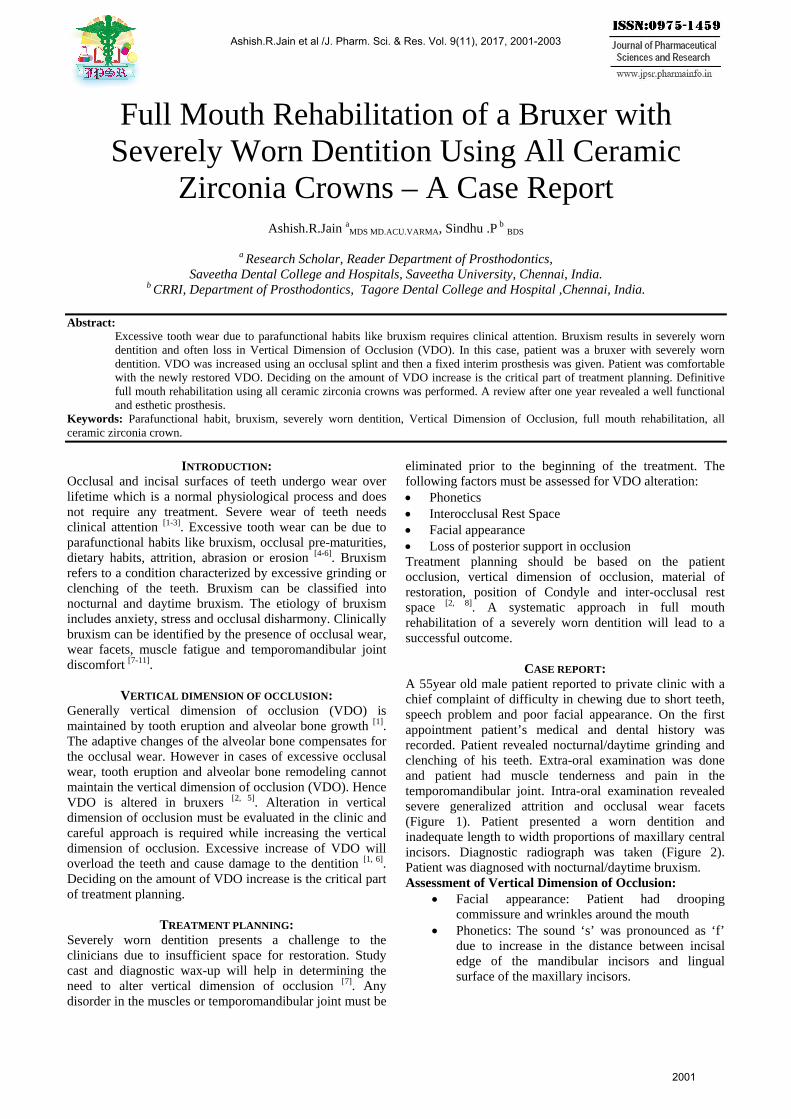
CASE REPORT: A 55year old male patient reported to private clinic with a chief complaint of difficulty in chewing due to short teeth, speech problem and poor facial appearance. On the first appointment patient’s medical and dental history was recorded. Patient revealed nocturnal/daytime grinding and clenching of his teeth. Extra-oral examination was done and patient had muscle tenderness and pain in the temporomandibular joint. Intra-oral examination revealed severe generalized attrition and occlusal wear facets (Figure 1). Patient presented a worn dentition and inadequate length to width proportions of maxillary central incisors. Diagnostic radiograph was taken (Figure 2). Patient was diagnosed with nocturnal/daytime bruxism. Assessment of Vertical Dimension of Occlusion:
Facial appearance: Patient had droopingcommissure and wrinkles around the mouth
Phonetics: The sound ‘s’ was pronounced as ‘f’due to increase in the distance between incisaledge of the mandibular incisors and lingualsurface of the maxillary incisors.
Ashish.R.Jain et al /J. Pharm. Sci. & Res. Vol. 9(11), 2017, 2001-2003
2001
Interocclusal Rest Space: Interocclusal Rest Space was found to be around 6-8mm which is more than the normal value (2-4mm).
Vertical Dimension of Occlusion was decided to be increased after the above examination. The following treatment option was explained to the patient for full mouth rehabilitation. Increasing the Vertical Dimension of Occlusion (VDO) using an occlusal overlay splint followed by a fixed interim prosthesis. Finally full mouth rehabilitation with Zirconia crowns at restored Vertical Dimension of Occlusion.
TREATMENT: 1. Study cast were fabricated and were articulated in a
semi-adjustable articulator using facebow and interocclusal record.
2. Extraction of 25, 26, 46 and 47 was planned, as the radiograph revealed poor prognosis and removal of porcelain fused to metal crowns in relation to 14, 15, 16 and 17 as they were poorly designed and fabricated.
3. The Vertical Dimension of Occlusion was increased by 4mm in the anterior region and posterior region.
4. An occlusal overlay splint was fabricated that provides bilateral contact in centric relation. Patient was monitored for one month. Patient did not experience any muscle tenderness or discomfort in the temporomandibular joint. He was comfortable at the new restored VDO.
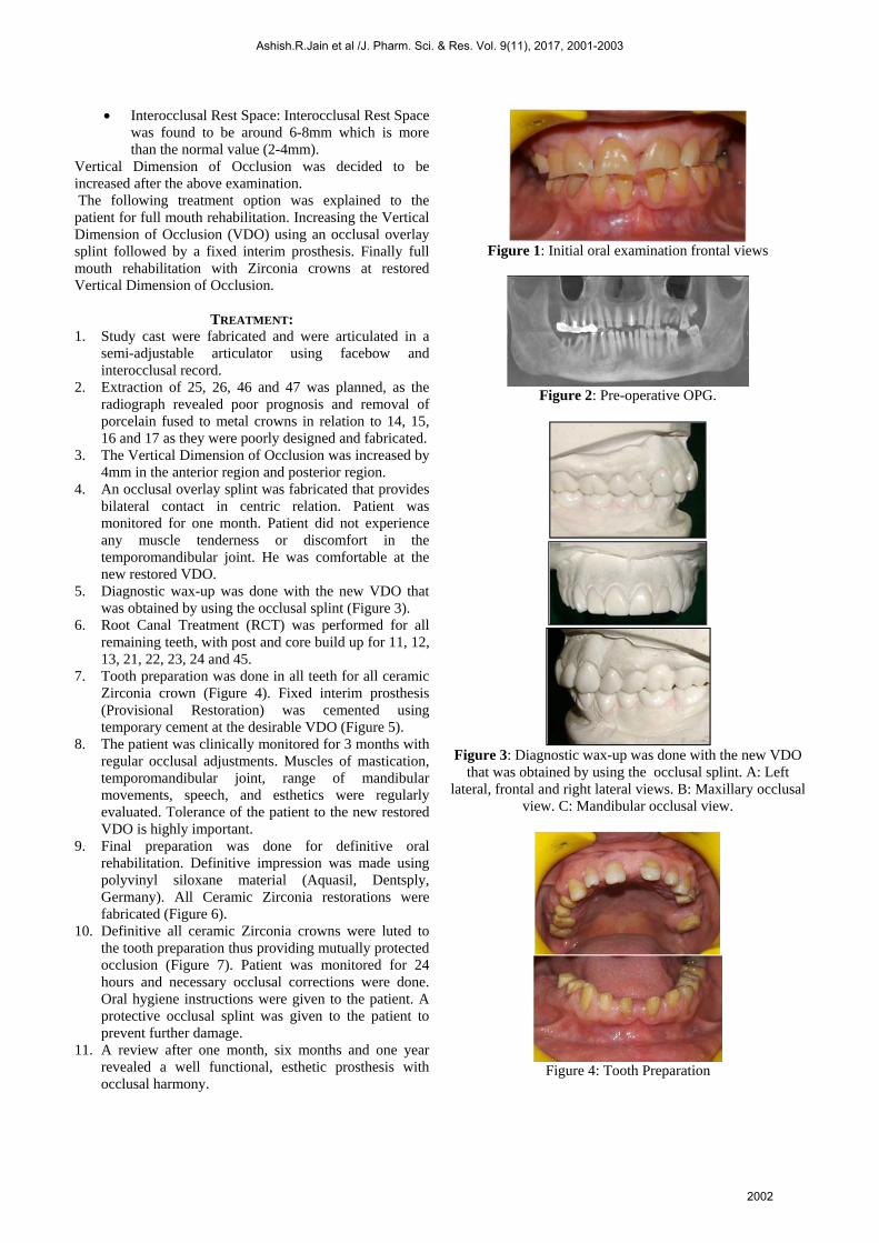
5. Diagnostic wax-up was done with the new VDO that was obtained by using the occlusal splint (Figure 3).
6. Root Canal Treatment (RCT) was performed for all remaining teeth, with post and core build up for 11, 12, 13, 21, 22, 23, 24 and 45.
7. Tooth preparation was done in all teeth for all ceramic Zirconia crown (Figure 4). Fixed interim prosthesis (Provisional Restoration) was cemented using temporary cement at the desirable VDO (Figure 5).
8. The patient was clinically monitored for 3 months with regular occlusal adjustments. Muscles of mastication, temporomandibular joint, range of mandibular movements, speech, and esthetics were regularly evaluated. Tolerance of the patient to the new restored VDO is highly important.
9. Final preparation was done for definitive oral rehabilitation. Definitive impression was made using polyvinyl siloxane material (Aquasil, Dentsply, Germany). All Ceramic Zirconia restorations were fabricated (Figure 6).
10. Definitive all ceramic Zirconia crowns were luted to the tooth preparation thus providing mutually protected occlusion (Figure 7). Patient was monitored for 24 hours and necessary occlusal corrections were done. Oral hygiene instructions were given to the patient. A protective occlusal splint was given to the patient to prevent further damage.
11. A review after one month, six months and one year revealed a well functional, esthetic prosthesis with occlusal harmony.
Figure 1: Initial oral examination frontal views
Figure 2: Pre-operative OPG.
Figure 3: Diagnostic wax-up was done with the new VDO
that was obtained by using the occlusal splint. A: Left lateral, frontal and right lateral views. B: Maxillary occlusal
view. C: Mandibular occlusal view.
Figure 4: Tooth Preparation
Ashish.R.Jain et al /J. Pharm. Sci. & Res. Vol. 9(11), 2017, 2001-2003
2002
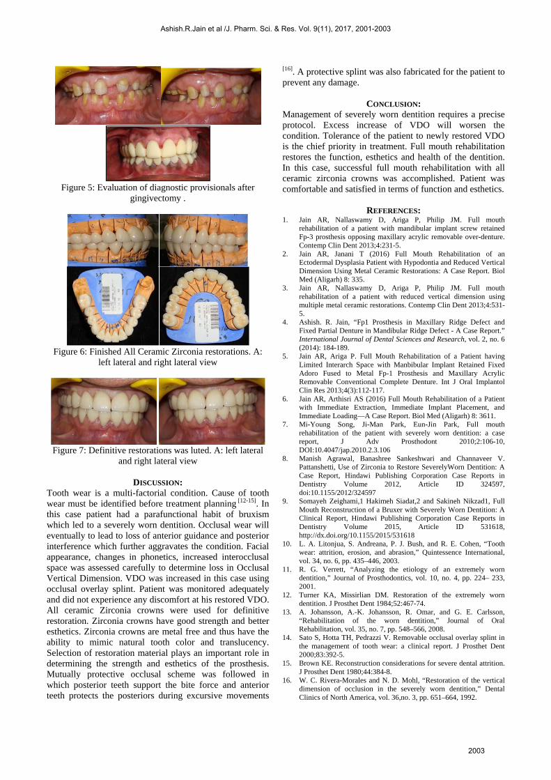
Figure 5: Evaluation of diagnostic provisionals after gingivectomy .
Figure 6: Finished All Ceramic Zirconia restorations. A: left lateral and right lateral view
Figure 7: Definitive restorations was luted. A: left lateral and right lateral view
DISCUSSION: Tooth wear is a multi-factorial condition. Cause of tooth wear must be identified before treatment planning [12-15]. In this case patient had a parafunctional habit of bruxism which led to a severely worn dentition. Occlusal wear will eventually to lead to loss of anterior guidance and posterior interference which further aggravates the condition. Facial appearance, changes in phonetics, increased interocclusal space was assessed carefully to determine loss in Occlusal Vertical Dimension. VDO was increased in this case using occlusal overlay splint. Patient was monitored adequately and did not experience any discomfort at his restored VDO. All ceramic Zirconia crowns were used for definitive restoration. Zirconia crowns have good strength and better esthetics. Zirconia crowns are metal free and thus have the ability to mimic natural tooth color and translucency. Selection of restoration material plays an important role in determining the strength and esthetics of the prosthesis. Mutually protective occlusal scheme was followed in which posterior teeth support the bite force and anterior teeth protects the posteriors during excursive movements
[16]. A protective splint was also fabricated for the patient to prevent any damage.
CONCLUSION: Management of severely worn dentition requires a precise protocol. Excess increase of VDO will worsen the condition. Tolerance of the patient to newly restored VDO is the chief priority in treatment. Full mouth rehabilitation restores the function, esthetics and health of the dentition. In this case, successful full mouth rehabilitation with all ceramic zirconia crowns was accomplished. Patient was comfortable and satisfied in terms of function and esthetics.
REFERENCES: 1. Jain AR, Nallaswamy D, Ariga P, Philip JM. Full mouth
rehabilitation of a patient with mandibular implant screw retainedFp-3 prosthesis opposing maxillary acrylic removable over-denture.Contemp Clin Dent 2013;4:231-5.
2. Jain AR, Janani T (2016) Full Mouth Rehabilitation of anEctodermal Dysplasia Patient with Hypodontia and Reduced VerticalDimension Using Metal Ceramic Restorations: A Case Report. BiolMed (Aligarh) 8: 335.
3. Jain AR, Nallaswamy D, Ariga P, Philip JM. Full mouthrehabilitation of a patient with reduced vertical dimension usingmultiple metal ceramic restorations. Contemp Clin Dent 2013;4:531-5.
4. Ashish. R. Jain, “Fp1 Prosthesis in Maxillary Ridge Defect andFixed Partial Denture in Mandibular Ridge Defect - A Case Report.”International Journal of Dental Sciences and Research, vol. 2, no. 6(2014): 184-189.
5. Jain AR, Ariga P. Full Mouth Rehabilitation of a Patient havingLimited Interarch Space with Manbibular Implant Retained FixedAdoro Fused to Metal Fp-1 Prosthesis and Maxillary AcrylicRemovable Conventional Complete Denture. Int J Oral ImplantolClin Res 2013;4(3):112-117.
6. Jain AR, Arthisri AS (2016) Full Mouth Rehabilitation of a Patientwith Immediate Extraction, Immediate Implant Placement, andImmediate Loading—A Case Report. Biol Med (Aligarh) 8: 3611.
7. Mi-Young Song, Ji-Man Park, Eun-Jin Park, Full mouthrehabilitation of the patient with severely worn dentition: a casereport, J Adv Prosthodont 2010;2:106-10,DOI:10.4047/jap.2010.2.3.106
8. Manish Agrawal, Banashree Sankeshwari and Channaveer V.Pattanshetti, Use of Zirconia to Restore SeverelyWorn Dentition: ACase Report, Hindawi Publishing Corporation Case Reports inDentistry Volume 2012, Article ID 324597,doi:10.1155/2012/324597
9. Somayeh Zeighami,1 Hakimeh Siadat,2 and Sakineh Nikzad1, FullMouth Reconstruction of a Bruxer with Severely Worn Dentition: AClinical Report, Hindawi Publishing Corporation Case Reports inDentistry Volume 2015, Article ID 531618,http://dx.doi.org/10.1155/2015/531618
10. L. A. Litonjua, S. Andreana, P. J. Bush, and R. E. Cohen, “Toothwear: attrition, erosion, and abrasion,” Quintessence International,vol. 34, no. 6, pp. 435–446, 2003.
11. R. G. Verrett, “Analyzing the etiology of an extremely worndentition,” Journal of Prosthodontics, vol. 10, no. 4, pp. 224– 233,2001.
12. Turner KA, Missirlian DM. Restoration of the extremely worndentition. J Prosthet Dent 1984;52:467-74.
13. A. Johansson, A.-K. Johansson, R. Omar, and G. E. Carlsson,“Rehabilitation of the worn dentition,” Journal of OralRehabilitation, vol. 35, no. 7, pp. 548–566, 2008.
14. Sato S, Hotta TH, Pedrazzi V. Removable occlusal overlay splint inthe management of tooth wear: a clinical report. J Prosthet Dent2000;83:392-5.
15. Brown KE. Reconstruction considerations for severe dental attrition.J Prosthet Dent 1980;44:384-8.
16. W. C. Rivera-Morales and N. D. Mohl, “Restoration of the verticaldimension of occlusion in the severely worn dentition,” DentalClinics of North America, vol. 36,no. 3, pp. 651–664, 1992.
Ashish.R.Jain et al /J. Pharm. Sci. & Res. Vol. 9(11), 2017, 2001-2003
2003


