Frontispiece. - link.springer.com978-1-4471-1369-0/1.pdf · Frontispiece. Leonardo da Vinci's...
12
Transcript of Frontispiece. - link.springer.com978-1-4471-1369-0/1.pdf · Frontispiece. Leonardo da Vinci's...


Frontispiece. Leonardo da Vinci's drawing of an embryo in utero (a detail). By gracious permission of Her Majesty The Queen.
N.B. This is one of the earliest illustrations of breech malposition, the foetal posture responsible for pre-natal hip displacement. It is a very accurate record of the left sacro-anterior lie, from the posterior aspect, being the commonest breech position; both hips are
flexed and laterally rotated (see Fig. 4.13, p. 55).

John A. Wilkinson
Congenital Displacement of the Hip Joint
With 168 figures
Springer-Verlag Berlin Heidelberg New York Tokyo

John A. Wilkinson, BSc, MCh, FRCS.
Hunterian Professor, RCS. ; Simpson Smith Prize; Robert Jones Gold Medal and ABC Fellow, BOA; Senior Consultant Orthopaedic Surgeon and Clinical Teacher Southampton University Hospitals, The Lord Mayor Treloar Orthopaedic Hospital, Alton, and the Channel Islands
ISBN-13:978-1-4471-1371-3 e-ISBN-13:978-1-4471-1369-0 001: 10.1007/978-1-4471-1369-0
library of Congress Cataloging in Publication Data Wilkinson, John Arthur, 1925- Congenital displacement of the hip joint. Includes bibliographies and index. 1. Hip joint-Dislocation, Congenital. I. Title. [DNLM: 1. Hip Dislocation, Congenital. WE 860 W686c] RD772.W55 1985 617'.376 85-2584 ISBN-13:978-1-4471-1371-3 (U.S.)
This work is subject to copyright. All rights are reserved, whether the whole or part of the material is concerned, specifically those of translation, reprinting, re-use of illustrations, broadcasting, reproduction by photocopying machine or similar means, and storage in data banks. Under §54 of the German Copyright Law where copies are made for other than private use, a fee is payable to 'Verwertungsgesellschaft Wort' Munich.
© Springer-Verlag Berlin Heidelberg 1985 Softcover reprint of the hardcover 1st edition 1985
The use of registered names, trademarks etc. in this publication does not imply, even in the absence of a specific statement, that such names are exempt from the relevant laws and regulations and therefore free for general use.
Product Liability: The publisher can give no guarantee for information about drug dosage and application thereof contained in this book. In every individual case the respective user must check its accuracy by consulting other pharmaceutical literature.
2128/3916543210

Acknowledgements
The preparation of a monograph involves the labours, co-operation and encouragement of many individuals.
Vernon Hart (1948)
It is my pleasure and privilege to acknowledge the efforts of many who have consciously and unconsciously contributed to the contents of this book. I am indebted to my two secretaries, Eileen Sizer and Christine Clearkin, who have worked so hard on the script, never complaining about their arduous tasks. The diagrams and photographs have been produced by the Department of Illustration, Southampton University Hospital, and I am more than grateful to Mr. David Whitcher for his advice and help, and to Mr. Peter Jack for his artistic impressions. Mrs. Janice Mayhew, the Librarian of the Wessex Regional Orthopaedic Library, has provided many of the references included in the text.
I am also indebted to the parents who have entrusted me with the care of their children, and to the nurses, physiotherapists and radiographers who have contributed to the clinical work, which has been performed at the Southampton University Hospital and the Lord Mayor Treloar Orthopaedic Hospital, Alton. I thank my colleagues who have referred their cases, and the success of my surgery has been entirely dependent upon Dr. Peter Baxter, my Senior Anaesthetist, for without his skill nothing could have been achieved.
Finally, I must address the words of John Hunter (1786) to my mentors Mr. A.a. Parker, Mr. David Trevor, Sir Herbert Seddon, Sir Denis Browne, Mr. Eric Lloyd, and Mr. George Lloyd-Roberts:
As the following observations were made in the course of those pursuits in which you have so warmly interested yourselves and promoted with the most friendly assistance, I should be wanting in gratitude were I not to address them to you as a public testimony of the friendship and esteem with which I am, dear sirs, you obliged and very humble servant.
Southampton, January 1985 John A. Wilkinson
Preface
If I have seen further, it is by standing on ye shoulders of giants. Sir Isaac Newton (1675)
Although congenital displacement of the hip has always been recognised as the commonest pre-natal deformity of the musculoskeletal system found in otherwise normal children, it is surprising to find that much of our present-day understanding concerning its origin and nature has been discovered only in the past 150 years and its successful management eventually emerged during the last 60 years, almost within the professional life time of our more senior colleagues.
Whereas Hippocrates (460-370 B.C.) appeared to recognise patients that had experienced dislocation of their hips in utero and identified them from acquired forms of displacement, according to Severin (1941) it was another 2000 years before Palleta (1820) first recorded a careful description of the deformity based on his observations at an autopsy performed on an II-day-old boy with bilateral dislocations; he concluded that the findings were not caused by injury at birth, but dated from a pre-natal stage. Soon after, Baron Dupuytren (1847) gave an accurate description of congenital dislocation in his contribution entitled "Memoire sur un deplacement original ou congenital de la tete des femurs". He noted the absence of abscesses and fistulae as seen in painful and cruel pathological dislocations, which led him to believe that these congenital displacements were not likely to be the result of foetal disease as the affected babies appeared quite healthy at birth. He concluded that they were probably due to an accidental displacement in utero which appeared to affect girls much more frequently than boys. With amazing foresight, Dupuytren came to the conclusion that these displacements were the result of the thighs being very much bent on the belly, from which it followed that the heads of the thigh bones were continuously pressing against the lower and back part of the capsular ligaments, "a circumstance which though without effect in well-formed individuals might, I apprehend, have an injurious influence in such as are weak or of lax resisting fibre". It is no wonder that Lorenz paid tribute to Dupuytren for setting a scientific landslide in motion (Severin 1941). Yet, knowledge concerning the prevalence of this congenital defOrmity remained fragmentary until Roentgen (1895) discovered the diagnostic value of radiology, and soon after Le Damany and Saigret (1910) published the first detailed study in a normal population.
With regard to treatment, Pravaz (1837) was the first person known for certain to have succeeded in redUCing a congenital dislocation of the hip, but again according to Severin he was unable to retain the femoral head in its new position. Poggi (1888) is thought to be the first person to have performed a successful surgical reduction, retaining the femoral
viii Preface
head by deepening the acetabulum. His techniques were later developed by Lorenz (1896), who undertook a thorough study of the surgical anatomy of the deformation; this led him to develop his closed reduction of congenital dislocations which was referred to as the "bloodless method" by Ludloff (1913). Lorenz was the first to realise that retention of reduction by splinting was just as important as the technique of manipulative reduction. At the same time, Paci described a method of closed reduction by circumduction of the hip, and this appeared to become the more popular method in parts of Europe and England. Whereas many children under 3 years were prevented by such conservative measures from being crippled, the majority of these patients reached hospital after this age and did not respond to this treatment. Their high prevalence of redislocations and stiff hips stimulated a popular return to surgical reduction in the earlier part of this century. Major contributions were made by Hoffa (1895) and Ludloff (1913) in Europe, with Hey Groves (1928) and Fairbank (1930) in Great Britain; whereas in North America, Sherman (1903), Hibbs (1915) and Galloway (1920) were also pioneers of similar techniques. Nathaniel Allison (1928) recorded a detailed account of the history of management in his historical contribution to the Robert Jones Birthday Volume.
Later it became acknowledged generally that the results of both conservative and surgical management could only be improved upon if more cases were treated earlier, and every effort was made to bring this about. Putti (1933) produced another scientific landslide, with his concept of early diagnosis within the first year of life. He described the radiological state of "predislocation" and sincerely believed that every new-born should be subjected to a routine radiological examination. He recommended that treatment should begin at the very moment the deformity is first observed, even if it be on the day of birth, and promised a 95% cure by simply applying his adjustable abduction apparatus.
Marino Ortolani (1948) was Putti's pupil and he took a major step forward in management, by identifying hip instability clinically in the new-born. Although he gave credence to the work of his mentor, Ortolani found that the "scatto" or snapping sign was a more accurate way of diagnosing "predislocation", as compared to radiographs or even arthrography in the first year of life, and this saved exposing many normal children to radiological surveys. He found that there were "embryonary" or complete degrees of dislocation at birth which proved resistant to simple treatment. Such forms were rare compared to the commoner forms of "predislocation" or "preluxation" which were thought to develop into postnatal dislocations, although he recognised that many recovered spontaneously without treatment. Palmen (1961) carried Ortolani's methods to Scandinavia and popularised the vetting of new-born babies for hip instability throughout Sweden. Von Rosen (1962) continued with this work, enthusiastically, and Barlow brought his teaching to Great Britain. They all believed that congenital dislocation of the hip was simply a case of hip instability at the time of birth, and could be treated successfully when splinted within the first 3 days of life. Thus Andren (1961) stated "it is clear that there is hardly any excuse for congenital dislocation of the hip not being recognised and not being effectively treated from birth" .
Against this background of development and modern thinking, I have gained a personal experience in the understanding of the diagnosis and treatment of this condition over the past 2 5 years and my findings are expounded in this monograph. As a young House Surgeon and Registrar in orthopaedic centres of excellence including the Prince of Wales Orthopaedic Hospital, the Royal National Orthopaedic Hospital, and the Hospital for Sick Children in London, I was encouraged by my mentors to discard" cant and dogma", and they stimulated me to think about the fundamental problems involved in the aetiology and management of this congenital deformity, in preference to teaching me their methods and techniques. I was directed to perform laboratory research in order to gain an understanding of the mechanism and the degrees of deformation of the normal hip joint, in immature animals exposed to the aetiological factors of hormonal joint laxity and breech malposition. The soft tissue and bony changes produced in these animals led to a basic understanding of the anatomical changes found in the hip joints of stillborn human foetuses. Thus with an intimate knowledge of the mechanism of intra-uterine hip displacement and the features and degrees of deformation, one was able to diagnose the different degrees of deformity at birth and understand the development of further changes during the first few years of life.
Preface ix
At that time the Ministry of Health initiated the national screening programme (1969), emphasising that "early diagnosis is therefore of greatest importance and in the earliest phases all that is required is to replace the head of the femur in the acetabulum, holding it there until the capsule shrinks and a satisfactory acetabulum forms". Such advice was not found to be fruitful and within a short time the difficulties of early diagnosis and management became apparent (Wilkinson 1972). In spite of a major effort to detect and treat affected new-born in a captive population, the prevalence of persistent hip displacement in the infant population was not reduced. Thus an original programme of treatment was evolved during my subsequent clinical experience in the Southampton University Hospitals and the Lord Mayor Treloar Orthopaedic Hospital, Alton.
Congenital displacement of the hip has been found to be partly the result of genetic and familial attributes combined with hormonal and mechanical intra-uterine factors affecting the near-normally developed foetal hip joint during the last months of pregnancy. The complexity and the degrees of deformation are not consistent, and, because of this, rigid orthodox management is doomed to failure in a large percentage of cases. It has become recognised that the primary deformation is a posterior displacement of the acetabulum affecting its three components, and a retroversion of the femur. This is associated with a recurrent displacement ofthe femoral head, in relation to the acetabulum, producing soft tissue deformation of the capsule and intra-articular structures.
"The term congenital dislocation of the hip which is deeply rooted in the literature does not accurately and comprehensively describe this deformity" (Gill 1948). It might be wiser to choose the term "congenital deformation of the hip" as suggested by Dunn (1976) to be a more accurate description, but this would also embrace such unrelated conditions as congenital coxa vara and other degrees of embryological failure. Therefore, I have chosen to use the terminology, "congenital displacement of the hip jOint," which was first coined by Sayre (1882) and has subsequently found popularity with many writers, including Beckett Howorth (1963), and more recently Edgar Somerville (1982). Its use highlights the primary displacement of the acetabulum on the pelvis, as well as the recurrent displacement of the femoral head and the secondary soft tissue deformation.
It is hoped that the chapters of the monograph will dispel the common belief that all congenital displacements of the hip are responsive to simple treatment at birth, and also prove that the incarcerated limbus is the frequent cause of persistent displacement and iatrogenic deformation. May the evidence supplied reveal the necessity for concentric reduction, attained through excision of the limbus and maintained by the efficiency of the Lorenz splint. Finally the results of treatment will illuminate the potentials of pelvic re-orientation and decompression, compared to the morbidity of femoral realignment, in the surgical attempts to overcome incongruity of the bony components of the deformation.
"Where shall I begin, please your Majesty!" he asked. "Begin at the beginning", the King said gravely, "and go on till you come to the end: then stop".
References
From Alice's Adventures in Wonderland by Lewis Carroll (1832-1898)
Andren L (1961) Aetiology and diagnosis of congenital dislocation of the hip in newborns. Radiologie (Berlin) 1:89-94
Allison N (1928) The open operations for congenital dislocation of the hip. Robert Jones Birthday Volume. Oxford University Press. Oxford
Central Health Services Council (1969) Screening for the Detection of C.D.H. in Infants. Her Majesty's Stationery Office, London
Dupuytren G (1847) Injuries and diseases of bones. Translated for the Sydenham SOciety. London Dunn P (1976) Perinatal observations on the aetiology of congenital dislocation of the hip. Clinical Orthopaedics
119: 11-21 Fairbank T (1930) Congenital dislocation of the hip with special reference to anatomy. Br J Surg 17 :380

x
Galloway HPH (1920) The open operation for congenital dislocation of the hip. J Orthop Surg 2(3 9c) Gill B (1948) Congenital dislocation of the hip. J Bone Joint Surg Editorial Hart VL (1948) Congenital dysplasia of the hip joint and sequelae. Charles E. Thomas, Illinois
References
Hey Groves E (1928) The treatment of congenital dislocation of the hip. Robert Jones Birthday Volume, Oxford Press
Hibbs RA (1915) quoted by Nathanial Allison (1928) The open operations for congenital dislocation of the hip. Robert Jones Birthday Volume, Oxford University Press, Oxford
Hippocrates The genuine works of Hippocrates. Translation from the Greek by Francis Adams, London (1849) Hoffa A (1895) quoted by Nathanial Allison (1928) The open operations for congenital dislocation of the hip.
Robert Jones Birthday Volume, Oxford University Press, Oxford Howorth B (1963) The etiology of congenital dislocation ofthe hip. Clin Orthop 29 : 164 Hunter J (1786) Letter to Sir Joseph Banks, President of the Royal Society Le Damany p, Saiget J (1910) quoted by J.W. Dickson (1969) Pierre Le Damany on congenital dysplasia of the
hip. Proc R SocMed 62 :575 Lorenz A (1896) Zur Prioritiit der unblutigen Reposition der angeborenen Hiiftverrenkung. Wien klin Wchschr
9:658 Ludloff K (1913) The open reduction ofthe congenital hip dislocation by an anterior incision. Am J Orthop Surg
10:438--454 Newton I (1675) Newton to Hook. The correspondence of Isaac Newton, vol 1 (1661-1675). Edited by HW
Turnbull, FRS Ortolani M (1948) La lussazione congenita dell'anca: Nuovi criteri diagnostici e profillattico-correttivi. Bologna,
Cappelli PalmenK (1961) Preluxationofthe hip jOint. Acta Paediatr 50, Suppl129 Poggi A (1888) Contributo alla cura cruenta della lussatione congenita. Arch Ortop p. 105 Pravaz CA (1837) Du traitment de la luxation congenitale. Bull Acad Med 2 : 5 79 Putti V (1933) Early treatment of congenital dislocation of the hip. J Bone Joint Surg 15 : 16 Roentgen WC (1895) Ueber eine neue Art von Strahlen (Vorlaeufige Mitteilung). Sitzungs-Berichte der
Physikalisch-medizinischen Gesellschaft zu Wiirzburg 9: 132-141 Sayre LA (1882) Orthopaedic surgery and diseases of the joints. Appleton-Century, New York Severin E (1941) Contribution to the knowledge of congenital dislocation of the hip jOint. Late results of closed
reduction and arthrographic studies of recent cases. Acta Coo Scand 84, Suppl 63 Sherman H (1903) quoted by Nathanial Allison (1928) The open operations for congenital dislocation of the hip.
Robert Jones Birthday Volume, Oxford University Press, Oxford Somerville EW (1982) Displacement of the hip in childhood. Springer-Verlag, Berlin, Heidelberg, New York Von Rosen S (1962) Diagnosis and treatment of congenital dislocation of the hip joint in the newborn. J Bone
Joint Surg 44B :284 Wilkinson JA (1972) A post-natal survey for congenital displacement of the hip. J Bone Joint Surg 52B:4--49
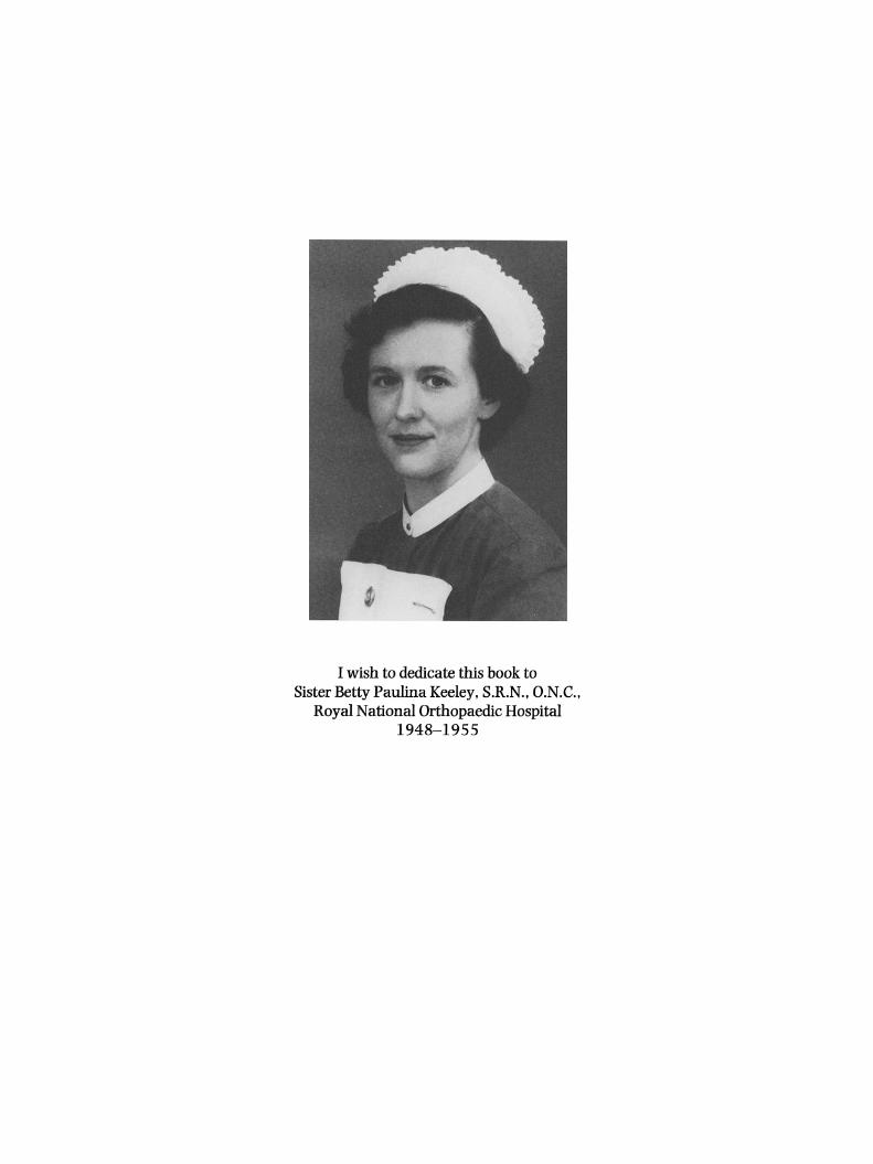
I wish to dedicate this book to Sister Betty Paulina Keeley, S.R.N., O.N.C.,
Royal National Orthopaedic Hospital 1948-1955

Contents
1 Aetiology .. . . . . . . . . . . . . . . . . . . . . . . . . . . . . . . . . . . . . . . . . . . . . . . . . . . . . . . . . . . . . . . . . 1
Normal Development .......................................................... 1 Abnormal Development . . . . . . . . . . . . . . . . . . . . . . . . . . . . . . . . . . . . . . . . . . . . . . . . . . . . . . 6 The Mechanism of CDH ........................................................ 8 Summary ...................................................................... 10 References ...................................................................... 10
2 Experimental Research .................................................... 13
Rotational Splinting. . . . . . . . . . . . . . . . . . . . . . . . . . . . . . . . . . . . . . . . . . . . . . . . . . . . . . . . . . . . 13 Compression Experiments . . . . . . . . . . . . . . . . . . . . . . . . . . . . . . . . . . . . . . . . . . . . . . . . . . . . 16 Experiments Studying the Effects of Breech Malposition and Hormonal Joint
Laxity ....... ...................... ... ................................ ........ 16 Summary ...................................................................... 23 References ...................................................................... 23
3 Anatomy of Congenital Displacement of the Hip .......................... 25
Normal Growth... ... ..... ........... ............... .... ..... ..... ... .......... 26 Abnormal Growth Due to Persistent Breech Malposition . . ... .. .. ... . .... . . . 26 The Development ofCDH ...................................................... 33 Post-natal Development . . . . . . . . . . . . . . . . . . . . . . . . . . . . . . . . . . . . . . . . . . . . . . . . . . . . . . 37 Summary ...................................................................... 42 References ...................................................................... 42
4 Genetic and Environmental Aetiological Factors and Family Studies in the Prevalence of CDH ..................................................... .
Developmental Factors ........................................................ 43 Pre-natal Environmental Factors .............................................. 48 Interaction of Pre-natal Factors .............................................. 49 Risk of CDH .................................................................... 50 Post-natal Environmental Factors . . . . . . . . . . . . . . . . . . . . . . . . . . . . . . . . . . . . . . . . . . . . 50

xiv Contents
Prevalence of CDR . . . . . . . . . . . . . . . . . . . . . . . . . . . . . . . . . . . . . . . . . . . . . . . . . . . . . . . . . . . . 51 Summary ...................................................................... 56 References ...................................................................... 57
5 CDR at Birth and in the First Ten Months of Life: Its Diagnosis and Management . . . . . . . . . . . . . . . . . . . . . . . . . . . . . . . . . . . . . . . . . . . . . . . . . . . . . . . . . . . . . . 59
History of Early Diagnosis...................................................... 59 Post-natal Presentation........................................................ 61 Congenital Displacement at Birth. . . . . . . . . . . . . . . . . . . . . . . . . . . . . . . . . . . . . . . . . . . . . . 62 Neonatal Displacement Between 1 and 10 Months .......................... 70 Management of Normal New-born . .......... .... ............................. 74 Management of New-born with CDR......... .... .... ............. ......... ... 75 Neonatal CDR Response to Treatment ........................................ 81 Summary ...................................................................... 84 References ...................................................................... 85
6 Infantile Displacement ofthe Rip (10 Months to 3 years) ................ 87
Parental Symptoms ............................................................ 87 Clinical Signs .. . . . . . . . . . . . . . . . . . . . . . . . . . . . . . . . . . . . . . . . . . . . . . . . . . . . . . . . . . . . . . . . . 88 Radiological Signs. . . . . . . . . . . . . . . . . . . . . . . . . . . . . . . . . . . . . . . . . . . . . . . . . . . . . . . . . . . . . . 90 Differential Diagnosis .......................................................... 97 Surgical Management . . . . . . . . . . . . . . . . . . . . . . . . . . . . . . . . . . . . . . . . . . . . . . . . . . . . . . . . 101 Summary ...................................................................... 123 References ...................................................................... 123
7 Juvenile Rip Displacement (3t Years to Skeletal Maturity) .............. 127
Missed CDR .................................................................... 127 Failed Infantile CDR. . . . .. .. . .. . .. . .. .. .. . .. .. .... . .. . .. . . .. ... . . .. . . .. . .. .. . .. . 127 Surgical Management . . . . . . . . . . . . . . . . . . . . . . . . . . . . . . . . . . . . . . . . . . . . . . . . . . . . . . . . 130 Summary ...................................................................... 140 References ...................................................................... 140
8 Adult Congenital Acetabular Dysplasia with Subluxation of the Rip Joint 141
Young Adults (15-25 Years) .................................................. 141 Older Age Group (34-45 Years) .............................................. 142 Management . . . . . . . . . . . . . . . . . . . . . . . . . . . . . . . . . . . . . . . . . . . . . . . . . . . . . . . . . . . . . . . . . . 144 Summary ...................................................................... 149 References...................................................................... 150
Subject Index .. . . . . . . . . . . . . . . . . . . . . . . . . . . . . . . . . . . . . . . . . . . . . . . . . . . . . . . . . . . . . . . . . 151