Frontal Brain Asymmetry and Immune Function Brain Asymmetry and Immune Function Duck-Hee Kang School...
10
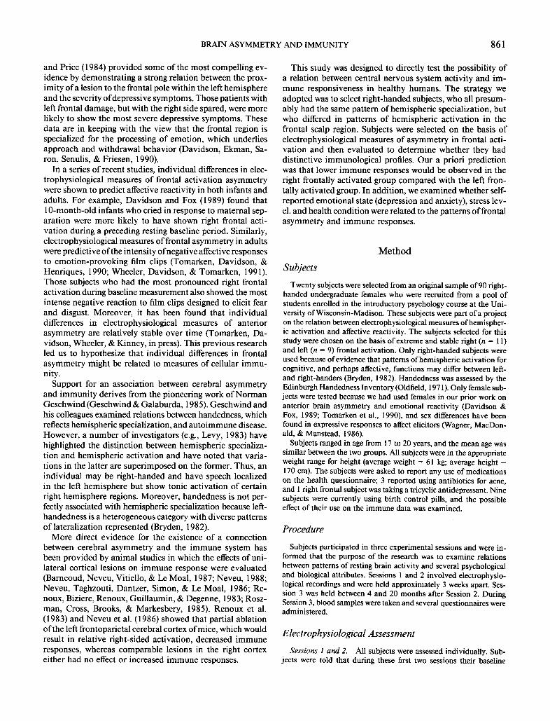
Behavioral Neuroscience Copyright 1991 by the American PsychologicalAssociation, Inc. 1991, Vol. 105, No. 6, 860-869 0735-7044/91/$3.00 Frontal Brain Asymmetry and Immune Function Duck-Hee Kang School of Nursing, University of Wisconsin-Madison Richard J. Davidson, Christopher L. Coe, Robert E. Wheeler, and Andrew J. Tomarken University of Wisconsin-Madison William B. Ershler Department of Medicine, University of Wisconsin-Madison The relation between brain activity and the immune system was evaluated by assessing immune responses in 20 healthy women who manifested extreme differences in the asymmetry of frontal cortex activation. One group showed extreme and stable left frontal activation; the other group showed extreme and stable right frontal activation. As predicted, women with extreme right frontal activation had significantly lower levels of natural killer cell activity (at effector:target cell ratios of 33:1 and 11:1) than did left frontally activated individuals. This difference did not extend to two other immune measures, lymphocyte proliferation and T-cell subsets. How- ever, higher immunoglobulin levels of the M class were observed in the right frontal group. In this study, the immune patterns could not be accounted for by plasma cortisol levels, anxiety- and depression-related symptomatology, or recent health histories. These findings support the hypothesis that there is a specific association between frontal brain asymmetry and certain immune responses. Growing interest in the field of psychoneuroimmunology over the last 2 decades has led many investigators to examine the influence of psychological and environmental events on immune responses. It is now known that a variety of stressful events can induce specific alterations in immune responses. For example, divorce, bereavement after loss of a spouse, and psychiatric illness have been shown to be associated with decreased lymphocyte proliferation responses and lower nat- ural killer cell (NK) activity (Bartrop, Lazarus, Luckhurst, Kiloh, & Penny, 1977; Irwin, Daniels, Bloom, Smith, & Wei- ner, 1987; Kiecolt-Glaser et al., 1987; Schleifer, Keller, Cam- erino, Thornton, & Stein, 1983; Schleifer et al., 1984; Stein, Keller, & Schleifer, 1985). Even milder stressors, such as final examinations in medical students, have been shown to cause decreases in these cellular immune responses and may lead to a reactivation of latent herpes viruses (Glaser et al., 1987; Kiecolt-Glaser et al., 1984; Kiecolt-Glaser & Glaser, 1986). Similar observations have been made in animal studies in This research was supported in part by National Institute of Mental Health (NIMH) Research Scientist Development Award MH00875, NIMH Grants MH40747 and MH 43454 to Richard J. Davidson, NIMH Grant 41659 and Office on Naval Research Contract N00014- 87-0227 to Christopher L. Coe, and National Institutes of Health Grants AG007831 and AG00451 to William B. Ershler. Duck-Hee Kang had a National Research Service Award predoctoral fellow- ship, Andrew J. Tomarken was supported by NIMH postdoctoral fellowship MH09678, and William B. Ershler had a Veterans Ad- ministration Merit Award. We thank R. Klopp for assistance with the immune assays, E. Nordheim for statistical consultation, and J. Senulis, R. Doss, L. Kinney, and A. Straus for their help during various phases of this research. Correspondence concerning this article should be addressed to Duck-Hee Kang, School of Nursing, University of Wisconsin-Madi- son, 600 Highland Avenue, Madison, Wisconsin 53792. which it has been possible to show that activation of the endocrine and autonomic nervous systems is the probable mediator of this effect of emotions on the immune system (Besedovsky, Del Rey, Sorkin, Da Prada, & Keller, 1979; Coe, Lubach, & Ershler, 1990; Riley, Fitzmaurice, & Spack- man, 1981; Shavit, Lewis, Terman, Gale, & Liebeskind, 1984). A notable fact about immunological studies, however, is the pronounced individual variability in both baseline mea- sures and the magnitude of the response to the stressor. One challenge for future research is to account for this individual variability. In only a few studies has it been possible to show that individual differences in any cognitive or emotional vari- able were significantly correlated with the degree of immu- nological change. One exception was a study of medical stu- dents in which the personality trait of loneliness was predictive of low NK activity both before and after examinations (Kie- colt-Glaser et al., 1984). Other studies, such as those on depressed patients, have found variable correlations between the severity of depressive symptoms and the measured im- mune responses (Irwin et al., 1987, 1990; Schleifer, Keller, Bond, Cohen, & Stein, 1989). Using an alternative approach, the major goal of the present study was to examine the degree to which a measure of activation asymmetry in the frontal brain region would be associated with individual differences in baseline immune responses. A growing body of evidence indicates that the anterior regions of the two cerebral hemispheres are differentially specialized for positive and negative emotions. Specifically, the data suggest that the left frontal region is activated during the experience and expression of certain positive emotions, whereas the right frontal region is activated during the ex- perience and expression of certain negative emotions (for reviews see Davidson, 1984; Davidson & Tomarken, 1989; Leventhal & Tomarken, 1986; Silberman & Weingartner, 1986). Support for this view is based on both normal and brain-damaged populations. Robinson, Kubos, Starr, Rao, 860
Transcript of Frontal Brain Asymmetry and Immune Function Brain Asymmetry and Immune Function Duck-Hee Kang School...
Behavioral Neuroscience Copyright 1991 by the American Psychological Association, Inc. 1991, Vol. 105, No. 6, 860-869 0735-7044/91/$3.00
Frontal Brain Asymmetry and Immune Function
Duck-Hee Kang School of Nursing, Universi ty of Wisconsin-Madison
Richard J. Davidson, Christopher L. Coe, Robert E. Wheeler, and Andrew J. Tomarken
Universi ty of Wisconsin-Madison
William B. Ershler Depar tment of Medicine, Universi ty of Wisconsin-Madison
The relation between brain activity and the immune system was evaluated by assessing immune responses in 20 healthy women who manifested extreme differences in the asymmetry of frontal cortex activation. One group showed extreme and stable left frontal activation; the other group showed extreme and stable right frontal activation. As predicted, women with extreme right frontal activation had significantly lower levels of natural killer cell activity (at effector:target cell ratios of 33:1 and 11:1) than did left frontally activated individuals. This difference did not extend to two other immune measures, lymphocyte proliferation and T-cell subsets. How- ever, higher immunoglobulin levels of the M class were observed in the right frontal group. In this study, the immune patterns could not be accounted for by plasma cortisol levels, anxiety- and depression-related symptomatology, or recent health histories. These findings support the hypothesis that there is a specific association between frontal brain asymmetry and certain immune responses.
Growing interest in the field of psychoneuroimmunology over the last 2 decades has led many investigators to examine the influence of psychological and environmental events on immune responses. It is now known that a variety of stressful events can induce specific alterations in immune responses. For example, divorce, bereavement after loss of a spouse, and psychiatric illness have been shown to be associated with decreased lymphocyte proliferation responses and lower nat- ural killer cell (NK) activity (Bartrop, Lazarus, Luckhurst, Kiloh, & Penny, 1977; Irwin, Daniels, Bloom, Smith, & Wei- ner, 1987; Kiecolt-Glaser et al., 1987; Schleifer, Keller, Cam- erino, Thornton, & Stein, 1983; Schleifer et al., 1984; Stein, Keller, & Schleifer, 1985). Even milder stressors, such as final examinat ions in medical students, have been shown to cause decreases in these cellular immune responses and may lead to a reactivation of latent herpes viruses (Glaser et al., 1987; Kiecolt-Glaser et al., 1984; Kiecolt-Glaser & Glaser, 1986). Similar observations have been made in animal studies in
This research was supported in part by National Institute of Mental Health (NIMH) Research Scientist Development Award MH00875, NIMH Grants MH40747 and MH 43454 to Richard J. Davidson, NIMH Grant 41659 and Office on Naval Research Contract N00014- 87-0227 to Christopher L. Coe, and National Institutes of Health Grants AG007831 and AG00451 to William B. Ershler. Duck-Hee Kang had a National Research Service Award predoctoral fellow- ship, Andrew J. Tomarken was supported by NIMH postdoctoral fellowship MH09678, and William B. Ershler had a Veterans Ad- ministration Merit Award.
We thank R. Klopp for assistance with the immune assays, E. Nordheim for statistical consultation, and J. Senulis, R. Doss, L. Kinney, and A. Straus for their help during various phases of this research.
Correspondence concerning this article should be addressed to Duck-Hee Kang, School of Nursing, University of Wisconsin-Madi- son, 600 Highland Avenue, Madison, Wisconsin 53792.
which it has been possible to show that activation of the endocrine and autonomic nervous systems is the probable mediator of this effect of emotions on the immune system (Besedovsky, Del Rey, Sorkin, Da Prada, & Keller, 1979; Coe, Lubach, & Ershler, 1990; Riley, Fitzmaurice, & Spack- man, 1981; Shavit, Lewis, Terman, Gale, & Liebeskind, 1984).
A notable fact about immunological studies, however, is the pronounced individual variabil i ty in both baseline mea- sures and the magnitude of the response to the stressor. One challenge for future research is to account for this individual variability. In only a few studies has it been possible to show that individual differences in any cognitive or emotional vari- able were significantly correlated with the degree of immu- nological change. One exception was a study of medical stu- dents in which the personality trait of loneliness was predictive of low N K activity both before and after examinations (Kie- colt-Glaser et al., 1984). Other studies, such as those on depressed patients, have found variable correlations between the severity of depressive symptoms and the measured im- mune responses (Irwin et al., 1987, 1990; Schleifer, Keller, Bond, Cohen, & Stein, 1989). Using an alternative approach, the major goal of the present study was to examine the degree to which a measure of activation asymmetry in the frontal brain region would be associated with individual differences in baseline immune responses.
A growing body of evidence indicates that the anterior regions of the two cerebral hemispheres are differentially specialized for positive and negative emotions. Specifically, the data suggest that the left frontal region is activated during the experience and expression of certain positive emotions, whereas the right frontal region is activated during the ex- perience and expression of certain negative emotions (for reviews see Davidson, 1984; Davidson & Tomarken, 1989; Leventhal & Tomarken, 1986; Silberman & Weingartner, 1986). Support for this view is based on both normal and brain-damaged populations. Robinson, Kubos, Starr, Rao,
860
BRAIN ASYMMETRY AND IMMUNITY 861
and Price (1984) provided some of the most compelling ev- idence by demonstrat ing a strong relation between the prox- imity of a lesion to the frontal pole within the left hemisphere and the severity o f depressive symptoms. Those patients with left frontal damage, but with the right side spared, were more likely to show the most severe depressive symptoms. These data are in keeping with the view that the frontal region is specialized for the processing of emotion, which underlies approach and withdrawal behavior (Davidson, Ekman, Sa- ron, Senulis, & Friesen, 1990).
In a series of recent studies, individual differences in elec- trophysiological measures of frontal activation asymmetry were shown to predict affective reactivity in both infants and adults. For example, Davidson and Fox (1989) found that 10-month-old infants who cried in response to maternal sep- aration were more likely to have shown right frontal acti- vat ion during a preceding resting baseline period. Similarly, electrophysiological measures of frontal asymmetry in adults were predictive of the intensity of negative affective responses to emot ion-provoking film clips (Tomarken, Davidson, & Henriques, 1990; Wheeler, Davidson, & Tomarken, 1991). Those subjects who had the most pronounced right frontal activation during baseline measurement also showed the most intense negative reaction to film clips designed to elicit fear and disgust. Moreover, it has been found that individual differences in electrophysiological measures of anterior asymmetry are relatively stable over t ime (Tomarken, Da- vidson, Wheeler, & Kinney, in press). This previous research led us to hypothesize that individual differences in frontal asymmetry might be related to measures of cellular immu- nity.
Support for an association between cerebral asymmetry and immuni ty derives from the pioneering work of Norman Geschwind (Geschwind & Galaburda, 1985). Geschwind and his colleagues examined relations between handedness, which reflects hemispheric specialization, and autoimmune disease. However, a number of investigators (e.g., Levy, 1983) have highlighted the distinction between hemispheric specializa- t ion and hemispheric activation and have noted that varia- t ions in the latter are superimposed on the former. Thus, an individual may be right-handed and have speech localized in the left hemisphere but show tonic activation of certain right hemisphere regions. Moreover, handedness is not per- fectly associated with hemispheric specialization because left- handedness is a heterogeneous category with diverse patterns of lateralization represented (Bryden, 1982).
More direct evidence for the existence of a connection between cerebral asymmetry and the immune system has been provided by animal studies in which the effects o f uni- lateral conical lesions on immune response were evaluated (Barneoud, Neveu, Vitiello, & Le Moal, 1987; Neveu, 1988; Neveu, Taghzouti, Dantzer, Simon, & Le Moal, 1986; Re- noux, Biziere, Renoux, Guil laumin, & Degenne, 1983; Rosz- man, Cross, Brooks, & Markesbery, 1985). Renoux et al. (1983) and Neveu et al. (1986) showed that partial ablat ion of the left frontoparietal cerebral cortex of mice, which would result in relative right-sided activation, decreased immune responses, whereas comparable lesions in the right cortex either had no effect or increased immune responses.
This study was designed to directly test the possibili ty of a relation between central nervous system activity and im- mune responsiveness in healthy humans. The strategy we adopted was to select r ight-handed subjects, who all presum- ably had the same pattern of hemispheric specialization, but who differed in patterns o f hemispheric activation in the frontal scalp region. Subjects were selected on the basis of electrophysiological measures o f asymmetry in frontal acti- vat ion and then evaluated to determine whether they had distinctive immunological profiles. Our a priori prediction was that lower immune responses would be observed in the right frontally act ivated group compared with the left fron- tally act ivated group. In addit ion, we examined whether self- reported emotional state (depression and anxiety), stress lev- el, and health condit ion were related to the patterns of frontal asymmetry and immune responses.
M e t h o d
Subjects
Twenty subjects were selected from an original sample of 90 right- handed undergraduate females who were recruited from a pool of students enrolled in the introductory psychology course at the Uni- versity of Wisconsin-Madison. These subjects were part of a project on the relation between electrophysiological measures of hemispher- ic activation and affective reactivity. The subjects selected for this study were chosen on the basis of extreme and stable right (n = l 1) and left (n = 9) frontal activation. Only right-handed subjects were used because of evidence that patterns of hemispheric activation for cognitive, and perhaps affective, functions may differ between left- and right-handers (Bryden, 1982). Handedness was assessed by the Edinburgh Handedness Inventory (Oldfield, 1971). Only female sub- jects were tested because we had used females in our prior work on anterior brain asymmetry and emotional reactivity (Davidson & Fox, 1989; Tomarken et al., 1990), and sex differences have been found in expressive responses to affect elicitors (Wagner, MacDon- ald, & Manstead, 1986).
Subjects ranged in age from 17 to 20 years, and the mean age was similar between the two groups. All subjects were in the appropriate weight range for height (average weight = 61 kg; average height = 170 cm). The subjects were asked to report any use of medications on the health questionnaire; 3 reported using antibiotics for acne, and 1 right frontal subject was taking a tricyclic antidepressant. Nine subjects were currently using birth control pills, and the possible effect of their use on the immune data was examined.
Procedure
Subjects participated in three experimental sessions and were in- formed that the purpose of the research was to examine relations between patterns of resting brain activity and several psychological and biological attributes. Sessions l and 2 involved electrophysio- logical recordings and were held approximately 3 weeks apart. Ses- sion 3 was held between 4 and 20 months after Session 2. During Session 3, blood samples were taken and several questionnaires were administered.
Electrophysiological Assessment
Sessions 1 and 2. All subjects were assessed individually. Sub- jects were told that during these first two sessions their baseline
862 KANG, DAVIDSON, COE, WHEELER, TOMARKEN, ERSHLER
pattern of brain activity would be assessed. After the procedures were explained in detail, electrodes were applied for the measure- ment of the electroencephalogram (EEG). After the experimenter left the room, instructions on a video monitor informed subjects that (a) there would be eight l-min resting baselines; (b) four baselines would be conducted with eyes open, and four would be conducted with eyes closed; and (c) during the resting baselines, they should try to minimize eyeblinks and movements.
E E G recording and quantification. Our EEG recording meth- odology has been previously presented in detail (Davidson, Chap- man, Chapman, & Henriques, 1990; Henriques & Davidson, 1990). Briefly, EEG was recorded using a lycra stretchable cap (Electro-Cap International, Inc., Eaton, OH) that was positioned on the subject's head using known anatomical landmarks. During Session 1, EEG was recorded from the left and right midfrontal (F3, F4) and anterior temporal (T3, T4) regions. In Session 2, because we were interested in electrophysiological changes induced by the emotional films, EEG was recorded from the same locations, plus 14 additional scalp regions. During recording, all four sites were referenced to vertex (Cz). Two additional channels were recorded to derive an averaged ears ref- erence: Cz-A1 and Cz-A2 (AI = left earlobe; A2 = right earlobe). For the purpose of selecting subjects on the basis of electrophysio- logical asymmetries, data from the average ears reference montage were used because this yielded the highest overall stability of mea- sures of alpha power asymmetry (Tomarken et al., in press). All electrode impedances were under 5,000 r , and the impedances for homologous sites were within 500 fl of each other. In both sessions, eye movements were also recorded from the external canthus to supraorbit of one eye to facilitate artifact-scoring of EEG.
In both sessions, EEG was amplifed, filtered, edited for artifact, and then digitized at 200 Hz by a PDP I 1/34A computer (Davidson, 1988). For each of the eight baselines in Sessions 1 and 2, all artifact- free chunks that were 2.05 s in duration were extracted through overlapping Hamming windows, which were used to prevent spu- rious estimates of spectral power (for a review see Dumermuth & Molinari, 1987). A fast Fourier transform, which decomposes a com- plex waveform into its constituent sine wave components, was then used to derive estimates of spectral power (in uV :) in different 1-Hz frequency bins. These power values were then averaged across each of the artifact-free chunks of a given resting baseline trial. Finally, power values were converted to power density (in ~tV2/Hz), a measure of the average power within a given frequency band or range, and then log-transformed to normalize their distributions.
We selected subjects for inclusion on the basis of asymmetry of alpha power (8-13 Hz). Alpha power is inversely related to activation so that asymmetries in alpha power can be taken to indicate asym- metries in regional hemispheric activation (Davidson, 1988; Sha- gass, 1972). In studies that have directly compared power values in different frequency bands, alpha power is consistently most highly associated with measures of affective and cognitive processes (Da- vidson, Chapman, Chapman, & Henriques, 1990; Davidson, Ek- man, Saron, Senulis, & Friesen, 1990). At the end of this process, then, for each of the four sites of interest (F3, F4, T3, and T4), there were three indices of log power density corresponding to the mean for Session 1, the mean for Session 2, and the grand mean pooled across Sessions 1 and 2. In the final data reduction step, measures of EEG asymmetry were derived. Asymmetry was computed as the difference between log alpha power density in the right hemisphere lead and log alpha power density in the left hemisphere lead (i.e., log R - log L alpha power). Higher scores on this index indicate greater relative left hemisphere activation.
To select subjects for this study, the following procedure was fol- lowed. Subjects who scored in the top 25th percentile (i.e., greater relative left hemisphere activation) of the distribution of frontal asymmetry scores in both sessions were assigned to the left frontal
group. Conversely, those subjects were scored in the bottom 25th percentile on both occasions (greater relative right hemisphere ac- tivation) were assigned to the right frontal group.
Immunological Assessment
Approximately 10-15 ml of blood was collected from each subject by peripheral venipuncture into heparinized vacutainers. Samples were collected from 4--6 subjects on a given day, with subjects from each group being sampled on the same day. To control for possible effects of school activities and final exams (Kiecolt-Glaser et at., 1984), Session 3 was conducted during the middle of the semester. Furthermore, all samples were collected between 8:30 a.m. and 10: 30 a.m. to control for diurnal variation in immune responses. Func- tional immune assays were conducted immediately after sample col- lection on the same day.
Cellpreparation. Blood was layered onto Ficoll-Hypaque (Sigma Chemical Co., St. Louis, MO) and centrifuged at 400 x g for 30 min. Mononuclear cells (MNC) at the interface of plasma and Ficoll were harvested and washed twice with sterile phosphate-buffered solution. The MNC were resuspended at a concentration of 2 x l06 cells/ml of complete media (RPMI 1640 supplemented with HEPES 25 mM, 5 ml glutamine, and gentamicin 50 t~g/ml). Plasma was stored separately and frozen for later determination of cortisol and immunoglobulin levels.
N K assay. A chromium-51 release cytotoxicity was used to de- terrnine NK activity. MNC (Ix l06) were incubated with target cells (K562 cells, a human erythroleukemia cell line) in three different effector:target cell ratios with 60% heat-inactivated human serum. The three effector:target cell ratios were 100:1, 33:1, and 11: I. Spon- taneous and maximal lysis were determined by incubating target cells with either media or 4% centrimide, respectively. The plates were centrifuged at 800-900 revolution per minute (rpm) for 6 min and incubated for 5 hr in 5% COs at 37 *C. Cytotoxicity was deter- mined by measuring the release of radiolabel into the culture supernatant with a gamma counter. Lysis was calculated with the following formula, where X is the experimental data point: % cyto- toxicity = ( X - spontaneous release)/(maximum - spontaneous release) x 100.
Lymphocyte proliferation. MNC (105) were cultured in triplicate in the presence of mitogen for 72 hr. Three different mitogens, each at three concentrations, were used: (a) concanavalin A at 10, 80, and 160 #g/ml; (b) phytohemagglutinin at 5, 40, and 160 #g/ml; and (c) pokeweed at 1, 8, and 24 #g/ml. These concentrations resulted in suboptimal, optimal, and supraoptimal stimulation of the cells by each mitogen. After 72 hr of incubation in humidified air with 5% CO2 at 37 *C, cells were treated with tritiated thymidine. After 18 hr, the cells were then harvested onto glass-fiber filters (MASH har- vester, Otto Hiller) and counted by a liquid scintillation counter (Packard Tricarb 300 CD). The results were expressed as net counts per minute (mitogen-stimulated counts minus counts obtained from unstimulated cells). The unstimulated cell counts were generated by using media instead of mitogen.
T cell subset. The percentages of helper-inducer cells and sup- pressor--cytotoxic cells were determined using Becton Dickinson monoclonal antibodies (Leu3a and Leu2a). The cells were initially frozen in liquid nitrogen on the day of collection and then thawed for analysis. Isolated lymphocytes (1 x 10⁶) were incubated in 20 ul of antibody for 45 min on ice. The cells were then washed with cold RPMI, centrifuged at - 1 0 *C at 1,000 rpm for 10 rain, and incubated with conjugated goat-anti-mouse IgG for an additional 30 min on ice. The cells were resuspended in RPMI and counted using an Ortho System fluorescence-activated cell sorter. In addition, a helper:suppressor cell ratio was computed for each subject.
BRAIN ASYMMETRY AND IMMUNITY 863
Plasma immunoglobulins. Immunoglobulin levels were deter- mined from each frozen plasma sample by radial immunodiffusion (RID) using Kallestad end©plates (Austin, TX). Five microliters of plasma were pipetted into wells of an agarose gel that contained specific antisera against IgG or IgM and incubated for 48 hr at room temperature. Precipitin ring-size diameters were quantified with a calibrating RID viewer, and the immunogiobulin levels were deter- mined by comparison to a curve generated from known IgG or IgM standards.
Plasma cortisol. Cortisol levels were determined by radioim- munoassay using a Clinical Assay Gamma Coat Cortisol Kit (Star Corp., Stillwater, MN). All samples were diluted so that final values fell within the standard curve.
Self-Report Psychological Measures
After the collection of blood samples, each subject was asked to fill out a number of psychological and health questionnaires. These instruments focused on anxiety, depression, and stress measures.
State-Trait Anxiety Inventory (STAI). The STAI is a 20-item inventory that provides data on subjective trait anxiety (Spielberger, Gorsuch, & Lushene, 1970). Each response to true--false statements indicative of anxiety received a score of 1, and the total score was used for comparison.
Beck Depression Inventory (BDI). The BDI contains 21 groups of statements describing behavioral manifestations of depression, and each statement in a group receives a weighted value of 0-3 (Beck, Ward, Mendelson, Mock, & Erbaugh, 1961). Subjects marked the statement that best described them for the past 2 weeks. A higher sum of scores was indicative of more depression.
Derogatis Stress Profile (DSP). To quantify the subject's relative sense of stress, the DSP was used (Derogatis, 1984). This is a 77- item inventory that purports to distinguish overall stress perception and three different domains of stress: personality mediators, envi- ronment, and emotion. Subjects rated whether each item was ap- plicable to them on a 0--4 scale. After evaluating the high correlation between subcategories, we opted to use the total scores on the DSP for this study.
Log R-Log L Alpha Power
0 . 2 m
0 , 1 - -
0 . 0 - -
-0.1 - -
-0.2--
© ©
CI3
©
Left-Activated Right -Activated
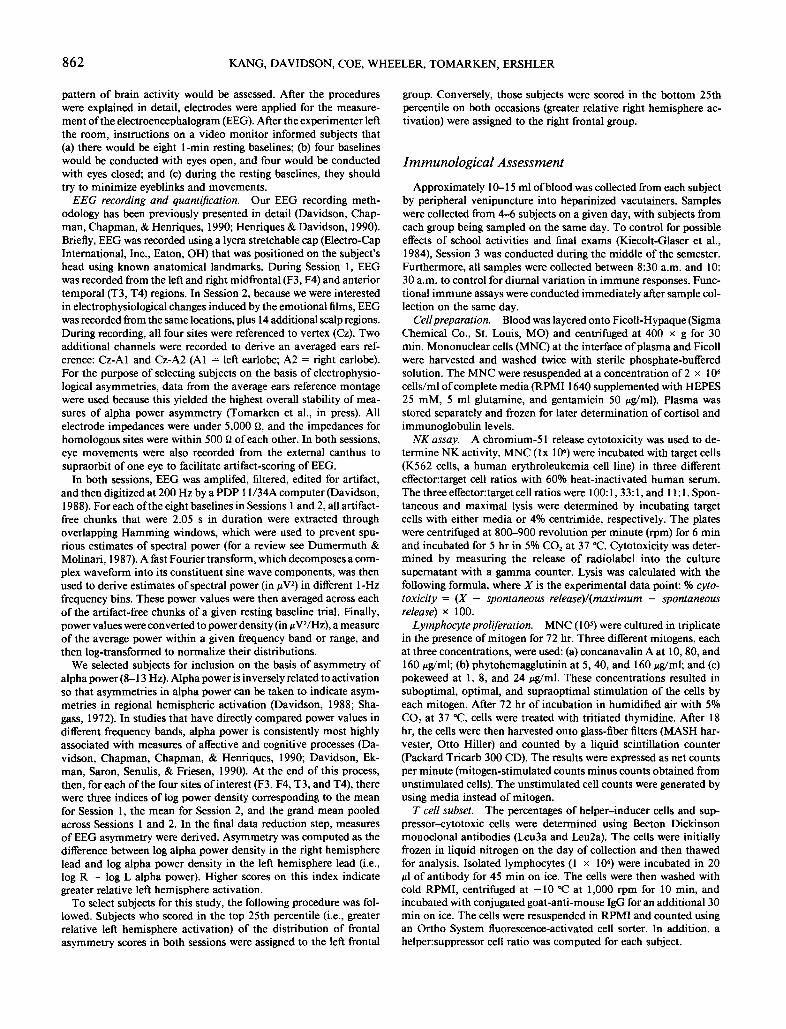
Figure 1. Frontal asymmetry scores (log right (R) - log left (L) alpha power) for each individual subject in the left and right frontal groups. (Positive numbers denote left frontal activation [i.e., less alpha power in the left frontal region than right frontal region], whereas negative numbers denote right frontal activation [i.e., less alpha power in the right frontal region than left frontal region].)
(Harris, 1985). Correlation coefficients were examined when appro- priate.
Results
Survey of Immunological and General Health
After examining a number of health questionnaires that have been commonly used in psychoneuroimmunological research, it became apparent that there were no suitable instruments that completely assessed the relevant health issues. Therefore, we developed our own health questionnaire that focused on the frequency of common ill- nesses that might reflect immune competence. The subject was asked to indicate the frequency of each illness condition during both the preceding 2- and 12-month periods. Health data from the preceding 12-month period were used primarily for verification of more recent health status. The illness categories included respiratory, viral (e.g., herpes, mononucleosis), and fungal (e.g., vaginal, athlete's foot) in- fections, as well as dermatological (e.g., eczema) and allergic (e.g., airborne and food) status. In addition, the subject was asked to indicate any family history of autoimmune diseases, the number of days absent from school or work due to illness, and personal drug u s e .
Statistical Analysis
Student's t test was the primary statistic used to analyze the data. For the analyses of multiple ratios of NK activity and three con- centrations of lymphocyte proliferation, Hotelling's 1 ̀2 was used
Electrophysiological Differentiation of the Groups
Figure 1 presents the frontal a symmet ry scores (averaged across Sessions 1 and 2) for subjects f rom the left and right frontal ly ac t iva ted groups. As can be seen, the two groups showed clear and comple te separation, as would be expected f rom the selection criteria. All left frontal subjects showed absolute left frontal ac t iva t ion (denoted by pos i t ive asym- met ry scores); all right frontal subjects showed absolute right frontal ac t iva t ion (denoted by negat ive a symmet ry scores).
N K Act iv i ty
N K act iv i ty p roved to be the mos t d iscr iminat ing i m m u n e measure in this study (Figure 2). A significant overal l differ- ence in cytotoxici ty was found when subjects wi th left frontal ac t iva t ion were c o m p a r e d with those with right frontal ac- t ivat ion, F(3, 16) = 4.53, p < .02. ' As Figure 2 illustrates,
' Because the distribution of HoteUing's T 2 has the same shape as the F distribution, the F test was used to determine the statistical significance of the observed value of T 2.
864 KANG, DAVIDSON, COE, WHEELER, TOMARKEN, ERSHLER
70-
6 0 -
.~_~' --=- Left Frontal • ~ 50 --B- Right Frontal
40
0 ~ 3o
20
10 , a , ,
1 0 0 : 1 3 3 : 1 11 :1
I = f f e c t o r : T a r g e t R a t i o
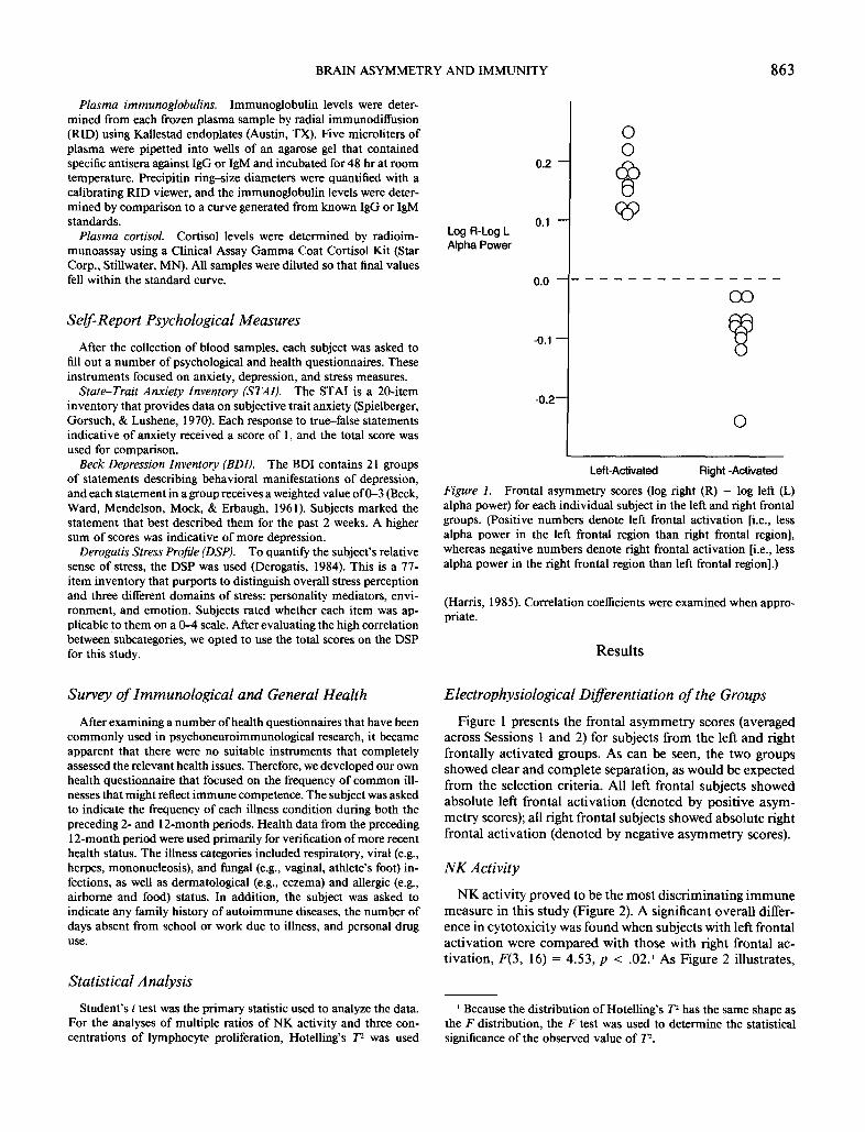
Figure 2. Natural killer cell activity (% cytotoxicity) at three dif- ferent effector:target cell ratios for the left and right frontal activation groups.
subjects in the right frontal group showed lower NK activity compared with their left frontal counterparts. The groups were further compared at each of the three ratios to specify the locus o f the group difference. Significant differences were found at the ratios o f 33:1 and 11:1, t(18) = 2.2, p < .05, and t(l 8) = 2.65, p < .02, respectively. At the highest ratio (100:1), no group difference was observed, t(18) = 0.24, ns. An additional analysis oflytic units, computed from the three ratios, also showed a significant group difference at 30% kill- ing, t(18) = 2.86, p = .01.
Lymphocyte Proliferation
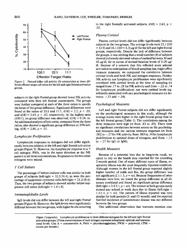
Lymphocyte responses to mitogens did not differ signifi- cantly between subjects in the left and right frontal activation groups (Figure 3). However, the lymphocyte response to a T cell mitogen, PHA, was in the same direction as the NK pattern at all three concentrations. Responses to the two other mitogens were mixed.
T CellSubsets
The percentage o f helper-inducer cells was similar in both groups of subjects (left:right = 32.5:34.4), as were the per- centage o f suppressor--cytotoxic cells (left:right = 21.2:18.4). Thus, the two groups of subjects showed similar helper:sup- pressor cell ratios (left:right = 1.6:1.8).
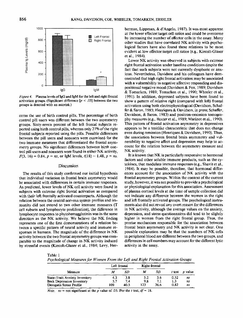
Immunoglobulin Levels
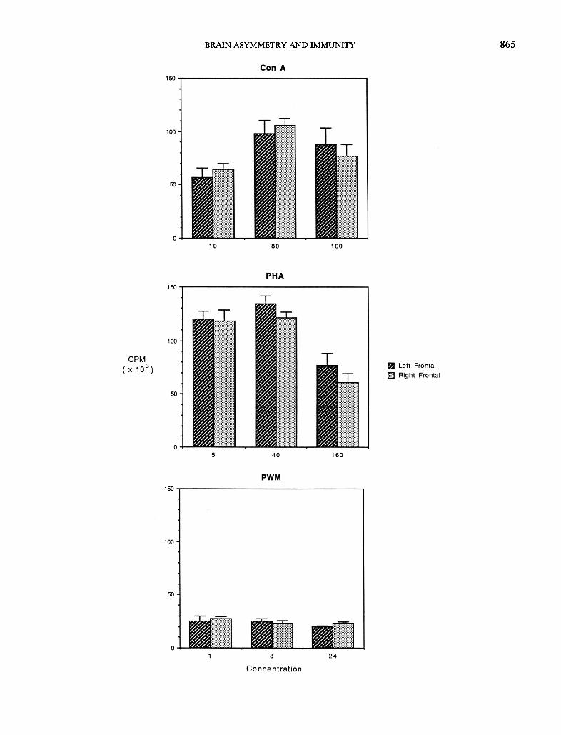
IgG levels did not differ between the left and right frontal groups (Figure 4). However, the IgM levels were significantly different between the two groups, with higher levels occurring
in the right frontally activated subjects, t(18) = 2.63, p < .02.
Plasma Cortisol
Plasma cortisol levels did not differ significantly between subjects in the two groups. The average levels were 23.1 (SD = 12.9) and 16.1 (SD = 6.3) ug/dl for the left and right frontal groups, respectively. Despite the lack o f difference between the groups, it was striking that a small number (4) of subjects showed extremely elevated values ofcortisol between 30 and 40 ug/dl, far in excess of normal baseline levels of 5-20 ug/ dl. Because of a concern that this reflected acute adrenal activation in anticipation o f blood sampling that might affect immune measures, we computed the correlation between cortisol levels and both N K and mitogen responses. Neither NK activity nor lymphocyte proliferation were significantly correlated with cortisol levels at the time of sampling (rs ranged f rom. 17 to .28 for NK activity and from - . 2 2 to . 14 for lymphocyte proliferation); nor were cortisol levels sig- nificantly associated with any psychological measures (rs be- tween - . 3 3 and - .34) .
Psychological Measures
Left and right frontal subjects did not differ significantly on the psychological measures in this study, although the average scores were higher in the right frontal group than in the left frontal group (Table 1). The correlations among the three measures were high (rs from .83 to .89). There were no significant correlations between scores on the psycholog- ical measures and the various immune responses (rs from .002 to - . 2 7 for NK activity, from .003 to .34 for lymphocyte proliferation to optimal doses o f mitogens, and from - . 13 to - . 2 7 for IgG or IgM).
Health Measures
Because o f a potential bias due to long-term recall, we opted to rely on the health data reported for the preceding 2-month period. Out o f many different types of illness, re- spiratory illness was the most frequently reported condition. Although women in the left frontal group reported slightly higher number o f colds and flus, the group difference was not significant (2.1:1.3, p = ns). Because frequencies of other illnesses were low, we tested the group difference in all ill- nesses combined and found no significant group difference (left:right = 3.9:2. l ,p = ns). The women in both groups rarely missed any school or work days due to illness (left:right = 1.5:1.1, p = ns). The pattern of illness reported was very similar for both periods (2- and 12-month), and the reported familial incidence o f autoimmune disease was not different between the two groups.
One additional observation that warrants mention con-
Figure 3 (opposite). Lymphocyte proliferation to three different mitogens for the left and fight frontal activation groups. (Three concentrations of each mitogen represent suboptimal, optimal, and supraop- timal levels. Con A = concanavalin A; PHA = phytohemagglutinin; PWM = pokeweed; CPM = counts per minute.)
BRAIN ASYMMETRY AND IMMUNITY 865
866 KANG, DAVIDSON, COE, WHEELER, TOMARKEN, ERSHLER
Figure 4. Plasma levels of IgG and IgM for the left and right frontal activation groups. (Significant difference [p < .05] between the two groups is denoted with an asterisk.)
cerns the use of birth control pills. The percentage of birth control pill users was different between the two asymmetry groups. Sixty-seven percent of the left frontal subjects re- ported using birth control pills, whereas only 27% of the right frontal subjects reported using the pills. Possible differences between the pill users and nonusers were examined for the two immune measures that differentiated the frontal asym- metry groups. No significant differences between birth con- trol pill users and nonusers were found in either NK activity, F(3, 16) = 0.84, p = ns, or IgM levels, t(18) = 1.48, p = ns.
Discuss ion
The results of this study confirmed our initial hypothesis that individual variation in frontal brain asymmetry would be associated with differences in certain immune responses. As predicted, lower levels of NK cell activity were found in subjects with extreme right frontal activation as compared with their left frontally activated counterparts. Although the relation between the central nervous system profiles and im- munity did not extend to two other immune measures (T cell subsets and lymphocyte proliferation), the difference in lymphocyte responses to phytohemagglutinin was in the same direction as the NK activity. We believe the NK finding represents one of the first demonstrations of a relation be- tween a specific pattern of neural activity and immune re- sponses in humans. The magnitude of the difference in NK activity between the two frontal asymmetry groups was com- parable to the magnitude o f change in NK activity induced by stressful events (Kiecolt-Glaser et al., 1984; Levy, Her-
berman, Lippman, & d'Angelo, 1987). It was most apparent at the lower effector:target cell ratios and could be overcome by increasing the number of effector cells in the assay. Many other studies that have correlated NK activity with psycho- logical factors have also found these relations to be most evident at low effector:target cell ratios (e.g., Kiecolt-Glaser et al., 1984).
Lower NK activity was observed in subjects with extreme right frontal activation under baseline conditions despite the fact that such subjects were not currently dysphoric or anx- ious. Nevertheless, Davidson and his colleagues have dem- onstrated that high right frontal activation may be associated with a vulnerability to negative affective responding and dis- positional negative mood (Davidson & Fox, 1989; Davidson & Tomarken, 1989; Tomarken et al., 1990; Wheeler et al., 1991). In addition, depressed subjects have been found to show a pattern of relative right (compared with left) frontal activation using both electrophysiological (Davidson, Schaf- fer & Saron, 1985; Henriques & Davidson, in press; Schaffer, Davidson, & Saron, 1983) and positron-emission tomogra- phy measures (e.g., Baxter et al., 1989; Marinot et al., 1990). This pattern of frontal activation asymmetry in depressives appears to be a traitlike characteristic that does not change even during remission (Henriques & Davidson, 1990). Thus, the association between frontal brain asymmetry and vul- nerability to negative affect and depression may help to ac- count for the relation between the asymmetry measure and NK activity.
It is known that NK is particularly responsive to hormonal factors and other soluble immune products, such as the cy- tokines, that modulate immune responses (e.g., Shavit et al., 1984). It may be possible, therefore, that hormonal differ- ences account for the association of NK activity with the frontal asymmetry groups. Within the context of the current study, however, it was not possible to provide a psychological or physiological explanation for this association. Assessment o f plasma cortisol levels at the time of sample collection did not indicate any difference between the women in the right and left frontally activated groups. The psychological instru- ments also did not reveal any overt reason for the differences in NK activity, although the average values on the anxiety, depression, and stress questionnaires did tend to be slightly higher in women from the right frontal group. Thus, the precise mechanism responsible for the association between frontal brain asymmetry and NK activity is not clear. One possible explanation may be that the numbers of NK cells in peripheral blood are different between the two groups, and differences in cell numbers may account for the different lytic activity in the assay.
Table 1 Psychological Measures for Women From the Left and Right Frontal Activation Groups
Left frontal Right frontal
Measure M SD M SD t test p value
State-Trait Anxiety Inventory 4.3 3.8 5.2 3.6 0.52 ns Beck Depression Inventory 5.7 7.4 9.8 7.1 1.3 ns Derogatis Stress Profile 109 40.5 123 36.6 0.82 ns
Note. ns = not significant at the p value of .05. For the t test, df= 18.
BRAIN ASYMMETRY AND IMMUNITY 867
Our findings are in general agreement with an increasing number o f studies indicating that there is a relation between brain asymmetry and the immune system. Much of the pre- vious evidence purporting to show such an association has come from studies o f relations between handedness and dis- orders of the immune system. For example, epidemiological surveys have suggested that there is a higher-than-expected likelihood of allergies, asthma, and autoimmune disease in left-handed families (Geschwind & Behan, 1982, 1984; Smith, 1987). Asthmatic women are also more likely to give birth to left-handed children (Weinstein & Pieper, 1988). It should be reiterated, however, that our population was composed of all right-handed individuals, and we assessed regional frontal activity, not hemispheric dominance.
The general difficulty in interpreting the data on handed- ness and immune function arises from the fact that patterns of hemispheric organization are variable among left-handed individuals (Bryden, 1982). Research over the past decade using both behavioral (Hellige, 1990; Levy, 1983) and elec- trophysiological (Davidson & Tomarken, 1989) measures of hemispheric activation has underscored the importance of individual variability in patterns of asymmetric activation within handedness groups. Such evidence suggests that the most fruitful approach to examining relations between asym- metry and immunity will come from studies that assess pat- terns of hemispheric activation, while holding hemispheric specialization constant. It is precisely this method that was adopted in the present study.
In this context, it should be noted that our approach is more similar to that used in the animal literature on this topic. Asymmetries in activation can be produced by uni- lateral lesions of discrete neocortical regions. Using such methods, researchers have shown that partial ablation o f the left cortex reduces immune responses in rodents, whereas equivalent ablation of the homologous region in the right neocortex results in increased immune responses (Neveu et al., 1986; Renoux et al., 1983). Of particular relevance was the observation that lesions in the left neocortex, which pre- sumably resulted in a shift toward greater right-sided acti- vation, led to decreases in NK activity (Renoux et al., 1983).
One other interesting observation in this study was the higher use o f birth control pills by the left frontal group. However, this could not have accounted for our findings because most recent studies have found very little or no effect o f birth control pills on immune responses (Baker et al., 1985; Bisset & Griffin, 1988a, 1988b). Indeed, it would have worked against our findings because the only significant effects on NK responses have been that pill use may decrease lytic activity slightly (Baker, Salvatore, & Milch, 1989). This as- sociation was not replicated in our analyses; nor did another study observe any effect of the menstrual cycle on cellular immune parameters (Caggiula et al., 1990).
The overall health significance of these biological differ- ences remains to be determined. Our health survey did not indicate any significant difference in illness frequency be- tween left and right frontal subjects during the preceding 2 months. The only indication of a possible difference between the women was the occurrence o f higher IgM levels in the right frontal group, which could reflect recent subclinical in-
fection, but further work would be required to determine antibody levels for specific illnesses. More dramatic health- related findings might emerge if one included individuals from families genetically predisposed for certain illnesses or from a left-handed population. It may be that the constel- lation o f high right frontal activation and low NK cell activity is a predisposing condition that could alter an individual's vulnerability for illness. However, such individual differ- ences in vulnerability would become evident only in response to stressful life events, after exposure to pathogenic agents, or at an older age. It has been argued previously that the trait o f right frontal activation represents a possible diathesis for affective or anxiety disorders (or both). Similarly, this con- dition could interact with the process of immune senescence to create a vulnerability to immunological disease at the end of the life span.
References
Baker, D. A., Hameed, C., Tejani, N., Milch, P., Thomas, J., Mon- heit, A. G., & Dattwyler, R. J. (1985). Lymphocyte subsets in women on low dose oral contraceptives. Contraception, 32, 377- 382.
Baker, D. A., Salvatore, W., & Milch, P. O. (1989). Effects of low- dose oral contraceptives on natural killer cell activity. Contracep- tion, 39, 119-124.
Barneoud, P., Neveu, P. J., VitieUo, S., & Le Moal, M. (1987). Functional heterogeneity of the right and left cerebral neocortex in the modulation of the immune system. Physiology and Behavior, 41, 525-530.
Bartrop, R. W., Lazars, L., Luckhurst, E., Kiloh, L. G., & Penny, R. (1977). Depressed lymphocyte function after bereavement. Lan- cet, 1, 834-836.
Baxter, L, R., Schwartz, J. M., Phelps, M. E., Mazziotta, J. C., Guze, B. H., Selin, C. E., Gerner, R. H., & Sumida, R. M. (1989). Re- duction of prefrontal cortex glucose metabolism common to three types of depression. Archives of General Psychiatry, 46, 243-250.
Beck, A. T., Ward, C. H., Mendelson, M., Mock, J., & Erbaugh, J. (1961). An inventory for measuring depression. Archives of Gen- eral Psychiatry, 4, 561-571.
Besedovsky, H. O., Del Rey, A., Sorkin, E., Da Prada, M., & Keller, H. H. (1979). Immunoregulation mediated by the sym- pathetic nervous system. Cell Immunology, 48, 346--355.
Bisset, L. R., & Griffin, J. F. T. (1988a). Humoral immunity in oral contraceptive users: I. Plasma immunoglobulin levels. Contra- ception, 38, 567-572.
Bisset, L. R., & Griffin, J. F. T. (1988b). Humoral immunity in oral contraceptive users: II. In vitro immunoglobulin production. Con- traception, 38, 573-578.
Bryden, M. P. (1982). Laterality." Functional asymmetry in the intact brain. San Diego, CA: Academic Press.
Caggiula, A. R., Stoney, C. M., Matthews, K. A., Owens, J. F., Davis, M. C., & Rabin, B. S. (1990). T-lymphocyte reactivity during the menstrual cycle in women. Clinical Immunology and Immuno- pathology, 56, 130-134.
Coe, C. L., Lubach, G. R., & Ershler, W. B. (1990). Immunological consequences of maternal separation in infant primates. In M. Lewis & J. Worobey (Eds.), New directions for child development: Infant stress and coping (pp. 65-92). San Francisco: Jossey Bass.
Davidson, R. J. (1984). Affect, cognition, and hemispheric special- ization. In C. E. Izard, J. Kagan, & R. Zajonc (Eds.), Emotion, cognition and behavior (pp. 320-365). New York: Cambridge Uni- versity Press.
868 KANG, DAVIDSON, COE, WHEELER, TOMARKEN, ERSHLER
Davidson, R. J. (1988). EEG measures of cerebral asymmetry: Con- ceptual and methodological issues. International Journal of Neu- roscience, 39, 71-89.
Davidson, R. J., Chapman, J. P., Chapman, L. J., & Henriques, J. B. (1990). Asymmetrical brain electrical activity discriminates between psychometrically-matched verbal and spatial cognitive tasks. Psychophysiology, 27, 528-543.
Davidson, R. J., Ekman, P., Saron, C. D., Senulis, J. A., & Friesen, W. V. (1990). Approach/withdrawal and cerebral asymmetry: Emotional expression and brain physiology I. Journal of Person- ality and Social Psychology, 58, 330-341.
Davidson, R. J., & Fox, N. A. (1989). Frontal brain asymmetry predicts infants' response to maternal separation. Journal of Ab- normal Psychology, 98, 127-131.
Davidson, R. J., Schaffer, C. E., & Saron, C. (1985). Effect of later- alized presentations of faces on self-reports of emotion and EEG asymmetry in depressed and non-depressed subjects. Psycho- physiology, 22, 353-364.
Davidson, R. J., & Tomarken, A. J. (1989). Laterality and emotion: an electrophysiologicat approach. In F. Boiler & J. Grafman (Eds.), Handbook of neuropsychology (pp. 419--441). New York: Elsevier Science.
Derogatis, L. R. (1984). The Derogatis Stress Profile (DSP): A sum- mary report. Baltimore, MD: Clinical Psychometric Research.
Dumermuth, G., & Molinari, L. (1987). Spectral analysis of EEG background activity. In A. S. Gevins & A. Remond (Eds.), Hand- book of electroencephalography and clinical neurophysiology: Methods of analysis of brain electrical and magnetic signals (Vol. 1, pp. 85-130). Amsterdam: Elsevier.
Geschwind, N., & Behan, P. O. (1982). Left-handedness: Association with immune disease, migraine, and developmental learning dis- order. Proceedings of the National Academy of Sciences USA, 79, 5097-5100.
Geschwind, N., & Behan, P. O. (1984). Laterality, hormones and immunity. In N. Geschwind & A. M. Galaburda (Eds.), Cerebral dominance: The biological foundations (pp. 211-224). Cambridge, MA: Harvard University Press.
Geschwind, N., & Galaburda, A. M. (1985). Cerebral tateralization: Biological mechanisms, associations, and pathology: I. A hypoth- esis and a program for research. Archives of Neurology, 42, 428- 459.
Glaser, R., Rice, J., Sheridan, J., Fertel, R., Stout, J., Speicher, C., Pinsky, D., Kotur, M., Post, A., Beck, M., & Kiecolt-Glaser, J. (1987). Stress-related immune suppression: Health implications. Brain, Behavior, and lmmunity, I, 7-20.
Harris, R. J. (1985). A primer of multivariate statistics (pp. 99-146). San Diego, CA: Academic Press.
Hellige, J. B. (1990). Hemispheric asymmetry. Annual Review of Psychology, 41, 55-80.
Henriques, J. B., & Davidson, R. J. (1990). Regional brain electrical asymmetries discriminates between previously depressed and healthy control subjects. Journal of Abnormal Psychology, 99, 22-31.
Henriques, J. B., & Davidson, R. J. (in press). Left frontal hypoac- tivation in depression. Journal of Abnormal Psychology.
Irwin, M., Daniels, M., Bloom, E. T., Smith, T. L., & Weiner, H. (1987). Life events, depressive symptoms, and immune function. American Journal of Psychiatry, 144, 437--441.
Irwin, M., Patterson, T., Smith, T. L., Caldwell, C., Brown, S. A., GiUin, C., & Grant, I. (1990). Reduction of immune function in life stress and depression. Biological Psychiatry, 27, 22-30.
Kiecolt-Glaser, J. K., Fisher, L., Ogrocki, P., Stout, J., Speicher, C. E., & Glaser, R. (1987). Marital quality, marital disruption, and immune function. Psychosomatic Medicine, 49, 13-35.
Kiecolt-Glaser, J. K., Garner, W., Speicher, C., Penn, G. M., Hol-
liday, J., & Glaser, R. (1984). Psychosocial modifiers of immu- nocompetence in medical students. Psychosomatic Medicine, 46, 7-13.
Kiecolt-Glaser, J. K., & Glaser, R. (1986). Psychological influences on immunity. Psychosomatics, 27, 621-624.
Leventhal, H., & Tomarken, A. J. (1986). Emotion: Today's prob- lems. Annual Review of Psychology, 37, 565-610.
Levy, J. (1983). Individual differences in cerebral asymmetry: The- oretical issues and experimental considerations. In J. B. Hellige (Ed.), Cerebral hemisphere asymmetry: Method, theory and ap- plication (pp. 465-5 t 5). New York: Praeger.
Levy, S., Herberman, R., Lippman, M., & d'Angelo, T. (1987). Cor- relation of stress factors with sustained depression of natural killer cell activity and predicted prognosis in patients with breast cancer. Journal of Clinical Oncology, 5, 348-353.
Martinot, J. L., Hardy, P., Feline, A., Huret, J. D., Mazoyer, B., Attar-Levy, D., Pappata, S., & Syrota, A. (1990). Left prefrontal glucose metabolism in the depressed state: A confirmation. Amer- ican Journal of Psychiatry, 147, 1313-1317.
Neveu, P. J. (1988). Minireview: Cerebral neocortex modulation of immune functions. Life Sciences, 42, 1917-1923.
Neveu, P. J., Taghzouti, K., Dantzer, R., Simon, H., & Le Moal, M. (1986). Modulation of mitogen-induced lymphoproliferation by cerebral neocortex. Life Sciences, 38, 1907-1913.
Oldfield, R. C. (1971). The assessment of analysis of handedness: The Edinburgh inventory. Neuropsychotogia, 9, 97-113.
Renoux, G., Biziere, K., Renoux, M., Guillaumin, J., & Degenne, D. (1983). A balanced brain asymmetry modulates T cell-mediated events. Journal of Neuroimmunology, 5, 227-238.
Riley, V., Fitzmaurice, M. A., & Spackman, D. H. (1981). Psycho- neuroimmunologic factors in neoplasia: Studies in animals. In R. Ader (Ed.), Psychoneuroimmunology(pp. 31-102). San Diego, CA: Academic Press.
Robinson, R. G., Kubos, K. L., Starr, L. B., Rao, K., Price, T. R. (1984). Mood disorders in stroke patients: Importance of location of lesion. Brain, 107, 81-93.
Roszman, T. L., Cross, R. J., Brooks, W. H., & Markesbery, W. R. (1985). Neuroimmunomodulation: Effects of neural lesions on cellular immunity. In R. Guillemin, M. Cohn, & T. Melnechuk (Eds.), Neural modulation of immunity (pp. 95-109). New York: Raven Press.
Schaffer, C. E., Davidson, R. J., & Saron, C. (t983). Frontal and parietal EEG asymmetries in depressed and non-depressed sub- jects. Biological Psychiatry, 18, 753-762.
Schleifer, S. J., Keller, S. E., Bond, R. N., Cohen, J., & Stein, M. (1989). Major depressive disorder and immunity. Archives of Gen- eral Psychiatry, 46, 81-87.
Schleifer, S. J., Keller, S. E., Camerino, M, Thornton, J. C., & Stein, M. (1983). Suppression of lymphocyte stimulation following be- reavement. Journal of the American Medical Association, 250, 374-377.
Schleifer, S. J., Keller, S. E., Meyerson, A. T., Raskin, M. J., Davis, K. L., & Stein, M. (1984). Lymphocyte function in major de- pressive disorder. Archives of General Psychiatry, 41, 484--486.
Shagass, C. (1972). Electrical activity of the brain. In N. S. Greenfield & R. A. Sternbach (Eds.), Handbook ofpsychophysiology(pp. 263- 328). New York: Holt, Rinehart & Winston.
Shavit, Y., Lewis, J. W., Terman, G. W., Gale, R. R., & Liebeskind, J. C. (1984). Opioid peptides mediate the suppressive effects of stress on natural killer cell cytotoxicity. Science, 223, 188-190.
Siiberman, E. K., & Weingartner, H. (1986). Hemispheric lateraliza- tion of functions related to emotion. Brain and Cognition, 5, 322- 353.
Smith, J. (1987). Left-handedness: Its association with allergic dis- ease. Neuropsychologia, 25, 665--674.
BRAIN ASYMMETRY AND IMMUNITY 869
Spielberger, C. D., Gorsuch, R. C., & Lushene, R. E. (1970). The state-trait anxiety inventory. Palo Alto, CA: Consulting Psychol- ogists Press.
Stein, M., Keller, S. E., & Schleifer, S. J. (1985). Immune system: Relationship to anxiety disorders. Psychiatric Clinics of North America, 11, 349-360.
Tomarken, A. J., Davidson, R. J., & Henriques, J. B. (1990). Resting frontal brain asymmetry predicts affective responses to films. Jour- nal of Personality and Social Psychology, 59, 791-801.
Tomarken, A. J., Davidson, R. J., Wheeler, R. W., & Kinney, L. (in press). Psychometric properties of resting anterior EEG asym° metry: Temporal stability and internal consistency. Psychophys- iology.
Wagner, H. L., MacDonald, C. J., & Manstead, A. S. R. (1986).
Communication of individual emotions by spontaneous facial ex- pressions. Journal of Personality and Social Psychology, 50, 737- 743.
Weinstein, R. E., & Pieper, D. R. (1988). Altered cerebral dominance in an atopic population. Brain, Behavior, and Immunity, 2, 235- 241.
Wheeler, R. E., Davidson, R. J., & Tomarken, A. J. (1991). Frontal brain asymmetry and emotional reactivity." A biological substrate ofaffective style. Manuscript submitted for publication.
Received January 16, 1991 Revision received May 24, 1991
Accepted June 19, 1991 •
Z a h n - W a x l e r A p p o i n t e d N e w E d i t o r , 1 9 9 3 - 1 9 9 8
The Publications and Communications Board of the American Psychological Association announces the appointment o f Carolyn Zahn-Waxler as editor of Developrnental Psychology. Zahn-Waxler is associated with the National Institute of Mental Health. As of January 1, 1992, manuscripts should be directed to
Carolyn Zahn-Waxler 4305 Dresden Street Kensington, Maryland 20895
Manuscript submission patterns make the precise date of completion of the 1992 volume uncertain. The current editor will receive and consider manuscripts through December 1991. Should the 1992 volume be completed before that date, manuscripts will be redirected to the incoming editor for consideration in the 1993 volume.