(From the Hospital of The Rockefeller Institute for Medical Research ...
19
ARTERIAL HYPERTENSION IN RATS II. EFFECTS ON-rm_m KIDNEYS BY HENRY A. SCRROEDER, M.D., AND CHARLES NEUMANN, M.D. (From the Hospital of The Rockefeller Institute for Medical Research) PLATES 13 TO 16 (Received for publication, February 9, 1942) The exact relation between disease of the renal arterioles and arterial hyper- tension is not understood. It is not known whether the observed sclerosis and necrosis of the arterioles of the kidneys cause renal ischemia and therefore hypertension or are merely a consequence of elevation of the blood pressure. In some animals the evidence gained in experiments suggests, however, that the appearance in the renal arterioles results from hypertension. By partially constricting one renal artery in rats Wilson and Byrom (1) were able to bring about elevation of blood pressure and necrotizing arteriolitis in the vessels of the other kidney, indicating that in rats, at least, arteriolar disease is oc- casioned by hypertension. Their work has not received the notice it deserves. If their findings were substantiated in other animals and in man, some of the factors contributing to arterial hypertension would be made more dear. Un- fortunately the experiment cannot be performed in dogs, as chronic hyperten- sion does not usually result from affecting one kidney when the other is intact. It appeared rewarding, on the plan of Wilson and Byrom, to attempt to ascertain further the rdation of arteriolar disease to arterial hypertension in rats and to observe the effects upon one kidney of various kinds of injury to the other. It has been shown that unilateral renal injury results in cardiac hyper- trophy, an indication of the presence of hypertension (2). The current study deals with changes in the renal arterioles associated with the latter condition. Methods The left kidneys of 80 rats were injured either by partial constriction of the renal artery, by the production of hydronephrosis, by trauma, or by placing cellophane around them (2). Eighteen more rats were injected with adrenalin in oil or pitressin and estradiol, and six with other substances (renin, dihydroxyphenylalanine, tyrosinase). The animals were killed, after a suitable interval, the hearts weighed as previously described (2), and the kidneys fixed in Zenker acetic acid solution. Sections were stained either with eosin-methylene blue, hematoxylin and eosin, or Mallory's connec- tive tissue stain. They were then examined by one of us who had had no previous knowledge of which of the rats had exhibited arterial hypertension. The degreeof the following anatomical changes ~n each kidney was estimated and recorded in terms 527 on February 13, 2018 jem.rupress.org Downloaded from
-
Upload
vuongkhuong -
Category
Documents
-
view
221 -
download
3
Transcript of (From the Hospital of The Rockefeller Institute for Medical Research ...

ARTERIAL HYPERTENSION IN RATS
II. EFFECTS ON-rm_m KIDNEYS
BY HENRY A. SCRROEDER, M.D., AND CHARLES NEUMANN, M.D.
(From the Hospital of The Rockefeller Institute for Medical Research)
PLATES 13 TO 16
(Received for publication, February 9, 1942)
The exact relation between disease of the renal arterioles and arterial hyper- tension is not understood. I t is not known whether the observed sclerosis and necrosis of the arterioles of the kidneys cause renal ischemia and therefore hypertension or are merely a consequence of elevation of the blood pressure. In some animals the evidence gained in experiments suggests, however, that the appearance in the renal arterioles results from hypertension. By partially constricting one renal artery in rats Wilson and Byrom (1) were able to bring about elevation of blood pressure and necrotizing arteriolitis in the vessels of the other kidney, indicating that in rats, at least, arteriolar disease is oc- casioned by hypertension. Their work has not received the notice it deserves. If their findings were substantiated in other animals and in man, some of the factors contributing to arterial hypertension would be made more dear. Un- fortunately the experiment cannot be performed in dogs, as chronic hyperten- sion does not usually result from affecting one kidney when the other is intact.
I t appeared rewarding, on the plan of Wilson and Byrom, to attempt to ascertain further the rdation of arteriolar disease to arterial hypertension in rats and to observe the effects upon one kidney of various kinds of injury to the other. I t has been shown that unilateral renal injury results in cardiac hyper- trophy, an indication of the presence of hypertension (2). The current study deals with changes in the renal arterioles associated with the latter condition.
Methods
The left kidneys of 80 rats were injured either by partial constriction of the renal artery, by the production of hydronephrosis, by trauma, or by placing cellophane around them (2). Eighteen more rats were injected with adrenalin in oil or pitressin and estradiol, and six with other substances (renin, dihydroxyphenylalanine, tyrosinase).
The animals were killed, after a suitable interval, the hearts weighed as previously described (2), and the kidneys fixed in Zenker acetic acid solution. Sections were stained either with eosin-methylene blue, hematoxylin and eosin, or Mallory's connec- tive tissue stain. They were then examined by one of us who had had no previous knowledge of which of the rats had exhibited arterial hypertension. The degreeof the following anatomical changes ~n each kidney was estimated and recorded in terms
527
on February 13, 2018
jem.rupress.org
Dow
nloaded from

528 ARTERIAL HYPERTENSION IN RATS. II
of one to four plus: the presence of hyalinization of the glomeruli singly or in groups, proliferation of the parietal layer of Bowman's capsule (crescent formation), infiltra- tion of the interstitial tissue by round cells, atrophy of the tubules, and replacement fibrosis. The condition of the vessels was estimated in a similar manner by ascertain- hag the presence or absence of the following: intimal proliferation of the arterioles, necrosis of their walls, hypertrophy of their muscles, hyaline degeneration, hyperplasia of the internal elastic membrane of the small arteries, and hyalinization of their walls. The presence of small hemorrhages in the substance of the kidney was noted, as was any other abnormality. A decision was then made as to the resemblance of these kidneys to those found in arterial hypertension particularly in so far as vascular change was concerned. The results of the microscopic examination were then com- pared with data obtained during the life of the animals, and with such changes as had taken place in the weights of their hearts.
RESULTS
The pathological findings were used to group the rats into two general classes. In the one, arteriolar lesions, including marked hypertrophy of the muscle, narrowing of the lumen, and partial to almost complete necrosis of the wall were the predominating features. These lesions bore a close resemblance to the arteriolar changes in the kidneys of rapidly progressive hypertension in human beings. In the other, arteriolar lesions were either non-existent or limited to variable but slight hypertrophy of the muscle. In this group the predominating pathological features were confined to the glomeruli, which showed thickening of the parietal layer of Bowman's capsule and various stages of hyalinization. Only the kidneys exhibiting arteriolar changes were believed to represent those of hypertensive animals. How closely this division ac- cording to pathological findings alone corresponded with other signs of hyper- tension such as the level of the blood pressure and cardiac weight found post- mortem will now be described.
Fifty-nine rats exhibited cardiac hypertrophy. The kidneys of 51 (86 per cent) showed changes in the renal arterioles of various degrees and in 3 others sfight lesions were noticed. In 45 cases there was no cardiac hyper- trophy; in 36 there were few or no renal arteriolar lesions (Tables I to V).
When one kidney was injured by hydronephrosis, by partial constriction of the renal artery, by trauma, or by cellophane, arteriolar changes occurred in the opposite kidney with great regularity (Figs. 1 to 13). Of 52 rats so treated which exhibited cardiac hypertrophy, renal arteriolar lesions occurred in 47. Cardiac hypertrophy did not occur in the cases of 25; yet in the unaffected kidneys of 9 of these similar lesions were seen in the arterioles (Tables I to IV).
Rats given various chemical compounds (in an attempt to induce hyperten- sion) and exhibiting cardiac hypertrophy developed renal arteriolar lesions with less regularity (Table V). Of 2 injected with pitressin and estradiol benzoate, renal changes occurred in 1. Of 4 injected with adrenalin in off,
on February 13, 2018
jem.rupress.org
Dow
nloaded from

T A B L E I
Effects of Unilateral Hydronepkrosis
g
mm.Hg days
14-0/11C 15 Both + +
i67/112 23 R +-I-
148/112 23 RL ++++
__140/105 1,5 Both + +
1~0/13s 30 ~oth + +
150/120 28 Both + +
148/112 27 L + + 11 + +
1-~/122" 109 L -{-'4- R + +
i68/128 22 Both +-l-
' - ~ 10.5/60 H 149§§ 33 L 11 +
H 147§§ -4-3 100/58 33 L 11 +
H143§§ --1 90/52 42 L 11
H141§§ --3 95/50 42 L 11
H 145§§ % 100/52 33 L 1l
Anatomical changes in kidneys
Arterioles o
~ ~ .~ ~ ~ R e ~ r ~
+ + + Yes Slight hydronephro- sis
q- + + Yes Slight hydronephro- sis
+ + + + + F Moderate hydrone- Yes phrosis
+ + + Yes Slight hydronephro- sis
+ + + + Yes Slight hydronephro- sis
+ + + + Yes Slight hydronephro- sis
-F ++] ++-F + + q- F Slight hydronephro- + + + + + F Yes sis
+ + + + ÷ + + + + + + + F and R: Destruction of kid- + + + + + + + + + Yes uey
+ + + + Yes Slight hydronephro- sis
:++-F+ + + + + + + F and R Destruction of kid- + No ney
++-F+ -I-+++ ++'4-+ +q-+q" F and R Destruction of kid- + -~- No ney
+ + ++-F Marked hydrone- + No phrosis
+ + + + + F Marked hydrone- + No phrosis
+ + + + F Destruction of kid- + + No hey
+ + + + + Marked hydrone- -4- No phrosis
§§ Tota l ureteral occlusion.
In the tables the significance of the symbols is as follows: * = based on the formulas of R y t a n d (5).
= measured by Hami l ton ' s optical manometer . The first figure represents the systolic, the second the diastolic pressure.
§ = i.e. proliferation of internal elastic membrane , hyalinization of walls, muscular hyper t rophy. [] -- i.e. singly or in large foci. ** = F indicates fibrosis, R indicates infiltration by round cells. :~ -- an est imation of the general appearance of the kidney and its resemblance to those observed
in hypertensive animals.
Changes are indicated by a plus ( + ) sign, and their degree es t imated on the basis of one to four plus. When no symbol is given, there were no lesions.
529
on February 13, 2018
jem.rupress.org
Dow
nloaded from
530 ARTERIAL a'Zl'S.RZ'ZNSXON m ItATS. n
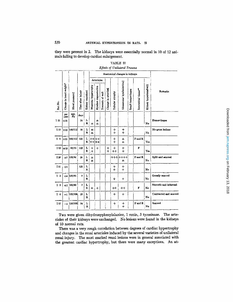
they were present in 2. The kidneys were essentially normal in 10 of 12 ani- mals failing to develop cardiac enlargement.
T A B L E I I
Efl*¢ls of Unilateral Trauma
Remsrks
Hemordxages
No gross lesions
Split Bad scarred
Grossly scarred
Necrotic ~nd infarcted
Contracted and scarred
Scarred
Two were given dihydroxyphenylalanine, 1 renin, 3 tyrosinase. The arte- rioles of their kidneys were unchanged. No lesions were found in the kidneys of 10 normal rats.
There was a very rough correlation between degrees of cardiac hypertrophy and changes in the renal arterioles induced by the several varieties of unilateral renal injury. The most marked renal lesions were in general associated with the greatest cardiac hypertrophy, but there were many exceptions. An at-
on February 13, 2018
jem.rupress.org
Dow
nloaded from
HENRY A. SCHROEDER AND CHARLES 17EUMANN 531
tempt was made to find whether there was a relation between the degree of renal damage in the untreated kidney and the number of days after the other was injured. Marked changes occurred as early as 7 to 14 days. Five animals developing "malignant hypertension" (1) were allowed to survive for 2 months or more and their kidneys were more severely damaged.
When elevation of the blood pressure was considered an indication of the hypertensive state, about the same relative number of animals (88 per cent) developed changes in their kidneys as when cardiac hypertrophy was taken as the sign (Table VI). Single estimations of blood pressure have little value, especially when the pressure is normal or low in the presence of cardiac hyper- trophy, since a state of shock is easily induced when such a condition obtains. But of the 32 animals which exhibited elevation of blood pressure 28 developed renal vascular lesions, and in 12 these included necrosis. Of 8 rats with normal hearts and elevated blood pressures, they were present in 6. When cardiac hypertrophy and elevation of blood pressure were considered together, roughly the same proportion (85 per cent) exhibited renal lesions and a similar one, necrosis of arterioles. Renal lesions were found in only 5 animals with normal hearts and normal blood pressures--an in rats subjected to partial constriction of a renal artery (Table VI).
The survival of renal tissue in the injured kidney at death was not always necessary for the development of hypertension or vascular lesions. The affected kidney was atrophic in 13 animals in which the renal artery had been constricted; 7 of these developed cardiac hypertrophy and 9 arteriolar lesions. On the other hand functioning of a partially injured hydronephrotic kidney seemed to be necessary for the development of hypertension and arteriolar lesions (Table I, Figs. 1 to 7).
Cardiac hypertrophy occurred in 3 cases when the left kidneys had been injured and the right ones remained normM. In 1 rat the injured organ was completed atrophied, in 1 full of hemorrhagic areas, and in the other the renal tubules were atrophied. In 2 cases after adrenalin, and 1 after pitressin and estradiol had been given, cardiac hypertrophy was found in the presence of normal kidneys.
Renal arteriolar lesions occurred without cardiac hypertrophy in 11 animals. The blood pressure was elevated in all but 5 in which renal arteries were con- stricted. I t was elevated without cardiac hypertrophy in only 2 instances when the kidney was considered normal.
Although in most instances glomeruli showed hyaline degeneration, in only 3 was there seen proliferation of the parietal layer of Bowman's capsule (cres- cent formation), such as occurs in glomerulonephritis. On the other hand, necrosis of the walls of arterioles was common, being found in 34 animals. Five of these did not exhibit cardiac hypertrophy although the blood pressures of 3 were elevated.
on February 13, 2018
jem.rupress.org
Dow
nloaded from

T A B L E I I I
Effects of Partial Constriction of One Renal Artery
J "r.,
per cent mm.Hg days
174 +77 59 L R
158 +75 168/135 35 L R
Anatomical changes in kidneys
Arterioles
m °~ '~
i.
$. .4 '~ ~' 4 ~ % "i Remarks
+ + + + + + + + + + + + + F Partially + + + + + + + + + + + + + F Yes atrophic
+ + + + + + + + + + + + + + F + + + + + + + + + + + + + + F Yes
lSl +52 110/80 35 L + + + + + + [ + + + + + + + + + F R + + + + + + + + + + + + + + + + + + F Yes
121 +48 118/90 10 L + + + + + + + + + + + F a n d R R + ÷ ÷ ÷ + ÷ R Yes
103 +4~ i t L + + + + + + + + + + + + + + F R + + + + + + + + + + + F Yes
107 +46 88/38 7 Both + + + + + + + + F and R Yes
156 +37 80/49 14 L + + + + + + + + + + + F
R + + + + + + +
168 ÷37 4o/22 61 L ÷ ÷ ÷ ÷ ÷ ÷÷÷÷ ÷÷÷ F and R + + + + + + + + + + + + + + + + + + + F Yes
123 +36 120/104 16 L R + + + + + +
110 +35130/106 41 L
R ÷ ÷ t ÷ I _ 106 +32 148/105 7 L + + + + + + + + F
R + + + + + + + +
175 +2~ 170/12~ 68 L R + + + + + + + +
154 +28 138/90 9 L + + R + + +
118 +27 146/12C 7 Both + + + +
108 +26 110/64 12 L + + + + g + + + +
112 +26 72/62 4 L R
116 +26 110/98 20 L
R + + +
+
+ + + + + + +
+ + + + + + +
+ + + + + + + + + + + +
+ + I
I + !
i
÷1
Par tially atrophic
Yes Enlarged
Completely Yes atrophic
Completely Yes atrophic
Yes
+
+ Yes
+ IF and R Yes
+ F Yes
! No
f I
I Yes
Completely Yes atrophic
Completely atrophic
Almost com- pletely fibrotic
532
on February 13, 2018
jem.rupress.org
Dow
nloaded from

TABLE HI--Conduded
%
• ~ ~ ~ o
per ~m.Hg days cent
122 +2~ 150/112 14 Both + +
167 +24 50/28 61 L
Anatomical changes in kidneys
Arterioles
+ + + +
R + + + + + + + + + + +
170 +1~!140/1~ 61 L
Yes
R +
104 +18160/134 32 Both + +
126 +15 112/80 8 L + + + R + + + +
152 +13 140/102 30 L + + R + + + + + + + + + +
176 +11 132/92 68 L R + + +
118 +8120/82 12 L R
169 +5 178/128 61 L R
172
+ + F Yes
162
161
153
160 - 130/10= 7 R + + +
166 --2 132/96 56 R + + + + +
164 --,5 88/42 36 L R
t ,62/128f, p L + + + +
+ No
+ + + FandR Yes
+ + + + + + + + + F and R + Yes
+ + + + + + + F + Yes
+ +
+ + +
+2 110/60 75 L R + + + + + + + + + + + +
+1 160/122 7 R + + + + + + + + + +
+1 160/130 70 L R + + + + + + + + + + + +
--1 120/82 35 L + + + + + + + + + + + + + + + + R + + + + + + + + +
+
+ + +
-4-
Uremia. Com- pletely atrophic
Com- pletely atro- phlc
Atrophic Yes
Atrophic No
Atrophic No Enlarged
Atrophic Yes
F Yes
Atrophic Yes
F Yes
Yes
Yes
Atrophic No
i Yes
§§ O p p o s i t e ( r igh t ) n e p h r e c t o m y p e r f o r m e d .
533
on February 13, 2018
jem.rupress.org
Dow
nloaded from
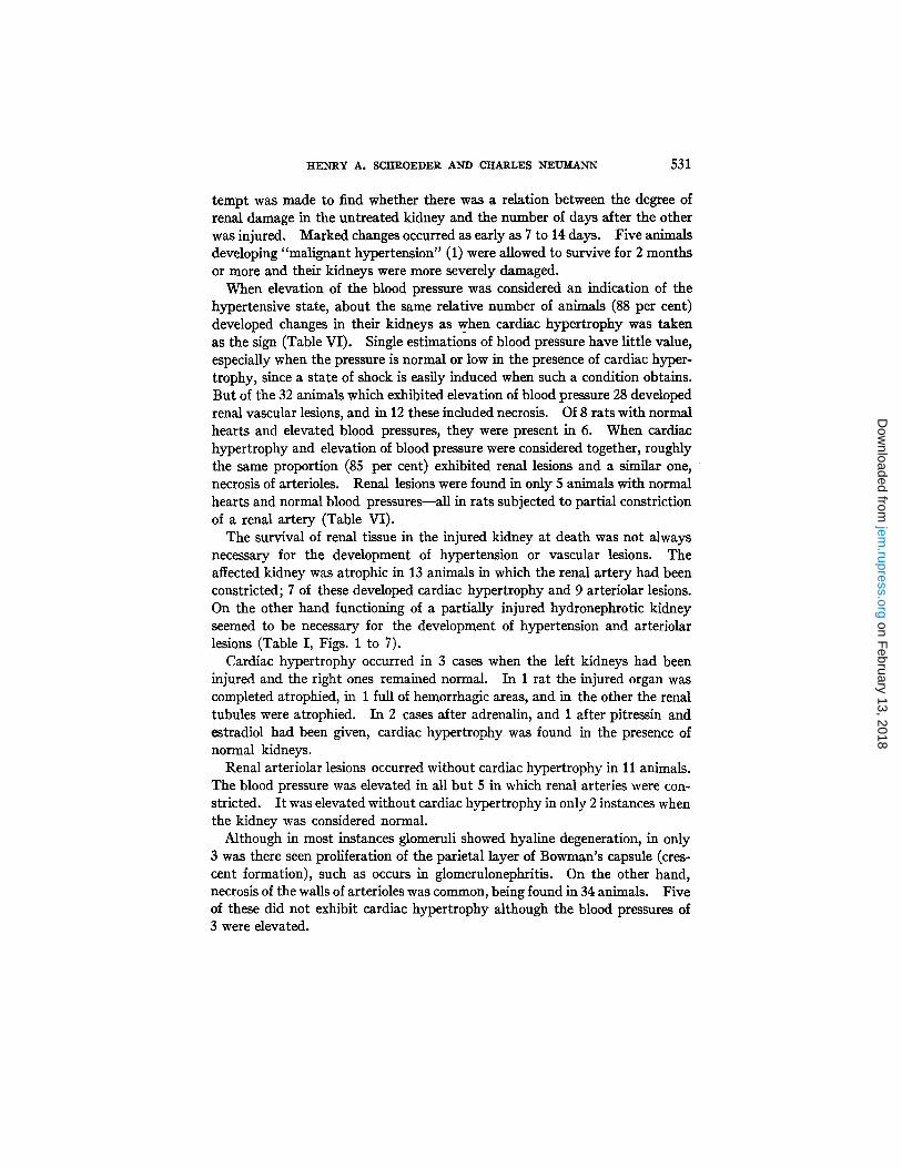
T A B L E I V
Effects of Unilateral Cellophane Perinephritls
Anatomical changes in kidneys
Arterioles %
per mm.H~ days cen~ C 9 +6{ 98/55 59 R + + + +
C20 +38 142/102 36 R + + + + +
C 6 +37 120/60 36 L
~" ~, .,, § o
Remarks
s
R + + + + + + + + + + + + +
+ + + + + + F Yes
+ 4- Yes
Infection F Yes Large cortical hemor-
rhage
C 5 +36 162/80 21 R + + + + + + + Yes
C 8 +33 160/120 73 L Infection R + + + + + + Yes
C 19 +27 62/28 33 R + + + + + + + + + + + F Yes
C 7 +25 120/86 60 L R + + + + + + + + + +
C 3 +24 170/106 27 L + + + + + + + + F & R Infection R + + + + + + + + + + + + + + F Yes ttydronephrotlc
C IO +24 90•70 16 R + + + + + + + + + + F Yes '
C 12 -+-23 172/98 16 L R + + + + + + + + + + + F Yes
C13 +20 160/130 31 R i + + + + + + + + + + + + + + + + + F Yes
C 14 +19 160/102 44 L + + + + + + + + + + + + R + + + + + + + Yes
C 15 ~+18 178/117 88 L R + + + + + + + + + + + + +
C 1 +17 14.5/102 59 L R + + + + + + + + + + + + + +
Slight infection Yes Cortical hemorrhnges
Severely scarred
Infection Yes
Infection F Yes Hydronephrotic
F Yes Cortical hemorrhages
Infection I Yes
No
Yes
No fibrous capsule F No
No fibrous capsule No
N No fibrous capsule o
C 17 +16 190/150 31 L + - b + + + + + - b - b + --t--{'-"{--+ R + + + + + + + + + + +
C 4 +61160/10¢ 28 L + + + "4-+ + + + + R + + + + + +
C l l +~ t8o/9o 38 R ±
C 16 --1 165/12~ 31 R --I--'F -'F-'F -'h ±
R2*, +e t48/10e 60 L R + + + +
R2S --~ n2/78 6o L R -4-
R 26 ~ 130/102 60 L R 4- -4,-
C ~ ce l l ophane capsu le . R = r a y o n capsu le .
5 3 4
on February 13, 2018
jem.rupress.org
Dow
nloaded from

T A B L E V
L
Anatomical changes in kidneys
Arterioles
Remarks
Effects of pitressin
psf ce,nt
P 21 +25
P 18 +18
P 13 -t-ll
P 7 -1-6
P 16 --I-4
P 14 --1
P 17 --8
P 19 --13
Hg
174/124
125/75
110/70
170/122
13o/9o
14o/98
142/88
days70 Both -I -++
7 L R
10 R
4 R
40 Both -I-
9 R
10 R
16 R
+ ++
+
+ +
-4-
4-
-4-
+ +++
-4-
.4-
-t-
F Yes
No
No
No
Yes
No
No
N o
Cortical scars
Cortical scars
Effects of adrenalin
A 20
A 13
A 17
A 14
A 22
A 21
A 18
A 19
A 15
A 16
+3_~o 1
+25 1
-[-16 14
-[-13 162/124 14
-.[-6 1
+ I 1 L
...[-1 134/104 14 L
0 1 L
-11158/11~ 16 L R
--3 ~155/11~ i 16 L
L ++ ++ ++
L ++ + +
L
L
L
+
+
++ +
+
.4-
+
4-
.4- • .
+
.4-
+
Yes
Yes
No
F No
No
No
No
No
N o
Yes
Small cortical hemorrhages
Small cortical hemorrhages
Small cortical hemorrhages
Small cortical hemorrhages
Small cortical hemorrhages
Effects of miscellaneous substances
N33 - 9 122/10 23 I Both
J N 34 --1 90/79 16 Both
N ~ -~TI12s/so Both !
N 47 +15 144/lO6 26 Both
N 70 i --7 140/92 9 Both
N 7 +4 116/75 9 Both
+
-4-
+
- H - +
+
+
.4-
4-
-4-
F
F
R
No
No
No
No
No
No
Given tyrosinase
Given tyrosinase
Given tyrosinase Spontaneous pyelonephrith
Given renin
Given dopa
Given dopa
535
on February 13, 2018
jem.rupress.org
Dow
nloaded from

536 ARTERIAL HYPERTENSION IN RATS. 12[
The injured kidney appeared to be included in the hypertensive process along with the uninjured. This was to be expected in the cases of those animals with kidneys affected by hydronephrosis, by trauma, and by cellophane pert- nephritis (Figs. 12 and 13), exposed as they were to the elevated blood pressure. When the renal artery was partially constricted, however, the affected kidney should theoretically have been "protected" by the constriction. This was not the case (Figs. 8 and 9). Of the 28 of 33 animals developing
TABLE VI
Summary of Results in 104 Rats
Type of injury
Iydronephrosis • rauma 'artial constriction of re-
nal artery :ellophane perine-
phritis§§ [ayon sac ~drenalin in oil 'itressin and estradiol ~iscellaneous 'otal 'er cent
15 10 34
18
3 10 8 6
104
Cardiac hypertrophy
{ z
9 9 1 4 2 0
24 22 17
15 15 13
0 0 -- 4 2 0 2 1 0 1 0 - -
5 9 51 31 86 61
Cardiac hyper- Elevation of trophy or eleva-
blood pressure tion of blood
° ~ 0
8 8 1 2 1 0 9 8 6
8 8 5
0 0 - 3 1 0 2 2 0 0 0 --
32 28 12 88 43
6
9 4
28
17
0 6 3 1
68
No cardiac hypertrophy
or elevation of pressure blood pressure
9 1 6 0 -- 2 0 6 0 --
25 19 7 5 1
17 13 1 0 --
0 -- 3 0 -- 3 0 4 0 -- 2 0 5 0 -- 0 -- 5 0 --
58 33 37 5 1
85 57 14 20
§§ In this series a diastolic tension when the systolic was upper limit of normal.
pressure of > 100 mm. Hg was considered to indicate hyper- > 150 (2). In all other cases 110 ram. Hg was taken as the
renal arteriolar changes, the affected kidneys were atrophic in 10, in 3 they were not examined, and in only 2 of the remaining 15 was there any marked difference in the arterioles of the injured and the uninjured sides (Figs. 10 and 11). One animal subjected to partial constriction of the left renal artery and right nephrectomy is included; lesions were present in the affected kidney (Table III).
DISCUSSION
In the light of these experiments it is clear that when one kidney of a rat is damaged by one of several methods renal vascular lesions occur with great
on February 13, 2018
jem.rupress.org
Dow
nloaded from

HENRY A. SCHROEDER AND CHARLES NEUMANN 537
regularity in the opposite one when signs of arterial hypertension result. I t is probable that vascular disease of this nature can, therefore, be caused by one of two factors; elevation of the blood pressure or some harmful substance released by the injured kidney. There is considerable ditficulty in deciding which is responsible for the changes observed and what is the sequence of events. Injury to one kidney may cause the release of some pressor substance which raises blood pressure. The vessels of the other kidney may then be included in a general process of vasoconstriction, and respond by becoming hypertrophied. Or, injury to one kidney may cause the release of some vas- cular toxin which occasions lesions in both; ischemia then itself results in releasing pressor substances which raise blood pressure. Or perhaps indeed, the injured kidney may release two or more substances--pressor substances and vascular toxins. In that event arterial hypertension results from one, and renal arteriolar lesions from others. Clearly, situations may then arise in which only one substance is released. The experiments of Winternitz (3) point to this possibility.
In most instances renal vascular disease in rats is closely associated with the hypertensive state, and results from procedures which occasion arterial hypertension. From these observations it is impossible to be certain whether vascular lesions themselves are the cause or the result of elevation of the blood pressure. Their presence (5 cases) when both indices of the hypertensive state were absent, and their absence when one was present (8 cases) suggest that more than one process is at work. The most likely explanation is that arte- riolar lesions are caused by the same disturbance whch results in hypertension and are merely a manifestation of its existence. That they in turn contribute toward the maintenance of renal ischemia and therefore hypertension, is prob- able. In those cases in which one kidney was completely destroyed by infec- tion, trauma, or infarction, and in which hypertension persisted, ischemia may have been maintained by the renal vascular disease present in the other. Re- moval of an injured kidney in rats need not accordingly cause hypertension to disappear (4).
I t is diflficult to explain why kidneys made ischemic by partial constriction of renal arteries were included in the vascular process. This is not the case in dogs. A circulating vascular toxin may have affected the arterioles of both kidneys. Perhaps rats are more susceptible to vascular damage than are other animals.
These results differ from those found by Wilson and Byrom (1), although the conclusions to be drawn are similar. They report few lesions in kidneys af- fected by partial constriction of a renal artery; many were found in this study. Glomerular changes were common in their animals; less so in ours. The syndrome "malignant hypertension" described by them was encountered only 5 times, and then only when a renal artery had been constricted.
on February 13, 2018
jem.rupress.org
Dow
nloaded from

538 ARTERIAL HYPERTENSION IN RATS. II
One difficulty has been the lack of reliable indices to establish the presence of the hypertensive s~ate. Attempts have been made (2) to learn the normal blood pressure of rats and so to ascertan the development of hypertension. To this end greater emphasis was placed upon the presence of cardiac hypertrophy than upon elevation of the blood pressure, since this was known to be easily influenced by environmental factors and by stages in the disease itself (1). If renal vascular disease is also an expression of arterial hypertension, it be- comes obvious that there is no single reliable way of establishing the existence of the hypertensive state. One or more of the phenomena, in all combinations, cardiac hypertrophy, elevation of blood pressure, and renal vascular disease occurred in a small proportion of the animals. Perhaps the best index of the hypertensive state is the presence of two of these three.
SV~ARY AND CONCLVSlO~S
1. When rats developed cardiac hypertrophy or elevation of blood pressure as a result of one of several methods designed to bring about arterial hyper- tension, renal vascular disease occurred frequently.
2. When injury to one kidney was followed by cardiac hypertrophy or elevation of blood pressure, vascular lesions were found with considerable regularity in the opposite one, as well as in the one injured.
3. Renal lesions rarely occurred in the absence of cardiac hypertrophy or elevated blood pressure.
4. Renal vascular lesions in rats are occasioned, therefore, by injury to one kidney and are usually associated with, and dependent on, the presence of arterial hypertension.
BIBLIOGRAPHY
1. Wilson, C., and Byrom, F. B., Lancet, 1939, 1, 136. 2. Schroeder, H. A., J. Exp. Med., 1942, 75, 513. 3. Winternitz, M. C., Nylon, E., Waters, L. L., and Katzenstein, R., Yale J. Biol.
and Med., 1940, 12, 623. 4. Friedman, B., Jarman, J., and Klemperer, P., Am. J. MeA. So., 1941, 9.02, 20. 5. Rytand, D. A., J. Clin. Inv., 1938, 17, 391.
EXPLANATION OF PLATES
These photographs were made by Mr. Joseph B. Haulenbeek.
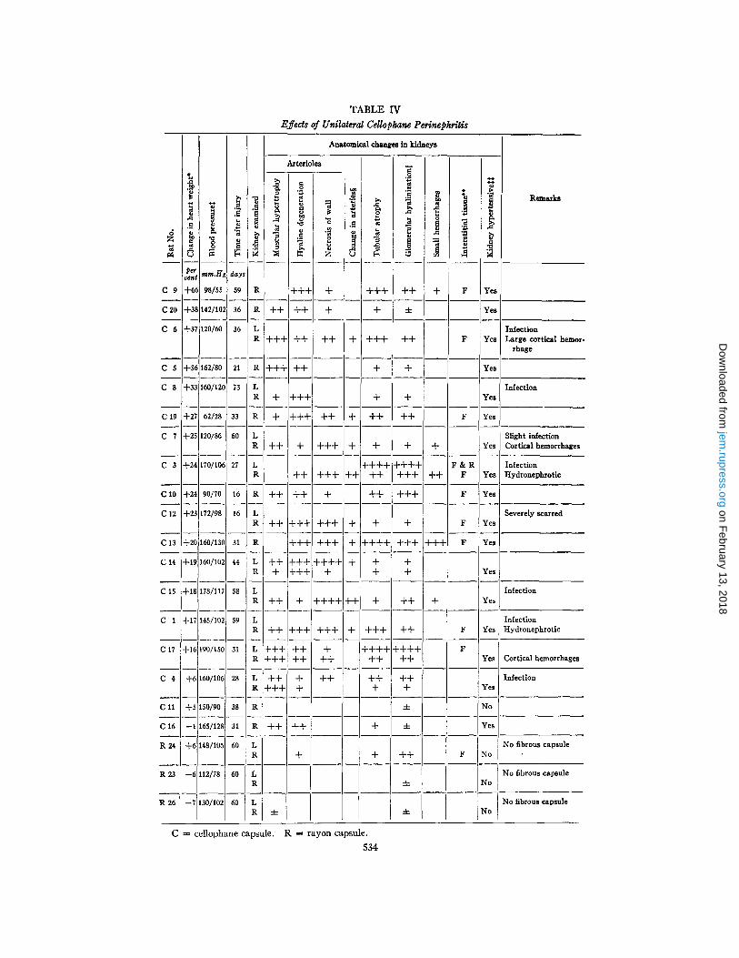
PLATE 13
FIGS. 1 to 6. Photographs of sections of the left kidneys of rats exhibiting hydro- nephrosis. Fig. 1, normal kidney; Fig. 2, rat H 94; Fig. 3, rat H 97; Fig. 4, rat H 100; Fig. 5, rat H 150; Fig. 6, rat H 147. The kidneys of all except that of No. H 147 functioned. Arterial hypertension was exhibited by all animals except No. H 147. Hematoxylin and eosin stain. X 2.
Fla. 7. Photomicrograph of right kidney of rat H 96, of which the left was hydro- nephrotic. Arterial hypertension was present. The glomerular arteriole is diseased, its wall thickened. Hematoxylin and eosin stain. X 500.
on February 13, 2018
jem.rupress.org
Dow
nloaded from
THE JOURNAL OF EXPERIMENTAL MEDICINE VOL. 75 PLATE 13
(Schroeder and Neumann : 2~-rterial hypertension in rats. II)
on February 13, 2018
jem.rupress.org
Dow
nloaded from

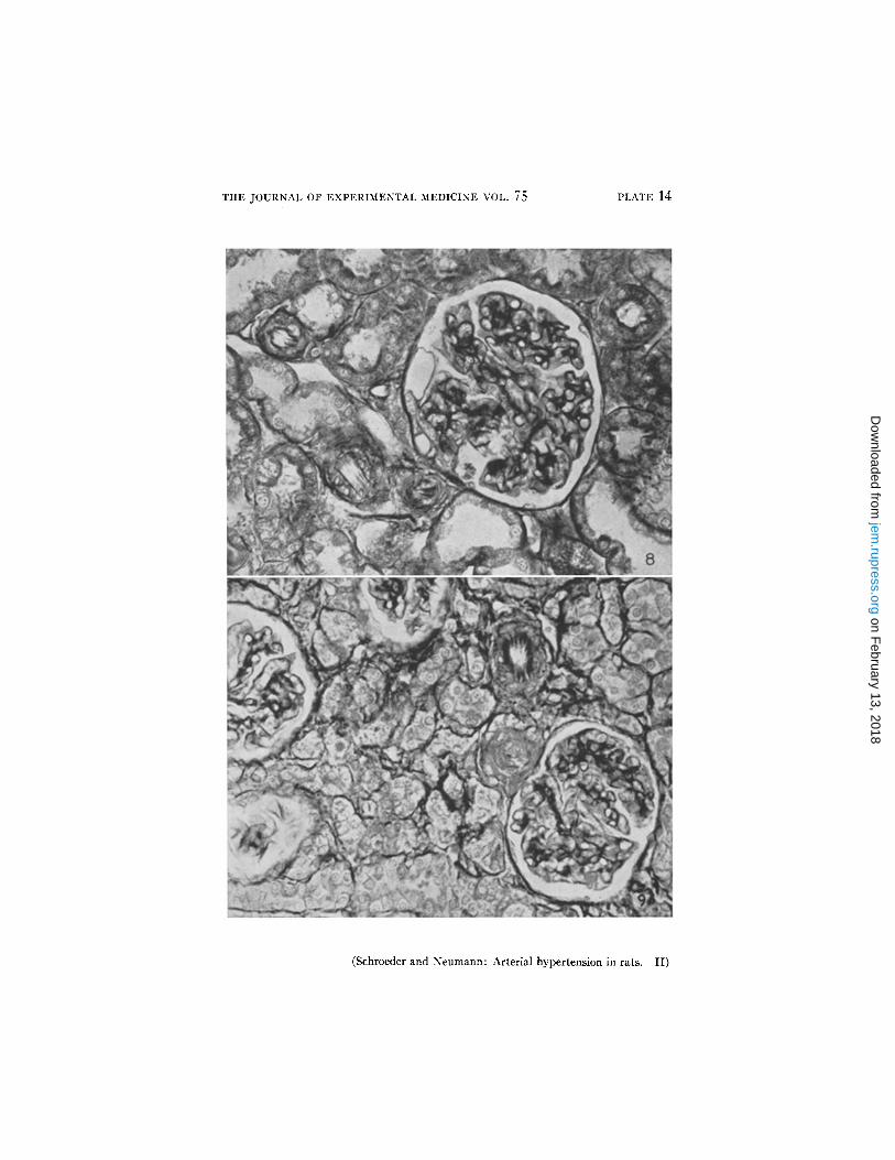
PLATE 14
FIOS. 8 and 9. Fig. 8, photomicrograph of section of right kidney, and Fig. 9, of left kidney of rat O 158. The left renal artery had been partially constricted 35 days previously, and the rat exhibited arterial hypertension. Vascular changes, hyaline degeneration, hypertrophy of muscle, and necrosis are present in the arterioles of both sections to a similar degree. Mallory connective tissue stain. X 400.
on February 13, 2018
jem.rupress.org
Dow
nloaded from
THE JOURNAL OF EXPERIMENTAL MEDICINE VOL. 75 PLATE 14
(Schroeder and Neumann: Arterial hypertension in rats. II)
on February 13, 2018
jem.rupress.org
Dow
nloaded from

PLATE 15
FIGS. 10 and 11. Fig. 10, photomicrograph of left kidney and Fig. 11, of right kidney of rat G 168. The left renal artery had been partially constricted 61 days previously. The animal exhibited cardiac hypertrophy, loss of weight, and convul- sions before death. In the left kidney there are moderate changes in the arterioles, in the right marked. Necrosis of the walls of the arterioles is present only in the right kidney. Mallory connective tissue stain. X 200.
on February 13, 2018
jem.rupress.org
Dow
nloaded from

THE JOURNAL OF EXPERIMENTAL MEDICINE VOL. 75 PLATE 15
(Schroeder and Neumann : Arterial hypertension in rats. II)
on February 13, 2018
jem.rupress.org
Dow
nloaded from

PLATE 16
FIGS. 12 and 13. Fig. 12, photomicrograph of section of left kidney, and Fig. 13, of right kidney of rat C 17. Cellophane perinephritis had been induced about the left kidney 31 days previously. The animal exhibited arterial hypertension. Vas- cular changes occurred in both kidneys to a similar degree. There is hyaline de- generation of the walls of the arterioles in both sections. Mallory connective tissue stain. X 200.
on February 13, 2018
jem.rupress.org
Dow
nloaded from

THE JOURNAL OF EXPERIMENTAL MEDICINE VOL. 75 PLATE 16
(Schroeder and Neumann: Arterial hypertension in rats. II)
on February 13, 2018
jem.rupress.org
Dow
nloaded from