(From ~he Department of Bacteriology and Experimental ...
23
THE ROLE OF THE "WAX" OF THE TUBERCLE BACILLUS IN ESTABLISHING DELAYED HYPERSENSITIVITY II. HYPERSENSITIVITY TO A PROTEIN ANTIGEN, EGG ALBUMIN* BY SIDNEY RAFFEL, M.D., LOUIS E. ARNAUD, C. DEAN DUKES, ProD., JWO S. HUANG (From ~heDepartment of Bacteriology and Experimental Pathology, Sckool of Medicine, Stanford University, Stanford) PLATES 1 Am) 2 (Received for publication, March 5, 1949) Recent publications (I, 2) have described the influence of the "wax" of the human tubercle bacillus in causing guinea pigs to respond with the delayed form of hypersensitivity to tuberculoprotein. In all essential features the hypersensitive state established by the use of these two bacteria-free compo- nents of the bacillus reproduced the allergy of infection, including cutaneous and systemic responsiveness to old tuberculin, a modified (non-necrotizing) Koch reactivity to the intracutancous injection of bacilli,and reactivity of ex- planted tissue to the presence of tuberculin. A natural sequence to these observations has been the question of the in- fluence of this lipoidal fraction in altering the type of hypersensitivity to other antigenic substances unrelated to the tubercle bacillus. Since tuberculopro- tcin alone performs immunologically like any other antigenic protein, inducing humoral antibody and anaphylactic sensitivity, and since these activities are extended by the addition of the wax to include induction of delayed allergy, it is reasonable to suppose that this function of the wax might reveal itselfas well with any antigen. This view has bccn implemented with evidence in the case of picryl chloride (3). Over twenty years ago Dicnes and his coworkers (4-6) showed that the in- jection of egg white into tuberculous guinea pigs, or into animals receiving injections of killcd tubercle bacilli,resulted in a type of reactivity to intra- cutaneous injection with this antigen which differed considerably from that ordinarily seen in animals sensitized with egg white. In the tuberculous ani- mals the dermal reactions to egg white persisted for 48 hours, and were often necrotic. In contrast, skin testsin sensitized non-tuberculous animals resulted in the usual edematous reactions of the Arthus type which reached their peak within a few hours and vanished or waned considerably within 24 hours. The former reaction resembles the tuberculin type of hypersensitive response; the * This work has been supported by grants from The National Tuberculosis Association, The CaliforniaTuberculosis and Health Association,and The Alameda County Tuberculosis and Health Association. 53 Downloaded from http://rupress.org/jem/article-pdf/90/1/53/1184229/53.pdf by guest on 20 December 2021
Transcript of (From ~he Department of Bacteriology and Experimental ...

THE ROLE OF THE "WAX" OF THE TUBERCLE BACILLUS IN ESTABLISHING DELAYED HYPERSENSITIVITY
II. HYPERSENSITIVITY TO A PROTEIN ANTIGEN, EGG ALBUMIN*
BY SIDNEY RAFFEL, M.D., LOUIS E. ARNAUD, C. DEAN DUKES, ProD., JWO S. HUANG
(From ~he Department of Bacteriology and Experimental Pathology, Sckool of Medicine, Stanford University, Stanford)
PLATES 1 Am) 2
(Received for publication, March 5, 1949)
Recent publications (I, 2) have described the influence of the "wax" of the human tubercle bacillus in causing guinea pigs to respond with the delayed form of hypersensitivity to tuberculoprotein. In all essential features the hypersensitive state established by the use of these two bacteria-free compo- nents of the bacillus reproduced the allergy of infection, including cutaneous and systemic responsiveness to old tuberculin, a modified (non-necrotizing) Koch reactivity to the intracutancous injection of bacilli, and reactivity of ex- planted tissue to the presence of tuberculin.
A natural sequence to these observations has been the question of the in- fluence of this lipoidal fraction in altering the type of hypersensitivity to other antigenic substances unrelated to the tubercle bacillus. Since tuberculopro- tcin alone performs immunologically like any other antigenic protein, inducing humoral antibody and anaphylactic sensitivity, and since these activities are extended by the addition of the wax to include induction of delayed allergy, it is reasonable to suppose that this function of the wax might reveal itself as well with any antigen. This view has bccn implemented with evidence in the case of picryl chloride (3).
Over twenty years ago Dicnes and his coworkers (4-6) showed that the in- jection of egg white into tuberculous guinea pigs, or into animals receiving injections of killcd tubercle bacilli, resulted in a type of reactivity to intra- cutaneous injection with this antigen which differed considerably from that ordinarily seen in animals sensitized with egg white. In the tuberculous ani- mals the dermal reactions to egg white persisted for 48 hours, and were often necrotic. In contrast, skin tests in sensitized non-tuberculous animals resulted in the usual edematous reactions of the Arthus type which reached their peak within a few hours and vanished or waned considerably within 24 hours. The former reaction resembles the tuberculin type of hypersensitive response; the
* This work has been supported by grants from The National Tuberculosis Association, The California Tuberculosis and Health Association, and The Alameda County Tuberculosis and Health Association.
53
Dow
nloaded from http://rupress.org/jem
/article-pdf/90/1/53/1184229/53.pdf by guest on 20 Decem
ber 2021

54 TUBERCLE BACILLUS WAX AND DELAYED HYPERSENSITMTY. II
latter is characteristic of the "immediate" form of dermal response. In line with this distinction, the first could not be transferred passively to normal animals by means of serum while the second could. This general type of demonstration of the influence of tubercle bacilli on the emergence of delayed sensitivity to various antigens has been added to by Hanks (7), Aronson (8), Landsteiner and Chase (9), Freund (10), and others.
In the present communication evidence is presented that the altered reac- tivity to egg albumin occasioned by the presence of living or killed tubercle bacilli is due to the same bacterial component which determines the occurrence of tuberculin hypersensitivity itself; i.e., the wax of the tubercle bacillus.
EXPERIMENTAL
Wa~.-~The wax employed here was obtained from two strains of tubercle bacilli of human type (H37Rv and Cutter bacilli) as described in previous papers (2, 3). The purified wax fraction was used. The chemical isolation procedures (11) and the precautions undertaken to assure the removal of bacterial bodies from this fraction have been described (2, 3). Dr. R. J. Anderson also supplied a sample of purified wax prepared from H37 bacilli in his own laboratory; this was freed of residual bacilli by the same ultrafiltration and prolonged ultra- centrifugation procedures employed with our own substances, and proved to have the same kind and degree of activity.
Phospha~ide.--Two samples of tubercle bacillary phosphatide were employed here as in our previous work (2); one was prepared by us from tubercle bacilli of human strains by the method of Anderson (12) while the other was supplied to us by Dr. Anderson as the purified phosphatide.
Egg Alburain.--Crystalline egg albumin was prepared from fresh hens' eggs. Separation of globulin was effected by half-saturation with ammonium sulfate. The albumin was re- crystallized three times by the addition of ammonium sulfate and acetic acid. The resultant product after dialysis was lyophilized and freshly dissolved for each use.
Walzr-in-Oil Eraulsion.--Tlfis was prepared in accordance with the procedure of Freund (10). Equal volumesof warm aquaphor and saline (containing egg albumin) were mixed in a mortar with the subsequent addition of three parts of paraffin oil.
Preparations/or Inje~t/on.--Egg albumin was dissolved in saline, 10 rag. per 0.5 ml. for each subcutaneous injection. When employed in water-in-oil emulsion, the same amount was dissolved in saline and incorporated in 0.5 mL of the completed emulsion.
Wax was emulsified in distilled water by the method of Sabin (13) and was employed in doses of 5 rag. per 0.5 ml., injected subcutaneously. Because the small residue of chloroform employed in preparing this emulsion was found to coagulate egg albumin, these substances were not injected together. Instead, the wax was inoculated into a marked subcutaneous site, and 2 hours later the egg albumin was injected into the same site.
When used in water-in-oil emulsion the wax was dissolved in the paraffin oil and the usual mixture made. Since in this case no chloroform was employed, i t was possible to incorporate the egg albumin in the same emulsion.
Phosphatide was cooled to 0°C. in a mortar and was emulsified, by light grinding, in saline. The egg albumin was incorporated during this process. Ten rag. of egg albumin was thus incorporated with 5 mg. of phosphatide in 0.5 ml. of saline. When combined in water-in-oil emulsion, the same procedure and quantities were employed as described for wax.
Culaneous Tests.--One or 2 rag. of egg albumin in 0.1 ml. of saline was employed as the skin test dose. The injection was made into the midflank. Readings were made at intervals
Dow
nloaded from http://rupress.org/jem
/article-pdf/90/1/53/1184229/53.pdf by guest on 20 Decem
ber 2021

S. RAF~'EL, L. E. ARNALrD, C. D. DUKES, AND J. S. HUANG 55
beginning at one-half hour after injection, and extending through 48 hours. Diameters were measured and the heights of reaction were estimated by palpation; both were recorded in millimeters.
Corneal Test~.--An egg albumin solution of 20 rag. per ml. in saline was used for corneal tests. Intracorneal injections were made through a 30 gauge needle with a 0.25 ml. tuber- culin syringe. Sufficient fluid was instilled centrally into the cornea to produce an area of clouding about 2 mm. in diameter. The volume of the individual injections could not be measured, but the average inocuinm was about 0.002 ml. Normal animals were tested at the same time as the experimental groups. The corneas were examined at 24 and 48 hours under a stereoscopic dissecting microscope (magnification X 27) with a strong light source at right angles to the visual plane. At 48 hours the animals were killed with ether and the eyes enucleated. Two parallel incisions were made on the superficial corneal layer of each eye to bracket the site of injection. The whole eyeball was then placed in Pere.nyi's fixative. Mter fixation the strip of cornea marked off by incisions was removed for histological process- ing. The corneal strips were sectioned parallel to the long axis, and alternate ribbons of sections were stained with hematoxylin and eosin. There was no possibility with this technic of failing to obtain sections through the area injected with antigen.
T~sue Cu/ture~.--These were set up by the hanging drop method as outlined previously (2), employing femoral bone marrow for explantation. The total volume of clot was 0.05 ml.; of this, 0.005 ml. was egg albumin in Simm's solution, 3 rag. per ml. Thus, 0.015 rag. of egg albumin was present in each preparation of 0.05 ml. volume.
Three explants of marrow from each animal were placed in each clot. Several slides were ordinarily prepared for simultaneous observation with and without addition of egg albumin. The latter was found to be without toxic effect on normal bone marrow explants.
Serological Tests.--Although it was desired to titrate sera by the optimal proportions method this was not feasible because of the small concentrations of antibody produced even by the most responsive guinea pigs. Complement fixation titrations were employed in- stead.
Dilutions of sera in 0.1 ml. amounts were titrated against 0.1 ml. volumes of 1:1,000 egg albumin. Incubation in the presence of 2 units of complement was carried out at 4°C. for 18 hours before the addition of sensitized erythrocytes. Adequate controls were included.
Passive Transfa, Test;.--These were carried out by the intracutaneous injection of 0.2 ml. amounts of the sera to be tested into guinea pigs of 250 to 450 gin. weight. Each animal received two different sera, one in each flank, and each serum was injected into two guinea pigs. Seventy-two hours later, 25 mg. of egg albumin was injected subcutaneously into the nuchal area. Readings of the local reactions were made at 0.5, 2, 4, 6, 24, and 48 hours.
RESULTS
1. Cutaneou3 Reactivity to Egg Albumin in Variously Sensitized Guinea Pigs.--In a p re l imina ry expe r imen t groups of two or three an imals were em-
p loyed for sensi t izat ion. One group consis ted of tubercu lous an imals which were
now sensi t ized dy subcu taneous in jec t ions of egg a lbumin . A second group was
compr i sed of gu inea pigs which had rece ived e ight in ject ions of tuberc le bac-
i l lary wax and were now g iven egg a lbumin into the sites of last wax inject ions.
A th i rd group of p rev ious ly u n t r e a t e d an imals was inocula ted wi th egg a lbumin
a long wi th bac i l l a ry wax. T h e las t group, also w i t h o u t p rev ious t r e a tmen t ,
was sensi t ized wi th egg a l b u m i n alone. Cu taneous tes ts wi th a lbumin re-
vea l ed a p ro longa t ion of responses in the tubercu lous and wax- t r ea t ed groups
Dow
nloaded from http://rupress.org/jem
/article-pdf/90/1/53/1184229/53.pdf by guest on 20 Decem
ber 2021

56 T U B E R C L E BACILLUS W A X AND DELAYED HYPERSENSITIVITY. I I
as compared with those occurring in the animals sensitized with egg albumin only. These preliminary results then agreed with previous observations (4-8, 10) concerning the influence of tubercle bacilli in determining the type of cu- taneous reactivity to protein antigens, and in addition suggested that the wax of the tubercle bacillus might be the element responsible for this alteration.
Experiments were now carried out with larger numbers of animals and with some changes in treatment. Groups of previously normal animals were em- ployed for the following injections:
Group/.--Egg albumin in saline. Group 2.--Egg albumin in water-in-oil emulsion. Group 3,--Egg albumin -b wax. Group 4.--Egg albumin Jr wax in water-in-oil emulsion. Group 5.--Egg albumin q- phosphatide. Group 6.--Untreated animals for skin test control. The first group received sensitizing injections of egg albumin alone in order to establish a
baseline of Arthus sensitivity/or comparative purposes. Group 2 was included in order to determine the possible influence of an ordinary immunologic adjuvant upon the type of sen- sitization. Groups 3 and 4 constituted the focal point of this experiment, and were tested to reveal the influence of tubercle bacillary wax upon the type of sensitization to egg albumin. Group 5 was added in order to demonstrate, as has been done with tuberculoprotein antigen (2), that another lipoidal constituent of the tubercle bacillus, the phosphatide, is not con, cemed in the induction of the delayed form of sensitivity.
Skin tests in these groups were carried out 2 weeks after each sensitizing inoculation, a total of five such injections being employed. Since cutaneous reactivity did not usually increase beyond the second sensitizing dose, we have presented a summarized version of the results of skin tests by combining the readings for each group after successive injections. These data are shown in Table I, and indicate that the responses in groups 3 and 4, which had received wax, were better maintained at the end of 24 and 48 hours than were those in the other, non-wax-treated, groups.
Obviously, the relative persistence of reactions in the wax-treated animals is insufficient grounds for judging the nature of the response, whether Arthus or delayed, since it is perfectly possible that more intense Arthus reactions might persist longer. However, examination of the levels of early responses (within the first 6 hours) disclosed that these were as marked in many of the animals in the non-wax-treated as in the wax-treated groups, but without the same persistence. An example is illustrated graphically in Text-fig. 1. In this case the non-wax-treated groups showed larger reactions at 1 hour than did the wax-treated; these were equalized at 2 hours, and by 6 hours the re- sponses in the wax groups became ascendant. At 24 hours the reactions in the non-wax-treated groups had fallen off considerably while those in the wax- treated groups were still at a high level, and this difference was seen also at 48 hours.
Dow
nloaded from http://rupress.org/jem
/article-pdf/90/1/53/1184229/53.pdf by guest on 20 Decem
ber 2021

TABLE I
A~age Cutaneous Reactions to Egg Albumin al Int~vats aflct Skin Testing
Sensitizing treatment
1 Egg albumin 2 Egg albumin
in water-in- oil
3 Egg albumin - ~ w a x
4 Egg albumin + wax in water-in-oil
5 Egg albumin + phospha- tide
6 Normal
181 45[ i
I
121 491
10i 43L
10i 31
141 14'
]R.e&ct~oB$"
1 hr. 2 hrs. 6 hrs. 24 hrs. 48 hrs.
15.2 2.0 14.1 1.7
14.2 1.5
14.7 1.4
No reading
10.2 1.2
15.6 1.8 12.9 1.5
17.3 1.9
16.2 1.7
21.9 2.1
8.9 0.9
19.0 1.9 19.5 2.2
22.3 2.2
24.2 2.1
2 4 . 8 2 . 3
6.8 0,7
8.5 0.9 10.3 1 . 0
16.9 1.8
18~4 1.8
7.7 0,8
2 . 4 0.3
1.9 0.3 1.9 0.3
9.1 1.1
11.5 1.5
2.9 0.3.
1.2 0,1
* The first figure for each reading is diameter in millimeters, the second is estimated height in millimeters.
film.
w,
m
A~DE A ]~ C])E ABC])Z.
g2 i
I
m
A B C I t E
5
m
A'BC DE. ABCDE.
24 4 8
m
t~OLtP5 T~xT-FIo. 1. Timed readings of intracutaneous tests with egg albumin. Group A, sensitized with egg albumin. Group B, sensitized with egg albumin in water-in-oil emulsion. Group C, sensitized with egg albumin + wax. Group D, sensitized with egg albumin + wax in water-in-oil emulsion. Group E, unsensitized controls. Width of column denotes average thickness of reaction for each group.
57
Dow
nloaded from http://rupress.org/jem
/article-pdf/90/1/53/1184229/53.pdf by guest on 20 Decem
ber 2021

5 8 TUBERCLE BACILLUS WAX AND DELAYED HYPERSENSITIVITY. II
I t appears then that the persistence of cutaneous reactions in the wax-treated groups denotes that delayed responses are superimposed upon initial Arthus reactions, which occur in all animals. Delayed reactivity is thus not a sub- stitutive form of response; it is additive. Analogy suggests this as a reasonable expectation; thus, tuberculous animals may show anaphylactic reactivity to tuberculoprotein as well as the tuberculin type of sensitivity.
Although suggestive, these observations of cutaneous responses cannot supply entirely objective evidence for the nature of sensitivity, and especially so when two types of reactivity may be coexistent. Other lines of evidence were there- fore pursued.
2, Relationship of the Delayed Cutaneous Response to the Corneal Reaction.- Several investigators have successfully employed the cornea for the study of hypersensitive reactions. This tissue, because of its avascularity, provides an excellent medium for distinguishing immediate from delayed types of hyper- sensitiveness. In the absence of blood vessels the Arthus response cannot occur here even though the cutaneous reactivity of the sensitized animal be very pronounced. Only when the corneal cells themselves are vulnerable to contact with antigen can a response be elicited in this tissue, and this con- dition appears to be met in the delayed type of sensitivity only.
Derick and Swift (14) with a non-hemolytic streptococcus and J'ulianelle (15) with the pneumococcus have provoked reactions in the scarified corneas of rabbits sen- sitized with intact bacterial cells and showing delayed skin responses. The latter author (16) was unable to demonstrate such reactions in rabbits sensitized with pneu- mococcal nudeoprotein or egg albumin and possessing only Arthus reactivity to these antigens. HoUey (17) and Rich and Follis (18) demonstrated the occurrence of de- layed reactions in the corneas of tuberculous animals following local injections of tuberculoprotein. These reactions were marked by edema, heavy cellular infiltra- tion, swelling and necrosis of corneal fibers and, in some instances, necrosis of the overlying epithelial cells.
The following experiment was carried out:
Eight guinea pigs which several months previously had been sensitized with albumin plus wax (in some instances in water-in-oil emulsion) were now reinjected with albumin plus wax in water-in-oil emulsion.
Nine guinea pigs were sensitized by three subcutaneous injections of egg albumin plus tubercle bacillary phosphatide in water-in-oil emulsion.
Eight guinea pigs which had been sensitized several months earlier by injections of egg albumin in saline or in water-in-oil emulsion were now reinjected with egg albumin in water- in-oil emulsion.
Seventeen days later all animals of these groups as well as several untreated guinea pigs were given skin tests. Early and late readings were made with results in each group conforming to the pattern already described; at 48 hours
Dow
nloaded from http://rupress.org/jem
/article-pdf/90/1/53/1184229/53.pdf by guest on 20 Decem
ber 2021

S. RAI~FEL, L. E. ARNAUD, C. D. DUKES, AND ~. S. HUANG 5~
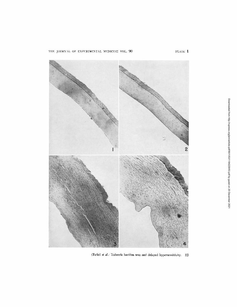
only the wax-treated animals revealed persisting reactions interpreted as de- layed responses. Simultaneously with the skin tests all the animals were ' inoculated centrally into the cornea of one or both eyes with a droplet of al- bumin solution. G~oss examination of the corneas at 24 and 48 hours revealed a striking distinction between group 1, which had been sensitized with albumin- wax, and the other groups, as illustrated diagrammatically in Text-fig. 2. At 24 hours seven of the eight wax-treated guinea pigs revealed corneal opacity, and this was seen in all eight at 48 hours. In contrast, the other groups at 24 hours showed slight evidence of trauma consisting of small areas of clouding about the site of needle penetration; this was less marked or had disappeared by 48 hours.
At 48 hours all animals were sacrificed and the corneas prepared for histo- logical examination. In the process of dissecting strips from the whole fixed corneas under the dissecting microscope, it was possible in every instance to distinguish the corneas of the wax-treated animals from those of the other groups. The former were markedly opaque and thickened while corneas from the non-wax-treated groups looked and felt entirely normal. Histological sec- tions confirmed these gross findings. In group 1, stained sections revealed thickening of individual fibers which resulted in an increased thickness of the cornea to two or three times that of the normal. Many corneal nuclei ap- peared to be swollen or degenerated, and in some instances the fibers them- selves appeared to have undergone necrosis. Cellular exudation was usually intense, extending throughout the tissue from limbus to limbus. In some in- stances patent capillaries were seen near the center of the cornea. In con- trast, corneas from animals of the other three groups showed either slight or no changes. Occasional mild cellular infiltration at the limbus was noted in some as the result of trauma. Edema or fiber thickening was not evident.
The microscopic observations are recorded in Text-fig. 2, and illustrated in Figs. 1 to 4.
This experiment was clear-cut in demonstrating that corneal reactivity to egg albumin is confined to those animals which had received tubercle bacillary wax along with the sensitizing injections of protein, and Which showed the protracted skin reactions to egg albumin.
One further comment may be of interest with respect to the delayed corneal reactivity seen here. We have observed corneal responses to tuberculopro- tein in guinea pigs with tuberculin hypersensitivity resulting from infection with virulent human bacilli and from inoculation of BCG. Skin tests revealed a high degree of sensitivity in the tuberculous animals, most showing necrotic reactions, and a moderate level of sensitivity in the BCG-vaccinated animals. Only the tuberculous group responded with corneal reactions; these were en- tirely comparable to those portrayed in Figs. 1 to 4. If we infer from this experience that a relatively high level of delayed hypersensitivity is necessary
Dow
nloaded from http://rupress.org/jem
/article-pdf/90/1/53/1184229/53.pdf by guest on 20 Decem
ber 2021

60" TUBERCLE BACILLUS WAX AND DELAYED HYPERSENSITIVITY. I I
I I ÷1+1 I +1 I ÷1 ÷1
-~:[~ e e e e e e e e m
@
~9 m W ~ UQ ~ m~
I _ 1
~ ~ ~ ÷ ÷ ÷ + +
8 o ~
@
$
r~
e,i
Dow
nloaded from http://rupress.org/jem
/article-pdf/90/1/53/1184229/53.pdf by guest on 20 Decem
ber 2021

S. RAF~FEL, L. E. ARNAUD, C. D. DUKES, AND J. S.. I-IUANG 6].
for the occurrence of the corneal response, the present results suggest that the wax treatment is very effective in this regard with egg albumin antigen.
3. Relationship of the Delayed Cutaneous Response to Reactions of Tissues i~, Culture.--Various investigations (8, 19-22) point to the usefulness of tissue. culture studies in providing a basis for differentiating types of hypersensitivity.. Explanted cells from animals with Arthus reactivity do not respond to antigen even in the presence of humoral antibody for the reasons discussed in connec- tion with corneal reactions. The cultured cells of the animal with delayed hypersensitivity, on the other hand, are directly affected by antigen, and upon exposure undergo injury or necrosis.
We have described the application of this procedure to the investigation of the tuberculin hypersensitivity induced in guinea pigs by means of isolated components of the tubercle bacillus (2). The same general methods were em- ployed here. Nineteen bone marrow explants from animals sensitized with egg albumin alone flourished in the presence of the antigen, whereas all 17 marrow fragments from guinea pigs sensitized with egg albumin plus tubercle bacillary wax showed some effect of exposure to antigen. In some instances this was manifested by poor growth and restricted migration of cells; in the majority of cases the tissues apparently died as judged by the relatively aceUular ap- pearances of the fragments, the granular debris about the sites, and the sparsity of intact cells. These results are portrayed in Figs. 5 to 10. I t is seen that' tissue from an egg albumin-sensitized animal grows normally in the presence of the antigen while under the same conditions the bone marrow of an animal sensitized to egg albumin with wax deteriorates,
Aronson (8) has investigated the comparative reactivities of tissues from guinea pigs sensitized in several ways. When tuberculous animals were sen- sitized to horse serum, by a procedure similar to that employed by Dienes, tissue explants subjected to the antigen failed to show the response described above. It seems possible that our success may be ascribed to higher levels of delayed reactivity in our animals. Not all animals reveal objective evidences of injury in experiments of this kind. Undoubtedly in these, as well as:in corneal tests, a certain minimal level of sensitivity is required in order that observable cellular changes may eventuate. In contrast, milder grades of sen- sitivity can be visualized in the cutaneous test because the occurrence of. in, flammation provides an index of reaction which is lacking in avascular tissue cultures or corneas.
4. Relationship of the Delayed Cutaneous Response to Anaphylactic Reactii~ ity.--Several animals in each of the four sensitized groups were tested for anaphy- lactic reactions to the intraperitoneal adminstration of egg albumin. A test dose of 10 mg. was chosen. I t was thought that with this dose and route it might be possible to distinguish levels of anaphylactic reactivity which would be ob- scured by the same dose given intravenously, or by a larger dose intraperitone.-
Dow
nloaded from http://rupress.org/jem
/article-pdf/90/1/53/1184229/53.pdf by guest on 20 Decem
ber 2021

62 TUBERCLE BACILLUS WAX AND DELAYED HYPERSENSITIVITY. I I
ally. This expectation was realized, as shown in Table II. Of twenty-two guinea pigs tested only eight developed the anaphylactic syndrome, and only in two of these was there a fatal outcome. I t happened that anaphylactic shock occurred only in animals of the non-wax-treated groups. This distri- bution is probably fortuitous, since there is no reason to believe that animals which receive wax develop less anaphylactic sensitivity than those which do not. The central point in this experiment is the fact that the persistent type of skin reactions occurred in those animals (wax-treated) which were at a lower level of anaphylactic sensitivity; this suggests that the occurrence of the per- sistent cutaneous response is not closely related to the level of anaphylactic seasitivity and hence may be independent of this manifestation of the im- mediate type of reactivity.
I t might be objected that the wax-treated animals failed to show anaphylactic shock with the dose and route employed because they possessed greater amounts
TABLE II Corrdagon of Skin Re.aa~ss with Anaphylaaiv Sensitinity
Group
Egg albumin . . . . . . . . . . . . . . . . . . . . . . . . . . . . . . . . . albumin + water-in-oil . . . . . . . . . . . . . . . . . . .
Egg albumin + wax . . . . . . . . . . . . . . . . . . . . . . . . . . Egg albumin + wax + water-in-oil . . . . . . . . . . . .
No. of guinea pigs
- - 7 6 6 5
Average skin &naphylactic sensitivity at reactions
48 hrs.
1.8 0.3 4* 0.5 0.1 4*
11.0 1.2 0 17.4 2.0 0
* 1 fatal.
of circulating antibody which combined with the antigen in the peritoneal cavity, protecting the animal from the generalized effects of antigen absorbed into the circulation. As shown in the next section, this objection has no va- lidity because the animals of group 3 (albumin + wax) possessed no greater concentrations of antibody than did those in groups 1 and 2 (albumin with- o u t w a x ) .
5. The Relationship of the Delayed Cutaneous Response to Humoral Anti- body.--
It is a well known criterion of hypersensitivities of the delayed type that they are entirely independent of the occurrence of antibody in the circulation. In contrast, Arthus reactivity in occurrence and intensity appears to parallel the antibody con- tent of the blood (23-26), although this view is not expressed unanimously (27, 28). The last statement refers to the rabbit; there is little information available for the guinea pig probably because this animal is a poor antibody producer and its serum is not readily assessed by quantitative methods. Cannon and Marshall (29) give data for three guinea pigs sensitized by subcutaneous injections of egg al-
Dow
nloaded from http://rupress.org/jem
/article-pdf/90/1/53/1184229/53.pdf by guest on 20 Decem
ber 2021

S. RAFFEL, L. E. ARNAUD, C. D. DUKES, AND J. S. HUANG 63
bumin. Arthus reactions were compared with serum antibody determined by the collodion particle technic. There is some indication of correlation although the small number of tests leaves the matter indefinite. Dienes (30) has stated that in general guinea pigs with high concentrations of precipitating antibody regularly develop hemorrhagic Arthus reactions, but it is evident from his earlier reports that this corre- lation is not absolute.
Our central interest in measuring humoral antibody was to determine whether the persistent cutaneous reactions were independent of circulating antibody levels. Since, however, we were dealing with animals possessing simultaneous Arthus reactivity, it was necessary to find out also whether these early oc- curring skin reactions were related to antibody concentrations.
Antibody determinations by the complement fixation method were carried out on sera obtained within 1 or 2 days of the time that skin tests were per- formed. The results are shown in Table III. In the first two groups, sen- sitized with egg albumin alone or in water-in-oil emulsion, the titers were low and occasional, and there was no relationship to the size of the early skin re- actions. Group 3, sensitized with wax plus egg albumin, showed the same paucity of positive serological results, again without relation to the severity of early skin reactions. In the last group, where wax and water-in-oil emulsion were combined with the antigen for sensitization, there were obviously in- creased antibody responses. Thus, in the three groups where the antibody responses were poor the early skin reactions were in general at the same levels as in the fourth group where the antibody concentrations were relatively good. Further, in this last group individual correlations of antibody to early skin reactions fail. A few results of precipitation titrations with the sera of these groups have shown the same lack of relationship of circulating antibody to Arthus reactivity.
I t is difficult to explain this failure of a relationship which is apparently characteristic in the rabbit. Possibly the serologic procedure employed is inadequate, but this seems unlikely in view of the sharp distinction it afforded between the antibody levels in group 4 as compared with the other groups. I t may be that the ranges of Arthus reactivity and of antibody in these animals were too narrow to permit a correlation to be established. More intense Arthus reactions might occur only in animals with higher concentrations of circulating antibody than seen here. If this were the case then it could be adduced that the persistent, sometimes necrotic, reactions occurring in the wax-treated animals were not simply severe Arthus responses, since they occurred in animals which failed to reveal any evidence of humoral antibody.
Beyond this speculation these results do not warrant ascribing critical value to the observation that the persistent skin reactions occurred without rela- tionship to the presence or concentration of humoral antibody.
One further point is pertinent here. The low titers seen in group 3, in ani-
Dow
nloaded from http://rupress.org/jem
/article-pdf/90/1/53/1184229/53.pdf by guest on 20 Decem
ber 2021

TABLE III
Corrdation of Skin Reactions with Serological Titrations (Complement Fixation)
Guinea pig No. Skin readings at
1-6 hrs. [ 24 hrs. [ 48 hrs.
Complement fixation titer
Group 1. Sensitized with egg albumin*
A380 B 19 E 83 E 81 E 8 4 E 82 E 8 0 E 83 E 82 E 8 4
23 19.5 19 17 17 12 11.5 11 10 9
2.5 22 2.0 2.5 6 0.5 2.0 0 0 3.0 8 1.5 2.5 7 1.0 1.5 12 1.0 1.5 14 1.5 1.0 11 1.0 1.5 7 1.0 1.0 13 1.5
13 0 0 0 0 0 0 0 0 6
1.5 0 0 0 0 0 0 0 0
1.0
Group 2. Sensitized with egg albumin in water-in-oil*
E 74 E 77 E 73 E 78 E 75 E 76 E 79 E 67 E 68
23 2.5 21.5 2.5 18 2.0 17 2.5 13 2.0 12 1.0 11 1.0 10 1.0 10 1.0
6 0 9
12 0 6
10 12 8
0.5 3 0 0
1.5 8 1.5 0
0 0 0.5 3 1.0 0 0.5 0 1.0 0
0.5 0
1.0 0 0
0.5 0 0 0
2 0 0 0 0 8 0 2 2
Group 3. Sensitized with egg albumin plus wax$
A 37 E 98 A377 E 71 E 98 E 72 E 70 E 97 A403 A404
20 2.0 21 2.0 18 2.0 15 1.5 18.5 2.0 22 3.0 19 2.0 16 1.5 15 1.5 12 1.5
23 2.0§ 23 2.0§ 20 2.0 36 3.0 12 1.5 22 2.5 24 2.0 11 1.0 11 1.5 8 1.5
26 2.5§ 18 2.0§ 17 2.0§ 15 1.5 11 1.5 9 1.5 6 1.0 5 0.5 0 0 0 0
Group 4. Sensitized with egg albumin plus wax in water-in-oil
E 51 E 4 9 E 4 8 E 67 B 14 E 61 E 62 E 88 E 6 3
21 2.0 19 2.0 20 2.0 20.5 2.0 26 2.0 20 2.0 14 1.5 22 2.0 24 2.0
24 2.0 28 2.5§ 22 2.5 21 2.5 30 2.5§ 20 2.5 28 2.0 8 1.5
28 2.5
23 2.5 22 2.5§ 20 2.5 18 3.0 18 2.5§ 18 2.0 12 2.0 8 1.5 6 0.5
* Arranged in order of descending early (1 to 6 hours skin reactions. Arranged in order of descending late (48 hours) skin reactions.
§ Central necrosis.
64
2 64
128 32 0 8
64 2 0
Dow
nloaded from http://rupress.org/jem
/article-pdf/90/1/53/1184229/53.pdf by guest on 20 Decem
ber 2021

S. RAFFEL, L. E. ARNAUD, C. D. DUKES, AND J. S. HUANG 65
mals treated with egg albumin and wax, reveal the inefficacy of wax as an ordinary immunologic adjuvant. Similar observations have been made with the sera of animals receiving tuberculoprotein or picryl chloride with wax. This indicates that the activity of wax in determining the hypersensitive response is not that of an adjuvant. On the other hand, when wax and water- in-oil emulsion were employed together, there was a pronounced adjuvant effect in addition to the alteration of the hypersensitive response, and this also has been seen as well with the two antigens mentioned above. The adjuvant
ac t iv i ty of this mixture is a general one; it intensifies the altered hypersensi- tivity as well as the elaboration of antibodies.
6. Passive Transfer Tests with the Sera of Variously Sensitized Guinea P igs . - For our particular purposes the passive transfer test, in order to serve as a criterion of the delayed nature of the cutaneous responses occurring in wax- treated animals, should result in the transfer of Arthus reactivity from members of all groups sensitized, while at the same time there should be no transfer of the persistent reactions at all. However, such tests could not settle the pos- sibility that the persistent cutaneous responses are merely prolonged Arthus reactions, since failure of persistence in the transfer sites might mean simply that insufficient antibody was transferred to effect an intense local sensitiza- tion. This interpretative difficulty springs from the nature of the present experiment; if one were working with animals in which only immediate or only delayed reactions occurred, passive transfer tests should be perfectly straight- forward.
Nevertheless, it was desirable to know what the results of such tests might be, and to this end an experiment was carried out employing methods favorable for the transfer of reactivity to the skin of normal animals by means of serum. The sera of fourteen guinea pigs sensitized with egg albumin, of fifteen sensi- tized with albumin and wax, and of three normal animals, were each injected in 0.2 ml. amounts intradermally into the flanks of two normal animals of 250 to 450 gin. weight. Three days later each recipient pig received an injec- tion of 25 rag. of egg albumin subcutaneously in the nuchal area. Within a few minutes most animals were in varying degrees of anaphylactic shock, and five of the total group of thirty-two animals died within 40 minutes of injec- tion. Since each guinea pig had received two different sensitizing sera, one on each flank, it was not possible to differentiate which sera may have been responsible for the transfer of anaphylaxis. I t was obvious, however, that the transfer of anaphylactic reactivity was a general phenomenon in contrast to the paucity of cutaneous reactions (Table IV). The latter were of the early type, none persisting to 24 hours. In a technical sense therefore, no transfer of the delayed cutaneous responses occurred, but this conclusion possesses questionable merit on the basis of the considerations discussed above, and in view of the few successful transfers of the early cutaneous reactivity. This
Dow
nloaded from http://rupress.org/jem
/article-pdf/90/1/53/1184229/53.pdf by guest on 20 Decem
ber 2021

66 TUBERCLE BACILLUS WAX AND DELAYED HYPERSENSITIVITY. I[
kind of experiment appears not to be well adapted to the conditions involved in this study.
7. Other Attempts to Establish Criteria of Delayed ttypersensitivity.--Several other attempts to characterize the hypersensitivity appearing in these various groups of animals are only briefly described here because they have contributed no definite evidence on the question. However, they may be of general in- terest in relation to the hypersensitive states with which we have been con- cerned.
(a) Systemic Delayed Reactivity.--Two experiments were carried out to de- termine whether animals with delayed reactivity induced by albumin and wax might show delayed systemic reactions analogous to that which follows the in- jection of tuberculin in the tuberculous subject.
In one experiment seven protein-wax-sensitized animals, eleven guinea pigs sensitized with albumin alone, and three normal controls were injected with
TABLE IV Passive Transfer of Cutaneous Reactivity from Variously Sensitized Guinea Pigs
Group sensitized with
E g g a l b u m i n . . . . . . . . . . . . . . . . . . . . . . . . . .
E g g a l b u m i n in wa te r - in -o i l . . . . . . . . . . . . .
E g g a l b u m i n + w a x . . . . . . . . . . . . . . . . . . .
E g g a l b u m i n + w a x in w a t e r - i n - o i l . . . . . .
N o r m a l . . . . . . . . . . . . . . . . . . . . . . . . . . . . . . .
No. of sers No. of reci]~ient guinea pigs
18 10 18 12 6
Passive skin reactions beginning
at i hour after injection
2/18 0/10 1/18 0/12 0/6
0.1 ml. of 1:1,000 epinephrine followed by 25 mg. of egg albumin subcutane- ously. Three hours later this was followed by the same dose of epinephrine and 35 rag. of albumin and again, 3 hours later, the same dose of epinephrine and 50 mg. of albumin. Epinephrine was employed in order to mitigate anaphylactic reactivity and so to afford an opportunity to visualize any delayed response to the antigen. No such reactions were seen. A second experiment designed to test the same point was carried out in animals unprotected by epinephrine, employing a smaller dose--10 mg.--of egg albumin by the intm- peritoneal route. Eleven animals sensitized with egg albumin and eleven sensitized with albumin plus wax were so tested. In the latter group several of the animals were markedly positive by skin test, yet in no instance was any sign of delayed illness seen.
Dienes and Schoenheit (31) reported their general failure to show systemic reactions of the delayed type in tuberculous guinea pigs sensitized with egg white, and dis- cussed the possible factors responsible. They suggest that in the ease of tuberculosis
Dow
nloaded from http://rupress.org/jem
/article-pdf/90/1/53/1184229/53.pdf by guest on 20 Decem
ber 2021

S. RA~'PEL, L. E. ARNAUD, C. D. DUKES, AND J. S. HUANG 67
the systemic reaction is caused by the absorption of toxic products from tuberculous foci, and that analogous circumstances are not present in the case of reactivity to egg white antigen. Rich (32) takes issue with this view, believing success or failure to depend solely upon the level of hypersensitivity of the subject tested. Whatever the case, certain it is that systemic reactions cannot be elicited in all animals with tuberculous (2) nor with streptococcal hypersensitivity (reference 14 and unpublished data from this laboratory). The failure to demonstrate it here, therefore, adds no conclusive information regarding the nature of the hypersensitivity with which we are dealing.
(b) The Use of Pyribenzaraine as an Aid to the Distinction of Immediate and Delayed Cutaneous Reactions.--Antihistaminic drugs effectively inhibit ur- ticarial reactions in most human beings with "atopic" hypersensitivity. The compounds may be applied locally to the site of antigen administration, or may be given orally (33). Such drugs (i.e., benadryl and pyribenzamine) are not similarly effective in inhibiting the Arthus reaction in rabbits sensitized to penicillin or horse serum (34). However, it was considered worthwhile to investigate the possible usefulness of one of these drugs on this basis: tha t it might inhibit the early cutaneous reactions in animals of all groups, while leaving intact the delayed reactions in those guinea pigs sensitized with al- bumin plus wax. To this end two experiments were carried out with pyri- benzamine.
In the first trial, 3 mg. d the drug w a s mixed with each skin test dose of albumin, and the mixtures injected intracutaneously in animals of the various experimental groups as well a s
normal controls. Pyribenzamine proved irritating to the tissues so that all responses were augmented over the levels ordinarily seen, but in their relative aspects the skin reactions were entirely similar to those previously encountered. There was no indication of inhibition of the immediate skin reactions by this drug.
In a second experiment pyribene.amine was administered by subcutaneous injection at a distance from the skln test area. Each animal received 3 mg. per kilo of the drug on the morning of skin testing, and one-half hour later the skin test injections were made. Two and 5 hours later the doses of drug were repeated, so that a continuing level of pyribenzamine w a s
maintained in these animals beginning prior to the injection of antigen and for some hours thereafter. Readings made at 1, 2, and 6 hours after the skin tests showed the same develop- ment of responses in all groups as ordinarily seen, and subsequent readings, at 24 and 48 hours, differed in no wise from those usually observed.
I t is apparent then that this drug fails to influence the development of cutaneous Arthus reactions in guinea pigs sensitized to egg albumin, and this despite the high concentrations of drug administered compared with the dosage effective in human beings. Pyribenzamine is thus not a suitable aid for differentiating between immediate and delayed cutaneous reactivity in animals in which both types of responsiveness may be present.
(c) Contact Tests.--Tests were carried out to determine whether guinea pigs sensitized with albumin plus wax might develop contact responses. Those
Dow
nloaded from http://rupress.org/jem
/article-pdf/90/1/53/1184229/53.pdf by guest on 20 Decem
ber 2021

68 T U B E R C L E BACILLUS W A X AND DELAYED HYPERSENSITIVITY. I I
hypersensitive states characterized by cutaneous contact reactions appear to fall into the category of delayed reactivities.
Ordinarily, contact reactions occur when sensitization has been mediated through the skin itself (e.g., chemicals, poison oak, etc.). Only occasionally will delayed hypersensitivity induced by some other route result in the sensi- tization of epidermal cells to contact with antigen (3, 35, 36). In the present experiments the route of sensitization was subcutaneous, hence there was no reason necessarily to expect the emergence of contact reactivity. Its occur- rence, however, would be an additional criterion for the delayed nature of the hypersensitivity induced by wax with albumin, and experiments were under- taken to determine this possibility.
In the first of two experiments, a mixture of 50 mg. of egg albumin per ml. of aquaphor was rubbed into the skin, with negative results. In the second, crystalline egg albumin was placed on small squares of slightly moistened filter paper. These were applied to the skin of the flank previously prepared by depilation with barium sulfide and washing with ether. Over the patch was placed a larger square of lead foil, and the whole bound down firmly with adhesive tape wrapped entirely around the animal. The larger square of foil ensured an area surrounding the patch unirritated by the adhesive tape. The patches were removed after 48 hours and in all cases they were found to have remained in intimate contact with the skin. Neither at this time nor during 3 subsequent days of observation was there any indication of response in seven animals sensitized with albumin plus wax and showing good reactions in intracutaneous tests.
DISCUSSION
The waxy lipid of the human tubercle bacillus, which in previous com- munications has been shown to determine the delayed type of hypersensitive response to tuberculoprotein (1, 2) and to picryl chloride (3), is now demon- strated to produce the same effect with a non-bacterial protein, egg albumin. This demonstration localizes to a particular component of the tubercle bacil- l u s - t h e wax--a function which has for some years been known to be a property of the entire bacillary cell as such (4-10). The information contained in this report was sought in order to generalize further the ability of the wax to effect the kind of hypersensitive response which is associated with the presence of the tubercle bacillus in the tissues.
In two previous reports of the same kind of experiments with tuberculopro- tein (2) and picryl chloride (3), there was no particular difficulty in distinguish- ing the delayed cutaneous reactions from Arthus responses. In the first case, this was aided by the use of old tuberculin as the test antigen, for preceding investigations as well as our own have shown that animals with Arthus reac- tivity induced by tuberculoprotein do not respond with skin reactions to the
Dow
nloaded from http://rupress.org/jem
/article-pdf/90/1/53/1184229/53.pdf by guest on 20 Decem
ber 2021
S. RAFFEL, L. E. ARNAUD, C. D. DUKES, AND ~. S. HUANG 6~
heated product (old tuberculin). In the case of picryl chloride we were unable to demonstrate the existence of Arthus reactivity in any of the animals, and it was necessary only to prove that the responses seen were of the delayed• type. In the present instance it was obvious that all animals sensitized with egg albumin antigen developed Arthus reactivity, but only in those treated with wax did the persistent type of reactions develop in addition. I t was then necessary to provide evidence that the persistent reactions were not simply prolongations of more intense Arthus responses, but were in fact superimposed reactions of the delayed ("infectious") type.
A variety of procedures were employed in order to establish this point. Positive results stem chiefly from observations of cutaneous tests, from the relationship of cutaneous reactions to anaphylactic responses, from determina- tions of corneal reactivity, and from tissue culture studies. Other modes of study, including humoral antibody titrations, passive transfer studies, contact tests, and the use of an antihistaminic agent, failed to provide definite evidence on the question. I t is interesting that methods which are definitive in ane instance are not applicable in another; for example, contact reactivity is striking in experiments with picryl chloride but entirely negative with egg albumin, while tests of delayed systemic reactivity are productive in some animals sensitized with tuberculoprotein and wax but were without effect in this case. Some of these differences, such as the last mentioned, may depend upon dif- ferences in the level of sensitivity attainable with various antigens, but others are not explainable simply on a quantitative basis. Through the experiments with the various antigenic systems there run, however, the common threads of delayed cutaneous reactivity and the occurrence of injury in avascular tissues (cornea, tissue explants).
We have speculated before without profit on the nature of the activity of the bacillary wax which induces this type of immunologic response to antigens, and at the present time we have no evidence which adds to the clarification of this point. As with the previous antigens employed, here also water-in-oil emulsion was without effect, as was the purified phosphatide fraction of the tubercle bacillus. This activity is not then a general property possessed by any lipid or liquid hydrocarbon, nor is it related to an adjuvant activity of wax in the ordinary immunologic sense.
I t is of some interest that egg albumin may be injected into a subcutaneous site 2 hours after the deposition of wax with positive results; this procedure was, in fact, employed as routine in the present experiments. I t was found, however, that if a 24 hour interval were allowed between the two injections, delayed reactivity did not result. If the r61e of the wax should depend upon its ability to induce cytologic changes which in turn account for the changed reactivity of the tissues to antigen, the 24 hour interval between injections should be suitable for the induction of delayed sensitivity, since these ch'anges
Dow
nloaded from http://rupress.org/jem
/article-pdf/90/1/53/1184229/53.pdf by guest on 20 Decem
ber 2021

70 TUBERCLE BACILLUS WAX AND DELAYED HYPERSENSITIVITY. II
would be under way by the time antigen was injected into the area. That this does not occur suggests some other basis for the activity of wax than its property of inciting a granulomatous cellular response; this tentative con- clusion also follows from certain other considerations (3).
SUMMARY
Guinea pigs sensitized with egg albumin along with the purified wax fraction of the human tubercle bacillus respond with delayed hypersensitive reactivity to the protein antigen. Previous publications have reported a similar activity of the wax with respect to tuberculoprotein and picryl chloride. The effect is not referable to an ordinary adjuvant activity of the bacillary wax, since anti- body titers are not increased in animals which receive it, and since a known adjuvant, water-in-oil emulsion, has no effect with respect to the induction of delayed hypersensitivity.
This report further extends the r61e of the tubercle bacillary wax in the induction of delayed hypersensitive states.
BIBLIOGRAPHY
1. Raffel, S., Am. I~ . Tuberc., 1946, 54, 564. 2. Raffel, S., J. Infect. Dis., 1948, 82, 267. 3. Raffel, S., and Fomey, J. E.., J Exp. Med., 1948, 88, 485. 4. Dienes, L., and Schoenheit, E. W., Prac. Soc. Exp. Biol. and Med., 1926, 24, 32. 5. Dienes, L., J. Ir~r, unol., 1927, 14, 61.
"61 Dienes, L., J. Ir~munol., 1928, 15, 153. 7. Hanks, J. H., J. I~nmunol., 1935, 28, 105. 8. Aronson, ]. D., J. Imr~unol., 1933, 25, 1. 9, Landsteiner, K., and Chase, M. W., J. Exp. MeA., 1940, 71, 237.
10. Freund, J., and McDermott, K., Proc. So¢. Exp. Biol. and Med., 1942, 49, 548. 11. Anderson, R. J., J. Biol. Chem., 1929, 83, 505. 12. Anderson, R. J., Lothrop, W. C., and Creighton, M. M., J. Biol. Clwm., 1938,
:125, 299. 13. Sabin, F. R., J. Exp. Med., 1935, 62, 751. 14. Derick, C. L., and Swift, H. F., J. Exp. Med., 1929, 49, 615. 15. JulianeUe, L. A., J. Exp. Meal., 1930, 51, 633. 16. JulianeUe, L. A., J. Exp. Meal., 1930, 51, 643. 17. Holley, S. W., Am. J. Path., 1935, ].1, 937. i8. Rich, A. R., and Follis, H. R., Jr., Bull. Johns Hopkins Hosp., 1940, 66, 106. 19. Rich, A. R., and Lewis, M. R., Proc. Soc. Exp. Biol. and Med., 1927-28, 25, 596. 20. Aronson, J. D., J. Exp. Meal., 1931, 54, 387. 21. Moen, J. K., and Swift, I-I. F., J. Exp. Med., 1936, 64, 339. 22. Moen, J. K., J. Exp. Meal., 1936, 64, 355. 23. Opie, E. L., J. Imrnunol., 1924, 9, 231. 24. Culbertson, J. T., J. Irnmunol., 1935, 29, 29. 25. Cannon, P. R., Physiol. Rev., 1940, 20, 89.
Dow
nloaded from http://rupress.org/jem
/article-pdf/90/1/53/1184229/53.pdf by guest on 20 Decem
ber 2021

S. :RAFFEL, L. E. ARNAUD, C. D. DUKES~ AND .]. S. HUANG ~]~
26. Fischel, E. E., and Kabat, E. A., J. Immunol., 1947, 55, 337. 27. Seibert, F. B., J. Infect. Dis., 1932, 51, 38.3. 28. Kahn, R. L., J. ImmunoL, 1933, 25, 307. 29. Cannon, P. R., and Marshall, C. E., J. Immunol., 1941, 40, 127. 30. Dienes, L., Arch. Path., 1936, 9.1, 357. 31. Dienes, L., and Schoenheit, E. W., Am. Rew. Tuberc., 1929, 20, 92. 32. Rich, A. R., The Pathogenesis of Tuberculosis, Springfield, Illinois, Charles C.
Thomas, 1946, 428-431. 33. Feinberg, S. M., J. Am. Med. Assn., 1946, 139., 703. 34. Dreisbach, R. H., J. Allergy, 1947, 18, 397. 35. Sulzberger, M B., J. Allergy, 1947, 18, 176. 36. Cooke, R. A., J. Allergy, 1944, 15, 203.
Dow
nloaded from http://rupress.org/jem
/article-pdf/90/1/53/1184229/53.pdf by guest on 20 Decem
ber 2021

~2 TUBERCLE BACILLUS WAX Alq~ DELAYED HYPERSENSITMTY. H
EXPLANATION OF PLATES
PLATE 1
FIo. 1. Cornea of normal guinea pig. Forty-eight hours after injection of egg alb-m~n. Hematoxylin and eosin. X 85.
FIo. 2. Cornea of guinea pig sensitized with egg albumin. Forty-eight hours after injection of egg albumin. Hematoxylin and eosin. X 85.
l~os. 3 and 4. Corneas of guinea pigs sensitized with egg albumin plus wax. Forty° eight hours after injection of egg albumin. Hematoxylin and eosin. X 85.
Dow
nloaded from http://rupress.org/jem
/article-pdf/90/1/53/1184229/53.pdf by guest on 20 Decem
ber 2021
THE JOURNAL OF EXPERIMENTAL MEDICINE VOL. 90 PLATE 1
(Raffel et al.: Tubercle bacillus wax and delayed hypersensitivity. II)
Dow
nloaded from http://rupress.org/jem
/article-pdf/90/1/53/1184229/53.pdf by guest on 20 Decem
ber 2021
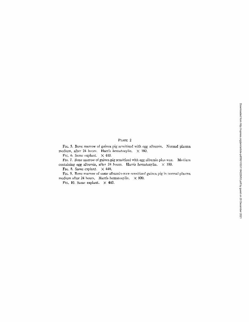
PLATE 2
FIG. 5. Bone marrow of guinea pig sensitized with egg albumin. Normal plasma medium, after 24 hours. Harris hematoxylin. X 100.
FIG. 6. Same explant. X 440. FIG. 7. Bone marrow of guinea pig sensitized with egg albumin plus wax. Medium
containing egg albumin, after 24 hours. Harris hematoxylin. × 100. Fro. 8. Same explant. × 440. FIG. 9. Bone marrow of same albumin-wax--sensitized guinea pig in normal plasma
medium after 24 hours. Harris hematoxylin. × 100. FIG. 10. Same explant. × 440.
Dow
nloaded from http://rupress.org/jem
/article-pdf/90/1/53/1184229/53.pdf by guest on 20 Decem
ber 2021

TItE JOURNAL OF EXPERIMENTAL MEDICINE VOL. 90 PLATE 2
(Raffel et al,: Tubercle bacillus wax and delayed hypersensitivity. II)
Dow
nloaded from http://rupress.org/jem
/article-pdf/90/1/53/1184229/53.pdf by guest on 20 Decem
ber 2021