From Department of Clinical Neuroscience, Div. of ...
66
From Department of Clinical Neuroscience, Div. of Neurology, Div. of Neurosurgery, Neuroimmunology Unit, Center for Molecular Medicine, Karolinska Institutet, Stockholm, Sweden ADULT NEURAL STEM CELLS AND THEIR DIFFERENTIATION CHOICES -examples from experimental inflammation and transplantation Cynthia Pérez Estrada Stockholm 2014
Transcript of From Department of Clinical Neuroscience, Div. of ...
From Department of Clinical Neuroscience, Div. of Neurology, Div. of Neurosurgery,
Neuroimmunology Unit, Center for Molecular Medicine, Karolinska Institutet, Stockholm,
Sweden
ADULT NEURAL STEM CELLS AND THEIR DIFFERENTIATION
CHOICES
-examples from experimental inflammation and transplantation
Cynthia Pérez Estrada
Stockholm 2014
Cover image:
Far left panel (blue and red), SVZ NPCs neurosphere stained with DAPI and Laminin B1.
The different colored variations in the following panels, represent the different NPC
specialization states, which they aquaire through interactions with their envrionment.
All previously published papers were reproduced with permission from the publisher.
Published by Karolinska Institutet.
Printed by Åtta.45 Digital Print AB
Karlsrogatan 2, 170 65 Solna
© Cynthia Pérez Estrada, 2014
ISBN 978-91-7549-642-9

TO MY ABUELITO, MOTHER, FATHER, AND MY FAMILY
ABSTRACT
The aim of this thesis is to study how inflammation influences the fate of NPCs, which can
impact the regenerative capacities of the CNS.
In paper I, we used a hypoglossal nerve avulsion model, to study if survival, differentiation, and
integration of transplanted NPCs could be achieved. Transplanted NPCs were present after 3
months post grafting, and expressed differentiation markers specific for neurons, astrocytes, and
oligodendrocytes. Motor neuronal cell survival was increased in injured animals which received
the NPC graft. This results show that transplanted NPCs can modulate the microenvironment
increasing motor neuronal survival. Further, in Paper II, we explored the same transplantation
setting in a transection injury of the hypoglossal nerve, i.e. an injury that yields significantly less
neuronal cell death in contrast to the more severe avulsion injury. Comparison of transplanted
NPCs into both injury models was then investigated. In the transection injury model we found
that NPC transplanted to the hypoglossal nucleus, maintained their Sox2 expression after three
months transplantation. Also, no differentiated progeny derived from the graft was observed.
Motor neuronal survival was not significantly different from non-transplanted animals,
indicating that the cues for NPCs differentiation provided from the environment were different
comparing to the avulsion model.
Reactive oxygen species (ROS), are an integral part of inflammation, so we next questioned
whether H2O2 a member of the ROS family would have an effect on the differentiation outcome
of NPC from the brain lateral ventricles. In paper III I found that NPCs exposed to H2O2 resulted
in more neurons and oligodendrocytes. H2O2 exposure influenced proliferation and the
expression of genes involved in chromatin remodeling and antioxidant defense. These results
indicate that H2O2 has a specific modulatory effect on the differentiation potential of NPCs.
In paper IV the global gene expression and differentiation outcome from NPCs from the brain
and the spinal cord were analyzed under normal conditions and after Experimental Allergic
Encephalomyelitis (EAE), a model for multiple sclerosis (MS). We found that NPC from naïve
rat brains were more neurogenic compared to spinal cord (SC) naïve NPCs. In contrast, NPCs
from the SC of EAE rats became more neurogenic comparing to healthy SC NPCs. Thus, the
inflammatory environment produced during EAE proves to be able to change NPC fate.
Overall the findings presented in this thesis suggest the inflammatory milieu as well as its
individual components, like H2O2, act as mediators of the regenerative properties of NPCs.
POPULAR SCIENCE ABSTRACT
In recent years the number of companies offering “the promise” that stem cells will regenerate
new organs, or even help us to live forever has become increasing, But, how much do we really
know about stem cells, and what can they really do for our health? In my thesis I concentrate on
a special type of stem cell, the adult neural stem cell. These cells are found in our brain and
spinal cord, and maintain limited potential to regenerate new brain cells throughout adulthood. I
investigate on how the environment instructs these stem cells to choose different fates, through a
process called differentiation. Differentiation allows these cells to become the brains three most
important cell types, neurons, astrocytes and oligodendrocytes. After injury an inflammatory
response arises, where several types of free radicals are present, causing various changes to the
microenvironment and to the neural stem cells. These changes can range from altered cellular
behavior to cell death. Understanding the role that inflammation and free radicals play in the
functionality of adult neural stem cells could help us to design better therapies to regenerate the
damaged adult central nervous system. My results show that free radicals can direct the NPCs to
become neurons; crucial for the communication of information in the brain, and
oligodendrocytes; necessary to ensure neuronal communication. Further I also show that after
injury there is an initial attempt for regeneration by the nervous system, instructing the stem
cells to become neurons, however inherent changes caused by inflammation, also provide cues
towards malfunctioning of these newly differentiated cells. Thus, to answer the initial questions,
we have to remain cautious on the use of stem cells for clinical therapies, as we still need to
learn further on applying and controlling stem cell potential in inflammatory conditions, to help
us optimize regenerative therapies. My thesis work, contributes to the path towards
understanding these possibilities envisioning a future where we can unleash the regenerative
potential of the CNS.
POPULAR SCIENCE ABSTRACT (SPANISH)
En los últimos años nuevas compañías que prometen regenerar nuevos órganos y hasta vida
eterna usando células madre ha incrementado, Pero, ¿cuánto sabemos realmente sobre las células
madre, y que pueden hacer estas células por nuestra salud? En mi tesis me concentro en una
clase especial de célula madre, las células madre neuronales adultas. Estas células se encuentran
en el cerebro y la médula espinal, y se mantienen con el potencial para regenerar nuevas células
cerebrales en la edad adulta. Así pues, he investigado los efectos que el microambiente tiene en
el comportamiento de estas células, influyendo la decisión que toman al elegir diferentes
destinos celulares, a través de un proceso llamado diferenciación. La diferenciación permite a
estas células transformarse en tres importantes tipos de células cerebrales; neuronas, atrocitas y
oligodendrocitos. Después de que una lesión ocurre en el sistema nervioso central (SNC), una
reacción inflamatoria ocurre, donde varios tipos de radicales libres están presentes, causando
diversos cambios en el microambiente y a las células progenitoras existentes. Estos cambios
pueden modificar el comportamiento celular o hasta causar la muerte de dichas células.
El entendimiento del rol que la inflamación y los radicales libres desempeñan en el
funcionamiento de las NPCs podría ayudarnos a diseñar mejores terapias para regenerar el SNC
adulto después de haber sido lesionado. En esta tesis investigue los efectos que los radicales
libres y la inflamación en el SNC tienen sobre la capacidad de diferenciación de las NPCs.
Mis resultados muestran que los radicales libres pueden instruir a las NPCs para convertirse en
neuronas; cruciales para la transferencia de información en el cerebro, y oligodendrocitos;
necesarios para garantizar la comunicación neuronal. Además, también muestro que después de
que ocurren daños en el SNC, el SNC hace in intento inicial para repararse, instruyendo a las
células madre para convertirse en neuronas, sin embargo cambios inherentes dentro de las
células en estas circunstancias, conllevan al inicio de mecanismos que pueden significar el mal
funcionamiento de estas células al madurar. Por lo tanto, respondiendo a las preguntas iniciales,
se tiene que mantener cautela en cuanto al uso de las células madre para terapias clínicas, ya que
todavía tenemos que aprender más sobre el funcionamiento y el control del potencial de éstas en
las enfermedades inflamatorias, para ayudar a optimizar terapias regenerativas. Mi trabajo de
tesis, contribuye al entendimiento de estas posibilidades imaginando un futuro en el que
podamos liberar el potencial de regeneración del Sistema Nervioso Central.
LIST OF PUBLICATIONS I. Neural stem/progenitor cells transplanted to the hypoglossal nucleus integrates with the host
CNS in adult rats and promotes motor neuron survival.
Michael Fagerlund*, Cynthia Pérez Estrada*, Nasren Jaff, Mikael Svensson and Lou Brundin
Cell Transplantation 2012, 21 (4):739-747
II. Integration differences of transplanted adult neural progenitors in two models of axotomy.
Cynthia Pérez Estrada*, Michael Fagerlund*, Nasren Jaff, Lou Brundin and Mikael Svensson
Manuscript
III. Oxidative stress increases neurogenesis and oligodendrogenesis in adult neural progenitor
cells.
Cynthia Pérez Estrada, Ruxandra Covacu, Mikael Svensson, Lou Brundin
Stem Cell and Development 2014, (Epub ahead of print).
IV. Change of fate commitment in adult neural progenitor cells subjected to chronic
inflammation.
Ruxandra Covacu*, Cynthia Pérez Estrada*, Lisa Arvidsson*, Mikael Svensson & Lou Brundin
The Journal of Neuroscience, 2014, 34 (35): 11571-11582
*These authors contributed equally
The author has also contributed to the following publications/manuscripts.
I. Nitric oxide-induced neuronal to glial lineage fate-change depends on NRSF/REST function in
neural progenitor cells
Bergsland M, Covacu R, Perez Estrada C, Svensson M, Brundin L.
Stem Cells, 2014, 32(9):2539-49
II. Pericytes generate scar tissue across multiple central nervous system disorders
David O. Dias *, Yildiz Kelahmetoglu *, Cynthia Pérez-Estrada **, Jemal Tatarishvili **, Aurélie Ernst
1, Zaal Kokaia , Olle Lindvall , Lou Brundin , Christian Göritz , Jonas Frisén
*These authors contributed equally to this work
**These authors contributed equally to this work
Manuscript
III. Altered gene expression and differentiation in spinal cord neural progenitor cells, after
exposure to low level inflammation.
Lisa Arvidsson, Ruxandra Covacu, Cynthia Peréz Estrada, Sreenivasa Raghavan Sankavaram, Mikael
Svensson, Lou Brundin
Manuscript
IV. Characterization of the response of endogenous spinal cord neural stem cells to EAE.
Ongoing collaboration with Jonas Frisen´s group.
TABLE OF CONTENTS
Introduction…………………………………………………………………………………...1
1. Adult Neurogenesis……………………………………………………………..…… ..……...2
Brain lateral sub-ventricular wall structure……….…………………….………3
Spinal cord central canal cyto-architechture…….…………………….………..3
Adult spinal cord radial glia has NPC properties….……………………..……..4
Filum Terminale……………………. ……………………………………… 4
The adult NPC niche…… ……………………………………………………5
2. Adult Neuroregeneration……………………………………………………..…..…….. .....…8
Regenerative potential in planarians (plathelmynthus)…………………………8
Salamander’s regenerative capacities…. ……………………………………...8
Human regenerative potential…………….. ……………………………….…..9
3. Nerve Avulsion and Transection Injuries………………………….….. .………………..…10
Nerve avulsion injuries……………. ………………….……………………..10
Transection injuries……… …….. ……………………………………….11
Hypoglossal nerve injury model………………………….. .. ……………….11
4. Stem Cell Transplantations………………………………………….….……………..……..12
NPC transplantation…………………….……………………………….…….12
5. Epigenetic Regulation of Adult progenitor Cells Differentiation..…………..……...……......13
Histone methylation and demethylation……………………………….. …….13
Histone Acetylation and Deacetylation……….……….. ……………………..14
6. Experimental Allergic Encephalomyelitis (EAE)…………………………...……...………..15
Rat clinical symptoms….………….………………………………………….15
Clinical similarities between EAE and MS..….………………………………16
NPC transplantation on the EAE model…..….……………………………….16
7. Reactive Oxygen Species and Their Role in NPC Signaling………….….…….……………16
Free Radicals……………………………………….………………………....17
Inflammation and H2O2….… …………………………………………….…..18
H2O2 and Neuroinflammation.…………… ……………………………….….18
ROS are necessary for the adult SVZ stem cell niche and NPCs function……18
The NPC niche and ROS….………………………………….…………….…19
NPC Antioxidant Mechanisms…………..……………………………………19
Super oxide dismutase (SOD)……….….………………………………….…20
Glutathione peroxidase (GPx)….…….…………………………………….…20
Catalase (CAT)…………………………………...…………………...………20
Peroxiredoxins (Prxs)…………………………………………… ………….21
8. Aims of The Study………………………………………………………….………….…..22
9. Results and Discussion…………………………………………………...………….……..23
Paper I.………………………………………………………..............…………..…….….23
Paper II...……………………………………………………..............……………….........26
Paper III....……………...……………………………………..............……….……….......29
PaperIV….…….…..................................................................................................................34
Concluding Remarks…………....................................................................................................42
Aknowledgements........................................................................................................................45
References.....................................................................................................................................46
Appendix: Papers 1-IV
LIST OF ABBREVIATIONS ALS
Ascl1
CAM
CAT
CC
CNS
CSF
DA
DAVID
DCX
DG
EAE
eGFP
GFAP
GFP
GPx
H2O2
HDACs
IHC
IPA
MeV
MOG
NAD
NO
NPC
Prxs
ROS
SC
SOD
SOX
SVZ
VEGF
WebGestalt
Amyotrophic Lateral Sclerosis
Achaete-scute homolog 1
Cell Adhesion Molecule
Catalase
Central canal
Central Nervous System
Cerebrospinal Fluid
Dark Agouti
Database for Annotation, Visualization and Integrated Discovery
Doublecortin
Dentate Gyrus
Experimental Allergic Encephalomyelitis
Enhanced –GFP
Glial Fibrillary Acidic Protein
Green Fluorescent Protein
Glutathione Peroxidase
Hydrogen Peroxide
Histone Deacetylases
Immunohistochemistry
Ingenuity Systems Pathway Analysis
Multiexperiment Viewer
Myelin Oligodendrocyte Glycoprotein
Nicotinamide Adenine Dinucleotide
Nitric Oxide
Neural progenitor cell
Peroxiredoxins
Reactive Oxygen Species
Spinal Cord
Super Oxide Dismutase
Sry-containing HMG box
Sub-Ventricular Zone
Vascular Endothelial Growth Factor
WEB-based Gene Set Analysis Toolkit

1
INTRODUCTION In the adult brain there is a cavity that persists through development, the ventricular system.
A lateral ventricle occupies each cerebral hemisphere. The lateral ventricles cell architecture,
as well as the spinal cord organization contain a special type of CNS cell, the neural
progenitor cell (NPC). NPCs were not discovered but until 1996 by Weiss et al. (Weiss et al.,
1996). NPC have the capacity to self-renew and to differentiate into specialized Central
Nervous System (CNS) cells (Doetsch et al., 1999, Weissman et al., 2001, Gage, 2000). CNS
pathologies ranging from neurodegenerative disorders to neuro trauma challenge the
regeneration and plasticity capacities of the adult mammalian CNS. In other organisms like
planarians and salamanders, the capacities for regenerating entire damaged nerve networks
are immense, however in mammals like humans and rodents, this capacity is limited or non
existant. Through evolution the mechanisms for loss of regeneration in humans could have
compensated a more intricate and complex nerve wiring for a lower regenerative capacity, as
rewiring existing circuits could represent a major challenge to guide, differentiate and
reconnect to the right terminals. There is increasing evidence that the adult CNS in mammals
retains poor plasticity, and neurogenesis takes place at a limited occurrence. The
environment surrounding the adult NPC is a strong regulator of NPC behavior. The focus of
my thesis centers on the interactions that the environment and the adult NPCs play together,
interactions which challenge the differentiation and integration of the NPC.
2
1 ADULT NEUROGENESIS
Exactly 101 years ago, in 1913 Ramón y Cajal introduced the prevalent notion that neuronal
birth exists only at embryonic stages, “In the adult centers, the nerve paths are something
fixed, ended, and immutable. Everything may die, nothing may be regenerated. It is for the
science of the future to change, if possible, this harsh decree.” (extract from Ramón y Cajal “
Estudios sobre la degeneración y regeneración del sistema nervioso”). Ramón y Cajal died
in 1934, only 28 years later in 1962 Joseph Altman, using H3-thymidine labeling, was able to
follow cells that had incorporated it into new chromosomal DNA, during mitotic cell
division. This lead to the first report on new neurons in the adult rat brain. More evidence
reporting adult neurogenesis in birds, rodents and in non-human primates in both the sub
ventricular zone (SVZ) and the dentate gyrus of the hippocampus would follow (Morshead et
al., 1994, Palmer et al., 1997, Nottebohm, 1989). Further Bernier et al, showed that the SVZ
NPCs contribute to neurogenesis in the amygdala of adult monkeys (Bernier et al., 2002), but
a central question remained, is there neurogenesis in the adult human CNS?
The first report on human adult neurogenesis came in 1998 by Eriksson et al,
who by using BrdU labeling found ongoing neurogenesis in the adult human dentate gyrus
(Eriksson et al., 1998). These results were confirmed by Jonas Frisen´s lab last year
(Spalding et al., 2013) and this year (Ernst et al., 2014). Further, in 2012 using C14 cell
dating, Bergman et al showed none or limited neurogenesis in the human olfactory bulb (OB)
(Bergmann et al., 2012). Given the presence of NPCs in the adult human SVZ (Quinones-
Hinojosa et al., 2006) this result re-opened the question, if there really is neurogenesis in the
adult human brain. In rodents the OB is vital for survival, as it plays a crucial role in their
interaction with the environment. Humans on the contrary, rely more on other brain functions
which may receive constant environmental stimuli, like learning and memory in the cortex.
However, Frisen et al using the C14 cell dating, found that there was no neurogenesis in the
adult human cortex (Spalding et al., 2005, Bhardwaj et al., 2006).
All together, these studies suggest adult neurogenesis in the adult human
brain occurs in specific locations of the adult brain, highlighting the SVZ and the Dentate
gyrus (DG) as neurogenic areas. In conclusion the adult human brain has some extent of
plasticity, further investigation on new born and integrated neurons in the adult human brain
will have to be addressed in the future.
3
I will next shortly describe the three areas in the CNS which harbor NPC niches
in the adult human and rat CNS; the SVZ of the brain, the sub granular area of the
hippocampus and the spinal cord and the filum terminale at the caudal end of the SC.
Brain lateral sub-ventricular wall structure
In the adult brain, the major germinal niche is in the sub-ventricular zone of the lateral
ventricles. Through development ependymal cells displace progenitor cells from the lateral
ventricle, into the sub-ventricular zone. The architecture of this area is populated by
ependymal cells which constitute the E1 and E2 cell types. They have cilia which contacts
the cerebrospinal fluid (CSF) and are found surrounding the radial glia (Mirzadeh et al.,
2008). Radial glia (RG) are the NPC during development (Malatesta and Gotz, 2013) and in
adulthood the neural progenitors cells of the SVZ, RG are known as the B1 cell type which
expresses GFAP+ (Doetsch et al., 1999) and similarly to E cells, the B1 cells also protrude
cilia contacting the CSF on one side and protruding with long specialized processes to
directly contact blood vessels (Fuentealba et al., 2012). B1 cells give rise to oligodendrocyte
progenitors (Olig2+) and transit amplifying type C cells (Ascl1+) (Parras et al., 2004), C
cells then further commit to become A type cells, expressing doublecourtin (DCX) (Nacher et
al., 2001), which are also known as the transit amplifying neural progenitor lying underneath
the B1 cells. Surrounding this germinal zone are microglial cells, blood vessels and CSF. It
becomes apparent the role that the environment may play in the behavior of the NPCs,
signaling through blood vessels and CSF.
Spinal cord central canal cyto-architechture
NPC in the spinal cord are located at the central canal (CC) region of the SC. This area is
divided on to two compartments an ependymal compartment and a sub-ependymal
compartment (Hamilton et al., 2009, Johansson et al., 1999). At the ependymal
compartment, ependymal cells in the CC are in closed contact to the vasculature, and have
from one to three cilia contacting the CSF, also some ependymal cells in this area are bi-
nucleated and have four cilia contacting the CSF (Alfaro-Cervello et al., 2012a). Ependymal
cells of the SC CC have been described to express Nestin (Meletis et al., 2008) but also
Afaro-Cervello et al have identified an ependymal cell population in the SC CC which is
Nestin negative (Alfaro-Cervello et al., 2012b), both of this described ependymal cell
populations possess neural progenitor properties. Ependymal cells have been described to
activate their stem cell potential after injury, where they were found to proliferate, migrate
and differentiate, in response to SC injury (Johansson et al., 1999, Brundin et al., 2003,
Meletis et al., 2008) . Dromard et al, (Dromard et al., 2008) describe a subset of ependymal
4
cells, the SC subependymal cells which express markers common to NPC from SVZ of the
adult brain and are able to differentiate into neurons, astrocytes and oligodendrocytes.
The cellular microenvironment organization of the SC CC consists of neurons
contacting the CSF (Alfaro-Cervello et al., 2012b), GFAP+ positive cells, and rare
proliferating ependymal cells which generate ependymal cell doublets. The sub-ependymal
layer’s ependymal cells, express GFAP, and are surrounded by neurons, both in contact with
the CSF and the microenvironment vasculature. Also, oligodendrocyte precursors are an
integral part of this niche (Hamilton et al., 2009).
It’s important to mention that NPC from the different regions of the SC
present distinct differentiation properties (Kulbatski and Tator, 2009, Shihabuddin et al.,
1997) (Paper IV), these differences could also indicate variations in the molecular and
physical constitution of the NPC niche at the different levels of the SC; cervical, thoracic,
and caudal which could define their characteristics.
Adult spinal cord radial glia has NPC properties
Within the adult rodent SC there is another cell type with NPC properties, these cells are a
heterogeneous population, sharing diverse genes with SVZ NPCs, and CC NPCs, the spinal
cord radial glia (RG) (Petit et al., 2011) (Table 1). The RG cells are found in the white matter
of the adult SC, these cells may proliferate in response to injury and differentiate in to
astrocytes (Bannerman et al., 2007), but they can also become oligodendrocytes and neurons
(Ohori et al., 2006). The identity of radial glia has been elusive, but they are known to
express several markers, PDGFβ, vimentin, NG2 and GFAP (Sabourin et al., 2009, Petit et
al., 2011). Some of these markers like PDGFb and vimentin are shared by other cell types
like pericytes, the scar building cells after SC injury (Goritz et al., 2011). It would definitely
be of help to find specific markers to define this cell population, to elucidate contributions to
neurogenesis and possible uses in regenerative therapies.
Filum terminale
At the most caudal part of the adult mammalian spinal cord, another NPC population resides,
as the CC narrows down to disappearing caudally, ependymal cells are found surrounded by
fibroblast, adipocytes, non-ciliated ependymal cell conglomerates, neuroblasts, neurons and
glial cells (Fontes et al., 2006, Jha et al., 2013). Culturing of the human and rodent filum
terminale area yields free floating neurospheres which differentiate into neurons, astrocytes
and oligonderocyes (Varghese et al., 2009, Arvidsson et al., 2011, Jha et al., 2013), however
5
the true identity of the NPC in this region has not yet been as well characterized as in the
SVZ and the CC of the SC.
All together the NPCs from the spinal cord show heterogenic qualities, further
characterization of these NPC populations would shed light into the roles of each of these
NPCs during inflammation and their impact in neuro repair.
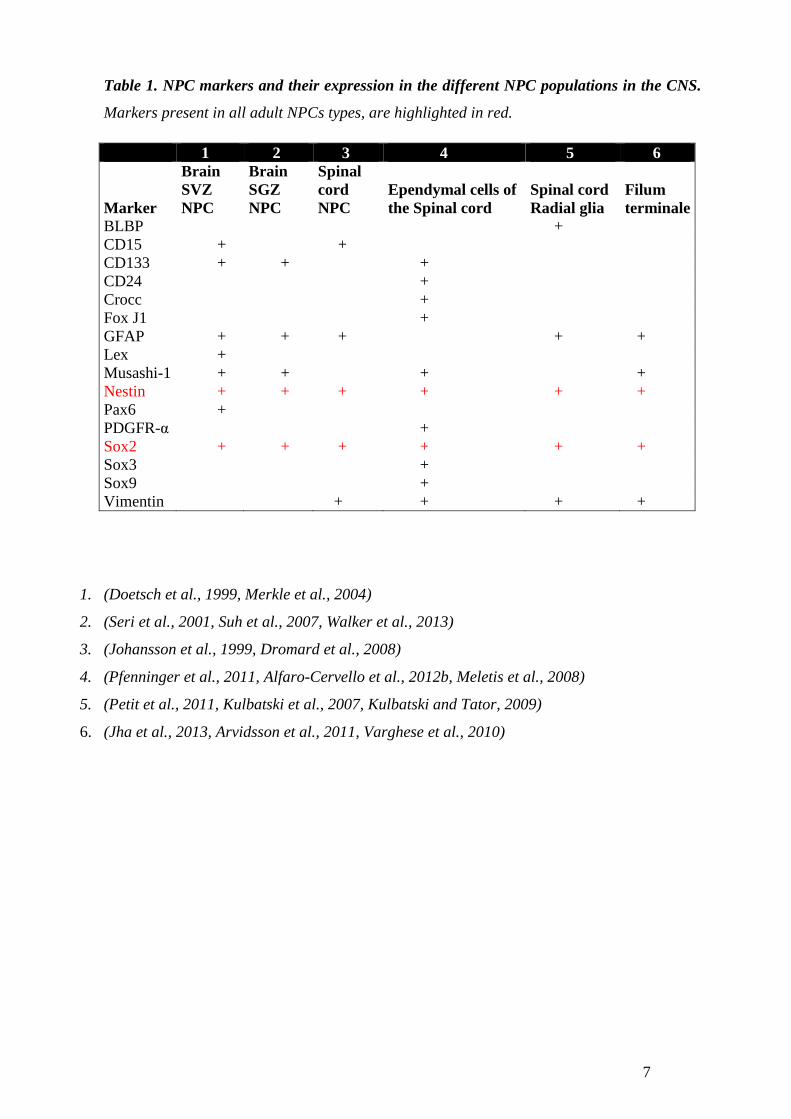
Table 1, shows the most commonly described NPC markers expressed in the heterogeneous
populations of the adult NPC in the CNS. Two markers are present among all the different
NPC populations, Sox2 and Nestin, being Sox2 the most widely used pluripotency marker.
Sox2 belongs to the group of genes SoxB1, crucially expressed during neurogenesis in the
embryo (Avilion et al., 2003, Masui et al., 2007) (For review see (Uchikawa et al., 2011)). In
the adult CNS Sox2 expression has been shown in NPCs (Brazel et al., 2005, Ellis et al.,
2004, Suh et al., 2007), and proven to be necessary for maintaining the undifferentiated state
(Taranova et al., 2006) as well as for neurogenesis in the adult CNS (Ferri et al., 2004). The
validation of Sox2 in these studies shows its importance in stablishing and maintaining
NPCs, and makes Sox2 a valuable marker for pluripotency.
The adult NPC niche
The adult NPC niche plays a crucial role in regulating stemness, cell proliferation, and
differentiation. The components of the NPC niche orchestrate a dynamic architecture capable
of providing the NPCs with the necessary environmental conditions to maintain their
properties. The study of the stem cell niche is an emerging field, next I provide a short
summary of some of the most important components of the adult NPC niche. The Basal
laminae extra cellular matrix, contains fractones (Mercier et al., 2002) which can
compartmentalize growth factors and cytokines (Bernfield et al., 1984) and play a special role
in the niche, where fractones sequester and hold mitogens like FGF, which bind to specific cell
types. Fractones contribute to an environment rich in stemness promoting factors (Kerever et
al., 2007), playing a crucial role in the maintenance of the NPCs pluripotent state.
Mechanical force. External forces on the stem cells are also part of the stem cell niche, some
groups have demonstrated that tension forces can regulate pluripotency in the presence of the
appropriate growth factors, suggesting the fine relationship between the chemical and the
physical environment that regulate stem cell behavior. (Nava et al., 2012) An example of this
quality are Cell Adhesion Molecules (CAMs) which are anchoring factors providing physical
tension, influencing cell geometry and cell differentiation, or loosing this physical tension and
allowing differentiation. Among all the CAMs, E-cadherin is considered a key molecule for
stem cell pluripotency (Li et al., 2012). Other important components of the niche, include other
6
cell types which participate of the architectural components of the niche; at the
supraependymal area of the SVZ there is a network of 5HT positive neurons, which innervates
the SVZ and may trigger proliferation of B1 and E1 cells upon release of 5HT (Tong et al.,
2014). B1 and E1 cells are also in close proximity to blood vessels, pericytes, endothelial cells,
and also microphages and fibroblasts (Mercier et al., 2002) which in turn could further provide
different molecular signals regulating stem cell behavior.
7
Table 1. NPC markers and their expression in the different NPC populations in the CNS.
Markers present in all adult NPCs types, are highlighted in red.
1. (Doetsch et al., 1999, Merkle et al., 2004)
2. (Seri et al., 2001, Suh et al., 2007, Walker et al., 2013)
3. (Johansson et al., 1999, Dromard et al., 2008)
4. (Pfenninger et al., 2011, Alfaro-Cervello et al., 2012b, Meletis et al., 2008)
5. (Petit et al., 2011, Kulbatski et al., 2007, Kulbatski and Tator, 2009)
6. (Jha et al., 2013, Arvidsson et al., 2011, Varghese et al., 2010)
1 2 3 4 5 6
Marker
Brain SVZ NPC
Brain SGZ NPC
Spinal cord NPC
Ependymal cells of the Spinal cord
Spinal cord Radial glia
Filum terminale
BLBP
+ CD15 +
+
CD133 + +
+ CD24
+
Crocc
+ Fox J1
+
GFAP + + +
+ + Lex +
Musashi-1 + +
+
+ Nestin + + + + + + Pax6 +
PDGFR-α
+ Sox2 + + + + + +
Sox3
+ Sox9
+
Vimentin
+ + + +
8
2. ADULT NEUROREGENERATION
It’s of importance to model CNS regeneration and find new candidate genes in organisms with
better regenerative capacities than humans. Evolutionary conserved genes have been identified
across species, like Drosophila M. genes present in human development and higher mammals,
allowing for the modeling and discovery of possible candidate genes in humans. Salamander
and planarians, possess copious regenerative capacities, and have therefore been widely
studied. Their regenerative capacity is very successful and could help us to model new
approaches to regenerate the mammalian CNS; Here I attempt to highlight some of the most
significant regenerative features from these organisms, to give a broader perspective to the
human regenerative features and possibilities.
Regenerative potential in planarians (plathelmynthus)
Planarians, members of the phylum Platyhelminthes, are widely studied due to their
outstanding regenerative capacities. Planarians can regenerate an entire new planarian from a
very small piece of tissue, and any cell type and organ of their adult body can be regenerated
from a single pluripotent adult stem cell type, known as the neoblast (Elliott and Sanchez
Alvarado, 2013, Reddien, 2013). There is crescent interest for finding the master genes for
pluripotency in the neoblast, since these genes could have analogues in mammals which could
allow for the discovery and implementation of regenerative therapies (Lobo et al., 2012).
Important gene candidates have already been identified, among which are genes homologues to
human genes like peroxiredoxin genes (Galloni, 2012), and possibly Oct4 and Sox2 (Onal et
al., 2012). Understanding the role of these factors in the topology of gene networks would give
us the opportunity to elucidate their possible role in mammalian regeneration.
Salamander’s regenerative capacities
Salamanders belong to the phylum cordata and have a remarkable potential for regeneration.
Salamanders are capable of regenerating the tail and limbs, (Simon and Tanaka, 2013) brain
(Parish et al., 2007) and spinal cord (Davis et al., 1990), but also the retina (Stone, 1950), lens
(Stone, 1967)and jaws (Morrison et al., 2006). The best studied regenerative area in
salamander is the limb blastema, which refers to the area from which a new limb grows back
after amputation. One of the mechanisms for limb regeneration, occurs after adult stem cells in
this area reverse to a more immature state (Sandoval-Guzman et al., 2014). Inflammation could
possibly initiate epigenetic mechanisms which unlock the cells dedifferentiation towards
9
rebuilding a new limb. There is a continuous effort to understand limb regeneration in
salamander and to develop new molecular markers to trace cell commitment in either
dedifferentiated cells or into the inherent stem cell pool which contribute to the limb formation.
Injured neurons and muscle cells may also exert specific secretomes orchestrating the
patterning and activation of the regenerative machinery (King and Newmark, 2012). Although
gene homologues to human genes have been identified like retinoic acid, Hox A genes (Nacu
and Tanaka, 2011) and Hdac genes (Taylor and Beck, 2012), many more remain to be unveiled
in this area.
Human regenerative potential
In adult humans some organs retain their ability for regeneration, like liver, bone and blood,
however CNS regeneration after injury is very limited. As mentioned earlier Eriksson et al
(Eriksson et al., 1998), and Frisen et al (Ernst et al., 2014) have reported neurogenesis in the
striatum and hippocampus, this findings imply a possible difference in between rodent
neurogenesis and human neurogenesis. Also Frisen´s lab, reported increased neurogenesis in
the striatum after stroke, however whether this young neuroblasts mature and integrate in the
environment is not known (Ernst et al., 2014). Another brain pathology which stimulates
neurogenesis is epilepsy, where neurogenesis occurs in the subgranular zone of the DG
(Ekdahl, 2012). Additionally, In the environment surrounding CNS injury, factors which
prevent regeneration have been found, like inhibitory molecules secreted by the CNS (Pernet
and Schwab, 2012), the immune system (Fukazawa et al., 2009) and in the scarring which
occurs after injury (Fawcett et al., 2012) among others. It is still debatable to at what extent the
immune system activation is beneficial or detrimental to regeneration (Monje et al., 2003)
(Paper III, Paper IV) but for example in amphibians (Fukazawa et al., 2009) and rats (Monje et
al., 2003) inhibiting immune cells activation after injury rescues regeneration. Also the
development of a thick glial scar after injury is now known to inhibit regeneration (Fawcett et
al., 2012), studies in axolotl salamanders, show wound healing occurs scar free (Seifert et al.,
2012).
Studying CNS regeneration in other organisms could help us find mechanisms and
pharmacological targets for improved regeneration in the adult mammalian CNS, and for the
possible discovery of silent genes present in humans with the potential to regenerate the CNS
after injury (Fig. 1).
10
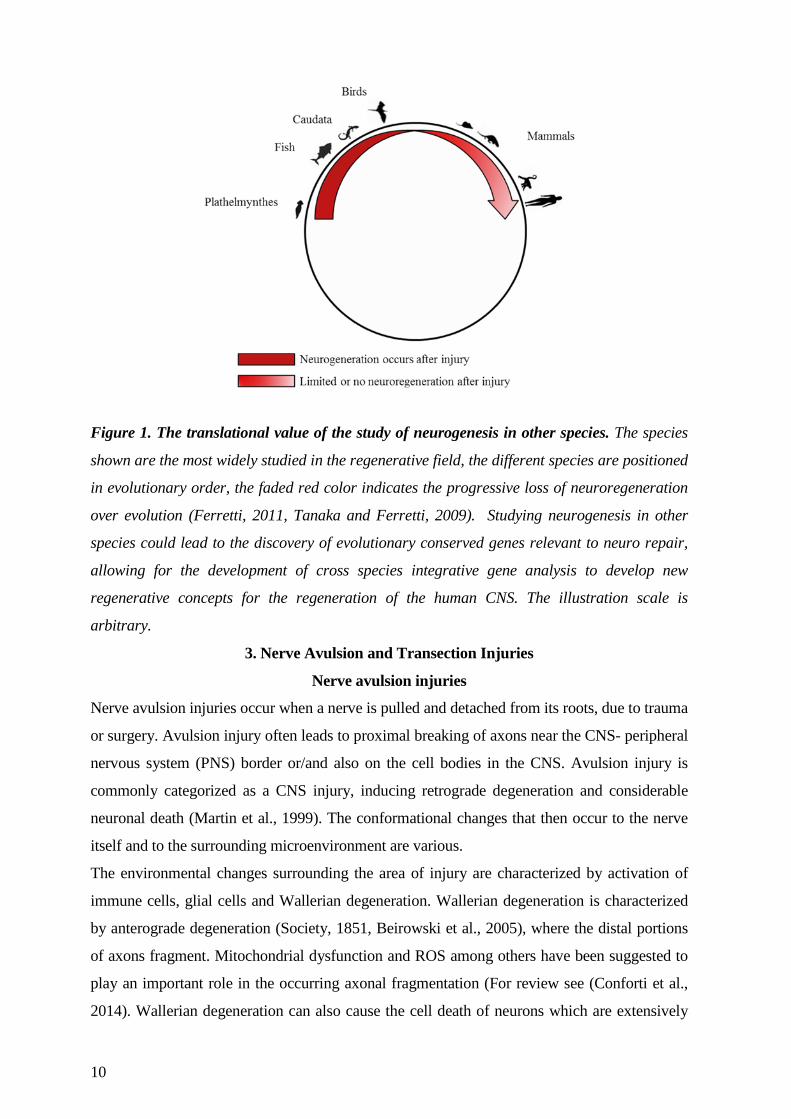
Figure 1. The translational value of the study of neurogenesis in other species. The species
shown are the most widely studied in the regenerative field, the different species are positioned
in evolutionary order, the faded red color indicates the progressive loss of neuroregeneration
over evolution (Ferretti, 2011, Tanaka and Ferretti, 2009). Studying neurogenesis in other
species could lead to the discovery of evolutionary conserved genes relevant to neuro repair,
allowing for the development of cross species integrative gene analysis to develop new
regenerative concepts for the regeneration of the human CNS. The illustration scale is
arbitrary.
3. Nerve Avulsion and Transection Injuries
Nerve avulsion injuries
Nerve avulsion injuries occur when a nerve is pulled and detached from its roots, due to trauma
or surgery. Avulsion injury often leads to proximal breaking of axons near the CNS- peripheral
nervous system (PNS) border or/and also on the cell bodies in the CNS. Avulsion injury is
commonly categorized as a CNS injury, inducing retrograde degeneration and considerable
neuronal death (Martin et al., 1999). The conformational changes that then occur to the nerve
itself and to the surrounding microenvironment are various.
The environmental changes surrounding the area of injury are characterized by activation of
immune cells, glial cells and Wallerian degeneration. Wallerian degeneration is characterized
by anterograde degeneration (Society, 1851, Beirowski et al., 2005), where the distal portions
of axons fragment. Mitochondrial dysfunction and ROS among others have been suggested to
play an important role in the occurring axonal fragmentation (For review see (Conforti et al.,
2014). Wallerian degeneration can also cause the cell death of neurons which are extensively
11
connected with the avulsed neuron, causing proximal cell death, or permanent atrophy
(Koliatsos et al., 1994, Jiang et al., 2000). Regenerative mechanisms are required to occur
across the verge of the CNS and the PNS.
Transection injuries
A transection injury leads to disruption of all of the components of the nerve, however in
contrast to the avulsion injury the number of motor neuronal death is lower. Importantly the
more distal the injury is to the site of the motor neuron, the lesser the motor neuronal death,
which in turn influences the load of inflammatory components into the environment (Yu, 1997,
Svensson and Aldskogius, 1993a, Mattsson et al., 1999). Transection injuries also lead to
Wallarian degeneration in the CNS, which as mentioned earlier is retrograde degeneration
occurs as described for the avulsion model (Conforti et al., 2014). Because of being in the
CNS-PNS border, both injury types generate reactions in the CNS and PNS. In the CNS
activation of glial cells occurs (Svensson and Aldskogius, 1993b), leading to ROS production
and conformational changes to the synapses, initiating synaptic stripping. The PNS reaction
involves, Schwann cell activation and proliferation, providing trophic support and clearing the
area from debris. The axon starts a regrowth mode, and in the best of cases may even
reestablish connectivity with other axons returning to a normal state (Brosius Lutz and Barres,
2014).
Hypoglossal nerve injury model
In order to be able to study if nerve regeneration and survival after nerve injury could be
improved using NPC transplantation, we used a model of nerve avulsion/transection of the
hypoglossal nerve. The hypoglossal nerve is the XII cranial nerve of the hypoglossal nucleus in
the brain stem. The hypoglossal nerve innervates the tongue muscle intrinsically and
extrinsically. This nerve is essential for swallowing, eating and in humans also for talking. The
anatomy of the area takes us to the upper part of the nucleus, protruding slightly in to the fourth
ventricle, adjacent to where the NPC niche is found. The lower part of the nucleus lies in close
proximity to the central canal, again near to a NPC niche. After avulsion injury limited
proliferation of ependymal cells is detected, and migration of NPCs is scarce (Fagerlund et al.,
2011).
There is a hypoglossal analogous nerve nucleus at each side of the CC. The anatomical
characteristics of this nerve allowed us to injure the hypoglossal nerve on one side of the CC,
keeping intact the analogous area, which served as a control for the injury. Thus the analysis
could be carried out in parallel, in the same section from the brain stem. The nuclei are also
ideal for microinjections, since the floor of the fourth ventricle adjacent to the nuclei is
12
surgically accessible, leading to minor additional trauma. Based on this, the hypoglossal injury
model became ideal for NPC transplantation delivered through microinjections. Furthermore,
the model can be extrapolated to a clinical setting, since it can be considered equivalent to
ventral nerve or root injury.
4. Stem Cell Transplantations
As discussed in section 2, mammalian regeneration is not successful (Fig. 1), therefore not only
the search for the application of evolutionary conserved genes to help CNS regeneration is
necessary, but also, to investigate how exogenous cell transplantation can contribute to repair.
There has been a plethora of attempts to achieve this primary goal, transplantation of
embryonic tissue, nerve grafts, mesenchymal stem cells and bone marrow stem cells. In my
thesis I will only discuss NPCs transplantation.
NPC transplantation
In order to alleviate incurable injuries and diseases NPC transplantation has been extensively
investigated, and have been shown to exert beneficial effects onto different neurodegenerative
conditions, like Parkinson’s disease (Gaillard and Jaber, 2011, Ziavra et al., 2012),
Amyotrophic Lateral Sclerosis (Xu et al., 2006, Lepore, 2011), MS or EAE (Pluchino et al.,
2003, Pluchino et al., 2009, Ben-Hur, 2008) and SC injury (Zhao et al., 2013). The beneficial
effects observed, have been considered to be achieved by direct cell replacement,
neuroprotection, or by the production of factors which can modulate the microenvironment,
like growth factors or immunomodulatory molecules like bone morphogenic protein-4, VEGF
and guidance molecules among others (De Feo et al., 2012, Lepore and Maragakis, 2007,
Fagerlund et al., 2012). Cell differentiation state for transplantation therapies is vital. Adult
NPCs grafting have shown no evidence of theratoma formation, comparing to cells from an
embryonic state (Brederlau et al., 2006). Hence, adult NPCs may be a better choice for
regenerative therapies. However the gathering of adult NPC tissue is scarce and challenging.
To solve this issue, the use of iPSC technology (Takahashi and Yamanaka, 2006) becomes a
viable option, which allows for the development of personalized regenerative therapies.
It is important to mention that even when cell transplantation is a promising therapy and should
continue to be investigated, reports on side effects exist (Bjorklund, 2004, Bjorklund and
Kordower, 2013), indicating further studies on the behavior of NPC transplantation in vivo are
required. Animal models where the study of transplantation cell state, type and numbers as
well as site and timing for transplantation are crucial to provide solid evidence to continue with
clinical trials.
13
5. Epigenetic Regulation of Adult Progenitor Cells Differentiation
The word epigenetic means on top of the DNA. Eukaryotic DNA is packaged in chromatin;
chromatin packaging is highly dynamic and its controlled by epigenetic changes which lead to
gene expression modulation (Berger et al., 2009). The fundamental unit of chromatin, is the
nucleosome, which is an octamere histone core, around which DNA is wrapped (Kouzarides,
2007). Epigenetic changes are regarded as changes to chromatin structure and not genomic
sequence alterations. Epigenetic changes can be inherited through generations (Berger et al.,
2009). In order to elucidate a mechanism by which H2O2 exposure in Paper III, resulted in
higher number of neurons and oligodendrocytes from NPCs, we were interested in studying the
gene expression of chromatin architecture enzymes. Chromatin architecture enzymes
contribute to variations in chromatin structure by influencing histone modifications, leading to
gene expression changes. Epigenetic regulation of NPC differentiation among others involves;
chromatin modification enzymes, like DNA methylation and demethylation, acetylation and
deacetylation, but also activation of micro RNAs, and complexes that modify chromatin
structure (Juliandi et al., 2010).
Intrinsic and environmental cues trigger epigenetic mechanisms. This environmental influence
has long lasting effects on chromatin structure. Thus, mircroenvironmental changes like
disease or medical treatment, may control transitions from one cellular state to another
(Mirbahai and Chipman, 2014). Here I will solely introduce methylation, demethylation and
acetylation, as these modifications are relevant to the chromatin architecture enzymes
expression we found affected by H2O2.
Histone methylation and demethylation
Methylation is processed by enzymes which add methyl groups to the histone tails of specific
nucleosomes, causing chromatin structure modifications (Chen and Riggs, 2011). Methyl
groups bound to DNA’s cytosine residues (CpG islands) can also mediate the recruitment of
other complexes, like histone deacetylases. Methyl groups are the bases of epigenetic gene
silencing. Methylation is reversible and methyl groups can be removed by demethylating
enzymes, although methyl transferases themselves may also initiate the demethylation process.
Specific mechanisms of active demethylation are yet not well understood (Ooi and Bestor,
2008). Methylation changes the structure of chromatin, modifying the interaction that DNA has
with different proteins, blocking the binding of such proteins to the DNA, and regulating
transcription activation, if this site incurs a promoter binding site (Jones and Takai, 2001).
14
Specific demethylating enzymes which take away methyl groups from specific histone sites,
play an important role between the self-renewal state transition of the NPC into the neurogenic
sate, it is also known that methylation of specific histone tails is necessary for neuronal
differentiation, allowing a dynamic interaction between specific transcription binding factors
which can transcribe neurogenic genes, for review see (Ma et al., 2010).
Histone Acetylation and Deacetylation
Acetylation and deacetylation of lysine residues in histone tails is mediated through the
enzymes histone acetyl transfrases (HATs) and histone deacetylases (HDACs) respectively.
Histone acetylation of specific sites of the core histones, occurs at lysine residues located at the
N-termini. In vivo, all core histones are acetylated and increased levels of acetylation, are often
correlated to increase transcriptional activity. In contrast to methylation, recruitment of HDAC
enzymes, and the consecutive deacetylation of N-Termini, results in repression of gene
expression (Cress and Seto, 2000) since DNA becomes more tightly compacted, preventing
transcription binding to promoter regions, for review see (Bose et al., 2014). During
neurogenesis acetylation has been recognized to play an important role (Sun et al., 2011),
leading to a more open chromatin state, allowing the binding of transcription factors to specific
sites which can initiate the gene transcription of neuronal genes. Differentiation of both
oligodendrocyte and neuronal lineages are dependent on HDACs activity (Juliandi et al.,
2010). Inhibition of HDACs at the right time window has been shown to decrease neurogenesis
by the silencing of specific neuronal genes (Shaked et al., 2008), whereas up-regulation of
HDACs could lead to oligodendrocyte differentiation (Ji et al., 2011).
HDACs family of enzymes, are composed of Class I, II and Class III HDACs. Here I will only
refer to Class III HDACs as Sirt2 the HDAC found relevant in Paper III, is part of this family.
Sirtuins are class III HDACs and are NAD+ dependent enzymes. Among the sirtuins, is Sirt2
which is found in the nucleus and in the cytoplasm of cells, Sirt2 has a strong expression in the
brain, and is highly expressed by oligodedrocytes. Sirt2 can be activated by cellular stress and
its upregulation its known to cause cell cycle exit (North and Verdin, 2007), and also to
influence differentiation towards the oligodercyte lineage (Ji et al., 2011). Specific chromatin
states can be correlated with specific differentiation status (Larson and Yuan, 2012). The
importance for the understanding of the orchestration of chromatin architecture could lead us to
develop better therapies by guiding the differentiation of NPC to the desired cell type.
15
6. Experimental Allergic Encephalomyelitis (EAE)
The lamentable accidental induction of symptoms which resembled multiple sclerosis (MS)
was observed after an anti-rabies vaccine in humans containing myelin-like components was
administered (Remlinger, 1905). This unfortunate event initiated the beginning of the
development of MS models like EAE. This condition may be induced by immunization of
myelin antigens in Freund adjuvant which enhances the immune response initiating
demyelination
(Wekerle and Lassmann, 1994). Widely, the EAE model is regarded as a helpful MS model, (t
Hart et al., 2011, Mix et al., 2010) and in the context of my thesis, to study NPCs in an
inflammatory environment, involving both CNS inflammation and demyelination.
Rat clinical symptoms
In our MOG induced EAE model in the Dark Agouti rat, clinical signs often begin at the 9th
day post immunization. An initial weight loss precedes the beginning of the symptoms which
are rated as follows; 0-no clinical symptoms; 1-tail weakness or tail paralysis; 2- hind-limb
paraparesis; 3- hind-limb paralysis; 4-tetraplegia; and 5-death. EAE data suggests that the
inflammatory cascade produced during disease may contain both, beneficial and detrimental
effector molecules, influencing disease progression (Olsson, 1995). Further, proteomic EAE
CSF analysis showed the up-regulation of members of the complement system and vitamin D
binding protein among others (Rosenling et al., 2012), inflammatory mediators have also been
detected, among which are TNF-α and IL-2 (Diab et al., 1997), all of which correlate with
disease severity. In the majority of rat EAE models, paralysis is accompanied by inflammation
and CNS infiltration of immune cells (Friese et al., 2014), the formation of reactive oxygen
species (ROS) (Gilgun-Sherki et al., 2004), demyelination, glial scar formation and
remyelination (Voskuhl et al., 2009). Clinical outcome may vary, as different rat strains carry
different genetic immune susceptibilities (Storch et al., 1998) (Weissert et al., 1998, Stefferl et
al., 1999, Sakuma et al., 2004) producing a broad spectrum of EAE clinical symptoms relating
to the clinical outcomes observed during human MS, similarities which I will review next.
Clinical similarities between EAE and MS
MS is a chronic inflammatory disease, characterized by demyelinating plaques in the CNS. MS
is most prevalent in females (Compston and Coles, 2002, Duquette et al., 1992) , a difference
which has also been stablished in mice EAE (Cruz-Orengo et al., 2014), Cruz-Orengo et al,
validated their data in humans showing increased gender susceptibility to MS and EAE is in
part attributed to higher expression of S1PR2 which increases BBB permeability in females.
16
MS clinical symptoms often start as a relapsing-remitting form of the disease. Already early in
the disease course neurons are lost (De Stefano et al., 2002) and CNS atrophy can be
measured. Inflammation is accompanied by neurodegeneration accumulating irreversible
neurological disabilities and worsening CNS deficits (Natowicz and Bejjani, 1994). DA rats
with MOG induced EAE, also developed relapsing-remitting disease with focal demyelination
in the spinal cord (Lorentzen et al., 1995).
High levels of inflammatory agents have been detected in MS CSF, comparable to
EAE, among which are NO (Svenningsson et al., 1999, Danilov et al., 2003b), TNF-α, IFNγ,
IL-2, and IL-17A (Duan et al., 2013) , but also other molecules important for immune system
regulation have been identified, like , vitamin D binding protein and the calcium regulating
protein Fetuin A (Olsson, 1995, Ottervald et al., 2010).
In this thesis work, I used the female DA rat strain which has shown EAE
susceptibility (Stepaniak et al., 1995). This MOG EAE model leads to a variety of clinical
symptoms representing a complex MS mimicking model (Storch et al., 1998).The
inflammatory milieu initiated by MOG- EAE, could have a potential effect on the inner NPC
pools, possibly affecting NPC behavior, inflicting regeneration (Nait-Oumesmar et al., 2007).
Importantly MS lesions are often found lining the lateral ventricles, suggesting possible
changes on the NPC nature may occur.
NPC transplantation on the EAE model
Several studies have reported the benefits of NPC transplantation on EAE animals, exerted via
modifying the inflammatory environment (Deboux et al., 2013, Pluchino et al., 2009). Also
some reports showed lower proliferation of NPC after EAE (Pluchino et al., 2008) and
differentiation changes from NPC after EAE (Picard-Riera et al., 2002, Kuhlmann et al., 2008).
Based on all the above, in this thesis the model of MOG-EAE on the female DA rat was ideal
to study NPC characteristics in an inflammatory environment, allowing us to produce data
which could be of possible clinical relevance.
7. Reactive oxygen species and their role in NPC signaling
Except for some anaerobic microorganisms, oxygen is necessary for the life of almost every
organism on earth. However, even though oxygen gives life to most organisms it also
represents a risk every time we use it, due to its highly reactive and mutagenic nature. We and
aerobic organisms only survive due to the antioxidant defense mechanisms we have evolved.
As oxygen levels fluctuated the atmosphere to the current value of 21% (Berner, 1999)
organisms became more complex, and adapted better mechanisms to produce oxygen energy
17
based products (Powell, 2010, Taylor and McElwain, 2010). In humans, tissue oxygen levels
are critical for normal embryonic development, cellular differentiation, and stem-cell
maintenance (Stamati et al., 2011). Also, the immune system has to be able to react and adapt
to environmental oxygen changes in order to generate the bioenergetics resources that allows it
to initiate an immune response/inflammation. Immune cells like neutrophils and macrophages,
have adapted mechanisms to combat infections which use oxygen consumption to produce
toxic products which can kill bacteria (Bedard and Krause, 2007). One of the most classical
immune mechanisms in which oxygen is used to produce toxic oxygen derivatives is the
oxidative burst. The membrane bound NADPH oxidase generates the super oxide anion (O2-)
(Bedard and Krause, 2007), which is converted to H2O2 by the enzyme super oxide dismutase
(SOD) , H2O2 is then enzymatically converted to more reactive chemical forms, like the
hydroxyl radical (HO.), hypochlorite (OCL-) and hypobromite (OBr-) (Yang et al., 2013).
These toxic oxygen derivatives are known as free radicals, and form part of a family of
molecules known as Reactive Oxygen Species (ROS).
Free Radicals
Free radicals are formed when any chemical species loses a single electron, which results in
one or more unpaired electrons. This condition of one or more unpaired electrons can produce
high reactivity, as the unpaired electron would make the chemical species more attracted to a
magnetic field. The oxygen molecule (O2) has two unpaired electrons, making it qualify as a
free radical, however this unpaired electrons have parallel spins, which makes the molecule
highly stable, being the form in which O2 exist in the air we breathe. Free radicals can react
differentially upon contact with different molecules, like hydrogen peroxide for example. H2O2
is a free radical which selectively reacts with other molecules, allowing it to also be used as a
signaling molecule by the cell. H2O2 can easily cross cell membranes, after which it reacts with
Fe and Cu, possibly by donating an electron to this elements (Prabhakar et al., 2004) to form
more reactive species, like HO. , which accounts for much of the DNA damage caused by H2O2
to cells.
Fe2+ + H2O2 → HO• + OH–+ Fe3+ (Gutteridge and Halliwell, 2007, Fourth Edition )
Cu2+ + H2O2 → HO• + OH–+ Cu+ (Simpson et al., 1988)
Simplified overview of H2O2 reacting with iron and Cu. The iron reaction its known as the
Fenton reaction after its discovery by H.J.H Fenton in 1894, where the oxidation of Fe2+ to
Fe3+ produces a hydroxyl radical and hydroxyl ion.
18
Inflammation and H2O2
Inflammation is characterized by the production of ROS among which we find nitric oxide
(NO.), super oxide O-., hydroxyl radical (HO.) and hydrogen peroxide (H2O2). Interestingly,
NO. and H2O2 can also play a signaling role in the cell as they can mediate cell to cell
communication and are classified as first type cell messengers (Holmquist et al., 2007).
In this thesis I will focus on H2O2 in relation to NPC differentiation. H2O2 may be produced by
neutrophils and macrophages during inflammation, where it can be found compartmentalized
in the cells mitochondria and the endoplasmic reticulum (Gough and Cotter, 2011). During
oxygen metabolism H2O2 contributes to the generation of energy in the cell mitochondria. Low
physiological levels of H2O2 are known to allow H2O2 signaling properties. As mentioned
earlier, comparing to other ROS, H2O2 is poorly reactive for example, depending on cell type,
oxidation of DNA, lipids and proteins does not occur at mM concentrations of H2O2
(Gutteridge and Halliwell, 2007, Fourth Edition ).
H2O2 and Neuroinflammation
In the brain Fe contents are low, however after brain injury and MS iron concentrations rise
rapidly (Mehta et al., 2013), Fe increase could then react with H2O2 and contribute to higher
levels of HO. and O-. in the cell increasing oxidative stress in the environment and contributing
further to cell toxicity. Further damage can continue by the formation of more H2O2 from
neurotransmitters like noradrenalin among others (Swaroop et al., 1983). The low oxygen
concentrations found in the brain, which are around 10 times lower than atmospheric levels
(Silver and Erecinska, 1998), are so perhaps to prevent formation of oxygen free radicals due
to the already high oxygen consumption by the brain. However a recent publication evaluates
ROS production from proliferating astrocytes after stroke, establishing an important
contribution to ROS levels in the brain (Walton et al., 2012), which in an inflammatory context
could further contribute to cell damage given the already hostile environment (Nikic et al.,
2011, Marti-Fabregas et al., 2010) and for review see (Rafalski and Brunet, 2011).
ROS are necessary for the adult SVZ stem cell niche and NPCs function
The NPCs population from the brain SVZ, are influenced by basal levels of ROS. ROS in
NPCs act as signaling mediators of self-renewal and normal neurogenesis, and are proven to be
necessary for NPCs normal functionality (Le Belle et al., 2011)(Paper III). Importantly ROS
are an important regulator of NPC proliferation and cell cycle entry (Chaudhari et al., 2012),
which leads to fate commitment and loss of stemness. Concomitantly the regulation of
19
chromatin architecture genes like the HDAC Sirt2, which can influence NPC fate decisions,
are also regulated by ROS (Nakagawa and Guarente, 2011, Perez Estrada et al., 2014) .
ROS are produced during energy production in the NPC by mitochondria (Zorov et al., 2014).
Mitochondrial activity is known to be higher in adult NPCs than in their differentiated progeny
yet, their ROS content in the undifferentiated state is low, the reason for this may be the high
concentration of antioxidants like glutathione peroxidase among others in this cells (Madhavan
et al., 2006), thus the adult NPCs show to have a higher metabolism with which they cope
efficiently by up-regulating antioxidant enzymes.
The NPC niche and ROS
Also the SVZ NPC niche has shown to be tightly regulated by ROS, since ROS is able to
control the anchoring molecules which give the NPC niche its cell architectural structure. The
B cells in-vivo structure depends on the dynamics of Cell Adhesion Molecules (CAMs)
(Kokovay et al., 2012). The close association of NPC to the neurovascular system (Shen et al.,
2008), may not only provide physical support but also, contribute to oxygen supply and or
signaling from the bloodstream. Disruption of the ROS levels could cause the malfuctionality
of molecules like V-CAM1 (Kokovay et al., 2012), and allow the proliferation of the NPC
which could in turn enfeeble the NPC population. Also, during ischemic conditions, oxygen
levels drop, low oxygen levels stimulate mitochondria to produce more ROS (Klimova and
Chandel, 2008). This oxygen increase can lead to the activation of the NPC pool, initiating
proliferation and cell cycle entry leading to NPC differentiation and as mentioned above, the
consequences of such activation could possibly result in the exhaustion of the NPC pool.
Moreover the hostile environment could make it difficult for the newly differentiated cells to
exert any beneficial properties onto the injury area.
NPC Antioxidant Mechanisms
NPCs cultures show lower profile of ROS than their or differentiated counterparts, possibly
through a more active antioxidant repertoire, where Glutathione peroxidase (GPx), super oxide
dismutase (SOD), and catalase (CAT) regulate the redox content of these cells (Madhavan et
al., 2006). Disruption of these defense mechanisms in the NPC pool could lead to
mitochondrial dysfunction, energy production deficiencies, and ultimately cell death affecting
the regenerative potential of the organism. Next I will shortly describe these three important
antioxidant enzymes.
20
Super oxide dismutase (SOD)
SODs are located in the cytosol and in the mitochondria of eukaryotic cells. The SODs family
of enzymes comprises SOD1 and SOD2. SOD1 which at its active site uses Cu and Zn, and is
localized in the cytosol. SOD2 uses Mn in its catalytic center (Fridovich, 1995) and is localized
in the cell mitochondrion, near by the mitochondrial respiratory chain, which leads to the
production of O2- as the end product . Further, SOD can inhibit the levels of the free radical
HO. produced by the Fenton reaction, by reducing O2- levels, however the dismutation of O2
-
results in H2O2 production (Luk et al., 2003). H2O2 then needs to be reduced, converting H2O2
into water by another antioxidant molecular set, the glutathione peroxidase family of enzymes.
Glutathione peroxidase (GPx)
A simplified way of describing GPx mode of action is as follows, GPx reduces lipid peroxides
to their alcohol more stable forms, and reduces H2O2 to water. GPx does that by using reduced
glutathione (GSH) as an electron donor, producing two H2O molecules as the end product;
H2O2 +2GSH = GS-SH+2H2O
GPxs are a family of enzymes, composed of four different GPxs, classically described by their
common location in the cell or in a specific organ. Gpx1is found in the cytosol, nucleus, and
mitochondria. GPx2 is found in the cytosol and nucleus, and is classically localized on the cells
lining the gastrointestinal tract. GPx3 is found in the cytosol. All of these enzymes use
selenium in their active site to perform their antioxidant function. The GPx4 which not only
takes care of H2O2 but also peroxidized fatty acid residues in the cell membrane, GPx4 is found
abundantly in testis, playing a role in sperm maturation, and in the cell it can be found in the
cytosol, nucleus, mitochondria and cell membrane (Gutteridge and Halliwell, 2007, Fourth
Edition ). More recently there have been additional forms of GPx described, GPx5,GPx6 and
GPx7 (Margis et al., 2008).
Catalase (CAT)
Another cell defense mechanism against H2O2 is the enzyme CAT, which catalyzes the direct
decomposition of H2O2 into O2. CAT is localized in the cell peroxisomes, CAT uses Fe at its
active site and its mode of action is similar to that of SOD, a dismutation reaction, reducing
H2O2 to H2O and O2. CAT function is optimal during high H2O2 levels, as the reaction velocity
and complete H2O2 clearance efficiency need two H2O2 molecules, when H2O2 concentrations
drop this efficiency becomes less likely, and CAT less efficient (Gutteridge and Halliwell,
2007, Fourth Edition ).

21
Peroxiredoxins (Prxs)
Are a family of peroxidases which reduce H2O2, peroxiredoxins redox reactions depend on
cysteine at their active site. There are known six different types of peroxiredoxins. Prxs are
very abundant in the cell, and can be found in different cellular compartments; Prx 1,2 and 6
found in the cytosol. There is the mitochondrial Prx3, the endoplasmic reticulum found Prx4 ,
and Prx5 found both in mitochondria and peroxisomes (Gutteridge and Halliwell, 2007, Fourth
Edition ).
22
AIMS
The aim of this thesis is to examine the NPCs survival, function and fate changes on a
transcriptional and functional level after exposure to three models of experimental
inflammation; A brain stem injury, in vitro exposure to free radicals, and EAE.

23
RESULTS AND DISCUSSION
PAPER I
NPCs Transplanted to the Hypoglossal Nucleus show signs of integration and promote
motor neuron survival
In this study we culture primary NPCs from the SVZ of the adult rat brain, isolated accordingly
to a modified protocol by Johansson et al. (Johansson et al., 1999). For NPC culture we used
the homozygote offspring from a backcrossed Lew rat which carries an eGFP insertion under
the ubiquitin promoter (Lois et al., 2002). These cells were transplanted into a hypoglossal
nerve avulsion injured wild type Lew rat sibling. We determined the survival, differentiation
and possible integration and or beneficial effect from the graft into the microenvironment of
the hypoglossal nerve. The advantage of the hypoglossal nerve avulsion model is that the
injured side is also in close vicinity to the corresponding uninjured side which serves as a
control and is present at each section. These characteristics allowed us to assess the potential
effects of NPCs transplantation onto the hypoglossal nucleus injured side, in contrast to its un-
injured site. After the second passage SVZ NPCs were prepared for transplantation in a single
cell suspension of 100,000 cells in 5ul. For the NPC transplantation the brain stem was
exposed and the obex identified. NPCs were injected into the right hypoglossal nucleus
through a glass micropipette sealed onto a Hamilton syringe. The experimental groups are
described in Fig. 2. NPCs were transplanted at the 9th day post injury in order to avoid the peak
of microglial reaction known to occur the first week after injury (Aldskogius and Svensson,
1993), and which could influence the transplantation result.
Transplanted NPCs survive, differentiate and express neuronal markers
To prevent transplantation rejection, all transplanted animals, including controls received
cyclosporine injections. 10mg/kg cyclosporine were injected daily, from 1d prior to
transplantation, and diminished to 3x/week after time point 28d. Animals were perfused after
2, 7, and 28 days and 3 months (n = 8 in each group and survival time). The brain stems were
removed and postfixed. The survival, differentiation, integration and possible beneficial
24
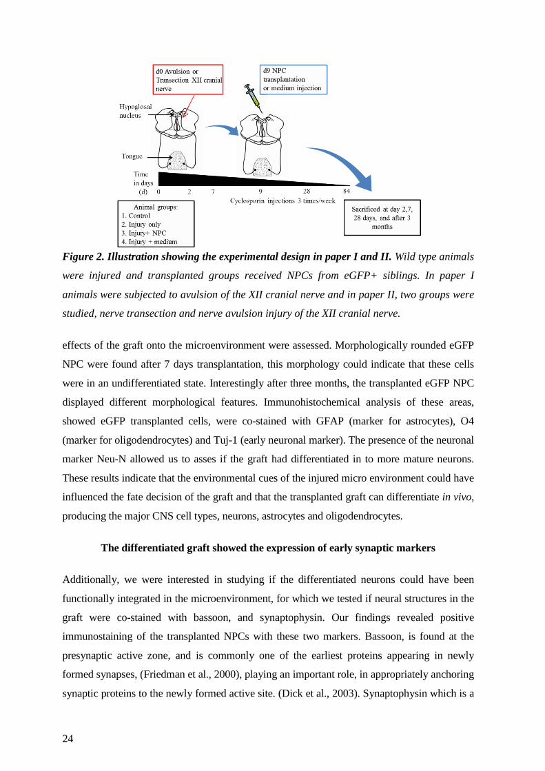
Figure 2. Illustration showing the experimental design in paper I and II. Wild type animals
were injured and transplanted groups received NPCs from eGFP+ siblings. In paper I
animals were subjected to avulsion of the XII cranial nerve and in paper II, two groups were
studied, nerve transection and nerve avulsion injury of the XII cranial nerve.
effects of the graft onto the microenvironment were assessed. Morphologically rounded eGFP
NPC were found after 7 days transplantation, this morphology could indicate that these cells
were in an undifferentiated state. Interestingly after three months, the transplanted eGFP NPC
displayed different morphological features. Immunohistochemical analysis of these areas,
showed eGFP transplanted cells, were co-stained with GFAP (marker for astrocytes), O4
(marker for oligodendrocytes) and Tuj-1 (early neuronal marker). The presence of the neuronal
marker Neu-N allowed us to asses if the graft had differentiated in to more mature neurons.
These results indicate that the environmental cues of the injured micro environment could have
influenced the fate decision of the graft and that the transplanted graft can differentiate in vivo,
producing the major CNS cell types, neurons, astrocytes and oligodendrocytes.
The differentiated graft showed the expression of early synaptic markers
Additionally, we were interested in studying if the differentiated neurons could have been
functionally integrated in the microenvironment, for which we tested if neural structures in the
graft were co-stained with bassoon, and synaptophysin. Our findings revealed positive
immunostaining of the transplanted NPCs with these two markers. Bassoon, is found at the
presynaptic active zone, and is commonly one of the earliest proteins appearing in newly
formed synapses, (Friedman et al., 2000), playing an important role, in appropriately anchoring
synaptic proteins to the newly formed active site. (Dick et al., 2003). Synaptophysin which is a
25
synaptic vesicle membrane protein, is widely used as a marker for presynaptic terminals, and
play several roles in the presynaptic terminals; endocytosis of vesicles, synapse formation and
exocytosis, highly relevant during neuronal activity (Becher et al., 1999). Together these
findings indicate that the transplanted NPC graft was capable of differentiating into astrocytes,
early oligodendrocytes and neurons, and that the newly differentiated neurons showed signs of
possible integration in to the new microenvironment, forming early synaptic signatures.
The number of surviving motor neurons was higher in transplanted animals
After nerve avulsion motor neuronal death is significant (Martin et al., 1999)(Paper I), with
numbers ranging from 80% after 3 months injury. Concomitantly we were interested to study if
the NPC graft would have any effect on motor neuronal viability. Quantification of motor
neurons showed that in animals which were injured but did not received NPC transplantation,
the number of surviving neurons kept decreasing with time and animals which were injured
and received the NPCs graft, showed a significantly higher number of surviving neurons after
nerve avulsion. Subsequent to nerve avulsion, the degeneration that follows is known as
Wallarian degeneration (described in Nerve avulsion injuries section page 10), our results then
indicate that the NPCs mediated a nursing effect on the motor neurons surrounding the lesion,
providing trophic support needed during this inflammatory condition. Also, this support was
able to be sustained for up to three months, as in the non NPC transplanted animals, cells
continued to degenerate whereas in the transplanted animals survival was consistent at this
time point.
Long lasting VEGF expression from the transplanted NPCs
The beneficial effects on motor neuron cell survival observed in the transplanted animals, lead
to another question; by which mechanism could the NPCs provide trophic support to the
injured microenvironment? We decided to study if the transplanted NPCs, could express
vascular endothelial growth factor (VEGF) on to the site of injury. VEGF is known to have
beneficial effects on different neurodegenerative landscapes, like during the devastating
disorder of amyotrophic lateral sclerosis (ALS), where motor neurons die irremediably. In this
context VEGF has been observed to reduce degeneration of motor neurons when infused onto
an animal model of ALS (Storkebaum et al., 2005). Also it has been shown that VEGF
treatment enhances neuronal survival and neurite outgrowth (Rosenstein et al., 2003) (Sondell
et al., 1999), and it can also protect neurons during hypoxic conditions (Jin et al., 2000). Our
results showed that NPCs transplanted to the avulsed injured animals, co-expressed VEGF
three months post-injury, suggesting a possible mechanism by which the NPCs could exert
their beneficial effect onto the injured area, supporting motor neuronal survival.
26
In Paper I, I present that transplanted adult NPCs can survive up to three months
after transplantation and are able to differentiate into astrocytes, oligodendrocytes, and neurons
on to the site of injury. Differentiated neurons expressed signs of newly formed synapses
indicating possible integration of the graft into the microenvironment. Besides differentiation,
the graft proved to be beneficial to the injured area, by decreasing the numbers of motor
neuronal cell death, a mechanism possibly influenced by VEGF. This study contributes to the
regenerative field by demonstrating that transplanted NPCs in to the injured CNS increased
motor neuronal survival, showing that external sources of NPCs can potentially contribute to
the repair of the CNS.
PAPER II
Transplanted NPCs showed activation differences depending on the injury severity
We were interested in studying whether the NPC activity was affected by the extent of
inflammation, which was modelled by two injury models: nerve transection and nerve avulsion
injury of the hypoglossal nerve.
A transection nerve injury differs from the avulsion model by causing less damage
to the site of injury (see page 11 of the introduction) and less neuronal death to the neurons in
close contact to the transected nerve. In this model the right side of the hypoglossal nerve was
transected at the level where it passes the carotid artery (groups II-IV). The experimental
design was the same as for Paper I (Figure 2). We speculated that the microenvironment of
inflammation could differ in the two different injury models and could also differentially
activate the transplanted NPCs and therefore affect the possible regeneration. After 9 days post
transection and avulsion injury, 100,000 primary eGFP NPCs were transplanted in injured
animals into the right hypoglossal nucleus as described in paper I. Our findings revealed
differences between the two injury models, with respect to transplanted NPCs distribution,
differentiation, and expression of the antioxidant defense molecule glutathione peroxidase
(Gpx).
Higher motor neuronal survival in nerve transected injury model
When quantifying the motor neuronal survival, we found that at 28 days post injury 89% of
motor neurons were persistent. In contrast as discussed in Paper I, motor neuronal survival in
the avulsion model is 25% at this time point.
27
Transplanted NPC distribution at the injury site
After three months we found that the transplanted NPCs density and morphology was different
between the transection model and the avulsion model. We observed 10% less eGFP+ NPCs
present in the transection injury model and scattered distant to the injury site, whereas in the
avulsion injury, higher numbers of eGFP+ cells were found and their distribution was observed
to remain at the site of injury, suggesting differences in instructive signals in each
inflammatory milieu directing the cells towards different distribution and survival patterns.
Transplanted NPC showed differentiation differences depending on injury type
Differentiation of the graft was observed three months post transplantation in paper I.
Immunohistochemistry for GFAP and Tuj-1 showed co-staining of eGFP+ NPCs to these
markers only in the avulsion model. The eGFP+ NPCs in the transection model, displayed
more rounded morphologies but also protrusions, however these cells did not show co-staining
with any of these differentiation markers. These differences in fate choice could be attributed
to components in the inflammatory milieu and differential regenerative needs in each injury
model. The nerve avulsion injury model is a more severe injury, which may involve different
instructive signals for the graft to express distinctive molecular components, like VEGF among
others. This could provide different cues to the NPCs to choose different lineage engagement,
however this will have to be further studied.
Interestingly at the later time point (3 months) we found several eGFP+ NPCs which were
Sox2 positive in the transection injury model (25-30%), whereas in the avulsion model, we
could not observe Sox2 expression from the grafted cells at this time point. Sox2 expression,
indicates that a portion of the transplanted cells, remained in an undifferentiated state, possibly
by the lack of environmental cues which could initiate the process for fate specification, or
exerting other beneficial effects to the microenvironment rather than integration, as has been
previously reported in other injury model (Bacigaluppi et al., 2009).
Transplanted NPCs expressed antioxidant markers
Despite the undifferentiated state of the NPC into the transected nerve injury, we wanted to
study the possible expression of other molecules which could benefit the inflamed
microenvironment, for which we tested the expression of glutathione peroxidase (Gpx1).
Gpx1 is an antioxidant enzyme which protects the cell from ROS, in particular from the
inflammation produced by H2O2, for review see (Brigelius-Flohe and Maiorino, 2013).
Immunohistochemical staining for this marker showed a higher expression of Gpx1 in the
avulsion model, than in the transection injury model. The distribution of the Gpx1 staining
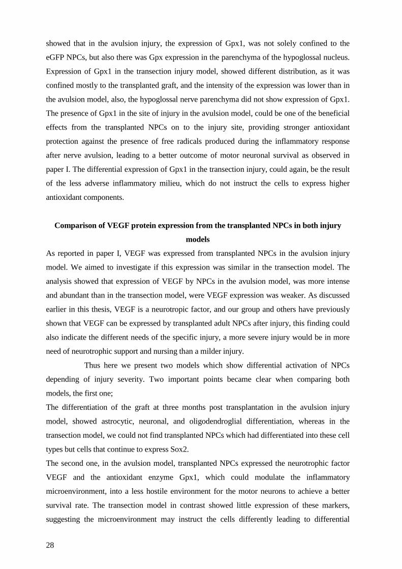
28
showed that in the avulsion injury, the expression of Gpx1, was not solely confined to the
eGFP NPCs, but also there was Gpx expression in the parenchyma of the hypoglossal nucleus.
Expression of Gpx1 in the transection injury model, showed different distribution, as it was
confined mostly to the transplanted graft, and the intensity of the expression was lower than in
the avulsion model, also, the hypoglossal nerve parenchyma did not show expression of Gpx1.
The presence of Gpx1 in the site of injury in the avulsion model, could be one of the beneficial
effects from the transplanted NPCs on to the injury site, providing stronger antioxidant
protection against the presence of free radicals produced during the inflammatory response
after nerve avulsion, leading to a better outcome of motor neuronal survival as observed in
paper I. The differential expression of Gpx1 in the transection injury, could again, be the result
of the less adverse inflammatory milieu, which do not instruct the cells to express higher
antioxidant components.
Comparison of VEGF protein expression from the transplanted NPCs in both injury
models
As reported in paper I, VEGF was expressed from transplanted NPCs in the avulsion injury
model. We aimed to investigate if this expression was similar in the transection model. The
analysis showed that expression of VEGF by NPCs in the avulsion model, was more intense
and abundant than in the transection model, were VEGF expression was weaker. As discussed
earlier in this thesis, VEGF is a neurotropic factor, and our group and others have previously
shown that VEGF can be expressed by transplanted adult NPCs after injury, this finding could
also indicate the different needs of the specific injury, a more severe injury would be in more
need of neurotrophic support and nursing than a milder injury.
Thus here we present two models which show differential activation of NPCs
depending of injury severity. Two important points became clear when comparing both
models, the first one;
The differentiation of the graft at three months post transplantation in the avulsion injury
model, showed astrocytic, neuronal, and oligodendroglial differentiation, whereas in the
transection model, we could not find transplanted NPCs which had differentiated into these cell
types but cells that continue to express Sox2.
The second one, in the avulsion model, transplanted NPCs expressed the neurotrophic factor
VEGF and the antioxidant enzyme Gpx1, which could modulate the inflammatory
microenvironment, into a less hostile environment for the motor neurons to achieve a better
survival rate. The transection model in contrast showed little expression of these markers,
suggesting the microenvironment may instruct the cells differently leading to differential
29
expression of these proteins. A comparison of study I and study II clearly show that a more
severe injury benefits more from NPC transplantation than a milder injury. Moreover, the
injury microenvironment plays an important role in dictating the transplanted NPCs to exert
different effects depending on injury type, this study could help us to elucidate better directed
therapies depending on injury type and to consider the environment as a major regulator of
NPC fate.
PAPER III
Oxidative stress influences the NPCs differentiation result by increasing numbers of
neurons and oligodendrocytes
In this study, we investigated the NPCs differentiation result after being exposed to the free
radical H2O2 a constituent part of the inflammatory cascade and immunity. We cultured
primary adult NPCs from the SVZ of adult DA rat brains as described in the above studies
(Johansson et al., 1999), and exposed them to different concentrations of H2O2. 100µM H2O2
was chosen since it did not have considerable effects in cell death, as was observed at 330mM
H2O2 a concentration level that has been measured during severe neuroinflammation (Nikic et
al., 2011). After 24h 100uM H2O2 exposure, NPCs were still viable. After exposure we
assessed, proliferation, cell viability (Lecoeur, 2002), expression of genes relevant during
differentiation, and immunohistochemistry and western blot analysis of the differentiation
result (Fig. 3).
Cell death and proliferation after H2O2 exposure
Ki67 labeling showed that proliferation was increased in NPC cultures exposed to H2O2.
These findings prompt us to investigate if there was a particular cell population proliferating
more than another in the exposed cultures. Cells death was increased in exposed samples
however this effect was not significant.
H2O2 exposure resulted in more neurons and oligodendrocytes
In differentiated NPCs derived from exposed cultures, we observed a significant increase in the
number of neurons and oligodendrocytes. This finding prompted us to characterize our
undifferentiated NPC culture in order to observe possible changes occurring immediately after
exposure and which could have led to this differentiation result.
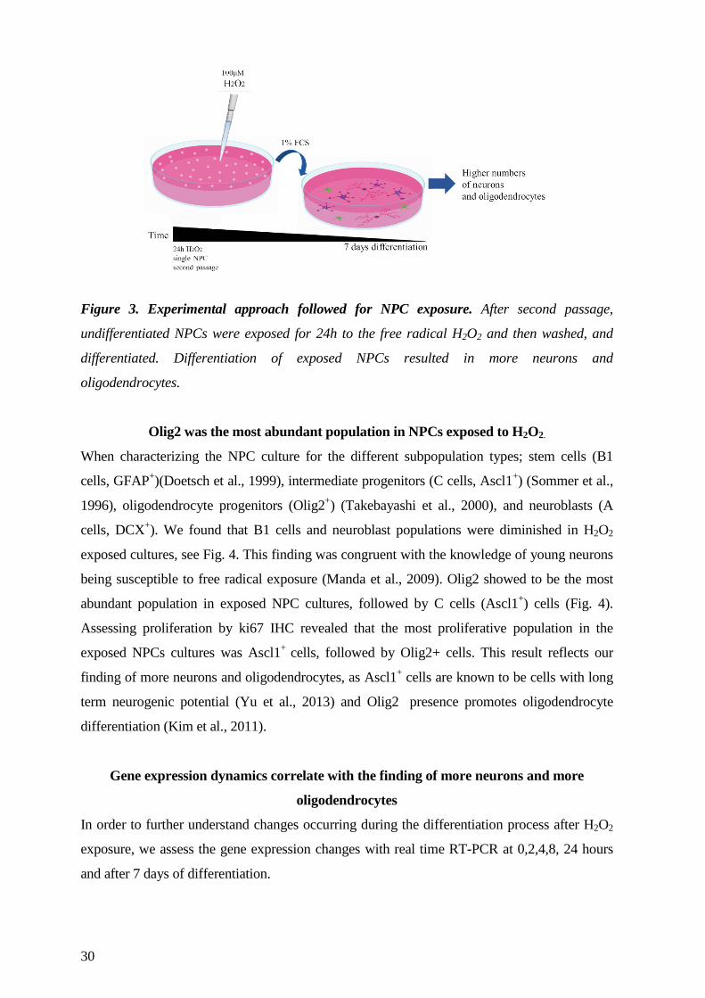
30
Figure 3. Experimental approach followed for NPC exposure. After second passage,
undifferentiated NPCs were exposed for 24h to the free radical H2O2 and then washed, and
differentiated. Differentiation of exposed NPCs resulted in more neurons and
oligodendrocytes.
Olig2 was the most abundant population in NPCs exposed to H2O2.
When characterizing the NPC culture for the different subpopulation types; stem cells (B1
cells, GFAP+)(Doetsch et al., 1999), intermediate progenitors (C cells, Ascl1+) (Sommer et al.,
1996), oligodendrocyte progenitors (Olig2+) (Takebayashi et al., 2000), and neuroblasts (A
cells, DCX+). We found that B1 cells and neuroblast populations were diminished in H2O2
exposed cultures, see Fig. 4. This finding was congruent with the knowledge of young neurons
being susceptible to free radical exposure (Manda et al., 2009). Olig2 showed to be the most
abundant population in exposed NPC cultures, followed by C cells (Ascl1+) cells (Fig. 4).
Assessing proliferation by ki67 IHC revealed that the most proliferative population in the
exposed NPCs cultures was Ascl1+ cells, followed by Olig2+ cells. This result reflects our
finding of more neurons and oligodendrocytes, as Ascl1+ cells are known to be cells with long
term neurogenic potential (Yu et al., 2013) and Olig2 presence promotes oligodendrocyte
differentiation (Kim et al., 2011).
Gene expression dynamics correlate with the finding of more neurons and more
oligodendrocytes
In order to further understand changes occurring during the differentiation process after H2O2
exposure, we assess the gene expression changes with real time RT-PCR at 0,2,4,8, 24 hours
and after 7 days of differentiation.
31
Regarding neuronal differentiation, we found the up-regulation of the proneural gene Ngn2 at 8
hours which is known to be highly express during the early neurogenic periods (Ma et al.,
1996) and concomitantly with this finding an increase in the neuronal marker βIII-tubulin at 7
days which was in agreement with the increased number of neurons we observed after 100 uM
H2O2.
The Notch1 gene expression known to be the brake from the immature state to the
differentiated state, but also necessary for oligodendrocyte differentiation (Park and Appel,
2003), was up-regulated at 24hours after H2O2 exposure. This up-regulation contrasted with the
down-regulation of Ngn-2, this expression dynamics are necessary for the orchestration of
oligodendrocyte differentiation. Sustained Notch1 expression is necessary for oligodendrocyte
production, which in turns leads to a down regulation of the pro neural gene Ngn2 (Zhou et al.,
2001). These results might suggest that, as during development, neurogenic events precede
oligodendrogenesis in H2O2 exposed NPCs, leading to increased numbers of neurons and
oligodendrocytes.
H2O2 exposures negatively impacts the antioxidant protection network of NPC
We were interested in studying possible effects of H2O2 on the antioxidant repertoire of the
NPCs. We used a real time RT-PCR gene arrays composed of a set of 84 genes related to
antioxidant defense components. Our findings demonstrated that H2O2 down regulated the
expression of the glutathione peroxidase pathway which reduces H2O2 to H2O. A less efficient
H2O2 detoxification could lead to a more effective H2O2 load in the cells.
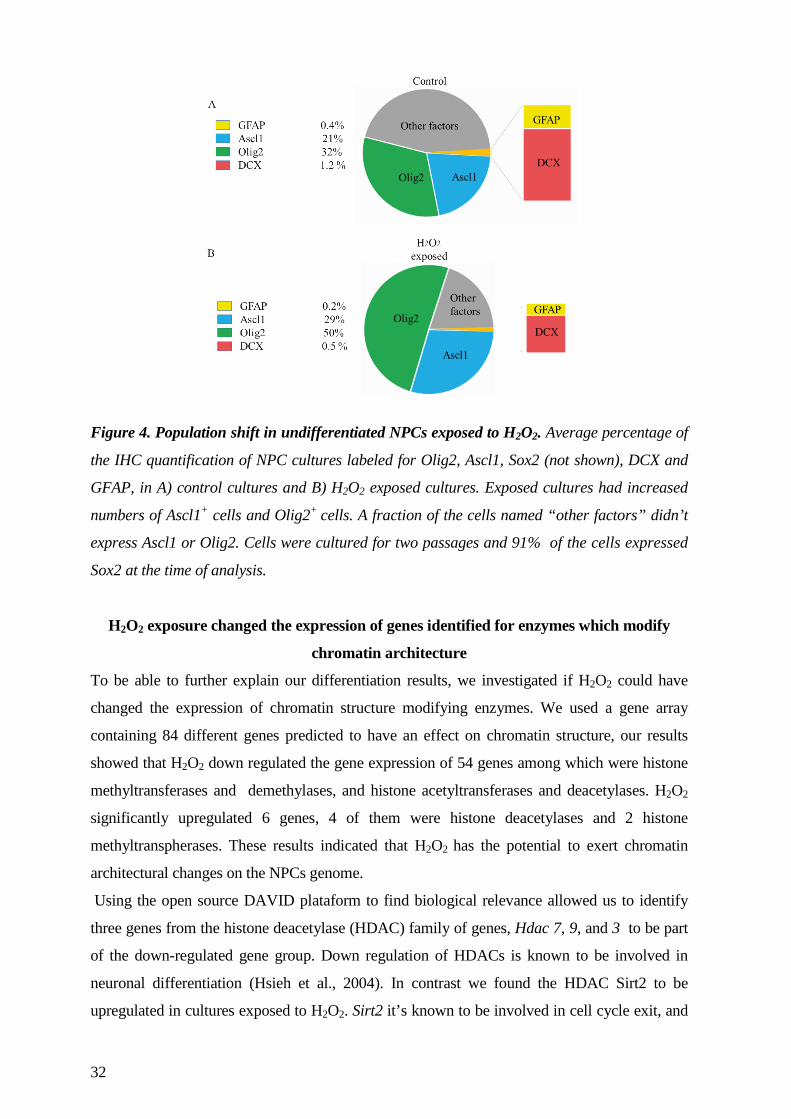
32
Figure 4. Population shift in undifferentiated NPCs exposed to H2O2. Average percentage of
the IHC quantification of NPC cultures labeled for Olig2, Ascl1, Sox2 (not shown), DCX and
GFAP, in A) control cultures and B) H2O2 exposed cultures. Exposed cultures had increased
numbers of Ascl1+ cells and Olig2+ cells. A fraction of the cells named “other factors” didn’t
express Ascl1 or Olig2. Cells were cultured for two passages and 91% of the cells expressed
Sox2 at the time of analysis.
H2O2 exposure changed the expression of genes identified for enzymes which modify
chromatin architecture
To be able to further explain our differentiation results, we investigated if H2O2 could have
changed the expression of chromatin structure modifying enzymes. We used a gene array
containing 84 different genes predicted to have an effect on chromatin structure, our results
showed that H2O2 down regulated the gene expression of 54 genes among which were histone
methyltransferases and demethylases, and histone acetyltransferases and deacetylases. H2O2
significantly upregulated 6 genes, 4 of them were histone deacetylases and 2 histone
methyltranspherases. These results indicated that H2O2 has the potential to exert chromatin
architectural changes on the NPCs genome.
Using the open source DAVID plataform to find biological relevance allowed us to identify
three genes from the histone deacetylase (HDAC) family of genes, Hdac 7, 9, and 3 to be part
of the down-regulated gene group. Down regulation of HDACs is known to be involved in
neuronal differentiation (Hsieh et al., 2004). In contrast we found the HDAC Sirt2 to be
upregulated in cultures exposed to H2O2. Sirt2 it’s known to be involved in cell cycle exit, and
33
oligodendrocyte differentiation (Li et al., 2007). Consequently, it became of interest to
investigate if H2O2 could affect Sirt2 expression and in turn inflict oligodendrocyte numbers.
To inhibit Sirt2 and to assess its requirement to oligodendrocyte differentiation, Sirtinol a Sirt2
inhibitor, was used in NPCs previously exposed to H2O2 and in unexposed NPCs. Sirtinol was
able to reduce the expression of Sirt2 in both conditions, and this effect was able to be
counteracted by the addition of H2O2 for 24h. The finding that Sirt2 was upregulated after
H2O2 exposure correlates with other studies reporting the Sirt2 expression triggered after cell
stressors are present in the cells microenvironment, (Zhang et al., 2013). When we assessed
oligodendrocyte numbers using IHC, in cultures exposed to sirtinol, we found that sirtinol was
able to decrease oligodendrocyte numbers and H2O2 exposure was able to restore them, these
results suggested a relationship between Sirt2 and oligodendrocyte differentiation. Further our
results confirm previous findings, demonstrating a role for HDACs in OPC maturation (Nave,
2008).
In paper III I describe the effect that H2O2 has on the NPCs differentiation potential, initiating
different fate dynamics in the NPC population, where the numbers of Olig2 and Ascl1
expressing cells increased. This increase lead to more oligodendrocytes and neuronal numbers.
Gene expression of chromatin modification enzymes was changed indicating DNA architecture
changes may allow the expression of genes favoring oligodendrocyte and neuronal
differentiation. In Fig. 5 I illustrate the network of molecules found relevant in this study for
NPC differentiation after H2O2 exposure.
In conclusion; Specific inflammatory components could offer specialized effects on the
regeneration of the CNS. Fine tuning of the inflammatory components could lead to the design
of new therapeutical approaches for regenerating the injured CNS.
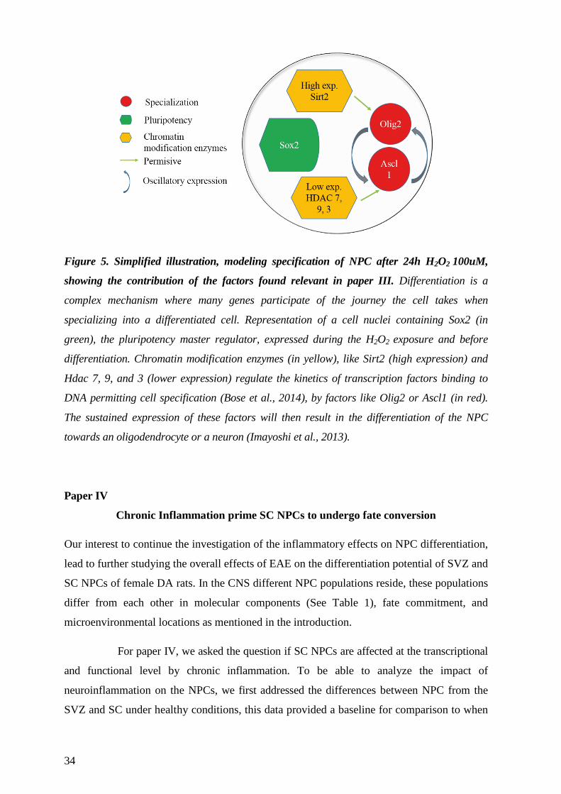
34
Figure 5. Simplified illustration, modeling specification of NPC after 24h H2O2 100uM,
showing the contribution of the factors found relevant in paper III. Differentiation is a
complex mechanism where many genes participate of the journey the cell takes when
specializing into a differentiated cell. Representation of a cell nuclei containing Sox2 (in
green), the pluripotency master regulator, expressed during the H2O2 exposure and before
differentiation. Chromatin modification enzymes (in yellow), like Sirt2 (high expression) and
Hdac 7, 9, and 3 (lower expression) regulate the kinetics of transcription factors binding to
DNA permitting cell specification (Bose et al., 2014), by factors like Olig2 or Ascl1 (in red).
The sustained expression of these factors will then result in the differentiation of the NPC
towards an oligodendrocyte or a neuron (Imayoshi et al., 2013).
Paper IV
Chronic Inflammation prime SC NPCs to undergo fate conversion
Our interest to continue the investigation of the inflammatory effects on NPC differentiation,
lead to further studying the overall effects of EAE on the differentiation potential of SVZ and
SC NPCs of female DA rats. In the CNS different NPC populations reside, these populations
differ from each other in molecular components (See Table 1), fate commitment, and
microenvironmental locations as mentioned in the introduction.
For paper IV, we asked the question if SC NPCs are affected at the transcriptional
and functional level by chronic inflammation. To be able to analyze the impact of
neuroinflammation on the NPCs, we first addressed the differences between NPC from the
SVZ and SC under healthy conditions, this data provided a baseline for comparison to when
35
these cells are in an inflamed environment. We studied the global gene expression
differences using an Affymetrix chip array, which allowed us to measure the global
expression of the genome in healthy NPCs and EAE derived NPCs, from both the SVZ and
the SC. For the Affymetrix experiment the experimental groups consisted of healthy derived
samples, and EAE derived samples, both groups with matched clinical scores of 3. We
analysed the cells in undifferentiated and differentiated conditions. The cells were extracted
from the SVZ and the cervical, thoracic and caudal parts of the spinal cord and from the SVZ
biopsy prior to NPC culturing. In parallel, NPC were cultured from the same CNS regional
areas and gene expression, immunohistochemistry, and western blot analysis were carried out
in undifferentiated and differentiated cells.
In healthy conditions the SVZ NPCs prove to be more neurogenic than the SC NPCs
which show higher gliogenic potential
In the undifferentiated NPC cultures, 187 of the genes were found to be differentially
expressed between the SVZ and the SC of healthy rats NPCs derived cultures, this expression
was higher in the SVZ comparing to the SC. The genes expressed in this fashion were
neurogenic, and some were associated with central nervous system development, generation of
neurons and neuronal differentiation. Gene expression analysis of the differentiated progeny of
the healthy SVZ revealed mature neuron functional gene expression validating our finding of a
neurogenic profile from the SVZ undifferentiated NPCs. The SC gene analysis profiling
presented a less neurogenic signature in both undifferentiated and differentiated NPCs.
Functional studies from both NPC regions, showed a significantly higher number of neurons
from the SVZ versus the SC, demonstrating the neurogenic nature of SVZ NPCs. The SC on
the other hand produced more oligodedrocytes than the SVZ, showing innate regional
differences in these two areas of the CNS.
EAE changed the genotypic characteristics of SC NPCs
We used 2 methods, Ingenuity System Pathway Analysis (IPA) and WebGestalt analysis
(Wang et al., 2013) to better understand the biological relevance of the global gene expression
data from NPCs derived from healthy and EAE SC comparison. The results revealed down
regulation of crucial genes participating of astrogliogenesis and oligodendrogenesis in the SC.
Functional studies of the spinal cord NPCs derived from EAE animals displayed lower
numbers of oligodendrocytes and higher numbers of neurons, in contrast to the healthy control
counterparts.
36
EAE increases the neurogenic potential of the SC NPCs as indicated by increased
numbers of Ascl1+ cells
As described in paper III Ascl1 and Olig 2 positive cells are known to generate neurons and
oligodendrocytes respectively, we investigated if there were differences in the numbers of
these cells in the SVZ and SC NPCs from healthy and from EAE animals. Quantification of the
SVZ NPCs showed a decreased in the numbers of Olig2+ cells in EAE derived NPCs, also a
trend towards increasing numbers of Ascl1+ cells was observed (Fig. 6). The analysis of the
SC-NPCs revealed an increase in the numbers of Ascl1 positive cells in EAE derived NPCs.
Olig2+ cell numbers were not different from the control cultures, however double labeling of
Olig2/Ascl1, revealed a significant higher number of Olig2+ cells which were also Ascl1+
confirming a newly acquired neurogenic signature by the Olig2 cells (Fig. 7). A dominant
expression of either Ascl1 or Olig2 will determine the final phenotype of these cells (Imayoshi
et al., 2013), which the functional analysis confirmed to be neuronal since we found an
increase in the numbers of these cells in EAE SC derived NPCs.
EAE SC derived NPCs develop a neurodegenerative signature
The transcriptome analysis revealed an increase in neurodegeneration related genes.
Importantly we observed a strong decrease in the expression of several functional groups
which control neurite developmental mechanisms, which could in turn affect the maturation
and integration of newly differentiated neurons. Another finding which can potentially
contribute to neuronal degeneration, is the observation of three functional groups predicted to
decreased functionality in lipid metabolism since lipid turnover and degradation are vital for
normal cellular mechanics, disruption of this system could have two outcomes, the instability
Figure 6. Quantification of Ascl1+ and Olig2+ cells in SVZ-NPCs from control and EAE.
Even though nitrite and nitrate levels were not detected in the SVZ-NPCs cultures,
quantification of Ascl1+ and Olig2+ cells revealed a significant difference in the Olig2
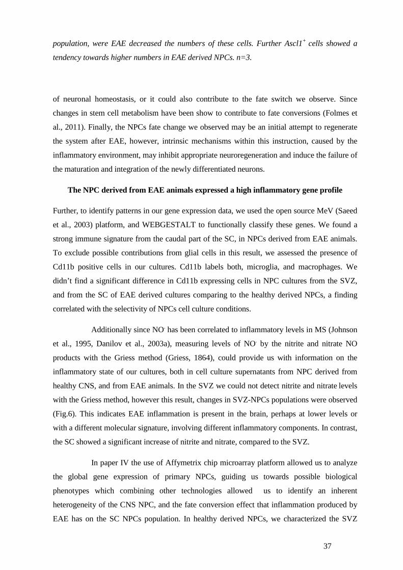
37
population, were EAE decreased the numbers of these cells. Further Ascl1+ cells showed a
tendency towards higher numbers in EAE derived NPCs. n=3.
of neuronal homeostasis, or it could also contribute to the fate switch we observe. Since
changes in stem cell metabolism have been show to contribute to fate conversions (Folmes et
al., 2011). Finally, the NPCs fate change we observed may be an initial attempt to regenerate
the system after EAE, however, intrinsic mechanisms within this instruction, caused by the
inflammatory environment, may inhibit appropriate neuroregeneration and induce the failure of
the maturation and integration of the newly differentiated neurons.
The NPC derived from EAE animals expressed a high inflammatory gene profile
Further, to identify patterns in our gene expression data, we used the open source MeV (Saeed
et al., 2003) platform, and WEBGESTALT to functionally classify these genes. We found a
strong immune signature from the caudal part of the SC, in NPCs derived from EAE animals.
To exclude possible contributions from glial cells in this result, we assessed the presence of
Cd11b positive cells in our cultures. Cd11b labels both, microglia, and macrophages. We
didn’t find a significant difference in Cd11b expressing cells in NPC cultures from the SVZ,
and from the SC of EAE derived cultures comparing to the healthy derived NPCs, a finding
correlated with the selectivity of NPCs cell culture conditions.
Additionally since NO. has been correlated to inflammatory levels in MS (Johnson
et al., 1995, Danilov et al., 2003a), measuring levels of NO. by the nitrite and nitrate NO
products with the Griess method (Griess, 1864), could provide us with information on the
inflammatory state of our cultures, both in cell culture supernatants from NPC derived from
healthy CNS, and from EAE animals. In the SVZ we could not detect nitrite and nitrate levels
with the Griess method, however this result, changes in SVZ-NPCs populations were observed
(Fig.6). This indicates EAE inflammation is present in the brain, perhaps at lower levels or
with a different molecular signature, involving different inflammatory components. In contrast,
the SC showed a significant increase of nitrite and nitrate, compared to the SVZ.
In paper IV the use of Affymetrix chip microarray platform allowed us to analyze
the global gene expression of primary NPCs, guiding us towards possible biological
phenotypes which combining other technologies allowed us to identify an inherent
heterogeneity of the CNS NPC, and the fate conversion effect that inflammation produced by
EAE has on the SC NPCs population. In healthy derived NPCs, we characterized the SVZ
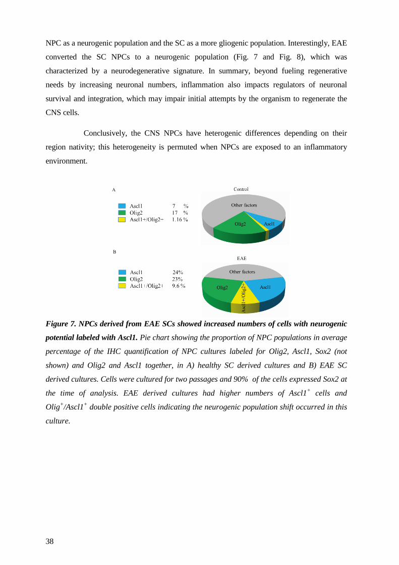
38
NPC as a neurogenic population and the SC as a more gliogenic population. Interestingly, EAE
converted the SC NPCs to a neurogenic population (Fig. 7 and Fig. 8), which was
characterized by a neurodegenerative signature. In summary, beyond fueling regenerative
needs by increasing neuronal numbers, inflammation also impacts regulators of neuronal
survival and integration, which may impair initial attempts by the organism to regenerate the
CNS cells.
Conclusively, the CNS NPCs have heterogenic differences depending on their
region nativity; this heterogeneity is permuted when NPCs are exposed to an inflammatory
environment.
Figure 7. NPCs derived from EAE SCs showed increased numbers of cells with neurogenic
potential labeled with Ascl1. Pie chart showing the proportion of NPC populations in average
percentage of the IHC quantification of NPC cultures labeled for Olig2, Ascl1, Sox2 (not
shown) and Olig2 and Ascl1 together, in A) healthy SC derived cultures and B) EAE SC
derived cultures. Cells were cultured for two passages and 90% of the cells expressed Sox2 at
the time of analysis. EAE derived cultures had higher numbers of Ascl1+ cells and
Olig+/Ascl1+ double positive cells indicating the neurogenic population shift occurred in this
culture.
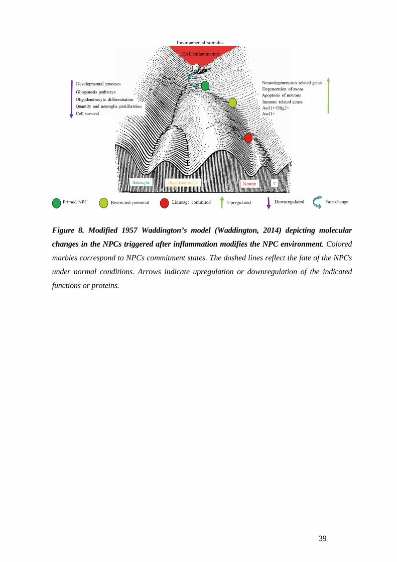
39
Figure 8. Modified 1957 Waddington’s model (Waddington, 2014) depicting molecular
changes in the NPCs triggered after inflammation modifies the NPC environment. Colored
marbles correspond to NPCs commitment states. The dashed lines reflect the fate of the NPCs
under normal conditions. Arrows indicate upregulation or downregulation of the indicated
functions or proteins.
40
CONCLUDING REMARKS
My thesis work shows that NPCs derived from the adult brain and SC have neuroregenerative
capacities in animal models of severe inflammation; a motor nerve injury model, and EAE.
Also the NPCs neurogenic potential is enhanced when cells are exposed to the inflammatory
component, the free radical H2O2. Three major points can be addressed from the present work;
a) CNS NPCs are heterogeneous, and present inherent genomic and functional differences
which are regionally dependent, where, under basal conditions the SVZ NPCs are more
neurogenic, and the SC more gliogenic. This NPC nature, may be changed during
neuroinflammation, proving the NPC plasticity in response to the surrounding
microenvironment.
b) Specific exposure of H2O2, a molecular component in the inflammatory milieu, activates the
neurogenic capacities of NPCs leading to more neurons as the differentiation result.
c) Different injury/inflammation degrees generate different regenerative responses from the
NPCs (Fig. 9). My work identifies severe inflammation as a permissive factor for NPC
neuronal differentiation, which at the same time also activates detrimental mechanisms within
the NPCs.
We are currently unable to ameliorate the degenerative processes occurring after
CNS injury. The CNS does activates a regenerative response after injury, where the
proliferation, migration and differentiation of NPCs occurs (Fagerlund et al., 2011, Danilov et
al., 2006), however this response is insufficient and defective. The more phylogenetically
sophisticated mammals are, and to this matter, humans, the more archaic the regenerative
potential is. Then, the need to empower our CNS regenerative potential becomes obvious. A
viable approach is the transplantation of exogenous NPCs which we and others demonstrate to
be beneficial to the injury site. Also inflammation, the first detectable reaction after injury, has
demonstrated to play a double role; Beneficial, as its not only capable of activating NPCs
(Danilov et al., 2006, Fagerlund et al., 2011), and diverting the NPC nature, allowing the
otherwise gliogenic SC NPCs to become neurogenic (Paper IV). But also an adverse role; as
inflammation may initiate intrinsic detrimental neuronal responses in the NPCs progeny, which
may result in cell death or poor cell integration into their new environment.
It becomes tempting to speculate that the latest is one of the mechanisms
responsible for a poor regenerative outcome in the CNS. Further, together with this micro
environmental determinants, we have the process of NPC differentiation. As described earlier
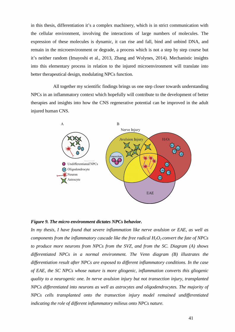
41
in this thesis, differentiation it’s a complex machinery, which is in strict communication with
the cellular environment, involving the interactions of large numbers of molecules. The
expression of these molecules is dynamic, it can rise and fall, bind and unbind DNA, and
remain in the microenvironment or degrade, a process which is not a step by step course but
it’s neither random (Imayoshi et al., 2013, Zhang and Wolynes, 2014). Mechanistic insights
into this elementary process in relation to the injured microenvironment will translate into
better therapeutical design, modulating NPCs function.
All together my scientific findings brings us one step closer towards understanding
NPCs in an inflammatory context which hopefully will contribute to the development of better
therapies and insights into how the CNS regenerative potential can be improved in the adult
injured human CNS.
Figure 9. The micro environment dictates NPCs behavior.
In my thesis, I have found that severe inflammation like nerve avulsion or EAE, as well as
components from the inflammatory cascade like the free radical H2O2 convert the fate of NPCs
to produce more neurons from NPCs from the SVZ, and from the SC. Diagram (A) shows
differentiated NPCs in a normal environment. The Venn diagram (B) illustrates the
differentiation result after NPCs are exposed to different inflammatory conditions. In the case
of EAE, the SC NPCs whose nature is more gliogenic, inflammation converts this gliogenic
quality to a neurogenic one. In nerve avulsion injury but not transection injury, transplanted
NPCs differentiated into neurons as well as astrocytes and oligodendrocytes. The majority of
NPCs cells transplanted onto the transection injury model remained undifferentiated
indicating the role of different inflammatory milieus onto NPCs nature.
42
Acknowledgements I would like to express my deepest appreciation to all the people who has touched my life positively
during my PhD studies time, friends, advisors and teachers.
Specially, it is with immense gratitude that I acknowledge the support and help of my advisor Lou
Brundin, who has taught me a lot during all these years. I admire your contagious commitment to
help patients, to impact their lives positively with our work. Thank you for being an excellent teacher
and for sharing great memories together; like trips where not only I learned a lot, but also, I had the
best times with you, discussing life and laughing together. Thank you for everything throughout these
years!
My co-supervisor Ruxandra Covacu, thank you for all the good times all these years, for teaching
me the NPC cultures, for fun scientific discussions and for our adventures trying out experiments.
Thanks for your friendship which I hope will grow well beyond my PhD studies.
My co-supervisor Mikael Svensson, I would like to thank you for being an excellent teacher, and for
your contagious curiosity and welcoming spirit. I admire your humbleness and I’m deeply thankful
for your help and contribution throughout my PhD studies time.
To my external supervisor Dan Larharmmar, It gives me great pleasure in acknowledging your
support throughout these years. Thank you for inspiring me, for introducing me to the amazingness of
the brain, after your course it was clear to me what I will be doing for the next years. Thank you for
all your support during this time.
To the neuroimmunology group; Thank you Tomas for putting together such a fun research group
and for you enthusiasm for Jazz and Science!, Bob, thanks so much for always being accessible to
discuss science and new ideas, Nada thanks for the amazing times in Istanbul, and for your
friendship, Pernilla always great to discuss science and life with you! Maja, thank you for your
support, and your contagious innovative spirit, I was very lucky to share the same lab with you,
Andre thank you for all the great times! Carl, Marie, Sabrina, Andreas, thank you guys for making
the lab more fun and full of life! Venus, thank you for all your efforts at the lab, it makes everything
roll, for your friendship, also without you my final experiments wouldn’t have been completed on
time, Xing Mei it’s great to have your positive energy around please keep it up! Petra, Hannes,
Roham, Harald, Sohel thanks for your huge support all these years. Thanks Rickard, Cecilia,
Mikael, Faiez, Fredrick and Shahin, for always being willing to discuss new ideas, for your good
sense of humor and the good times in and out of the lab. Sevi, Lara and Elian thanks for all the fun
times and interesting discussions, im sure your projects will deliver great results. Mohsen, thank you
for your kindness and all the care and work you do for the lab which makes the lab work easier,
Ingrid thank you for your kindness and scientific discussions, and also for letting me stay with you
and your family when I was in between apartments, it was great!
43
Also how to forget the past members of the Neuroimmunology group , Melanie, Ammenai, Johan,
Olle, Alexandra, Magda , Emilie, Magnus, Karin, Allan, Maggan, Åsa thank you All for the
enormous support throughout the years. To the Rheumatology unit, thank you All, for making the
lab a friendly and nice place to work, and always been helpful at any time.
To my office girls, Nånis, Mei, Brinda, and Hulda thank you for great conversations, your endless
support and for always being helpful and ready to have fun.
To my CMB collaborators Marie, Maggie, Jonas F., Christian G, David and Yildiz it’s been great
to work together, thank you for an exiting collaboration.
To my Brundin-Svensson group, Britt thank you for all your support and help, the good times at the
lab and outside, Micke and Lisa thanks for co-authorship and good times. Jonathan, Per, Erik,
Christian and Bomme thank your for the good times in and outside the lab, as well for your great
support throughout my PhD. Arvid thanks for your enthusiasm and help, especially those crazy days
at the microscope room, it’s always great to discuss science with you, Sreeni, thanks for the good
times and friendship, I’m sure your projects will give excellent results. Jonas, thanks for the support,
and fun discussions, good luck with your starting project, Sebastian thanks for your help in the
confocal and good scientific discussions. Maria, thank you for all the fun, the great wine nights and
your friendship, thank you for teaching me so much! Nasren thank you for your friendship and all
our laughs together, for being my coauthor and endless support, Pendar and Ramil thanks for all the
nice scientific discussions, and the fun in and out the lab, good luck with your projects!
To my wonderful and amazing friends in México, Sweden and elsewhere in the world, Cristian,
Ashley, thank you for always been there, for all the amazing times, and the ones to come, Marie B.
I’m so happy to still count you among my friends, since our Guanajuato times! Karl-Henrik,
Camilla, thank you for being a family to me in Stockholm, Sara E., Helena S., Analu, Frida,
Helena K. It wouldn’t have been the same without you my fav. Girls! Mai, Leif and Andreas, thank
you for your loving friendship all these years, Anna L. and Therese S. you have been an amazing
and happy support through my years in Sweden. Caroline and Francesca my cool and lovely
scientific friends, thanks for always being there! Sara F. thanks for all the fun and your friendship
and for your help with my cover image, Mariana P. Argentina-Mexico always together, thanks for
your support and friendship. Maria P. te quiero mi querida gracias por siempre estar. Liliana and
Auris without you none of this would have been possible. Peter D. and Ray K. in CA, thank you for
being such inspirational forces, SU brought back my old dreams.
Per thank you for all your love and patience, you have given me strength and smiles, I’m almost
there!
44
2 REFERENCES
ALDSKOGIUS, H. & SVENSSON, M. 1993. Neuronal and glial cell responses to axon injury.
Advances in structural biology, 2, 191-223. ALFARO-CERVELLO, C., SORIANO-NAVARRO, M., MIRZADEH, Z., ALVAREZ-BUYLLA, A.
& GARCIA-VERDUGO, J. M. 2012a. Biciliated ependymal cell proliferation contributes to spinal cord growth. J Comp Neurol, 520, 3528-52.
ALFARO-CERVELLO, C., SORIANO-NAVARRO, M., MIRZADEH, Z., ALVAREZ-BUYLLA, A. & GARCIA-VERDUGO, J. M. 2012b. Biciliated ependymal cell proliferation contributes to spinal cord growth. The Journal of comparative neurology, 520, 3528-52.
ARVIDSSON, L., FAGERLUND, M., JAFF, N., OSSOINAK, A., JANSSON, K., HAGERSTRAND, A., JOHANSSON, C. B., BRUNDIN, L. & SVENSSON, M. 2011. Distribution and characterization of progenitor cells within the human filum terminale. PloS one, 6, e27393.
AVILION, A. A., NICOLIS, S. K., PEVNY, L. H., PEREZ, L., VIVIAN, N. & LOVELL-BADGE, R. 2003. Multipotent cell lineages in early mouse development depend on SOX2 function. Genes & development, 17, 126-40.
BACIGALUPPI, M., PLUCHINO, S., PERUZZOTTI-JAMETTI, L., KILIC, E., KILIC, U., SALANI, G., BRAMBILLA, E., WEST, M. J., COMI, G., MARTINO, G. & HERMANN, D. M. 2009. Delayed post-ischaemic neuroprotection following systemic neural stem cell transplantation involves multiple mechanisms. Brain : a journal of neurology, 132, 2239-51.
BANNERMAN, P., HAHN, A., SOULIKA, A., GALLO, V. & PLEASURE, D. 2007. Astrogliosis in EAE spinal cord: derivation from radial glia, and relationships to oligodendroglia. Glia, 55, 57-64.
BECHER, A., DRENCKHAHN, A., PAHNER, I., MARGITTAI, M., JAHN, R. & AHNERT-HILGER, G. 1999. The synaptophysin-synaptobrevin complex: a hallmark of synaptic vesicle maturation. The Journal of neuroscience : the official journal of the Society for Neuroscience, 19, 1922-31.
BEDARD, K. & KRAUSE, K. H. 2007. The NOX family of ROS-generating NADPH oxidases: physiology and pathophysiology. Physiological reviews, 87, 245-313.
BEIROWSKI, B., ADALBERT, R., WAGNER, D., GRUMME, D. S., ADDICKS, K., RIBCHESTER, R. R. & COLEMAN, M. P. 2005. The progressive nature of Wallerian degeneration in wild-type and slow Wallerian degeneration (WldS) nerves. BMC Neurosci, 6, 6.
BEN-HUR, T. 2008. Immunomodulation by neural stem cells. Journal of the neurological sciences, 265, 102-4.
BERGER, S. L., KOUZARIDES, T., SHIEKHATTAR, R. & SHILATIFARD, A. 2009. An operational definition of epigenetics. Genes Dev, 23, 781-3.
BERGMANN, O., LIEBL, J., BERNARD, S., ALKASS, K., YEUNG, M. S., STEIER, P., KUTSCHERA, W., JOHNSON, L., LANDEN, M., DRUID, H., SPALDING, K. L. & FRISEN, J. 2012. The age of olfactory bulb neurons in humans. Neuron, 74, 634-9.
BERNER, R. A. 1999. Atmospheric oxygen over Phanerozoic time. Proceedings of the National Academy of Sciences of the United States of America, 96, 10955-7.
BERNFIELD, M., BANERJEE, S., KODA, J. & RAPRAEGER, A. 1984. Remodeling of the basement membrane as a mechanism of morphogenetic tissue interaction. The role of extracellular matrix in development. Liss, New York, 545-572.
BERNIER, P. J., BEDARD, A., VINET, J., LEVESQUE, M. & PARENT, A. 2002. Newly generated neurons in the amygdala and adjoining cortex of adult primates. Proceedings of the National Academy of Sciences of the United States of America, 99, 11464-9.
BHARDWAJ, R. D., CURTIS, M. A., SPALDING, K. L., BUCHHOLZ, B. A., FINK, D., BJORK-ERIKSSON, T., NORDBORG, C., GAGE, F. H., DRUID, H., ERIKSSON, P. S. & FRISEN, J. 2006. Neocortical neurogenesis in humans is restricted to development. Proceedings of the National Academy of Sciences of the United States of America, 103, 12564-8.
BJORKLUND, A. & KORDOWER, J. H. 2013. Cell therapy for Parkinson's disease: what next? Movement disorders : official journal of the Movement Disorder Society, 28, 110-5.
BJORKLUND, L. M. 2004. Stem cell therapy for Parkinson's disease. Dialogues in clinical neuroscience, 6, 303-11.
BOSE, P., DAI, Y. & GRANT, S. 2014. Histone deacetylase inhibitor (HDACI) mechanisms of action: Emerging insights. Pharmacol Ther.
45
BRAZEL, C. Y., LIMKE, T. L., OSBORNE, J. K., MIURA, T., CAI, J., PEVNY, L. & RAO, M. S. 2005. Sox2 expression defines a heterogeneous population of neurosphere-forming cells in the adult murine brain. Aging cell, 4, 197-207.
BREDERLAU, A., CORREIA, A. S., ANISIMOV, S. V., ELMI, M., PAUL, G., ROYBON, L., MORIZANE, A., BERGQUIST, F., RIEBE, I., NANNMARK, U., CARTA, M., HANSE, E., TAKAHASHI, J., SASAI, Y., FUNA, K., BRUNDIN, P., ERIKSSON, P. S. & LI, J. Y. 2006. Transplantation of human embryonic stem cell-derived cells to a rat model of Parkinson's disease: effect of in vitro differentiation on graft survival and teratoma formation. Stem cells, 24, 1433-40.
BRIGELIUS-FLOHE, R. & MAIORINO, M. 2013. Glutathione peroxidases. Biochim Biophys Acta, 1830, 3289-303.
BROSIUS LUTZ, A. & BARRES, B. A. 2014. Contrasting the glial response to axon injury in the central and peripheral nervous systems. Dev Cell, 28, 7-17.
BRUNDIN, L., BRISMAR, H., DANILOV, A. I., OLSSON, T. & JOHANSSON, C. B. 2003. Neural stem cells: a potential source for remyelination in neuroinflammatory disease. Brain Pathol, 13, 322-8.
CHAUDHARI, P., YE, Z. & JANG, Y. Y. 2012. Roles of Reactive Oxygen Species in the Fate of Stem Cells. Antioxidants & redox signaling.
CHEN, Z. X. & RIGGS, A. D. 2011. DNA methylation and demethylation in mammals. The Journal of biological chemistry, 286, 18347-53.
COMPSTON, A. & COLES, A. 2002. Multiple sclerosis. Lancet, 359, 1221-31. CONFORTI, L., GILLEY, J. & COLEMAN, M. P. 2014. Wallerian degeneration: an emerging axon
death pathway linking injury and disease. Nat Rev Neurosci, 15, 394-409. CRESS, W. D. & SETO, E. 2000. Histone deacetylases, transcriptional control, and cancer. Journal of
cellular physiology, 184, 1-16. CRUZ-ORENGO, L., DANIELS, B. P., DORSEY, D., BASAK, S. A., GRAJALES-REYES, J. G.,
MCCANDLESS, E. E., PICCIO, L., SCHMIDT, R. E., CROSS, A. H., CROSBY, S. D. & KLEIN, R. S. 2014. Enhanced sphingosine-1-phosphate receptor 2 expression underlies female CNS autoimmunity susceptibility. J Clin Invest, 124, 2571-84.
DANILOV, A. I., ANDERSSON, M., BAVAND, N., WIKLUND, N. P., OLSSON, T. & BRUNDIN, L. 2003a. Nitric oxide metabolite determinations reveal continuous inflammation in multiple sclerosis. J Neuroimmunol, 136, 112-8.
DANILOV, A. I., ANDERSSON, M., BAVAND, N., WIKLUND, N. P., OLSSON, T. & BRUNDIN, L. 2003b. Nitric oxide metabolite determinations reveal continuous inflammation in multiple sclerosis. Journal of neuroimmunology, 136, 112-8.
DANILOV, A. I., COVACU, R., MOE, M. C., LANGMOEN, I. A., JOHANSSON, C. B., OLSSON, T. & BRUNDIN, L. 2006. Neurogenesis in the adult spinal cord in an experimental model of multiple sclerosis. The European journal of neuroscience, 23, 394-400.
DAVIS, B. M., AYERS, J. L., KORAN, L., CARLSON, J., ANDERSON, M. C. & SIMPSON, S. B., JR. 1990. Time course of salamander spinal cord regeneration and recovery of swimming: HRP retrograde pathway tracing and kinematic analysis. Exp Neurol, 108, 198-213.
DE FEO, D., MERLINI, A., LATERZA, C. & MARTINO, G. 2012. Neural stem cell transplantation in central nervous system disorders: from cell replacement to neuroprotection. Current opinion in neurology, 25, 322-33.
DE STEFANO, N., NARAYANAN, S., FRANCIS, S. J., SMITH, S., MORTILLA, M., TARTAGLIA, M. C., BARTOLOZZI, M. L., GUIDI, L., FEDERICO, A. & ARNOLD, D. L. 2002. Diffuse axonal and tissue injury in patients with multiple sclerosis with low cerebral lesion load and no disability. Archives of neurology, 59, 1565-71.
DEBOUX, C., LADRAA, S., CAZAUBON, S., GHRIBI-MALLAH, S., WEISS, N., CHAVEROT, N., COURAUD, P. O. & EVERCOOREN, A. B. 2013. Overexpression of CD44 in Neural Precursor Cells Improves Trans- Endothelial Migration and Facilitates Their Invasion of Perivascular Tissues In Vivo. PloS one, 8, e57430.
DIAB, A., ZHU, J., XIAO, B. G., MUSTAFA, M. & LINK, H. 1997. High IL-6 and low IL-10 in the central nervous system are associated with protracted relapsing EAE in DA rats. Journal of neuropathology and experimental neurology, 56, 641-50.
DICK, O., TOM DIECK, S., ALTROCK, W. D., AMMERMULLER, J., WEILER, R., GARNER, C. C., GUNDELFINGER, E. D. & BRANDSTATTER, J. H. 2003. The presynaptic active zone protein bassoon is essential for photoreceptor ribbon synapse formation in the retina. Neuron, 37, 775-86.
46
DOETSCH, F., CAILLE, I., LIM, D. A., GARCIA-VERDUGO, J. M. & ALVAREZ-BUYLLA, A. 1999. Subventricular zone astrocytes are neural stem cells in the adult mammalian brain. Cell, 97, 703-16.
DROMARD, C., GUILLON, H., RIGAU, V., RIPOLL, C., SABOURIN, J. C., PERRIN, F. E., SCAMPS, F., BOZZA, S., SABATIER, P., LONJON, N., DUFFAU, H., VACHIERY-LAHAYE, F., PRIETO, M., TRAN VAN BA, C., DELEYROLLE, L., BOULARAN, A., LANGLEY, K., GAVIRIA, M., PRIVAT, A., HUGNOT, J. P. & BAUCHET, L. 2008. Adult human spinal cord harbors neural precursor cells that generate neurons and glial cells in vitro. Journal of neuroscience research, 86, 1916-26.
DUAN, H., LUO, Y., HAO, H., FENG, L., ZHANG, Y., LU, D., XING, S., FENG, J., YANG, D., SONG, L. & YAN, X. 2013. Soluble CD146 in cerebrospinal fluid of active multiple sclerosis. Neuroscience, 235, 16-26.
DUQUETTE, P., PLEINES, J., GIRARD, M., CHAREST, L., SENECAL-QUEVILLON, M. & MASSE, C. 1992. The increased susceptibility of women to multiple sclerosis. The Canadian journal of neurological sciences. Le journal canadien des sciences neurologiques, 19, 466-71.
EKDAHL, C. T. 2012. Microglial activation - tuning and pruning adult neurogenesis. Frontiers in pharmacology, 3, 41.
ELLIOTT, S. A. & SANCHEZ ALVARADO, A. 2013. The history and enduring contributions of planarians to the study of animal regeneration. Wiley Interdiscip Rev Dev Biol, 2, 301-26.
ELLIS, P., FAGAN, B. M., MAGNESS, S. T., HUTTON, S., TARANOVA, O., HAYASHI, S., MCMAHON, A., RAO, M. & PEVNY, L. 2004. SOX2, a persistent marker for multipotential neural stem cells derived from embryonic stem cells, the embryo or the adult. Dev Neurosci, 26, 148-65.
ERIKSSON, P. S., PERFILIEVA, E., BJORK-ERIKSSON, T., ALBORN, A. M., NORDBORG, C., PETERSON, D. A. & GAGE, F. H. 1998. Neurogenesis in the adult human hippocampus. Nature medicine, 4, 1313-7.
ERNST, A., ALKASS, K., BERNARD, S., SALEHPOUR, M., PERL, S., TISDALE, J., POSSNERT, G., DRUID, H. & FRISEN, J. 2014. Neurogenesis in the Striatum of the Adult Human Brain. Cell.
FAGERLUND, M., JAFF, N., DANILOV, A. I., PEREDO, I., BRUNDIN, L. & SVENSSON, M. 2011. Proliferation, migration and differentiation of ependymal region neural progenitor cells in the brainstem after hypoglossal nerve avulsion. Restor Neurol Neurosci, 29, 47-59.
FAGERLUND, M., PEREZ ESTRADA, C., JAFF, N., SVENSSON, M. & BRUNDIN, L. 2012. Neural stem/progenitor cells transplanted to the hypoglossal nucleus integrates with the host CNS in adult rats and promotes motor neuron survival. Cell Transplant, 21, 739-47.
FAWCETT, J. W., SCHWAB, M. E., MONTANI, L., BRAZDA, N. & MULLER, H. W. 2012. Defeating inhibition of regeneration by scar and myelin components. Handbook of clinical neurology, 109, 503-22.
FERRETTI, P. 2011. Is there a relationship between adult neurogenesis and neuron generation following injury across evolution? The European journal of neuroscience, 34, 951-62.
FERRI, A. L., CAVALLARO, M., BRAIDA, D., DI CRISTOFANO, A., CANTA, A., VEZZANI, A., OTTOLENGHI, S., PANDOLFI, P. P., SALA, M., DEBIASI, S. & NICOLIS, S. K. 2004. Sox2 deficiency causes neurodegeneration and impaired neurogenesis in the adult mouse brain. Development, 131, 3805-19.
FOLMES, C. D., NELSON, T. J., MARTINEZ-FERNANDEZ, A., ARRELL, D. K., LINDOR, J. Z., DZEJA, P. P., IKEDA, Y., PEREZ-TERZIC, C. & TERZIC, A. 2011. Somatic oxidative bioenergetics transitions into pluripotency-dependent glycolysis to facilitate nuclear reprogramming. Cell Metab, 14, 264-71.
FONTES, R. B., SAAD, F., SOARES, M. S., DE OLIVEIRA, F., PINTO, F. C. & LIBERTI, E. A. 2006. Ultrastructural study of the filum terminale and its elastic fibers. Neurosurgery, 58, 978-84; discussion 978-84.
FRIDOVICH, I. 1995. Superoxide radical and superoxide dismutases. Annual review of biochemistry, 64, 97-112.
FRIEDMAN, H. V., BRESLER, T., GARNER, C. C. & ZIV, N. E. 2000. Assembly of new individual excitatory synapses: time course and temporal order of synaptic molecule recruitment. Neuron, 27, 57-69.
FRIESE, M. A., SCHATTLING, B. & FUGGER, L. 2014. Mechanisms of neurodegeneration and axonal dysfunction in multiple sclerosis. Nature reviews. Neurology, 10, 225-38.
FUENTEALBA, L. C., OBERNIER, K. & ALVAREZ-BUYLLA, A. 2012. Adult neural stem cells bridge their niche. Cell Stem Cell, 10, 698-708.
47
FUKAZAWA, T., NAORA, Y., KUNIEDA, T. & KUBO, T. 2009. Suppression of the immune response potentiates tadpole tail regeneration during the refractory period. Development, 136, 2323-7.
GAGE, F. H. 2000. Mammalian neural stem cells. Science, 287, 1433-8. GAILLARD, A. & JABER, M. 2011. Rewiring the brain with cell transplantation in Parkinson's
disease. Trends in neurosciences, 34, 124-33. GALLONI, M. 2012. Global irradiation effects, stem cell genes and rare transcripts in the planarian
transcriptome. The International journal of developmental biology, 56, 103-16. GILGUN-SHERKI, Y., MELAMED, E. & OFFEN, D. 2004. The role of oxidative stress in the
pathogenesis of multiple sclerosis: the need for effective antioxidant therapy. J Neurol, 251, 261-8.
GORITZ, C., DIAS, D. O., TOMILIN, N., BARBACID, M., SHUPLIAKOV, O. & FRISEN, J. 2011. A pericyte origin of spinal cord scar tissue. Science, 333, 238-42.
GOUGH, D. R. & COTTER, T. G. 2011. Hydrogen peroxide: a Jekyll and Hyde signalling molecule. Cell death & disease, 2, e213.
GRIESS, P. 1864. On a new series of bodies in which nitrogen is substituted for hydrogen. Philosophical Transactions of the Royal Society of London, 667-731.
GUTTERIDGE, J. & HALLIWELL, B. 2007, Fourth Edition Free radicals in biology and medicine, Oxford Oxford University Press, New York.
HAMILTON, L. K., TRUONG, M. K., BEDNARCZYK, M. R., AUMONT, A. & FERNANDES, K. J. 2009. Cellular organization of the central canal ependymal zone, a niche of latent neural stem cells in the adult mammalian spinal cord. Neuroscience, 164, 1044-56.
HOLMQUIST, L., STUCHBURY, G., STEELE, M. & MUNCH, G. 2007. Hydrogen peroxide is a true first messenger. Journal of neural transmission. Supplementum, 39-41.
HSIEH, J., NAKASHIMA, K., KUWABARA, T., MEJIA, E. & GAGE, F. H. 2004. Histone deacetylase inhibition-mediated neuronal differentiation of multipotent adult neural progenitor cells. Proceedings of the National Academy of Sciences of the United States of America, 101, 16659-64.
IMAYOSHI, I., ISOMURA, A., HARIMA, Y., KAWAGUCHI, K., KORI, H., MIYACHI, H., FUJIWARA, T., ISHIDATE, F. & KAGEYAMA, R. 2013. Oscillatory control of factors determining multipotency and fate in mouse neural progenitors. Science, 342, 1203-8.
JHA, R. M., LIU, X., CHRENEK, R., MADSEN, J. R. & CARDOZO, D. L. 2013. The postnatal human filum terminale is a source of autologous multipotent neurospheres capable of generating motor neurons. Neurosurgery, 72, 118-29; discussion 129.
JI, S., DOUCETTE, J. R. & NAZARALI, A. J. 2011. Sirt2 is a novel in vivo downstream target of Nkx2.2 and enhances oligodendroglial cell differentiation. Journal of molecular cell biology, 3, 351-9.
JIANG, Y., ZHANG, M., KOISHI, K. & MCLENNAN, I. S. 2000. TGF-beta 2 attenuates the injury-induced death of mature motoneurons. J Neurosci Res, 62, 809-13.
JIN, K. L., MAO, X. O. & GREENBERG, D. A. 2000. Vascular endothelial growth factor: direct neuroprotective effect in in vitro ischemia. Proceedings of the National Academy of Sciences of the United States of America, 97, 10242-7.
JOHANSSON, C. B., MOMMA, S., CLARKE, D. L., RISLING, M., LENDAHL, U. & FRISEN, J. 1999. Identification of a neural stem cell in the adult mammalian central nervous system. Cell, 96, 25-34.
JOHNSON, A. W., LAND, J. M., THOMPSON, E. J., BOLANOS, J. P., CLARK, J. B. & HEALES, S. J. 1995. Evidence for increased nitric oxide production in multiple sclerosis. J Neurol Neurosurg Psychiatry, 58, 107.
JONES, P. A. & TAKAI, D. 2001. The role of DNA methylation in mammalian epigenetics. Science, 293, 1068-70.
JULIANDI, B., ABEMATSU, M. & NAKASHIMA, K. 2010. Epigenetic regulation in neural stem cell differentiation. Development, growth & differentiation, 52, 493-504.
KEREVER, A., SCHNACK, J., VELLINGA, D., ICHIKAWA, N., MOON, C., ARIKAWA-HIRASAWA, E., EFIRD, J. T. & MERCIER, F. 2007. Novel extracellular matrix structures in the neural stem cell niche capture the neurogenic factor fibroblast growth factor 2 from the extracellular milieu. Stem cells, 25, 2146-57.
KIM, E. J., ABLES, J. L., DICKEL, L. K., EISCH, A. J. & JOHNSON, J. E. 2011. Ascl1 (Mash1) defines cells with long-term neurogenic potential in subgranular and subventricular zones in adult mouse brain. PloS one, 6, e18472.
KING, R. S. & NEWMARK, P. A. 2012. The cell biology of regeneration. J Cell Biol, 196, 553-62.
48
KLIMOVA, T. & CHANDEL, N. S. 2008. Mitochondrial complex III regulates hypoxic activation of HIF. Cell death and differentiation, 15, 660-6.
KOKOVAY, E., WANG, Y., KUSEK, G., WURSTER, R., LEDERMAN, P., LOWRY, N., SHEN, Q. & TEMPLE, S. 2012. VCAM1 is essential to maintain the structure of the SVZ niche and acts as an environmental sensor to regulate SVZ lineage progression. Cell stem cell, 11, 220-30.
KOLIATSOS, V. E., PRICE, W. L., PARDO, C. A. & PRICE, D. L. 1994. Ventral root avulsion: an experimental model of death of adult motor neurons. J Comp Neurol, 342, 35-44.
KOUZARIDES, T. 2007. Chromatin modifications and their function. Cell, 128, 693-705. KUHLMANN, T., MIRON, V., CUI, Q., WEGNER, C., ANTEL, J. & BRUCK, W. 2008.
Differentiation block of oligodendroglial progenitor cells as a cause for remyelination failure in chronic multiple sclerosis. Brain : a journal of neurology, 131, 1749-58.
KULBATSKI, I., MOTHE, A. J., KEATING, A., HAKAMATA, Y., KOBAYASHI, E. & TATOR, C. H. 2007. Oligodendrocytes and radial glia derived from adult rat spinal cord progenitors: morphological and immunocytochemical characterization. The journal of histochemistry and cytochemistry : official journal of the Histochemistry Society, 55, 209-22.
KULBATSKI, I. & TATOR, C. H. 2009. Region-specific differentiation potential of adult rat spinal cord neural stem/precursors and their plasticity in response to in vitro manipulation. The journal of histochemistry and cytochemistry : official journal of the Histochemistry Society, 57, 405-23.
LARSON, J. L. & YUAN, G. C. 2012. Chromatin states accurately classify cell differentiation stages. PloS one, 7, e31414.
LE BELLE, J. E., OROZCO, N. M., PAUCAR, A. A., SAXE, J. P., MOTTAHEDEH, J., PYLE, A. D., WU, H. & KORNBLUM, H. I. 2011. Proliferative neural stem cells have high endogenous ROS levels that regulate self-renewal and neurogenesis in a PI3K/Akt-dependant manner. Cell stem cell, 8, 59-71.
LECOEUR, H. 2002. Nuclear apoptosis detection by flow cytometry: influence of endogenous endonucleases. Experimental cell research, 277, 1-14.
LEPORE, A. C. 2011. Intraspinal cell transplantation for targeting cervical ventral horn in amyotrophic lateral sclerosis and traumatic spinal cord injury. J Vis Exp.
LEPORE, A. C. & MARAGAKIS, N. J. 2007. Targeted stem cell transplantation strategies in ALS. Neurochemistry international, 50, 966-75.
LI, L., BENNETT, S. A. & WANG, L. 2012. Role of E-cadherin and other cell adhesion molecules in survival and differentiation of human pluripotent stem cells. Cell adhesion & migration, 6, 59-70.
LI, W., ZHANG, B., TANG, J., CAO, Q., WU, Y., WU, C., GUO, J., LING, E. A. & LIANG, F. 2007. Sirtuin 2, a mammalian homolog of yeast silent information regulator-2 longevity regulator, is an oligodendroglial protein that decelerates cell differentiation through deacetylating alpha-tubulin. J Neurosci, 27, 2606-16.
LOBO, D., BEANE, W. S. & LEVIN, M. 2012. Modeling planarian regeneration: a primer for reverse-engineering the worm. PLoS Comput Biol, 8, e1002481.
LOIS, C., HONG, E. J., PEASE, S., BROWN, E. J. & BALTIMORE, D. 2002. Germline transmission and tissue-specific expression of transgenes delivered by lentiviral vectors. Science, 295, 868-72.
LORENTZEN, J. C., ISSAZADEH, S., STORCH, M., MUSTAFA, M. I., LASSMAN, H., LININGTON, C., KLARESKOG, L. & OLSSON, T. 1995. Protracted, relapsing and demyelinating experimental autoimmune encephalomyelitis in DA rats immunized with syngeneic spinal cord and incomplete Freund's adjuvant. Journal of neuroimmunology, 63, 193-205.
LUK, E., CARROLL, M., BAKER, M. & CULOTTA, V. C. 2003. Manganese activation of superoxide dismutase 2 in Saccharomyces cerevisiae requires MTM1, a member of the mitochondrial carrier family. Proceedings of the National Academy of Sciences of the United States of America, 100, 10353-7.
MA, D. K., MARCHETTO, M. C., GUO, J. U., MING, G. L., GAGE, F. H. & SONG, H. 2010. Epigenetic choreographers of neurogenesis in the adult mammalian brain. Nature neuroscience, 13, 1338-44.
MA, Q., KINTNER, C. & ANDERSON, D. J. 1996. Identification of neurogenin, a vertebrate neuronal determination gene. Cell, 87, 43-52.
MADHAVAN, L., OUREDNIK, V. & OUREDNIK, J. 2006. Increased "vigilance" of antioxidant mechanisms in neural stem cells potentiates their capability to resist oxidative stress. Stem cells, 24, 2110-9.
49
MALATESTA, P. & GOTZ, M. 2013. Radial glia - from boring cables to stem cell stars. Development, 140, 483-6.
MANDA, K., UENO, M. & ANZAI, K. 2009. Cranial irradiation-induced inhibition of neurogenesis in hippocampal dentate gyrus of adult mice: attenuation by melatonin pretreatment. J Pineal Res, 46, 71-8.
MARGIS, R., DUNAND, C., TEIXEIRA, F. K. & MARGIS-PINHEIRO, M. 2008. Glutathione peroxidase family - an evolutionary overview. The FEBS journal, 275, 3959-70.
MARTI-FABREGAS, J., ROMAGUERA-ROS, M., GOMEZ-PINEDO, U., MARTINEZ-RAMIREZ, S., JIMENEZ-XARRIE, E., MARIN, R., MARTI-VILALTA, J. L. & GARCIA-VERDUGO, J. M. 2010. Proliferation in the human ipsilateral subventricular zone after ischemic stroke. Neurology, 74, 357-65.
MARTIN, L. J., KAISER, A. & PRICE, A. C. 1999. Motor neuron degeneration after sciatic nerve avulsion in adult rat evolves with oxidative stress and is apoptosis. J Neurobiol, 40, 185-201.
MASUI, S., NAKATAKE, Y., TOYOOKA, Y., SHIMOSATO, D., YAGI, R., TAKAHASHI, K., OKOCHI, H., OKUDA, A., MATOBA, R., SHAROV, A. A., KO, M. S. & NIWA, H. 2007. Pluripotency governed by Sox2 via regulation of Oct3/4 expression in mouse embryonic stem cells. Nature cell biology, 9, 625-35.
MATTSSON, P., MEIJER, B. & SVENSSON, M. 1999. Extensive neuronal cell death following intracranial transection of the facial nerve in the adult rat. Brain Res Bull, 49, 333-41.
MEHTA, V., PEI, W., YANG, G., LI, S., SWAMY, E., BOSTER, A., SCHMALBROCK, P. & PITT, D. 2013. Iron is a sensitive biomarker for inflammation in multiple sclerosis lesions. PloS one, 8, e57573.
MELETIS, K., BARNABE-HEIDER, F., CARLEN, M., EVERGREN, E., TOMILIN, N., SHUPLIAKOV, O. & FRISEN, J. 2008. Spinal cord injury reveals multilineage differentiation of ependymal cells. PLoS biology, 6, e182.
MERCIER, F., KITASAKO, J. T. & HATTON, G. I. 2002. Anatomy of the brain neurogenic zones revisited: fractones and the fibroblast/macrophage network. The Journal of comparative neurology, 451, 170-88.
MERKLE, F. T., TRAMONTIN, A. D., GARCIA-VERDUGO, J. M. & ALVAREZ-BUYLLA, A. 2004. Radial glia give rise to adult neural stem cells in the subventricular zone. Proceedings of the National Academy of Sciences of the United States of America, 101, 17528-32.
MIRBAHAI, L. & CHIPMAN, J. K. 2014. Epigenetic memory of environmental organisms: a reflection of lifetime stressor exposures. Mutat Res Genet Toxicol Environ Mutagen, 764-765, 10-7.
MIRZADEH, Z., MERKLE, F. T., SORIANO-NAVARRO, M., GARCIA-VERDUGO, J. M. & ALVAREZ-BUYLLA, A. 2008. Neural stem cells confer unique pinwheel architecture to the ventricular surface in neurogenic regions of the adult brain. Cell Stem Cell, 3, 265-78.
MIX, E., MEYER-RIENECKER, H., HARTUNG, H. P. & ZETTL, U. K. 2010. Animal models of multiple sclerosis--potentials and limitations. Progress in neurobiology, 92, 386-404.
MONJE, M. L., TODA, H. & PALMER, T. D. 2003. Inflammatory blockade restores adult hippocampal neurogenesis. Science, 302, 1760-5.
MORRISON, J. I., LOOF, S., HE, P. & SIMON, A. 2006. Salamander limb regeneration involves the activation of a multipotent skeletal muscle satellite cell population. J Cell Biol, 172, 433-40.
MORSHEAD, C. M., REYNOLDS, B. A., CRAIG, C. G., MCBURNEY, M. W., STAINES, W. A., MORASSUTTI, D., WEISS, S. & VAN DER KOOY, D. 1994. Neural stem cells in the adult mammalian forebrain: a relatively quiescent subpopulation of subependymal cells. Neuron, 13, 1071-1082.
NACHER, J., CRESPO, C. & MCEWEN, B. S. 2001. Doublecortin expression in the adult rat telencephalon. Eur J Neurosci, 14, 629-44.
NACU, E. & TANAKA, E. M. 2011. Limb regeneration: a new development? Annual review of cell and developmental biology, 27, 409-40.
NAIT-OUMESMAR, B., PICARD-RIERA, N., KERNINON, C., DECKER, L., SEILHEAN, D., HOGLINGER, G. U., HIRSCH, E. C., REYNOLDS, R. & BARON-VAN EVERCOOREN, A. 2007. Activation of the subventricular zone in multiple sclerosis: evidence for early glial progenitors. Proceedings of the National Academy of Sciences of the United States of America, 104, 4694-9.
NAKAGAWA, T. & GUARENTE, L. 2011. Sirtuins at a glance. Journal of cell science, 124, 833-8. NATOWICZ, M. R. & BEJJANI, B. 1994. Genetic disorders that masquerade as multiple sclerosis.
American journal of medical genetics, 49, 149-69.
50
NAVA, M. M., RAIMONDI, M. T. & PIETRABISSA, R. 2012. Controlling self-renewal and differentiation of stem cells via mechanical cues. Journal of biomedicine & biotechnology, 2012, 797410.
NAVE, K. A. 2008. Neuroscience: An ageing view of myelin repair. Nature, 455, 478-9. NIKIC, I., MERKLER, D., SORBARA, C., BRINKOETTER, M., KREUTZFELDT, M., BAREYRE,
F. M., BRUCK, W., BISHOP, D., MISGELD, T. & KERSCHENSTEINER, M. 2011. A reversible form of axon damage in experimental autoimmune encephalomyelitis and multiple sclerosis. Nature medicine, 17, 495-9.
NORTH, B. J. & VERDIN, E. 2007. Mitotic regulation of SIRT2 by cyclin-dependent kinase 1-dependent phosphorylation. The Journal of biological chemistry, 282, 19546-55.
NOTTEBOHM, F. 1989. From bird song to neurogenesis. Scientific American, 260, 74-9. OHORI, Y., YAMAMOTO, S., NAGAO, M., SUGIMORI, M., YAMAMOTO, N., NAKAMURA, K.
& NAKAFUKU, M. 2006. Growth factor treatment and genetic manipulation stimulate neurogenesis and oligodendrogenesis by endogenous neural progenitors in the injured adult spinal cord. The Journal of neuroscience : the official journal of the Society for Neuroscience, 26, 11948-60.
OLSSON, T. 1995. Critical influences of the cytokine orchestration on the outcome of myelin antigen-specific T-cell autoimmunity in experimental autoimmune encephalomyelitis and multiple sclerosis. Immunological reviews, 144, 245-68.
ONAL, P., GRUN, D., ADAMIDI, C., RYBAK, A., SOLANA, J., MASTROBUONI, G., WANG, Y., RAHN, H. P., CHEN, W., KEMPA, S., ZIEBOLD, U. & RAJEWSKY, N. 2012. Gene expression of pluripotency determinants is conserved between mammalian and planarian stem cells. The EMBO journal, 31, 2755-69.
OOI, S. K. & BESTOR, T. H. 2008. The colorful history of active DNA demethylation. Cell, 133, 1145-8.
OTTERVALD, J., FRANZEN, B., NILSSON, K., ANDERSSON, L. I., KHADEMI, M., ERIKSSON, B., KJELLSTROM, S., MARKO-VARGA, G., VEGVARI, A., HARRIS, R. A., LAURELL, T., MILIOTIS, T., MATUSEVICIUS, D., SALTER, H., FERM, M. & OLSSON, T. 2010. Multiple sclerosis: Identification and clinical evaluation of novel CSF biomarkers. Journal of proteomics, 73, 1117-32.
PALMER, T. D., TAKAHASHI, J. & GAGE, F. H. 1997. The adult rat hippocampus contains primordial neural stem cells. Molecular and cellular neurosciences, 8, 389-404.
PARISH, C. L., BELJAJEVA, A., ARENAS, E. & SIMON, A. 2007. Midbrain dopaminergic neurogenesis and behavioural recovery in a salamander lesion-induced regeneration model. Development, 134, 2881-7.
PARK, H. C. & APPEL, B. 2003. Delta-Notch signaling regulates oligodendrocyte specification. Development, 130, 3747-55.
PARRAS, C. M., GALLI, R., BRITZ, O., SOARES, S., GALICHET, C., BATTISTE, J., JOHNSON, J. E., NAKAFUKU, M., VESCOVI, A. & GUILLEMOT, F. 2004. Mash1 specifies neurons and oligodendrocytes in the postnatal brain. EMBO J, 23, 4495-505.
PEREZ ESTRADA, C., COVACU, R., SANKAVARAM, S. R., SVENSSON, M. & BRUNDIN, L. 2014. Oxidative stress increases neurogenesis and oligodendrogenesis in adult neural progenitor cells. Stem cells and development.
PERNET, V. & SCHWAB, M. E. 2012. The role of Nogo-A in axonal plasticity, regrowth and repair. Cell and tissue research, 349, 97-104.
PETIT, A., SANDERS, A. D., KENNEDY, T. E., TETZLAFF, W., GLATTFELDER, K. J., DALLEY, R. A., PUCHALSKI, R. B., JONES, A. R. & ROSKAMS, A. J. 2011. Adult spinal cord radial glia display a unique progenitor phenotype. PloS one, 6, e24538.
PFENNINGER, C. V., STEINHOFF, C., HERTWIG, F. & NUBER, U. A. 2011. Prospectively isolated CD133/CD24-positive ependymal cells from the adult spinal cord and lateral ventricle wall differ in their long-term in vitro self-renewal and in vivo gene expression. Glia, 59, 68-81.
PICARD-RIERA, N., DECKER, L., DELARASSE, C., GOUDE, K., NAIT-OUMESMAR, B., LIBLAU, R., PHAM-DINH, D. & BARON-VAN EVERCOOREN, A. 2002. Experimental autoimmune encephalomyelitis mobilizes neural progenitors from the subventricular zone to undergo oligodendrogenesis in adult mice. Proceedings of the National Academy of Sciences of the United States of America, 99, 13211-6.
PLUCHINO, S., GRITTI, A., BLEZER, E., AMADIO, S., BRAMBILLA, E., BORSELLINO, G., COSSETTI, C., DEL CARRO, U., COMI, G., T HART, B., VESCOVI, A. & MARTINO, G. 2009. Human neural stem cells ameliorate autoimmune encephalomyelitis in non-human primates. Annals of neurology, 66, 343-54.
51
PLUCHINO, S., MUZIO, L., IMITOLA, J., DELEIDI, M., ALFARO-CERVELLO, C., SALANI, G., PORCHERI, C., BRAMBILLA, E., CAVASINNI, F., BERGAMASCHI, A., GARCIA-VERDUGO, J. M., COMI, G., KHOURY, S. J. & MARTINO, G. 2008. Persistent inflammation alters the function of the endogenous brain stem cell compartment. Brain : a journal of neurology, 131, 2564-78.
PLUCHINO, S., QUATTRINI, A., BRAMBILLA, E., GRITTI, A., SALANI, G., DINA, G., GALLI, R., DEL CARRO, U., AMADIO, S., BERGAMI, A., FURLAN, R., COMI, G., VESCOVI, A. L. & MARTINO, G. 2003. Injection of adult neurospheres induces recovery in a chronic model of multiple sclerosis. Nature, 422, 688-94.
POWELL, F. L. 2010. Studying biological responses to global change in atmospheric oxygen. Respiratory physiology & neurobiology, 173 Suppl, S6-12.
PRABHAKAR, R., SIEGBAHN, P. E., MINAEV, B. F. & ÅGREN, H. 2004. Spin transition during H2O2 formation in the oxidative half-reaction of copper amine oxidases. The Journal of Physical Chemistry B, 108, 13882-13892.
QUINONES-HINOJOSA, A., SANAI, N., SORIANO-NAVARRO, M., GONZALEZ-PEREZ, O., MIRZADEH, Z., GIL-PEROTIN, S., ROMERO-RODRIGUEZ, R., BERGER, M. S., GARCIA-VERDUGO, J. M. & ALVAREZ-BUYLLA, A. 2006. Cellular composition and cytoarchitecture of the adult human subventricular zone: a niche of neural stem cells. The Journal of comparative neurology, 494, 415-34.
RAFALSKI, V. A. & BRUNET, A. 2011. Energy metabolism in adult neural stem cell fate. Progress in neurobiology, 93, 182-203.
REDDIEN, P. W. 2013. Specialized progenitors and regeneration. Development, 140, 951-7. REMLINGER, P. 1905. Accidents paralytiques au cours du traitement antirabique. ROSENLING, T., STOOP, M. P., ATTALI, A., VAN AKEN, H., SUIDGEEST, E., CHRISTIN, C.,
STINGL, C., SUITS, F., HORVATOVICH, P., HINTZEN, R. Q., TUINSTRA, T., BISCHOFF, R. & LUIDER, T. M. 2012. Profiling and identification of cerebrospinal fluid proteins in a rat EAE model of multiple sclerosis. Journal of proteome research, 11, 2048-60.
ROSENSTEIN, J. M., MANI, N., KHAIBULLINA, A. & KRUM, J. M. 2003. Neurotrophic effects of vascular endothelial growth factor on organotypic cortical explants and primary cortical neurons. The Journal of neuroscience : the official journal of the Society for Neuroscience, 23, 11036-44.
SABOURIN, J. C., ACKEMA, K. B., OHAYON, D., GUICHET, P. O., PERRIN, F. E., GARCES, A., RIPOLL, C., CHARITE, J., SIMONNEAU, L., KETTENMANN, H., ZINE, A., PRIVAT, A., VALMIER, J., PATTYN, A. & HUGNOT, J. P. 2009. A mesenchymal-like ZEB1(+) niche harbors dorsal radial glial fibrillary acidic protein-positive stem cells in the spinal cord. Stem cells, 27, 2722-33.
SAEED, A. I., SHAROV, V., WHITE, J., LI, J., LIANG, W., BHAGABATI, N., BRAISTED, J., KLAPA, M., CURRIER, T., THIAGARAJAN, M., STURN, A., SNUFFIN, M., REZANTSEV, A., POPOV, D., RYLTSOV, A., KOSTUKOVICH, E., BORISOVSKY, I., LIU, Z., VINSAVICH, A., TRUSH, V. & QUACKENBUSH, J. 2003. TM4: a free, open-source system for microarray data management and analysis. Biotechniques, 34, 374-8.
SAKUMA, H., KOHYAMA, K., PARK, I. K., MIYAKOSHI, A., TANUMA, N. & MATSUMOTO, Y. 2004. Clinicopathological study of a myelin oligodendrocyte glycoprotein-induced demyelinating disease in LEW.1AV1 rats. Brain : a journal of neurology, 127, 2201-13.
SANDOVAL-GUZMAN, T., WANG, H., KHATTAK, S., SCHUEZ, M., ROENSCH, K., NACU, E., TAZAKI, A., JOVEN, A., TANAKA, E. M. & SIMON, A. 2014. Fundamental differences in dedifferentiation and stem cell recruitment during skeletal muscle regeneration in two salamander species. Cell Stem Cell, 14, 174-87.
SEIFERT, A. W., MONAGHAN, J. R., VOSS, S. R. & MADEN, M. 2012. Skin regeneration in adult axolotls: a blueprint for scar-free healing in vertebrates. PloS one, 7, e32875.
SERI, B., GARCIA-VERDUGO, J. M., MCEWEN, B. S. & ALVAREZ-BUYLLA, A. 2001. Astrocytes give rise to new neurons in the adult mammalian hippocampus. The Journal of neuroscience : the official journal of the Society for Neuroscience, 21, 7153-60.
SHAKED, M., WEISSMULLER, K., SVOBODA, H., HORTSCHANSKY, P., NISHINO, N., WOLFL, S. & TUCKER, K. L. 2008. Histone deacetylases control neurogenesis in embryonic brain by inhibition of BMP2/4 signaling. PloS one, 3, e2668.
SHEN, Q., WANG, Y., KOKOVAY, E., LIN, G., CHUANG, S. M., GODERIE, S. K., ROYSAM, B. & TEMPLE, S. 2008. Adult SVZ stem cells lie in a vascular niche: a quantitative analysis of niche cell-cell interactions. Cell stem cell, 3, 289-300.
52
SHIHABUDDIN, L. S., RAY, J. & GAGE, F. H. 1997. FGF-2 is sufficient to isolate progenitors found in the adult mammalian spinal cord. Experimental neurology, 148, 577-86.
SILVER, I. & ERECINSKA, M. 1998. Oxygen and ion concentrations in normoxic and hypoxic brain cells. Advances in experimental medicine and biology, 454, 7-16.
SIMON, A. & TANAKA, E. M. 2013. Limb regeneration. Wiley Interdiscip Rev Dev Biol, 2, 291-300. SIMPSON, J. A., CHEESEMAN, K. H., SMITH, S. E. & DEAN, R. T. 1988. Free-radical generation
by copper ions and hydrogen peroxide. Stimulation by Hepes buffer. Biochem J, 254, 519-23. SOCIETY, R. 1851. Abstracts of the Papers Communicated to the Royal Society of London: 1843-
1850, Printed by Richard Taylor. SOMMER, L., MA, Q. & ANDERSON, D. J. 1996. neurogenins, a novel family of atonal-related
bHLH transcription factors, are putative mammalian neuronal determination genes that reveal progenitor cell heterogeneity in the developing CNS and PNS. Molecular and cellular neurosciences, 8, 221-41.
SONDELL, M., LUNDBORG, G. & KANJE, M. 1999. Vascular endothelial growth factor has neurotrophic activity and stimulates axonal outgrowth, enhancing cell survival and Schwann cell proliferation in the peripheral nervous system. The Journal of neuroscience, 19, 5731-5740.
SPALDING, K. L., BERGMANN, O., ALKASS, K., BERNARD, S., SALEHPOUR, M., HUTTNER, H. B., BOSTROM, E., WESTERLUND, I., VIAL, C., BUCHHOLZ, B. A., POSSNERT, G., MASH, D. C., DRUID, H. & FRISEN, J. 2013. Dynamics of hippocampal neurogenesis in adult humans. Cell, 153, 1219-27.
SPALDING, K. L., BHARDWAJ, R. D., BUCHHOLZ, B. A., DRUID, H. & FRISEN, J. 2005. Retrospective birth dating of cells in humans. Cell, 122, 133-43.
STAMATI, K., MUDERA, V. & CHEEMA, U. 2011. Evolution of oxygen utilization in multicellular organisms and implications for cell signalling in tissue engineering. Journal of tissue engineering, 2, 2041731411432365.
STEFFERL, A., BREHM, U., STORCH, M., LAMBRACHT-WASHINGTON, D., BOURQUIN, C., WONIGEIT, K., LASSMANN, H. & LININGTON, C. 1999. Myelin oligodendrocyte glycoprotein induces experimental autoimmune encephalomyelitis in the "resistant" Brown Norway rat: disease susceptibility is determined by MHC and MHC-linked effects on the B cell response. Journal of immunology, 163, 40-9.
STEPANIAK, J. A., GOULD, K. E., SUN, D. & SWANBORG, R. H. 1995. A comparative study of experimental autoimmune encephalomyelitis in Lewis and DA rats. Journal of immunology, 155, 2762-9.
STONE, L. S. 1950. Neural retina degeneration followed by regeneration from surviving retinal pigment cells in grafted adult salamander eyes. Anat Rec, 106, 89-109.
STONE, L. S. 1967. An investigation recording all salamanders which can and cannot regenerate a lens from the dorsal iris. J Exp Zool, 164, 87-103.
STORCH, M. K., STEFFERL, A., BREHM, U., WEISSERT, R., WALLSTROM, E., KERSCHENSTEINER, M., OLSSON, T., LININGTON, C. & LASSMANN, H. 1998. Autoimmunity to myelin oligodendrocyte glycoprotein in rats mimics the spectrum of multiple sclerosis pathology. Brain Pathol, 8, 681-94.
STORKEBAUM, E., LAMBRECHTS, D., DEWERCHIN, M., MORENO-MURCIANO, M. P., APPELMANS, S., OH, H., VAN DAMME, P., RUTTEN, B., MAN, W. Y., DE MOL, M., WYNS, S., MANKA, D., VERMEULEN, K., VAN DEN BOSCH, L., MERTENS, N., SCHMITZ, C., ROBBERECHT, W., CONWAY, E. M., COLLEN, D., MOONS, L. & CARMELIET, P. 2005. Treatment of motoneuron degeneration by intracerebroventricular delivery of VEGF in a rat model of ALS. Nature neuroscience, 8, 85-92.
SUH, H., CONSIGLIO, A., RAY, J., SAWAI, T., D'AMOUR, K. A. & GAGE, F. H. 2007. In vivo fate analysis reveals the multipotent and self-renewal capacities of Sox2+ neural stem cells in the adult hippocampus. Cell stem cell, 1, 515-28.
SUN, J., SUN, J., MING, G. L. & SONG, H. 2011. Epigenetic regulation of neurogenesis in the adult mammalian brain. Eur J Neurosci, 33, 1087-93.
SWAROOP, A., PATOLE, M. S., PURANAM, R. S. & RAMASARMA, T. 1983. Noradrenaline treatment of rats stimulates H2O2 generation in liver mitochondria. Biochem J, 214, 745-50.
SVENNINGSSON, A., PETERSSON, A. S., ANDERSEN, O. & HANSSON, G. K. 1999. Nitric oxide metabolites in CSF of patients with MS are related to clinical disease course. Neurology, 53, 1880-2.
SVENSSON, M. & ALDSKOGIUS, H. 1993a. Infusion of cytosine-arabinoside into the cerebrospinal fluid of the rat brain inhibits the microglial cell proliferation after hypoglossal nerve injury. Glia, 7, 286-98.
53
SVENSSON, M. & ALDSKOGIUS, H. 1993b. Regeneration of hypoglossal nerve axons following blockade of the axotomy-induced microglial cell reaction in the rat. Eur J Neurosci, 5, 85-94.
T HART, B. A., GRAN, B. & WEISSERT, R. 2011. EAE: imperfect but useful models of multiple sclerosis. Trends Mol Med, 17, 119-25.
TAKAHASHI, K. & YAMANAKA, S. 2006. Induction of pluripotent stem cells from mouse embryonic and adult fibroblast cultures by defined factors. Cell, 126, 663-76.
TAKEBAYASHI, H., YOSHIDA, S., SUGIMORI, M., KOSAKO, H., KOMINAMI, R., NAKAFUKU, M. & NABESHIMA, Y. 2000. Dynamic expression of basic helix-loop-helix Olig family members: implication of Olig2 in neuron and oligodendrocyte differentiation and identification of a new member, Olig3. Mechanisms of development, 99, 143-8.
TANAKA, E. M. & FERRETTI, P. 2009. Considering the evolution of regeneration in the central nervous system. Nature reviews. Neuroscience, 10, 713-23.
TARANOVA, O. V., MAGNESS, S. T., FAGAN, B. M., WU, Y., SURZENKO, N., HUTTON, S. R. & PEVNY, L. H. 2006. SOX2 is a dose-dependent regulator of retinal neural progenitor competence. Genes & development, 20, 1187-202.
TAYLOR, A. J. & BECK, C. W. 2012. Histone deacetylases are required for amphibian tail and limb regeneration but not development. Mechanisms of development, 129, 208-18.
TAYLOR, C. T. & MCELWAIN, J. C. 2010. Ancient atmospheres and the evolution of oxygen sensing via the hypoxia-inducible factor in metazoans. Physiology, 25, 272-9.
TONG, C. K., CHEN, J., CEBRIAN-SILLA, A., MIRZADEH, Z., OBERNIER, K., GUINTO, C. D., TECOTT, L. H., GARCIA-VERDUGO, J. M., KRIEGSTEIN, A. & ALVAREZ-BUYLLA, A. 2014. Axonal Control of the Adult Neural Stem Cell Niche. Cell stem cell.
UCHIKAWA, M., YOSHIDA, M., IWAFUCHI-DOI, M., MATSUDA, K., ISHIDA, Y., TAKEMOTO, T. & KONDOH, H. 2011. B1 and B2 Sox gene expression during neural plate development in chicken and mouse embryos: universal versus species-dependent features. Development, growth & differentiation, 53, 761-71.
WADDINGTON, C. H. 2014. The strategy of the genes, Routledge. WALKER, T. L., WIERICK, A., SYKES, A. M., WALDAU, B., CORBEIL, D., CARMELIET, P. &
KEMPERMANN, G. 2013. Prominin-1 allows prospective isolation of neural stem cells from the adult murine hippocampus. The Journal of neuroscience : the official journal of the Society for Neuroscience, 33, 3010-24.
WALTON, N. M., SHIN, R., TAJINDA, K., HEUSNER, C. L., KOGAN, J. H., MIYAKE, S., CHEN, Q., TAMURA, K. & MATSUMOTO, M. 2012. Adult neurogenesis transiently generates oxidative stress. PloS one, 7, e35264.
WANG, J., DUNCAN, D., SHI, Z. & ZHANG, B. 2013. WEB-based GEne SeT AnaLysis Toolkit (WebGestalt): update 2013. Nucleic Acids Res, 41, W77-83.
VARGHESE, M., OLSTORN, H., BERG-JOHNSEN, J., MOE, M. C., MURRELL, W. & LANGMOEN, I. A. 2009. Isolation of human multipotent neural progenitors from adult filum terminale. Stem cells and development, 18, 603-13.
VARGHESE, M., OLSTORN, H., MURRELL, W. & LANGMOEN, I. A. 2010. Exploring atypical locations of mammalian neural stem cells: the human filum terminale. Archives italiennes de biologie, 148, 85-94.
WEISS, S., DUNNE, C., HEWSON, J., WOHL, C., WHEATLEY, M., PETERSON, A. C. & REYNOLDS, B. A. 1996. Multipotent CNS stem cells are present in the adult mammalian spinal cord and ventricular neuroaxis. The Journal of neuroscience : the official journal of the Society for Neuroscience, 16, 7599-609.
WEISSERT, R., WALLSTROM, E., STORCH, M. K., STEFFERL, A., LORENTZEN, J., LASSMANN, H., LININGTON, C. & OLSSON, T. 1998. MHC haplotype-dependent regulation of MOG-induced EAE in rats. The Journal of clinical investigation, 102, 1265-73.
WEISSMAN, I. L., ANDERSON, D. J. & GAGE, F. 2001. Stem and progenitor cells: origins, phenotypes, lineage commitments, and transdifferentiations. Annual review of cell and developmental biology, 17, 387-403.
WEKERLE, H. & LASSMANN, H. 1994. Contra: evidence against a primary lesion in the target organ in autoimmune disease. International archives of allergy and immunology, 103, 328-331.
VOSKUHL, R. R., PETERSON, R. S., SONG, B., AO, Y., MORALES, L. B., TIWARI-WOODRUFF, S. & SOFRONIEW, M. V. 2009. Reactive astrocytes form scar-like perivascular barriers to leukocytes during adaptive immune inflammation of the CNS. The Journal of neuroscience : the official journal of the Society for Neuroscience, 29, 11511-22.
54
XU, L., YAN, J., CHEN, D., WELSH, A. M., HAZEL, T., JOHE, K., HATFIELD, G. & KOLIATSOS, V. E. 2006. Human neural stem cell grafts ameliorate motor neuron disease in SOD-1 transgenic rats. Transplantation, 82, 865-75.
YANG, Y., BAZHIN, A. V., WERNER, J. & KARAKHANOVA, S. 2013. Reactive oxygen species in the immune system. International reviews of immunology, 32, 249-70.
YU, W. H. 1997. Regulation of nitric oxide synthase expression in motoneurons following nerve injury. Dev Neurosci, 19, 247-54.
YU, Y., CHEN, Y., KIM, B., WANG, H., ZHAO, C., HE, X., LIU, L., LIU, W., WU, L. M., MAO, M., CHAN, J. R., WU, J. & LU, Q. R. 2013. Olig2 targets chromatin remodelers to enhancers to initiate oligodendrocyte differentiation. Cell, 152, 248-61.
ZHANG, B. & WOLYNES, P. G. 2014. Stem cell differentiation as a many-body problem. Proceedings of the National Academy of Sciences of the United States of America, 111, 10185-90.
ZHANG, H., PARK, S. H., PANTAZIDES, B. G., KARPIUK, O., WARREN, M. D., HARDY, C. W., DUONG, D. M., PARK, S. J., KIM, H. S., VASSILOPOULOS, A., SEYFRIED, N. T., JOHNSEN, S. A., GIUS, D. & YU, D. S. 2013. SIRT2 directs the replication stress response through CDK9 deacetylation. Proceedings of the National Academy of Sciences of the United States of America, 110, 13546-51.
ZHAO, X., YAO, G. S., LIU, Y., WANG, J., SATKUNENDRARAJAH, K. & FEHLINGS, M. 2013. The role of neural precursor cells and self assembling peptides in nerve regeneration. Journal of otolaryngology - head & neck surgery = Le Journal d'oto-rhino-laryngologie et de chirurgie cervico-faciale, 42, 60.
ZHOU, Q., CHOI, G. & ANDERSON, D. J. 2001. The bHLH transcription factor Olig2 promotes oligodendrocyte differentiation in collaboration with Nkx2.2. Neuron, 31, 791-807.
ZIAVRA, D., MAKRI, G., GIOMPRES, P., TARAVIRAS, S., THOMAIDOU, D., MATSAS, R., MITSACOS, A. & KOUVELAS, E. D. 2012. Neural stem cells transplanted in a mouse model of Parkinson's disease differentiate to neuronal phenotypes and reduce rotational deficit. CNS & neurological disorders drug targets, 11, 829-35.
ZOROV, D. B., JUHASZOVA, M. & SOLLOTT, S. J. 2014. Mitochondrial Reactive Oxygen Species (ROS) and ROS-Induced ROS Release. Physiological reviews, 94, 909-950.