From A Practical Guide to Neonatal Volume Guarantee ...manuelosses.cl/BNN/guia_VG_english.pdf ·...
14
From Journal of Perinatology : Official Journal of the California Perinatal Association A Practical Guide to Neonatal Volume Guarantee Ventilation C Klingenberg; K I Wheeler; P G Davis; C J Morley Authors and Disclosures Posted: 09/15/2011; J Perinatol. 2011;31(9):575-585. © 2011 Nature Publishing Group Abstract A recent systematic review and meta-analysis shows that volume-targeted ventilation (VTV) compared with pressure- limited ventilation (PLV) reduce death and bronchopulmonary dysplasia, pneumothorax, hypocarbia and severe cranial ultrasound abnormalities. In this paper, we present published research and our experience with volume guarantee (VG) ventilation, a VTV mode available on the Dräger Babylog 8000plus and VN500 ventilators. The VG algorithm measures the expired tidal volume (V T ) for each inflation and adjusts the peak inflating pressure for the next inflation to deliver a V T set by the clinician. The advantage of controlling expired V T is that this is less influenced by endotracheal tube leak than inspired V T . VG ventilation can be used with an endotracheal tube leak up to ~50%. Initial set V T for infants with respiratory distress syndrome should be 4.0 to 5.0 ml kg −1 . The set V T should be adjusted to maintain normocapnoea. Setting the peak inflating pressure limit well above the working pressure is important to enable the ventilator to deliver the set V T , and to avoid frequent alarms. This paper provides a practical guide on how to use VG ventilation. Introduction Mechanical ventilation is required to manage neonates with severe respiratory failure. Pressure-limited ventilation (PLV), delivering a fixed-peak inflating pressure (PIP), has traditionally been used to control the arterial carbon dioxide (PaCO 2 ). During PLV the tidal volume (V T ) fluctuates widely due to the baby's breathing, changes in lung mechanics and variable endotracheal tube (ETT) leak. [1,2] As high V T (volutrauma), and not pressure per se, causes lung injury, controlling V T rather than PIP is a logical strategy for ventilating preterm infants. [3–5] Modern microprocessor-controlled ventilators allow volume-targeted ventilation (VTV) even in very preterm infants. VTV modes vary in how they measure and control V T delivery. [6,7] Measurements are only accurate with the flow sensor placed at the Y-piece. [8,9] The flow-sensors measure inspired and expired V T , and ETT leak is calculated. The advantage of targeting inspired V T is that the ventilator controls the V T as it is delivered. The major disadvantage is that variable ETT leak alters the delivered V T . The advantage of using the expired V T is that this most accurately reflects the V T that entered the infant's lung, and is less influenced by ETT leaks unless they are very large. Volume guarantee (VG) ventilation is a VTV-mode controlling the expired V T . In Australasia and the Nordic countries, 50% of level 3 neonatal units routinely use VTV modes, and 80% of these used VG. [10] In contrast, a recent cross-sectional study in Europe revealed that only 11% of ventilated infants received VTV modes. [11] The current paper presents practical guidance about using VG ventilation drawing upon research and our experience with the Dräger Babylog 8000plus and our understanding about the Dräger Babylog VN500. Why Control Tidal Volume? The purpose of mechanical ventilation is to ventilate the lungs with an appropriate tidal volume. VTV reduces the variability of V T delivery compared with PLV. [1,2] Avoiding a high V T should reduce volutrauma. [5] Decreasing V T fluctuations, and reducing variations in minute volume, leads to a more stable PaCO 2 and less hypocarbia. [1,21,22] This reduces fluctuations in cerebral blood flow and decreases the risk of brain injury. [23,24] Avoiding a very low V T may reduce risks of atelectotrauma and hypercarbia. [21,25,26]
Transcript of From A Practical Guide to Neonatal Volume Guarantee ...manuelosses.cl/BNN/guia_VG_english.pdf ·...

From Journal of Perinatology : Official Journal of the California Perinatal Association
A Practical Guide to Neonatal Volume Guarantee Ventilation C Klingenberg; K I Wheeler; P G Davis; C J Morley
Authors and Disclosures Posted: 09/15/2011; J Perinatol. 2011;31(9):575-585. © 2011 Nature Publishing Group
Abstract
A recent systematic review and meta-analysis shows that volume-targeted ventilation (VTV) compared with pressure-limited ventilation (PLV) reduce death and bronchopulmonary dysplasia, pneumothorax, hypocarbia and severe cranial ultrasound abnormalities. In this paper, we present published research and our experience with volume guarantee (VG)
ventilation, a VTV mode available on the Dräger Babylog 8000plus and VN500 ventilators. The VG algorithm measures the expired tidal volume (VT) for each inflation and adjusts the peak inflating pressure for the next inflation to deliver a VT set by the clinician. The advantage of controlling expired VT is that this is less influenced by endotracheal tube leak than
inspired VT. VG ventilation can be used with an endotracheal tube leak up to ~50%. Initial set VT for infants with respiratory distress syndrome should be 4.0 to 5.0 ml kg−1. The set VT should be adjusted to maintain normocapnoea.
Setting the peak inflating pressure limit well above the working pressure is important to enable the ventilator to deliver the set VT, and to avoid frequent alarms. This paper provides a practical guide on how to use VG ventilation.
Introduction
Mechanical ventilation is required to manage neonates with severe respiratory failure. Pressure-limited ventilation (PLV), delivering a fixed-peak inflating pressure (PIP), has traditionally been used to control the arterial carbon dioxide (PaCO2). During PLV the tidal volume (VT) fluctuates widely due to the baby's breathing, changes in lung mechanics and variable
endotracheal tube (ETT) leak.[1,2] As high VT (volutrauma), and not pressure per se, causes lung injury, controlling VT rather than PIP is a logical strategy for ventilating preterm infants.[3–5]
Modern microprocessor-controlled ventilators allow volume-targeted ventilation (VTV) even in very preterm infants. VTV modes vary in how they measure and control VT delivery.[6,7] Measurements are only accurate with the flow sensor
placed at the Y-piece.[8,9] The flow-sensors measure inspired and expired VT, and ETT leak is calculated. The advantage of targeting inspired VT is that the ventilator controls the VT as it is delivered. The major disadvantage is that variable ETT
leak alters the delivered VT. The advantage of using the expired VT is that this most accurately reflects the VT that entered the infant's lung, and is less influenced by ETT leaks unless they are very large.
Volume guarantee (VG) ventilation is a VTV-mode controlling the expired VT. In Australasia and the Nordic countries, 50% of level 3 neonatal units routinely use VTV modes, and 80% of these used VG.[10] In contrast, a recent cross-sectional study in Europe revealed that only 11% of ventilated infants received VTV modes.[11] The current paper presents practical guidance about using VG ventilation drawing upon research and our experience with the Dräger Babylog 8000plus and
our understanding about the Dräger Babylog VN500.
Why Control Tidal Volume? The purpose of mechanical ventilation is to ventilate the lungs with an appropriate tidal volume. VTV reduces the variability of VT delivery compared with PLV.[1,2] Avoiding a high VT should reduce volutrauma.[5] Decreasing VT
fluctuations, and reducing variations in minute volume, leads to a more stable PaCO2 and less hypocarbia.[1,21,22] This reduces fluctuations in cerebral blood flow and decreases the risk of brain injury.[23,24] Avoiding a very low VT may reduce
risks of atelectotrauma and hypercarbia.[21,25,26]

How Does Volume Guarantee (VG) Work?
The operator sets a target expired VT (set VT). The ventilator measures the expired VT for each inflation and automatically adjusts the PIP for the next inflation of the same type, triggered or untriggered, aiming to deliver the VT around the set
level
(Figure 1, Table 1).
The volume guarantee (VG) software adjusts the peak inflating pressure (PIP) for the next inflation based on measurement of the expired VT of the previous inflation. This figure shows nine triggered inflations from a 750-g baby ventilated with assist-control VG. The three waves are from top to bottom flow (ml sec−1), pressure (cm H2O) and VT (ml). The set VT of 3.2 ml is shown by the vertical arrows after each VT. It shows the expired VT is slightly larger than the set VT for all inflations and so the PIP is reduced for each subsequent inflation.
How Accurate is the Control of Expired Tidal Volume With Volume Guarantee?
The expired VT may vary during VG ventilation but the ventilator algorithm makes automatic adjustments to the PIP for each inflation to keep the delivered VT as close as possible to the set VT. Keszler and Abubakar[1] reported that when using VG ventilation >60% of breaths were within target range of set VT. In another study of 6540 inflations from 10 preterm babies ventilated with VG, the mean expired VT, as percentage of the set VT, was 102% for triggered inflations and 98%

for untriggered inflations.[15] This study showed that VG maintains the average expired VT accurately. Reasons for intermittently higher or lower VT delivery are discussed below.
The Choice of Ventilator Modes
The Dräger Babylog 8000plus permits the use of VG only with triggered modes for example, synchronised intermittent mandatory ventilation, assist control (AC/SIPPV) or pressure support ventilation (PSV).[27] The new VN500 also offers the
use of VG with non-triggered continuous mandatory ventilation. Breaths unsupported by an inflation during continuous mandatory ventilation+VG or synchronised intermittent mandatory ventilation+VG are not volume targeted. We prefer using VG combined with AC or PSV because these modes support all spontaneous breaths. AC+VG is associated with
more stable expired VT, better oxygenation and reduced tachypnoea when compared with synchronised intermittent mandatory ventilation+VG.[28] No trials have directly compared PSV with AC during VG ventilation. However, the
difference between AC and PSV is probably small unless inflation times during PSV become very short.
Triggered and Untriggered Inflations
Ventilator inflations may be triggered by the infant or initiated by the ventilator in the absence of any effort from the infant ('untriggered inflations'). Babylog 8000plus and VN500 control the PIP for triggered and untriggered inflations
separately, and adjust the PIP according to the expired VT of the preceding inflation of the same type. Between adjacent inflations of the same type the change in PIP is usually well below 3 cm H2O (Table 1).[14] Between triggered and
untriggered inflations, the PIP can vary much more than this as it depends on the PIP used for the previous inflation of that type. When an infant contributes to the VT during a triggered inflation, the average PIP is 4 cm H2O lower than during
an untriggered inflation (Figure 2).[14]
Figure 2.

This figure shows a recording from an 850-g baby with ten inflations, illustrating the effect of triggered and untriggered inflations occurring close together because the back-up ventilator rate was too close to the baby's spontaneous rate. The three waves are from top to bottom flow (l min−1), pressure (cm H2O) and tidal volume (ml). The triggered inflations and their VT are indicated by T. The untriggered inflations
are indicated by UT. The inflating pressure for each inflation depends on expired VT of the preceding inflation of the same type. Note that although there is a large difference in the inflation pressures, there is
relatively little difference in the delivered VT.
Selecting Target VT for Different Clinical Conditions
CO2 exchange is determined by the alveolar minute ventilation (AMV=(VT−VDeadspace) × rate). The anatomical dead space is ~2 to 2.5 ml kg−1.[29,30] A VT around twice this is needed for adequate ventilation.[30] Data on the 'appropriate' VT to set for
different conditions are limited.
Spontaneously breathing infants <32 weeks' gestation, receiving continuous positive airway pressure, had a mean (range) VT of 4.4 (2.6 to 7.2) ml kg−1.[31] Vilstrup[32] reported that preterm infants with respiratory distress syndrome (RDS) have a
functional residual capacity of ~11 ml kg−1 and a total lung capacity of ~19 ml kg−1. A VT of 4 to 6 ml kg−1 is often considered appropriate for infants with RDS and experts suggest a VT>8 ml kg−1 may cause volutrauma.[27, 33] In infants <800 g, a VT of ~5 ml kg was required to achieve normocapnoea in the first week of life.[34] The VT per kg required for
normocapnoea was inversely related to birthweight suggesting that the flow sensor contributes a modestly increased fixed dead space in the smallest infants.[35] Lista et al.[25] compared a set VT of 5 ml kg−1 with 3 ml kg−1 in infants with RDS. The 3 ml kg−1 group showed higher levels of pro-inflammatory cytokines in tracheal aspirate and required a longer duration of
ventilation.
If the set VT is below the spontaneous VT, the VG mode lowers the PIP close to the positive end expiratory pressure (PEEP).[26] We have observed that with a set VT of 3.5 ml kg−1, many spontaneously breathing infants actually generate a
VT closer to 4 ml kg−1.[18]
For infants with RDS we recommend starting VG ventilation with set VT of 4.5 to 5.0 ml kg−1 if birthweight <1000 g and 4.0 to 4.5 ml kg−1 for infants>1000 g. Subsequent adjustments are made in increments of 0.5 ml kg−1, usually within the
range 4.0 to 6.0 ml kg−1, to achieve acceptable PaCO2 values.
Infants receiving prolonged mechanical ventilation can develop tracheal dilatation, increasing the dead space.[36] Ventilator-dependent infants >2 to 3 weeks postnatal age may require a higher set VT (5 to 8 ml kg−1) to control their
PaCO2.[34]
Infants with congenital diaphragmatic hernia have pulmonary hypoplasia, but require the same minute ventilation as healthy infants to achieve normocapnoea.[37] The mean (s.d.) spontaneous VT of infants with congenital diaphragmatic hernia immediately after birth was 3.8 (1.9) ml kg−1.[38] We suggest using a set VT~4 ml kg−1 and high ventilator rates if
necessary.
Setting the PIP Limit (Pmax) During VG the set PIP is not the same as the delivered PIP because the VG mode continuously alters the delivered PIP to achieve the set VT. The set PIP limit needs to be high enough to allow fluctuations around the average or 'working' PIP.[19] With Babylog 8000plus, the Pinsp control knob is used to set the PIP limit. The VN500 has a specific Pmax setting. If Pmax is too close to the working PIP, 'low tidal volume' alarms will occur frequently because the set VT is often not achieved (Table 1). If the Pmax is much higher than the working PIP, there may not be an early warning of major changes in lung

mechanics. However, the VG mode has safety features to prevent the delivery of excessive VT, despite a high Pmax (Table 1).[14] It should be noted that if the manual inflation button is pressed, the inflation is delivered with the Pmax because manual inflations are not volume targeted. Furthermore, closed endotracheal suction causes negative intratracheal pressure. Transiently higher PIP and VT may follow a suction procedure.[39] This effect should be considered when setting the PIP limit during VG ventilation.
We recommend starting VG ventilation with a PIP limit (Pmax) of ~25 to 30 cm H2O. This enables the ventilator to choose a PIP lower than this to deliver the set VT. Thereafter, the Pmax can be adjusted to at least 5 to 10 cm H2O above the working PIP, allowing the ventilator flexibility to deliver the set VT during variable ETT leaks and untriggered inflations.[27] The patient and ventilator should be assessed if the working PIP progressively increases, is persistently high (for example >30 cm H2O), or if the ventilator frequently alarms 'low tidal volume'. The main causes are: increasing ETT leak, baby splinting its abdominal muscles against ventilator inflations, untriggered inflations, worsening lung mechanics, air leaks, the ETT slipping down the right main bronchus or ETT kinking.[27]
Low Tidal Volume Alarm A 'low tidal volume' alarm occurs, if the expired VT is <90% of the set VT for the duration of the set alarm delay (Table 1). The number of these alarms may be reduced by increasing the Pmax or by setting the alarm delay >10 s. Occasionally, the circuit gas flow may need to be increased in order to achieve the required PIP within the inflation time
Setting the Trigger Sensitivity
The flow-sensor trigger threshold should be set at its greatest sensitivity (Table 1). If sensitivity is decreased, triggering is delayed and reduces synchrony between the baby's inspiration and ventilator inflation, and increases work of breathing. Movement of water within the circuit (rain out) is often misinterpreted by the flow sensor as the onset of a breath and
inappropriately trigger inflations out of synchrony with the infant. The circuit must be kept free of condensed water. The flow sensor should be positioned with the plug pointing upwards to reduce condensation in the sensor. It is inappropriate
to reduce the humidity in the circuit to prevent rain out. The gas must be delivered to the airways at 37 °C and 100% humidity to maintain cilial and mucus function and prevent epithelial dehydration.[40] Water vapour permeable ventilator
circuits should be used to prevent condensation.[41]
Setting the Inflation Time (Ti) Sufficient Ti is required for the pressure to rise to the PIP selected by the ventilator and form a small plateau. If the Ti is too short, the PIP will not rise to a level sufficient to deliver the set VT and a 'low tidal volume' alarm occurs. In an AC
mode we recommend setting the Ti~0.3 s for babies with RDS; similar to their spontaneous Ti.[42–44] Infants with other lung pathologies may need a longer Ti. The appropriateness of the Ti can be evaluated by observing the ventilator graphics (Figure 3). If Ti is set too long, the pressure plateau is held after the inflation flow has stopped and there is no further
increase in VT. The PSV mode allows the infant to control and vary its Ti by triggering the onset of expiration (Table 1). Figure 3.

This figure shows a photo from the ventilator screen of five inflations. The three waves are from top to bottom pressure (cm H2O), flow (l min−1) and VT (ml). It illustrates two main problems. First, the inflation
time is too short therefore the flow wave ends abruptly at the end of inflation before reaching the baseline, and the tidal volume curve has a sharp peak. This is shown by the arrows. Second, the expired
time is too short so the flow wave ends abruptly during expiration. This is shown by the asterisks.
Setting the Ventilator Rate
When using AC or PSV modes the clinician selects a ventilator back-up rate (BUR). The BUR delivers untriggered inflations if the infant's breathing rate falls below this rate. If the BUR is high, the baby has less opportunity to trigger
inflations before the ventilator delivers untriggered inflations (Figure 2).[14] In a study comparing BURs of 30, 40 and 50 per min in ventilated spontaneously breathing infants, there were most triggered inflations at a BUR of 30 per min.[45] It is important that infants trigger as many inflations as they need because their breathing contributes to the VT, and therefore, the PIP required is lower. However, for infants with apnoea, or a poor respiratory drive, a BUR about 50 to 60 per min
may be needed to maintain minute volume.
Setting the Circuit Gas Flow
With the Babylog 8000plus, the clinician sets the circuit gas flow. This alters the slope of the pressure wave (Figure 4). The VT will be limited if circuit gas flow is too low. An adequate flow is indicated by a short plateau of the pressure
waveform during inflation. In contrast, a high circuit gas flow causes a rapid pressure upstroke, which may provoke active expiration/asynchrony and injure the lungs.[46,47] We recommend a circuit gas flow of 6 to 8 l min−1, which is usually
enough for PIPs up to 30 cm H2O if used with Ti 0.3 s. If there is no pressure plateau during inflation, the flow rate or Ti
should be increased and a reversible ETT leak is corrected. If the pressure wave has a plateau longer than the time needed to complete the inflation consider decreasing the circuit flow, or the Ti. During PSV, a high circuit gas flow shortens the Ti (Figure 4).[47] Thus, in the PSV mode the circuit gas flow may need to be reduced if Ti's become very short (for example, <0.25 s). However, too low circuit flow (<4 to 6 l min−1) may lead to difficulty in achieving the set VT, particularly in the
presence of ETT leak.[20]
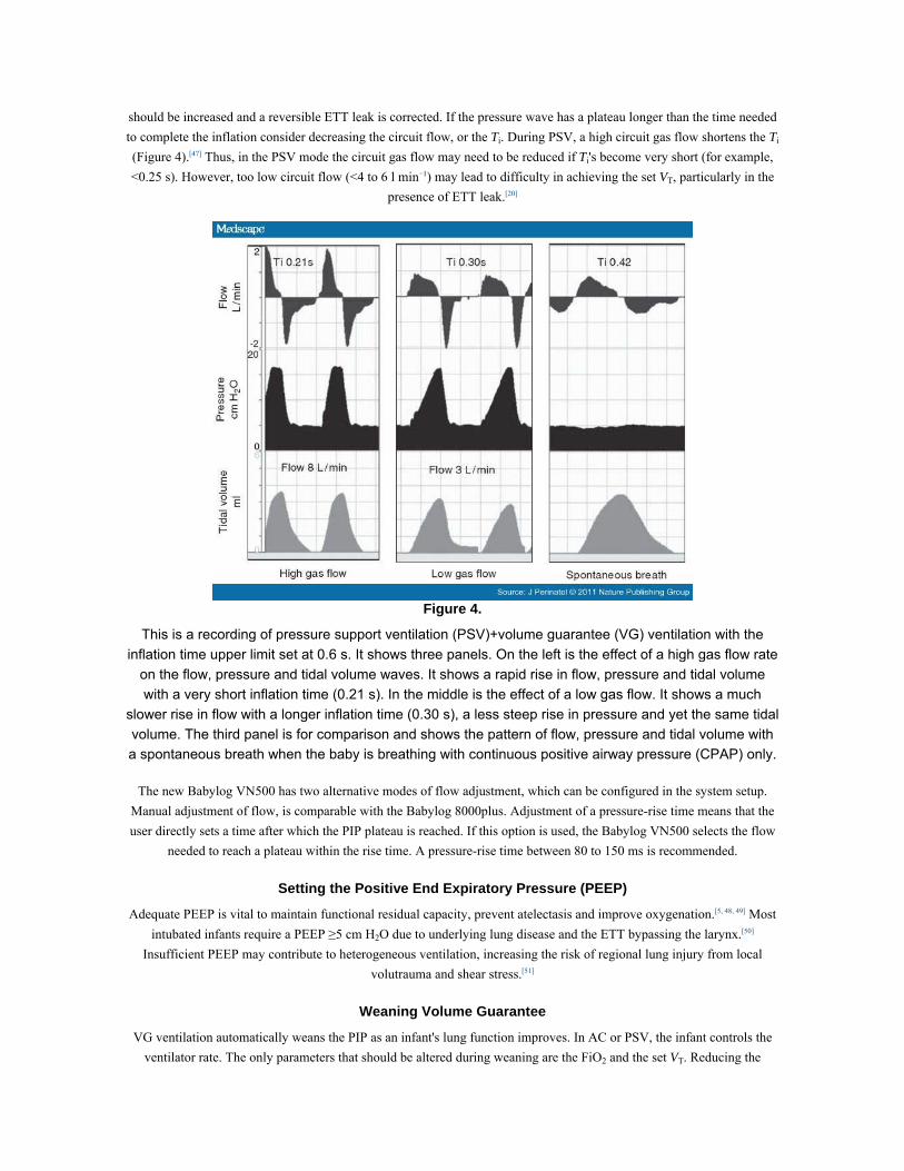
Figure 4.
This is a recording of pressure support ventilation (PSV)+volume guarantee (VG) ventilation with the inflation time upper limit set at 0.6 s. It shows three panels. On the left is the effect of a high gas flow rate
on the flow, pressure and tidal volume waves. It shows a rapid rise in flow, pressure and tidal volume with a very short inflation time (0.21 s). In the middle is the effect of a low gas flow. It shows a much
slower rise in flow with a longer inflation time (0.30 s), a less steep rise in pressure and yet the same tidal volume. The third panel is for comparison and shows the pattern of flow, pressure and tidal volume with a spontaneous breath when the baby is breathing with continuous positive airway pressure (CPAP) only.
The new Babylog VN500 has two alternative modes of flow adjustment, which can be configured in the system setup. Manual adjustment of flow, is comparable with the Babylog 8000plus. Adjustment of a pressure-rise time means that the user directly sets a time after which the PIP plateau is reached. If this option is used, the Babylog VN500 selects the flow
needed to reach a plateau within the rise time. A pressure-rise time between 80 to 150 ms is recommended.
Setting the Positive End Expiratory Pressure (PEEP)
Adequate PEEP is vital to maintain functional residual capacity, prevent atelectasis and improve oxygenation.[5, 48, 49] Most intubated infants require a PEEP ≥5 cm H2O due to underlying lung disease and the ETT bypassing the larynx.[50]
Insufficient PEEP may contribute to heterogeneous ventilation, increasing the risk of regional lung injury from local volutrauma and shear stress.[51]
Weaning Volume Guarantee
VG ventilation automatically weans the PIP as an infant's lung function improves. In AC or PSV, the infant controls the ventilator rate. The only parameters that should be altered during weaning are the FiO2 and the set VT. Reducing the

ventilator rate during AC or PSV has no effect on delivered rate unless respiratory effort is poor or the BUR is greater than the infant's breathing rate.
When the set VT is below, the infant's spontaneously generated VT the PIP will be reduced therefore the infant will be breathing on ETT continuous positive airway pressure, potentially increasing work of breathing and the risk of subsequent extubation failure due to fatigue. If this is observed for more than short periods, extubation should be considered. We do not recommend weaning VT below 3.5 ml kg−1.[18,25,26] We consider extubation if the mean airway pressure is consistently <8 to 10 cm H2O with set VT 3.5 to 4.5 ml kg−1 and blood gases are satisfactory. Readiness for extubation can be assessed
using the spontaneous breathing test.[52]
Endotracheal Tube Leak
Gas leak around an uncuffed neonatal ETT varies with its diameter, the applied airway pressure and the duration of ventilation.[53] It is largest during inflation. If there is an ETT leak, some of the gas volume passing down the ETT will be
lost before entering the lungs. Compared with inspired VT, the advantage of targeting expired VT is that it more closely reflects the actual VT that entered the infant's lungs. However, as ETT leak increases, expired VT becomes less accurate, because there is a leak during expiration, and the ventilator increasingly underestimates the delivered VT. In general, VG ventilation can be used with ETT leaks up to ~50%[27] because the ventilator automatically adjusts the PIP to deliver the
set VT (Table 1). An increasing ETT leak with a concomitant rise in PIP may be misinterpreted as worsening lung disease.[27] A very large leak may lead to hypocarbia due to underestimation of the delivered VT.
In a ventilated infant who is breathing well and has adequate blood gases despite having a large ETT leak, extubation may be appropriate. Otherwise, if an infant has a large ETT leak it is usually technically easy to place a larger ETT. We
recommend re-intubating with an appropriately sized ETT rather than turning off VG and leaving VT delivery uncontrolled.
Large Tidal Volumes
During periods of crying, breathing hard or gasping, the spontaneous VT may exceed the set VT.[14] VG permits infants to take large breaths but does not augment these inflations due to inbuilt safety features (Figure 5, Table 1).

Figure 5.
This recording of assist control (AC)+volume guarantee (VG) ventilation of a 1000-g baby illustrates the effect of the baby taking large spontaneous breaths. The three waves are from top to bottom flow (l min−1), pressure (cm H2O) and tidal volume (ml). During the first six inflations, the baby is breathing quietly and triggering inflations. During the next eight inflations the baby starts breathing hard. The inspired VT then exceeds the set VT, shown by the horizontal dotted line, by more than 130%. The
inflating pressure is stopped early and therefore reduced for each successive inflation over 130% of the set VT until the baby stops breathing. During the last three inflations, the baby is not breathing and three
untriggered back-up inflations are delivered.
Surfactant Treatment
Surfactant administration often causes brief increases in airway resistance and ETT obstruction.[16] To maintain the set VT, the PIP limit should be set 10 cm H2O or more above the pre-surfactant PIP to enable the set VT to be delivered.[16] Within
30 to 60 min, the working PIP automatically falls as lung mechanics improve.[16]
Forced Expiration, 'Splinting' and Episodes With Low Tidal Volume Babies can actively expire and then tighten their abdominal muscles so hard such that they prevent gas entering the lungs during an inflation; often termed 'splinting'.[54] Forced expiration and splinting cause hypoxemic episodes due to low lung volume and low VT delivery,[55] causing 'ETT obstructed' and 'low tidal volume' alarms (Figure 6). A higher Pmax setting (for example, 35 cm H2O) may allow the ventilator to increase the PIP and overcome the obstruction more quickly. The use of a higher target VT (6.0 ml kg−1) only slightly reduced hypoxemic episodes in studies with other VTV modes.[55, 56]

Figure 6.
This is a recording from a 1000-g baby ventilated with assist control (AC)+volume guarantee (VG) ventilation at a rate of 50 per min, a set peak inflating pressure (PIP) of 40 cm H2O, and a set VT 5 ml.
The three waves are from top to bottom flow (l min−1), pressure (cm H2O) and tidal volume (ml). It illustrates the effect on the inflating pressure when the baby tightens the abdominal muscles enough to
temporarily stop inflation. This is preceded by active expiration. During the first ten inflations, the pressure is modulated to maintain the expired VT. During inflations 7, 8 and 9 the expired VT is larger than set VT and so the pressure is reduced. At inflation 10 there is a very small VT, and therefore the
pressure is increased by 3 cm H2O for each inflation for the next five untriggered inflations until a VT is produced. This is then followed by triggered inflations at a similar inflating pressure to the start of this
recording, with one untriggered inflation in between.
Diaphragmatic Braking
Diaphragmatic braking causes an early interruption in expiratory flow (Figure 7) and is sometimes associated with a small inspiration as the diaphragm contracts.[15, 54] If this occurs within the 0.2 s refractory period of the ventilator an inflation is
not triggered. However, the VG software underestimates the expired VT by incorrectly interpreting the small initial expired VT as the total expired VT, and increases the PIP for the next inflation. Despite a higher PIP, the safety features help to prevent delivery of large VT (Table 1). Periods with expiratory braking occur with <5% of inflations and less
during triggered than untriggered inflations. These episodes are usually short lived and revert spontaneously to normal ventilation.

Figure 7.
This illustrates the normal volume guarantee (VG) pattern (left) and the diaphragmatic braking pattern (right). The three waves are from top to bottom flow (l min−1), pressure (cm H2O) and tidal volume (ml).
Diaphragmatic braking is characterised by an early interruption in expiratory flow, due to a small inspiration (arrow). This small inspiration starts shortly after the end of the inflation and separates the
expiratory flow into two parts.
Effect of the Flow-sensor Dead Space
The small dead space of the Babylog flow sensor (Table 1) has little effect on ventilation.[35] One reason for this is that with ETT leaks the dead space is continuously flushed with fresh gas. Also, during neonatal ventilation gas movement is more complicated than simple bulk flow, minimizing the effect of dead space.[35] In babies <1000 g, the extra dead space may slightly increase PaCO2 levels.[21, 34] The advantages of using flow sensors for monitoring, volume targeting and flow
triggering, outweigh the small effect on PaCO2.
Troubleshooting VG
If, during VG ventilation, an infant becomes tachypnoeic with low oxygen saturations or has a persistently low PIP with suboptimal blood gases, the condition of the infant and the ventilator must both be thoroughly assessed. Inadequate PEEP, very short Ti during PSV, a set VT which is too low, or a large ETT leak need to be ruled out. Important points to consider
for successful VG ventilation are listed in Box 1.

Conclusion
A Cochrane systematic review supports the use of VTV for ventilated preterm infants in need of mechanical ventilation.[12,
13] The VG mode controls the expired VT and provides breath-by-breath adjustments of the PIP to achieve the set VT. By controlling the expired VT, this mode is less influenced by endotracheal tube leak and can be used with ETT leaks up to
~50%. The initial set VT for infants with RDS should be 4.0 to 5.0 ml kg−1, but may need adjustments to maintain acceptable PaCO2 values. Very high (>8 ml kg−1) and very low (<3.5 ml kg−1) set VT may cause harm. Setting the PIP limit
well above the working pressure is important to enable the ventilator alter the PIP to deliver the set VT, and to avoid frequent low tidal volume alarms. We recommend combining VG with triggered modes supporting all inflations (AC or
PSV modes). A ventilator BUR <40 per min permits the infant to trigger most inflations. VG automatically weans the PIP as the baby's lung compliance and respiratory effort improves.
This state-of-the-art review on VG ventilation is intended to be used with the Dräger Babylog 8000plus and VN500 ventilators, which have a similar VG mode. We have insufficient experience with other ventilators to make
recommendations for their use. Some of the principles apply to other ventilators targeting expired VT but caution should be applied, as these ventilators do not work exactly in the same way (Table 2). We urge the clinicians to report their
experiences with all VTV modes and the manufacturers to describe the technical details of VTV modes in new neonatal ventilators.
References
1. Keszler M, Abubakar K. Volume guarantee: stability of tidal volume and incidence of hypocarbia. Pediatr Pulmonol 2004; 38: 240–245.
2. Swamy R, Gupta S, Singh J, Donn SM, Sinha SK. Tidal volume delivery and peak inspiratory pressure in babies receiving volume targeted or time cycled, pressure limited ventilation: a randomized controlled trial. J
Neonatal-Perinatal Med 2008; 2: 239–243. 3. Hernandez LA, Peevy KJ, Moise AA, Parker JC. Chest wall restriction limits high airway pressure-induced lung
injury in young rabbits. J Appl Physiol 1989; 66: 2364–2368. 4. Bjorklund LJ, Ingimarsson J, Curstedt T, John J, Robertson B, Werner O et al. Manual ventilation with a few
large breaths at birth compromises the therapeutic effect of subsequent surfactant replacement in immature lambs. Pediatr Res 1997; 42: 348–355.
5. Clark RH, Gerstmann DR, Jobe AH, Moffitt ST, Slutsky AS, Yoder BA. Lung injury in neonates: causes, strategies for prevention, and long-term consequences. J Pediatr 2001; 139: 478–486.
6. Sharma A, Milner AD, Greenough A. Performance of neonatal ventilators in volume targeted ventilation mode. Acta Paediatr 2007; 96: 176–180.
7. Jaecklin T, Morel DR, Rimensberger PC. Volume-targeted modes of modern neonatal ventilators: how stable is the delivered tidal volume? Intensive Care Med 2007; 33: 326–335.
8. Cappa P, Sciuto SA, Silvestri S. Experimental evaluation of errors in the measurement of respiratory parameters of the newborn performed by a continuous flow neonatal ventilator. J Med Eng Technol 2006; 30: 31–40.
9. Chow LC, Vanderhal A, Raber J, Sola A. Are tidal volume measurements in neonatal pressure-controlled ventilation accurate? Pediatr Pulmonol 2002; 34: 196–202.
10. van Kaam AH, Rimensberger PC, Borensztajn D, De Jaegere AP. Ventilation practices in the neonatal intensive care unit: a cross-sectional study. J Pediatr 2010; 157: 767–771.
11. Klingenberg C, Wheeler KI, Owen LS, Kaaresen PI, Davis PG. An international survey of volume-targeted neonatal ventilation. Arch Dis Child Fetal Neonatal Ed 2011; 96: F146–F148.
12. Wheeler K, Klingenberg C, McCallion N, Morley CJ, Davis PG. Volume-targeted versus pressure-limited ventilation in the neonate. Cochrane Database Syst Rev 2010; 11: CD003666.
13. Wheeler K, Klingenberg C, Morley CJ, Davis PG. Volume-targeted versus pressure-limited ventilation for preterm infants: a systematic review and meta-analysis. Neonatology 2011; 100: 219–227.

14. McCallion N, Lau R, Morley CJ, Dargaville PA. Neonatal volume guarantee ventilation: effects of spontaneous breathing, triggered and untriggered inflations. Arch Dis Child Fetal Neonatal Ed 2008; 93: F36–F39.
15. McCallion N, Lau R, Dargaville PA, Morley CJ. Volume guarantee ventilation, interrupted expiration, and expiratory braking. Arch Dis Child 2005; 90: 865–870.
16. Wheeler KI, Davis PG, Kamlin CO, Morley CJ. Assist control volume guarantee ventilation during surfactant administration. Arch Dis Child Fetal Neonatal Ed 2009; 94: F336–F338.
17. Wheeler KI, Morley CJ, Kamlin CO, Davis PG. Volume-guarantee ventilation: pressure may decrease during obstructed flow. Arch Dis Child Fetal Neonatal Ed 2009; 94: F84–F86.
18. Wheeler KI, Wong C, Morley CJ, Davis PG. Volume guarantee ventilation: how low should we go? J Paediatr Child Health 2011; 47(Suppl. 1): 60–116. Abstract nr.
19. Wheeler KI, Wong C, Morley CJ, Davis PG. Selecting the PIP limit for infants ventilated using assist control Volume Guarantee mode. J Paediatr Child Health 2011; 47(Suppl. 1): 60–116. Abstract nr.
20. Wheeler KI, Wong C, Morley CJ, Davis PG. Selecting the circuit flow for infants ventilated using assist control Volume Guarantee. J Paediatr Child Health 2010; 46(Suppl. 1): 7–55.
21. Cheema IU, Sinha AK, Kempley ST, Ahluwalia JS. Impact of volume guarantee ventilation on arterial carbon dioxide tension in newborn infants: a randomised controlled trial. Early Hum Dev 2007; 83: 183–189.
22. Dawson C, Davies MW. Volume-targeted ventilation and arterial carbon dioxide in neonates. J Paediatr Child Health 2005; 41: 518–521.
23. Erickson SJ, Grauaug A, Gurrin L, Swaminathan M. Hypocarbia in the ventilated preterm infant and its effect on intraventricular haemorrhage and bronchopulmonary dysplasia. J Paediatr Child Health 2002; 38: 560–562.
24. Fritz KI, Delivoria-Papadopoulos M. Mechanisms of injury to the newborn brain. Clin Perinatol 2006; 33: 573–591.
25. Lista G, Castoldi F, Fontana P, Reali R, Reggiani A, Bianchi S et al. Lung inflammation in preterm infants with respiratory distress syndrome: effects of ventilation with different tidal volumes. Pediatr Pulmonol 2006; 41:
357–363. 26. Herrera CM, Gerhardt T, Claure N, Everett R, Musante G, Thomas C et al. Effects of volume-guaranteed
synchronized intermittent mandatory ventilation in preterm infants recovering from respiratory failure. Pediatrics 2002; 110: 529–533.
27. Keszler M, Abubakar KM. Volume guarantee ventilation. Clin Perinatol 2007; 34: 107–116. 28. Abubakar K, Keszler M. Effect of volume guarantee combined with assist/control vs synchronized intermittent
mandatory ventilation. J Perinatol 2005; 25: 638–642. 29. Sandberg K, Sjoqvist BA, Hjalmarson O, Olsson T. Analysis of alveolar ventilation in the newborn. Arch Dis
Child 1984; 59: 542–547. 30. Milner AD, Vyas H, Hopkin IE. Efficacy of facemask resuscitation at birth. Br Med J (Clin Res Ed) 1984; 289:
1563–1565. 31. te Pas AB, Davis PG, Kamlin CO, Dawson J, O'Donnell CP, Morley CJ. Spontaneous breathing patterns of very
preterm infants treated with continuous positive airway pressure at birth. Pediatr Res 2008; 64: 281–285. 32. Vilstrup CT, Bjorklund LJ, Werner O, Larsson A. Lung volumes and pressure-volume relations of the respiratory system in small ventilated neonates with severe respiratory distress syndrome. Pediatr Res 1996; 39:
127–133. 33. van Kaam AH, Rimensberger PC. Lung-protective ventilation strategies in neonatology: what do we know--
what do we need to know? Crit Care Med 2007; 35: 925–931. 34. Keszler M, Nassabeh-Montazami S, Abubakar K. Evolution of tidal volume requirement during the first 3
weeks of life in infants <800 g ventilated with Volume Guarantee. Arch Dis Child Fetal Neonatal Ed 2009; 94: F279–F282.
35. Nassabeh-Montazami S, Abubakar KM, Keszler M. The impact of instrumental dead-space in volume-targeted ventilation of the extremely low birth weight (ELBW) infant. Pediatr Pulmonol 2009; 44: 128–133.

36. Bhutani VK, Ritchie WG, Shaffer TH. Acquired tracheomegaly in very preterm neonates. Am J Dis Child 1986; 140: 449–452.
37. Sharma S, Abubakar K, Keszler M. Tidal Volume in Infants with Congenital Diaphragmatic Hernia. Pediatric Academic Society (PAS), Vancouver, Canada, 2010. Abstract 1466.146.
38. te Pas AB, Kamlin CO, Dawson JA, O'Donnell C, Sokol J, Stewart M et al. Ventilation and spontaneous breathing at birth of infants with congenital diaphragmatic hernia. J Pediatr 2009; 154: 369–373.
39. Kiraly NJ, Tingay DG, Mills JF, Dargaville PA, Copnell B. Volume not guaranteed: closed endotracheal suction compromises ventilation in volume-targeted mode. Neonatology 2011; 99: 78–82.
40. Schulze A. Respiratory gas conditioning and humidification. Clin Perinatol 2007; 34: 19–33. 41. Deakins K, Di Fiore J. Evaluation of Condensation in Three Commercially Available Heated Wire Ventilator
Circuits. American Association of Respiratory Care: San Antonio, Texas, USA, 2005. Abstract. 42. Beck J, Reilly M, Grasselli G, Mirabella L, Slutsky AS, Dunn MS et al. Patient-ventilator interaction during
neurally adjusted ventilatory assist in low birth weight infants. Pediatr Res 2009; 65: 663–668. 43. South M, Morley CJ. Respiratory timing in intubated neonates with respiratory distress syndrome. Arch Dis
Child 1992; 67: 446–448. 44. Hird M, Greenough A. Inflation time in mechanical ventilation of preterm neonates. Eur J Pediatr 1991; 150:
440–443. 45. Wheeler KI, Morley CJ, Hooper SB, Davis PG. Lower back-up rates improve ventilator triggering during assist-
control ventilation: a randomized crossover trial. J Perinatol 2 Jun 2011 (e-pub ahead of print). 46. Greenough A. The premature infant's respiratory response to mechanical ventilation. Early Hum Dev 1988; 17:
1–5. 47. Bach KP, Kuschel CA, Oliver MH, Bloomfield FH. Ventilator gas flow rates affect inspiratory time and
ventilator efficiency index in term lambs. Neonatology 2009; 96: 259–264. 48. Monkman S, Kirpalani H. PEEP—a 'cheap' and effective lung protection. Paediatr Respir Rev 2003; 4: 15–20. 49. Monkman SL, Andersen CC, Nahmias C, Ghaffer H, Bourgeois JM, Roberts RS et al. Positive end-expiratory
pressure above lower inflection point minimizes influx of activated neutrophils into lung. Crit Care Med 2004; 32: 2471–2475.
50. Keszler M. State of the art in conventional mechanical ventilation. J Perinatol 2009; 29: 262–275. 51. Otto CM, Markstaller K, Kajikawa O, Karmrodt J, Syring RS, Pfeiffer B et al. Spatial and temporal heterogeneity of ventilator-associated lung injury after surfactant depletion. J Appl Physiol 2008; 104: 1485–
1494. 52. Kamlin CO, Davis PG, Morley CJ. Predicting successful extubation of very low birthweight infants. Arch Dis
Child Fetal Neonatal Ed 2006; 91: F180–F183. 53. Greenough A, Milner AD. Pulmonary disease of the newborn: acute respiratory disease. In: Rennie JM (ed).
Roberton's Textbook of Neonatology. 4th edn. London, England: Elsevier, 2005 pp 468–553. 54. te Pas AB, Wong C, Kamlin CO, Dawson JA, Morley CJ, Davis PG. Breathing patterns in preterm and term
infants immediately after birth. Pediatr Res 2009; 65: 352–356. 55. Hummler HD, Engelmann A, Pohlandt F, Franz AR. Volume-controlled intermittent mandatory ventilation in
preterm infants with hypoxemic episodes. Intensive Care Med 2006; 32: 577–584. 56. Polimeni V, Claure N, D'Ugard C, Bancalari E. Effects of volume-targeted synchronized intermittent mandatory
ventilation on spontaneous episodes of hypoxemia in preterm infants. Biol Neonate 2006; 89: 50–55.