
From a Physician's Perspective in Upper Extremity Injuries …€¦ · · 2007-05-22The...
7
Journal of Sport Rehabilitation, 1996, 5, 64-70 O 1996 Human Kinetics Publishers, Inc. The Significance of Closed Chain Kinetics in Upper Extremity Injuries From a Physician's Perspective James R. Andrews, James M. Dennison, and Kevin E. Wilk Most physicians, trainers, and therapists are accustomed to thinking of open and closed kinetic chain terminology in terms of exercise and its application in rehabilitation protocols. This terminology can also be used to describe the mechanism by which injuries occur. Categorizing upper extremity injuries in this way not only provides vital insight into the mechanism of the injuries and helps identify possible injured structures but also allows the clinician to better develop treatment protocols. In this article, this categorization is applied to common shoulder and elbow injuries to provide insight into the nature of these injuries. In order to fully understand the nature of an injury, it is first necessary to understand the mechanism by which the injury occurred. Often, the treatment strategy a physician chooses to manage a particular injury is based on this understanding. Recently, it has become popular to develop rehabilitation protocols based on open and closed kinetic chain exercises. However, from a physician's perspective, open and closed kinetic chain terminology can also be an extremely useful means to categorize the mechanism by which injuries occur. A basic tenet of medicine is that with a thorough history and physical examination, the majority of injuries can be understood and a treatment protocol designed without ancillary studies needed. This is obviously true of injuries involving the upper extremity. A thorough history provides a means by which to delineate the mechanism of the injury. This history allows the physician to define the structures that were likely injured and facilitates the development of a rational treatment plan. It allows the physician to develop a plan not only for the protection of the injured structures but also for the repair or reconstruction of these injured tissues if surgical intervention is necessary. Understanding the mechanism by which the injury occurred also allows the physician to develop a plan to prevent recurrence. Categorizing the mechanism of injury to the upper extremity as closed James R. Andrews and James M. Dennison are with the American Sports Medicine Institute, Birmingham, AL 35205. Kevin E. Wilk is with HEALTHSOUTH Sports Medi- cine & Rehabilitation Center, Birmingham, AL 35205.
-
Upload
nguyentram -
Category
Documents
-
view
220 -
download
1
Transcript of From a Physician's Perspective in Upper Extremity Injuries …€¦ · · 2007-05-22The...
Journal of Sport Rehabilitation, 1996, 5, 64-70 O 1996 Human Kinetics Publishers, Inc.
The Significance of Closed Chain Kinetics in Upper Extremity Injuries
From a Physician's Perspective
James R. Andrews, James M. Dennison, and Kevin E. Wilk
Most physicians, trainers, and therapists are accustomed to thinking of open and closed kinetic chain terminology in terms of exercise and its application in rehabilitation protocols. This terminology can also be used to describe the mechanism by which injuries occur. Categorizing upper extremity injuries in this way not only provides vital insight into the mechanism of the injuries and helps identify possible injured structures but also allows the clinician to better develop treatment protocols. In this article, this categorization is applied to common shoulder and elbow injuries to provide insight into the nature of these injuries.
In order to fully understand the nature of an injury, it is first necessary to understand the mechanism by which the injury occurred. Often, the treatment strategy a physician chooses to manage a particular injury is based on this understanding. Recently, it has become popular to develop rehabilitation protocols based on open and closed kinetic chain exercises. However, from a physician's perspective, open and closed kinetic chain terminology can also be an extremely useful means to categorize the mechanism by which injuries occur.
A basic tenet of medicine is that with a thorough history and physical examination, the majority of injuries can be understood and a treatment protocol designed without ancillary studies needed. This is obviously true of injuries involving the upper extremity. A thorough history provides a means by which to delineate the mechanism of the injury. This history allows the physician to define the structures that were likely injured and facilitates the development of a rational treatment plan. It allows the physician to develop a plan not only for the protection of the injured structures but also for the repair or reconstruction of these injured tissues if surgical intervention is necessary. Understanding the mechanism by which the injury occurred also allows the physician to develop a plan to prevent recurrence.
Categorizing the mechanism of injury to the upper extremity as closed
James R. Andrews and James M. Dennison are with the American Sports Medicine Institute, Birmingham, AL 35205. Kevin E. Wilk is with HEALTHSOUTH Sports Medi- cine & Rehabilitation Center, Birmingham, AL 35205.

Physician's Perspective 65
kinetic chain or open kinetic chain provides insight into the nature of these injuries. Obviously, it is necessary to understand the definitions of these terms. This terminology was derived by Steindler and is based on whether the terminal joint of the extremity is fixed or can move freely (6). By definition, a closed kinetic chain upper extremity injury is one in which the distal segment (i.e., hand or elbow) is fixed. It usually involves an impact or compression-type deforming force and most typically results in an acute, traumatic injury pattern. By contrast, an open kinetic chain upper extremity injury is one in which the hand or elbow is free. Such an injury is quite often caused by traction or tensile-type deforming force, and it likely involves an overuse injury pattern with resultant microtrauma. In addition, open kinetic chain activities are believed to create higher shear forces against the articulating surfaces. See Table 1 for a classification of upper extremity injuries.
Shoulder Injuries
To understand the utility of categorizing upper extremity injuries by whether they occurred by an open or a closed kinetic chain mechanism, it is useful to consider specific examples and injury patterns. The authors will consider some common shoulder injuries and see how this categorization can be applied and what useful information it can provide. This is not an attempt to provide an encyclopedic breakdown of all shoulder injuries but rather is an attempt to highlight common shoulder injuries and show the utility of this categorization.
A useful example is shoulder instability, which is often difficult to diagnose and correctly treat. When considering shoulder instability, the physician is faced with the difficult and confusing task of ascertaining not only the mechanism of the instability but also the direction and the chronicity of the injury, as well as the best treatment option. If the physician considers whether the mechanism of the injury was an open or a closed kinetic chain, it is possible to simplify and better understand shoulder instability. For example, traumatic anterior-inferior
Table 1 Classification of Upper Extremity Injuries
Closed kinetic chain Open kinetic chain
Shoulder Traumatic anterior-inferior instability Multidirectional instability Posterior instability Chronic rotator cuff tendinitis Acute rotator cuff tendinitis Superior labrum anteriodposte- Traumatic labial tears rior lesion in throwers Acromioclavicular joint sprains InternaI impingement syndrome
Elbow Dislocations Ulnar collateral ligament injury Fractures Ulnar neuritis Tendinitis Osteochondritis dess-icans of ca-
pitellum , Posterior medial osteophytes
66 Andrews, Dennison, and Wilk
dislocation of the shoulder can occur by direct trauma; however, indirect force is the most common cause (7). The mechanism of injury in this case is a load applied to the upper extremity with the hand fixed and the shoulder in abduction and external rotation; therefore, this represents a closed kinetic chain type of injury. A tremendous amount of force is necessary to cause this injury, and often a portion of the anterior-inferior labrum (i.e., a Bankart lesion) tears off the glenoid with or without some associated capsular attenuation. The recurrence rate of this injury is relatively high, depending primarily on the patient's age at the time of initial injury. Given the high recurrence rate, surgery is often required if appropriate nonoperative treatment fails.
Contrast the preceding description with a multidirectional instability pattem, which most typically occurs through an open kinetic chain mechanism. In an athletic population, multidirectional instability occurs commonly from a repetitive microtrauma type of injury. This injury occurs not only in young, sedentary patients but also quite commonly in an athletic population where the patient's natural shoulder laxity is converted into true instability about the glenohumeral joint. A prime example is throwing athletes, patients who often develop true patholaxity around their glenohumeral joints without any specific history of trauma. This instability around the glenohumeral joint then creates secondary pathology, such as rotator cuff tendinitis and impingement-type symptoms due to the dynamic constraints around the shoulder attempting to stabilize the lax joint. The mechanism of this injury is of the open kinetic chain variety. This has obvious implications from a treatment standpoint, as rehabilitation is the mainstay for the problem and is often effective in alleviating symptoms. If it is not, surgical reconstruction becomes an alternative.
Regarding posterior glenohumeral instability, it is useful to realize that this typically occurs from a closed kinetic chain mechanism of injury. A typical history would be a fall onto an outstretched hand in which the shoulder is adducted and internally rotated. This may cause either a frank dislocation or subluxation of the glenohumeral joint posteriorly. Since posterior dislocations are commonly missed on radiographic evaluation, understanding the mechanism of the patient's injury can heighten a physician's awareness regarding the possibility of posterior dislocation and hence can make the diagnosis more likely. It also allows the physician to better understand any secondary pathology that might accompany this type of injury (e.g., a posterior labral tear).
These three examples regarding glenohumeral instability demonstrate how categorizing the mechanism of the injury into an open or a closed kinetic chain pattern can be useful to the physician. Another example involving the glenohu- meral joint is rotator cuff tendinitis, which can occur by a variety of mechanisms. An acute injury often is caused by a closed kinetic chain movement in which the patient falls or dives onto the outstretched hand in the forward flex position. This may initially cause tendinitis, which if allowed to progress can lead to frank tearing of the cuff. The treatment protocol should be devised around active rest of the arm until the symptoms improve. Contrast this with chronic rotator cuff tendinitis in athletes, which can typically occur from repetitive overuse. This is particularly true in throwing athletes, where the mechanism is an open kinetic chain injury where rotator cuff fatigue can lead to malfunction and pain. In throwing athletes, impingement syndrome is often secondary to rotator cuff malfunction and/or an associated instability pattem. So, by understanding the
Physician's Perspective 67
mechanism surrounding the injury, the physician and rehabilitation specialist can better devise a treatment plan for the patient regarding rotator cuff tendinitis and any associated impingement symptoms.
The categorization of shoulder injuries into open and closed kinetic chain mechanisms can also be applied to better understand glenoid labral pathology. When a patient suffers a closed kinetic chain injury, such as a fall or a dive onto the outstretched hand, the humeral head can sublux and impact onto the labrum, disrupting the normal glenoid labral complex. In Snyder et al.'s original descrip- tion of superior labrum anteriorlposterior (SLAP) lesions, it was stated that the most common mechanism of injury was a fall onto an outstretched arm, with the shoulder abducted and forward flexed (8). This injury may manifest itself with persistent pain, catching, and grinding. If these symptoms persist, then the patient may require arthroscopic intervention.
Contrast this closed kinetic chain injury pattern with the mechanism by which labral injuries occur in throwing athletes. Isolated superior labral pathology often occurs in the throwing athlete as a result of overuse. Numerous etiologies for the development of SLAP lesions in throwing athletes have been postulated. Among these is the belief that forces imparted by the biceps during the follow- through phase of throwing are responsible for the development of this labral pathology (1). This would represent an open kinetic chain mechanism of injury resulting in a superior labral tear. For this injury, a thrower's rehabilitation protocol would be the primary treatment, and only if this failed would surgical intervention be entertained. Such a lesion would also heighten the physician's concern about the possibility of an underlying, subtle glenohumeral instability pattern.
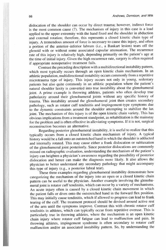
Understanding open kinetic chain injuries of the upper extremity aids in understanding posterior (internal) impingement syndrome. This refers to the articular side of the infraspinatus impinging on the posterior/superior glenoid labral rim in extremes of external rotation (Figure 1). This problem is peculiar to the throwing athlete and is usually secondary to the excessive external rotation that occurs about the glenohumeral joint. Nonoperative treatment for this type of pathology concentrates on improving dynamic stability of the glenohumeral joint to prevent excessive anterior humeral displacement. Operative treatment for this includes arthroscopic debridement of the infraspinatus and glenoid labral fraying, as well as a thrower's rehabilitation protocol.
Finally, the acromioclavicular (AC) joint is a frequently injured joint, especially in the athlete involved in contact sports. The most common mechanism of injury is a direct force applied to the AC joint. Frequently, the injury occurs in a closed kinetic chain fashion due to a fall onto the point of the shoulder (lateral acromion) with the arm adducted. This injury mechanism is extremely common in sports such as football and wrestling. The force imparted onto the acromion displaces the AC joint, resulting in a sequential failure of various tissues based on force applied. Initially,the AC joint ligament's capsule is injured, then the coracoclavicular ligaments (i.e., conoid and trapezoid) are injured, and then, if the force continues, the muscular attachments of the deltoid and trapezius muscles tear away, resulting in a severe AC joint sprain. The severity of the injury can be categorized according to Rockwood's ion scheme, which categorizes six types of AC sprains (9).

Andrews, Dennison, and Wilk
Figure 1 -Posterior impingement of the shoulder. Diagram showing the infraspi- natus impinging against the posterior labrum with arm in external rotation. (Re- printed with permission from G. Walch, P. Boileau, E. Noel, and S.T. Donell: Impingement of the deep surface of the supraspinatus tendon on the posterosuperior glenoid rim: An arthroscopic study. J. Shoulder Elbow Surg. 1(5):238-245, 1992.)
Elbow Injuries
The same categorization system can be used to better understand common elbow injuries, and it illustrates why the elbow pathology seen in throwing athletes is different from that seen in nonthrowing patients.
The three most common types of closed kinetic chain injuries about the ,, elbow are dislocations, fractures, and tendinitis. While there are several different
types of elbow dislocations, undoubtedly the most common is posterior. The mechanism of injury for most posterior elbow dislocations is a fall onto the outstretched hand or wrist. There is debate in the literature as to the exact mechanism that causes this dislocation (3). Regardless of the force that causes the dislocation, such a traumatic injury will often disrupt the normal ligamentous attachments around the elbow joint. The majority of these injuries are treated conservatively, and the patients are left with no residual instability. However, a small percentage of patients may have persistent posterolateral rotatory instability after such a traumatic injury (4, 5).
Other common elbow injuries are fractures about the elbow joint that occur secondary to a closed kinetic chain mechanism. These fractures may or may not be associated with a dislocation, and it is important to rule out both gross and subtle ligamentous instability. Common fracture patterns about the elbow joint include radial head, capitellar, and olecranon process fractures.
Finally, tendinitis is a common type of closed kinetic chain injury of the elbow. An example is triceps tendinitis that occurs after an overuse injury with weight training. Understanding the mechanism of this injury allows the physician to treat this patient conservatively with good results.
Open kinetic chain injuries about the elbow often occur from an overuse pattern and are particularly common in the athlete who uses overhead motion, especially the thrower. This theme was seen previously when we considered

Physician's Perspective 69
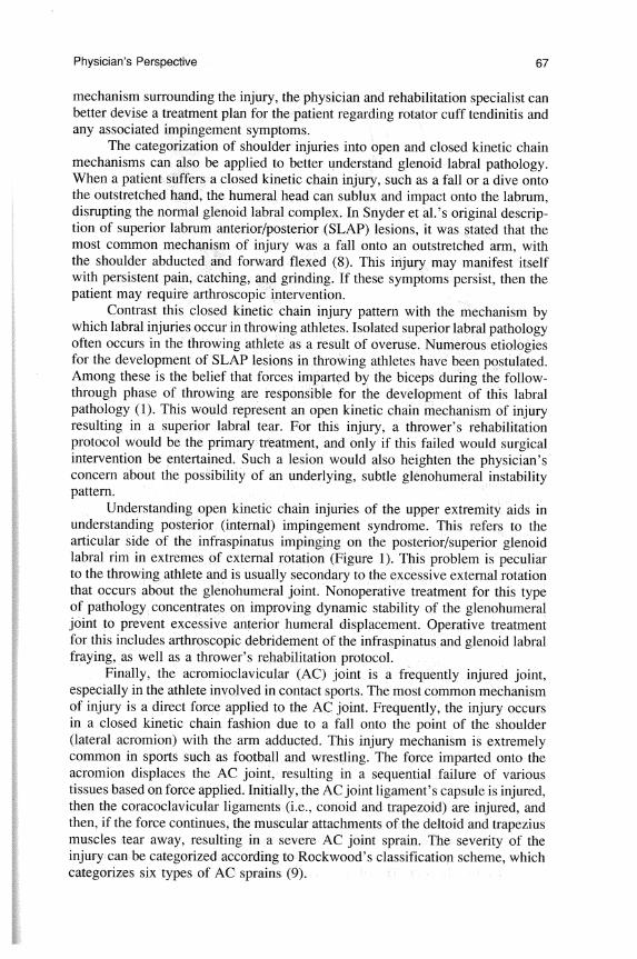
Figure 2 -The acceleration phase of pitching results in medial tension, lateral compression, and posterior overload on the elbow joint. (Reprinted with permission from J.A. Whiteside and J.R. Andrews. Common elbow problems in the recreational athlete. J. Musculoskeletal Med. 6:17-34, 1989.)
shoulder pathology. The common open kinetic chain injuries around the elbow joint in throwing athletes include medial tension injuries, lateral compression injuries, and extension overload pathology (Figure 2).
Medial tension injuries primarily involve injury or attenuation of the ulnar collateral ligament and ulnar neuritis. The ulnar collateral ligament is made up of the anterior oblique ligament, posterior oblique ligament, and transverse ligament. The anterior band of the ulnar collateral ligament provides roughly 55% of the stabilization against valgus stress (2). Chronic, repetitive valgus stress in a thrower's elbow is caused by an open kinetic chain mechanism in which the anterior oblique band of the collateral ligament becomes attenuated. The patient presents with pa5n over the ulnar collateral ligament, and if the patient does not respond to nonoperative management, reconstructive surgery may be necessary.
Ulnar neuropathy at the elbow can also occur from a medial tension type of injury. Three major etiologic factors include traction, friction, and compression Y2). Traction injuries occur secondary to a valgus stress with throwing. It 5s important to remember that ulnar nerve compression newpathy can coexist with ulnar collateral ligament pathology and occurs by the same mechanism.
It has been established that lateral compression injuries in the thrower's

70 Andrews, Dennison, and Wilk
elbow occur during late cocking and acceleration phases of pitching. These lateral compression injuries include the development of Panner's disease, which is osteochondritis dessicans of the capitellum. This osteochondral lesion is the result of an excessive valgus stress applying an abnormal compressive load across the radial capitellar joint. Finally, regarding the throwing elbow, the patient may develop an extension overload, where he or she develops osteophytes along the posteromedial tip of the olecranon and a trochlear chondromalacic kissing lesion. This pathology results from abnormal throwing mechanics in which there is excessive valgus thrust as the elbow nears its full extension.
Conclusion
The advantages of conceptualizing injuries of the upper extremity in terms of kinetic chain terminology are numerous. It allows the clinician to better predict the injury pattern as well as the pathology that the patient will present with. By identifying the structures most likely to be injured, the clinician can then devise a plan to prevent recurrence and further injury. This also allows the clinician to design strategies to correct the pathology through both nonoperative and operative means.
The goal of this paper was to present a way clinicians can view and analyze upper extremity injuries in terms of kinetic chain terminology. The authors feel this classification may be beneficial in analysis of athletic injuries, but the conventional methods remain the mainstay of the classification scheme.
References
1. Andrews, J.R., W.G. Carson, Jr., and W.D. McLeod. Glenoid labrum tears related to the long head of the biceps. Am. J . Sports Med. 13(5):337, 1985.
2. Andrews, J.R., and K. Meister. Overuse injuries in the athletes. In Orthopaedic Knowledge Update, Sports Medicine, L.Y. Griffin (Ed.). Rosemont, IL: American Academy of Orthopaedic Surgeons, 1994, pp. 182-183.
3. Hotchkiss, R.O., and D.A. Green. Fractures and dislocations of the elbow. In Fractures in Adults (3rd ed., Vol. I), C.A. Rockwood, Jr., D.P. Green, and R.W. Bulchoz (Eds.). New York: Lippincott, 1991, p. 781.
4. O'Driscoll, S.W., D.F. Bell, and B.F. Morrey. Posterolateral rotatory instability of the elbow. J . Bone Joint Surg. 73-A(3):440, 1991.
5. O'Driscoll, S.W., B.F. Morrey, S. Korinek, and K-N. An. Elbow subluxation and dislocation. Clin. Orthop. 280:186-197, 1992.
6. Palmitier, R.A. Kinetic chain exercises in knee rehabilitation. In Current Therapy in Sports Medicine (3rd ed.), J.S. Torg (Ed.). St. Louis, MO: Mosby-Year Book, 1995, p. 380.
7. Rockwood, Jr., C.A., S.C. Thomas, and F.A. Matsen, 111. Subluxation and dislocation about the glenohumeral joint. In Fractures in Adults (3rd ed., Vol. I), C.A. Rockwood, Jr., D.P. Green, and R.W. Bulchoz (Eds.). New York: Lippincott, 1991, p. 1046.
8. Snyder, S.J., R.P. Darzel, W. Del Pizzo, R.D. Ferkel, and M.J. Friedman. SLAP lesions of the shoulder. Arthroscopy 6(4):274-279, 1990.
9. Williams, G.R., V.D. Nguyen, and C.A. Rockwood, Jr. Classification and radiographic analysis of acromioclavicular dislocations. Appl. Radiol. 29-34, Feb. 1989.