Frog Dissection - Upstartihzephyr.weebly.com/uploads/2/2/6/3/22638828/frog_dissection_pkt... · All...
17
Frog Dissection Diagrams & Questions Name: ________________ Teacher: ________________ Hour: ____
Transcript of Frog Dissection - Upstartihzephyr.weebly.com/uploads/2/2/6/3/22638828/frog_dissection_pkt... · All...
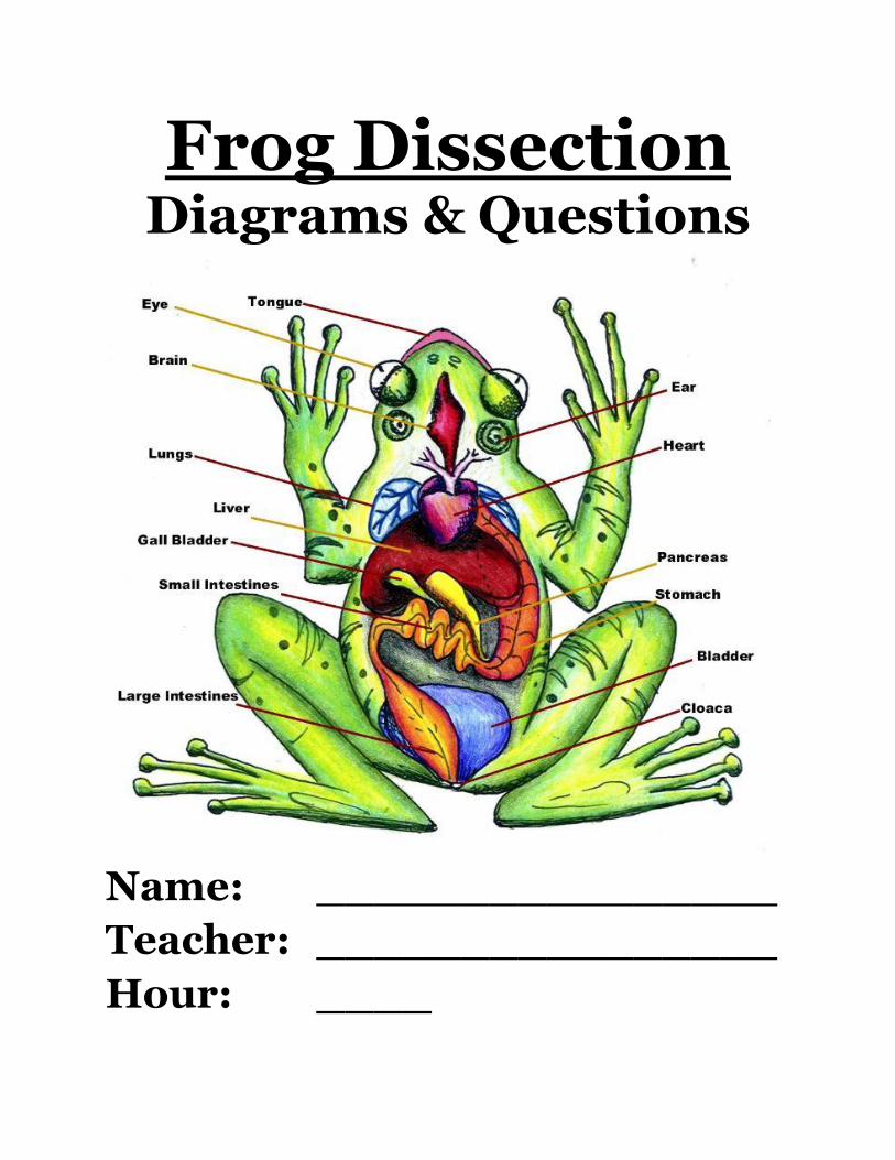
Frog Dissection Diagrams & Questions
Name: ________________
Teacher: ________________
Hour: ____
2
The frogs that we are going to observe have never lived in the wild. These frogs have
been raised on a frog farm. They were raised for only one function—to be dissected in a
science class. They have been stored in a preservative. Your frog will have been soaked
in water to get most of the preservative out.
At the end of each science class, we will store them in plastic bags after wrapping them
in wet paper towels. Before you receive your frog, there are a few additional thoughts
and statements that you should realize and understand. Please read the following:
a. You will follow the directions for this lab to the letter. Also, pay close
attention to additional instructions on the board or from your teacher. b. There is a lot of material in this lab packet and in the directions. Please take
the personal responsibility for learning this material. You will have
to take this packet out of class (home or study hall) to review various terms
and to prepare for the next class. c. Only do the work in the packet and on your frog that you are
supposed to do. If you rush ahead, you may destroy a part of the frog’s
internal anatomy that you will need later on. d. You will be working with another person in the lab. You are responsible
for your own work, but may work together to answer any questions. e. No one will leave the room until everything is clean and put away.
You will be dismissed by the teacher from your lab table. f. All frogs and frog parts will stay in this room. When you receive your frog, you will have to label it. To do so, you will use an ink
pen and write your initials, your partner’s initials, and your class period on a piece of
masking tape or name tag. The masking tape or name tag will then be placed on your
plastic storage bag and then placed in one of the trays which has been numbered
according to science classes.
Before we can learn about the internal anatomy of the frog, we must observe the
external anatomy of the frog. By doing so, we will study some relationships between
the internal and external anatomy of the frog.
We will also find out that some of the structures of the frog will explain the living
characteristics and tendencies of the frog. The frog’s body structures will also explain
why the frog lives in the environment that it does.
3
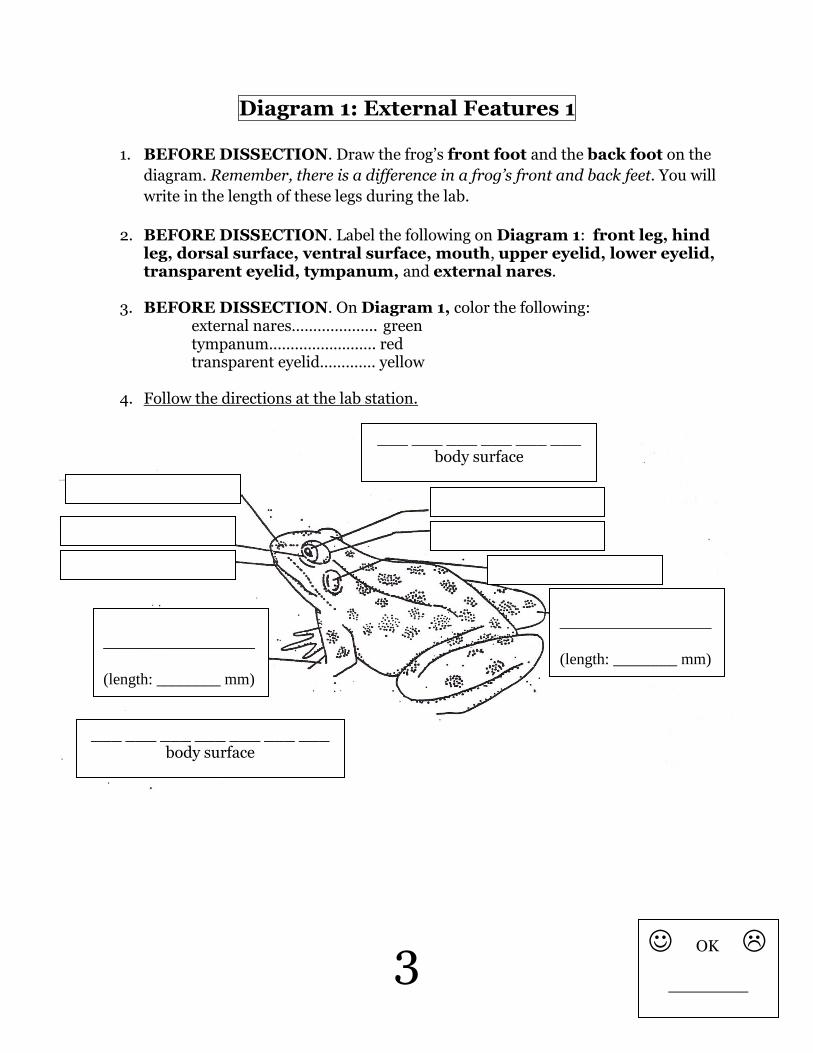
Diagram 1: External Features 1
1. BEFORE DISSECTION. Draw the frog’s front foot and the back foot on the
diagram. Remember, there is a difference in a frog’s front and back feet. You will
write in the length of these legs during the lab.
2. BEFORE DISSECTION. Label the following on Diagram 1: front leg, hind leg, dorsal surface, ventral surface, mouth, upper eyelid, lower eyelid, transparent eyelid, tympanum, and external nares.
3. BEFORE DISSECTION. On Diagram 1, color the following:
external nares……………….. green tympanum……………………. red transparent eyelid…………. yellow
4. Follow the directions at the lab station.
___ ___ ___ ___ ___ ___ ___ body surface
___ ___ ___ ___ ___ ___ body surface
___________________
(length: ________ mm)
___________________
(length: ________ mm)
OK
__________
4
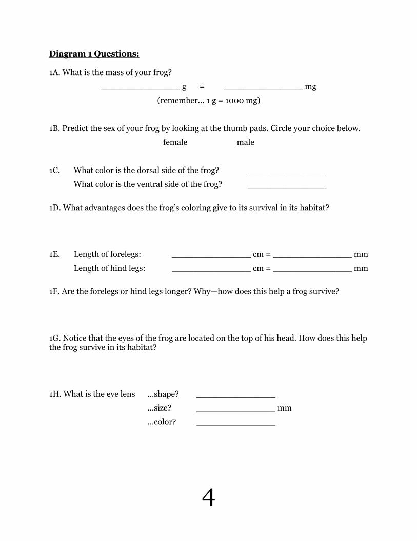
Diagram 1 Questions: 1A. What is the mass of your frog?
_______________ g = _______________ mg
(remember… 1 g = 1000 mg)
1B. Predict the sex of your frog by looking at the thumb pads. Circle your choice below.
female male
1C. What color is the dorsal side of the frog? _______________
What color is the ventral side of the frog? _______________
1D. What advantages does the frog’s coloring give to its survival in its habitat? 1E. Length of forelegs: _______________ cm = _______________ mm
Length of hind legs: _______________ cm = _______________ mm
1F. Are the forelegs or hind legs longer? Why—how does this help a frog survive? 1G. Notice that the eyes of the frog are located on the top of his head. How does this help the frog survive in its habitat? 1H. What is the eye lens …shape? _______________
…size? _______________ mm
…color? _______________
5
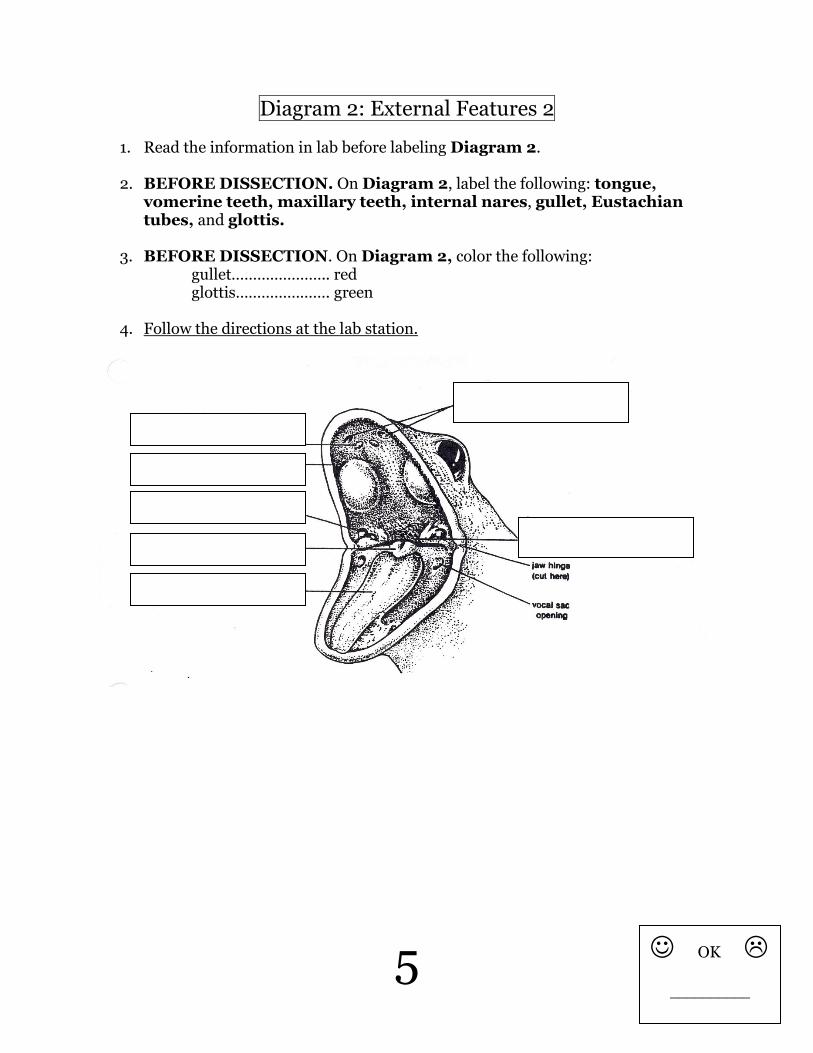
Diagram 2: External Features 2
1. Read the information in lab before labeling Diagram 2.
2. BEFORE DISSECTION. On Diagram 2, label the following: tongue, vomerine teeth, maxillary teeth, internal nares, gullet, Eustachian tubes, and glottis.
3. BEFORE DISSECTION. On Diagram 2, color the following: gullet………………….. red glottis…………………. green
4. Follow the directions at the lab station.
OK
__________

6
Diagram 2 Questions:
2A. Where is the frog’s tongue attached in the frog’s mouth?
2B. When the frog is living, its tongue is very sticky. Why do you think this is?
2C. Length of tongue: _______________ cm = _______________ mm
2D. Why are the frog’s teeth shaped so differently than the teeth of a human?
2E. The small opening at the beginning of the esophagus is the ________________. 2F. To what organ does the esophagus lead? ___________________________ 2G. The small opening at the beginning of the trachea is the __________________.
2H. What material (food/air) does the trachea transport? __________________ 2I. When you swallow, does the epiglottis cover the opening of the trachea or the opening of the esophagus? 2J. What would happen if some material accidently entered your trachea instead of your esophagus? 2K. In your body, what happens when your ears pop? (Why do they pop?)
7
Opening the Frog
Questions:
- On the inside of the skin layer, you should have noticed some brown lines. What are these?
- What is the purpose of blood vessels?
- How is the frog’s skin different from human skin? (hint: what does the frog use
its skin for?)
8
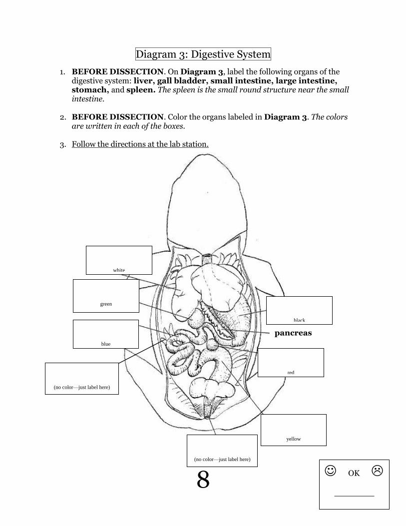
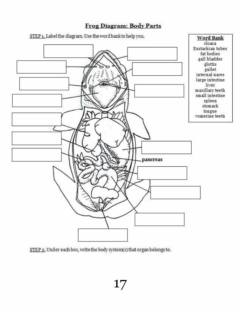
Diagram 3: Digestive System
1. BEFORE DISSECTION. On Diagram 3, label the following organs of the digestive system: liver, gall bladder, small intestine, large intestine, stomach, and spleen. The spleen is the small round structure near the small intestine.
2. BEFORE DISSECTION. Color the organs labeled in Diagram 3. The colors are written in each of the boxes.
3. Follow the directions at the lab station.
pancreas
black
red
yellow
(no color—just label here)
blue
green
white
(no color—just label here)
OK
__________
9
Diagram 3 Questions: 3A. What is the function of the liver?
3B. Why is the liver part of the digestive system?
3C. What does bile do?
3D. What is the diameter of the gall bladder? _____ mm
3E. How long is the stomach? _______________ cm = _______________ mm
3F. What does insulin do?
3G. What is the importance of mesenteries?
3H. What would happen to our bodies if mesenteries weren’t present?
3I. Why is the small intestine coiled?
10
3J. Could we say that the digestive tract is simply a long tube that runs from mouth to the anus of the animal? Circle below. yes no
3K. Why or why not?
3L.The esophagus leads from the _______________ (opening at the back of the
throat) to the _______________. The small intestine leads from the
______________ to the _______________. The large intestine leads from the
______________ to the _______________ (place where wastes exit the body).
11
Digestive System—Part 2
- How is the inside of the stomach different than the outside of the stomach?
- Why is the lining of the stomach the way that it is? (remember, the frog does not
chew its food.)
- What is the length of the small intestine?
_______________ cm = _______________ mm
- What is the function of the villi?
- How is the function of the villi in the small intestine different than the function
of the villi in the small intestine?
12
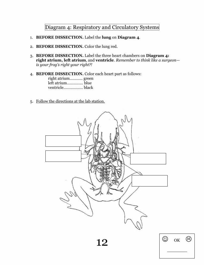
Diagram 4: Respiratory and Circulatory Systems
1. BEFORE DISSECTION. Label the lung on Diagram 4.
2. BEFORE DISSECTION. Color the lung red.
3. BEFORE DISSECTION. Label the three heart chambers on Diagram 4: right atrium, left atrium, and ventricle. Remember to think like a surgeon—is your frog’s right your right?!
4. BEFORE DISSECTION. Color each heart part as follows: right atrium………… green left atrium…………... blue ventricle……………… black
5. Follow the directions at the lab station.
OK
__________
13
Diagram 4 Questions:
4A. What is the function of the heart?
4B. What is the function of the lungs?
4C. What organ system do the heart, lungs, and blood vessels belong to?
4D. What does it mean to have “blue” blood?
4E. What does it mean to have “red” blood?
14
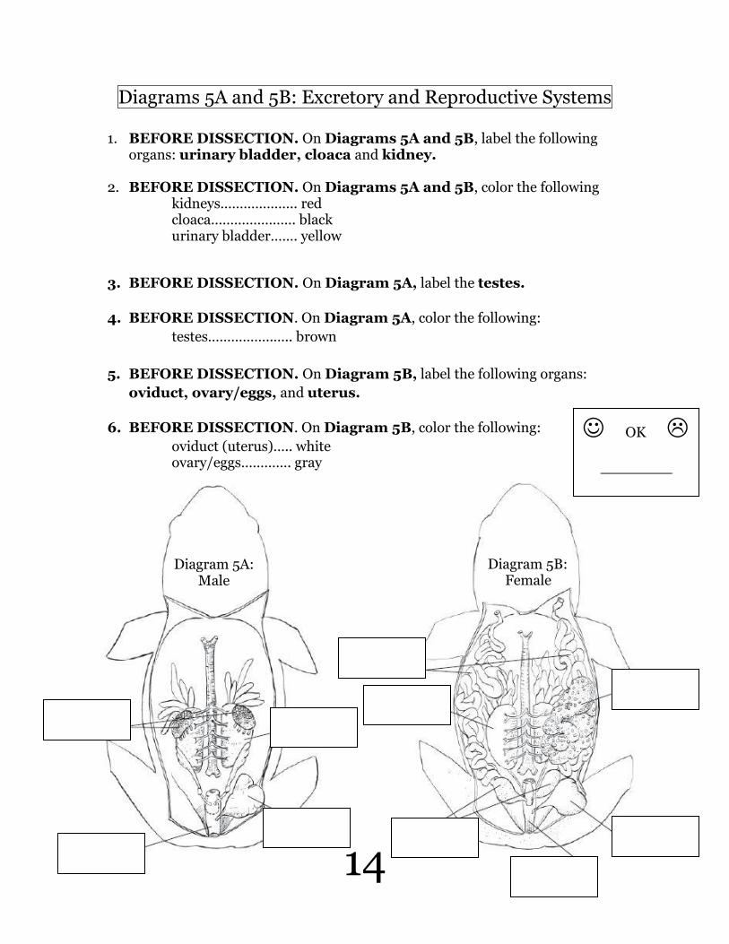
Diagrams 5A and 5B: Excretory and Reproductive Systems
1. BEFORE DISSECTION. On Diagrams 5A and 5B, label the following organs: urinary bladder, cloaca and kidney.
2. BEFORE DISSECTION. On Diagrams 5A and 5B, color the following kidneys……………….. red
cloaca…………………. black urinary bladder……. yellow
3. BEFORE DISSECTION. On Diagram 5A, label the testes.
4. BEFORE DISSECTION. On Diagram 5A, color the following:
testes……………..….. brown
5. BEFORE DISSECTION. On Diagram 5B, label the following organs:
oviduct, ovary/eggs, and uterus.
6. BEFORE DISSECTION. On Diagram 5B, color the following:
oviduct (uterus)….. white ovary/eggs…………. gray
Diagram 5A: Male
Diagram 5B: Female
OK
__________

15
Diagram 5 Questions:
5A. What is the function of the kidneys?
5B. What is the function of the ovaries?
5C. What is the function of the testes?
5D. Look back at question 1B. Were you correct in predicting the sex of your frog? Circle
below.
yes no
16
Dissection Questions:
Directions: Answer the following questions using the information you learned during
the frog dissection.
1. A frog is eating a grasshopper. Follow the grasshopper through the frog, placing
the organs that are used for digestion in order.
2. A 5-yr old boy sees a frog, chases it, and grabs it forcefully by squeezing the
ventral side.
A. What does ventral mean?
B. Which organs could have been damaged due to the forceful squeeze? List
three.
3. Compare the overall structure of a frog to the overall structure of a human. Write
at least 3-5 sentences.
17