SBI3U - The Circulatory System Introduction and Human Circulatory System.
description

Frog circulatory systemFROG CIRCULATORY SYSTEM

Function: TRANSPORT gases, nutritive materials, hormones, blood proteins, metabolic wastes to and from different parts of the body
Blood circulation in vertebrates are similar in principle but different in details depending on complexity of the heart and respiratory organs used.

Cardiovascular system a. Heart b. Blood vessels ( arteries , veins,
capillaries) c. Blood Lymphatic system
a. Heart b. Lymph c. Lymph vessels

A. FROG’s HEART

- pear shaped muscular organ -location: anterior part of the coelom ventral to the liver- Lying within 2-layered perichondrium-3 -chambered 2 atria (thin walls) 1 ventricle (thick walls)- right side : receives deoxygenated blood from the body- left side : receives oxygenated blood from the lungs-


DORSAL VIEW VENTRAL VIEW
sinus venosus (triangular) conus arteriosus continues -recieves blood from to trunchus arteriosus (l and r) 3 caval veins

Sinus venosus (common termination of all veins going to the heart)
Conus arteriolus Ventricle Right auricle
Truncus arteriosus (common origin of all arteries leaving the heart)
Pulmonar artery Pulmo-cutaneous artery

Capillaries of the lungs Pulmonary vein Ventricle Left auricle
Conus arteriolus Trunchus arteriosus Carotid artery

Pulmonary circulation-circulation inside the heart -flow of blood to and from the lungs- RIGHT : UNOXYGENATED- LEFT : OXYGENATED

B. Blood vessels1. Veins- return blood from the capillary
network of the organs to the heart 2. Arteries – convey blood from the heart into
the minute capillaries of the organ 3. Capillaries- smallest of the blood vessels
- bring nutrients and oxygen to the tissues and absorbs carbon dioxide and other waste products

THE VENOUS SYSTEM
Veins are grouped into the ff:A.Systemic – non-oxygenated blood
flows directly to the heart
B.Portal - non- oxygenated blood passes first through the capillaries of certain organs
C.Pulmonary – oxygenated blood flows from lungs to heart

TONGUE LOWER JAW SHOULDER BRAIN,HEAD HYOID SPINAL
CORD LINGUAL MAXILLARY SUBSCAPULAR INTERNAL
JUGULAR
External Jugular INNOMINATE
SUBCLAVIAN PRECAVA (anterior +forelimbs)
PRACHIAL SINUS VENOSUS(HEART)
FORELIMBS all orange are the 3 tributaries
vv

PORTAL SYSTEM - does not return blood directly to the
heart but to the capillary system through the liver or kidneys
A. HEPATIC PORTAL VEIN - conveys blood to capillaries of LIVERB. RENAL PORTAL VEIN - paired - conveys blood to capillaries of KIDNEYS - not found in man

HINDLIMBS STOMACH PANCREAS SPLEEN
SCIATIC FEMORAL GASTRIC PANCREATIC SPLEENIC
PELVIC INTESTINE
RENALPORTAL ANTERIOR ABDOMINAL INTESTINAL
LIVER HEPATIC PORTAL
KIDNEY DORSAL
HEPATIC BODY WALLRENAL
LUMBAR GONADS
POSTCAVA
SINUS VENOSUS UROGENITAL

THE ARTERIAL SYSTEMA pair of big arteries , the TRUNCHUS ARTERIOLUS, leave the heart each dividing into 3 branches
A.Common carotid – to the head regionB.Systemic- to the appendages, internal organs &parts posterior to the heartC. Pulmo-cutaneous- non-oxygenated blood to the organs where blood may undergo oxygenation

TRUNCUS ARTERIOSUSCOMMON CAROTID SYSTEMIC PULMO- CUTANEOUS INTERNAL EXTERNAL (non-oxygenated)
TONGUE
PALATINE EPHTHALMIC CEREBRAL
ROOF OF EYE BRAIN MOUTH
PULMONARY CUTANEOUS
LUNGS SKIN TYMPANUM

SYSTEMICESOPHAGEAL OCCIPITO-VERTEBRAL SUBCLAVIANESOPHAGUS SPINAL CORD brachial
FORELIMB DORSAL AORTA (unite w/each other)
COMMON ILIAC
EPIGASTRICO-VESICAL SCIATIC FEMORAL
EPIGASTRIC RECTO-VESICAL HINDLIMB
VENTRAL ABDOMINAL RECTUM,URINARY BLADDER
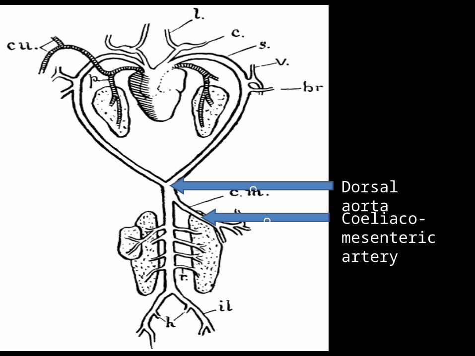
c Dorsal aortac Coeliaco-
mesenteric artery

DORSAL AORTA
COELIACO-MESENTERIC
COELIC ANTERIOR MESENTERIC
LEFT RIGHT HEPATIC SPLEENIC INTESTINAL GASTRIC
SPLEEN SMALL INTESTINESTOMACH LIVER
ANTERIOR HEMORRHOIDALSTOMACH,PANCREAS LARGE INTESTINE
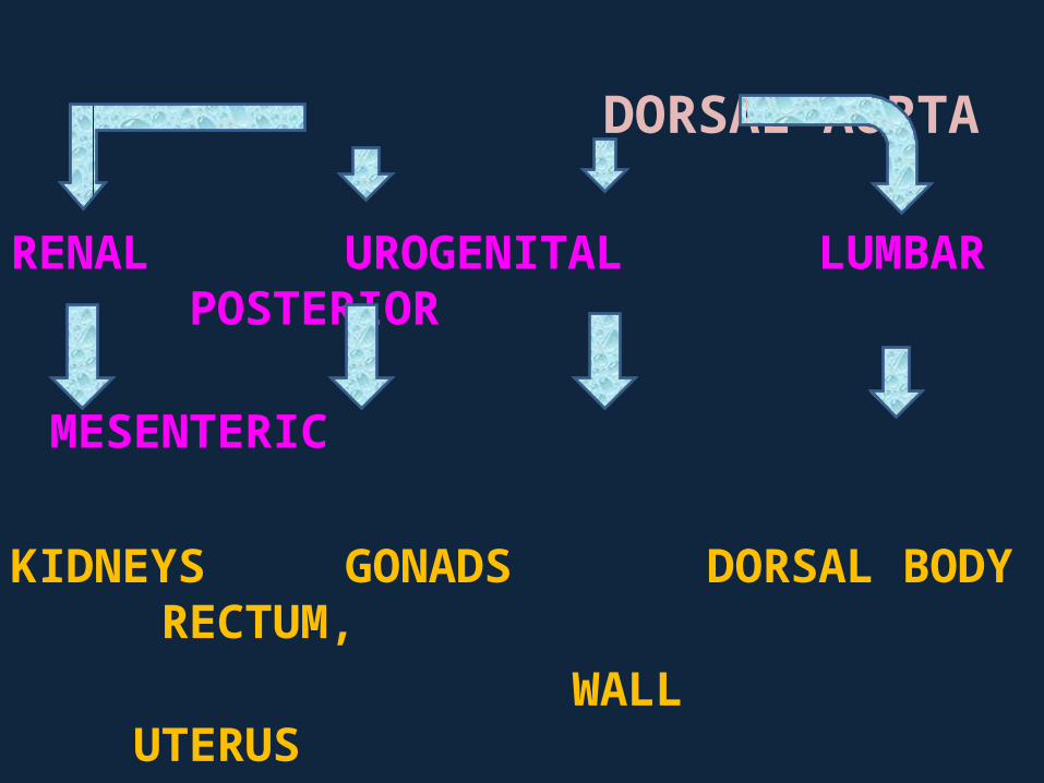
DORSAL AORTA
RENAL UROGENITAL LUMBAR POSTERIOR
MESENTERIC
KIDNEYS GONADS DORSAL BODY RECTUM,WALL UTERUS
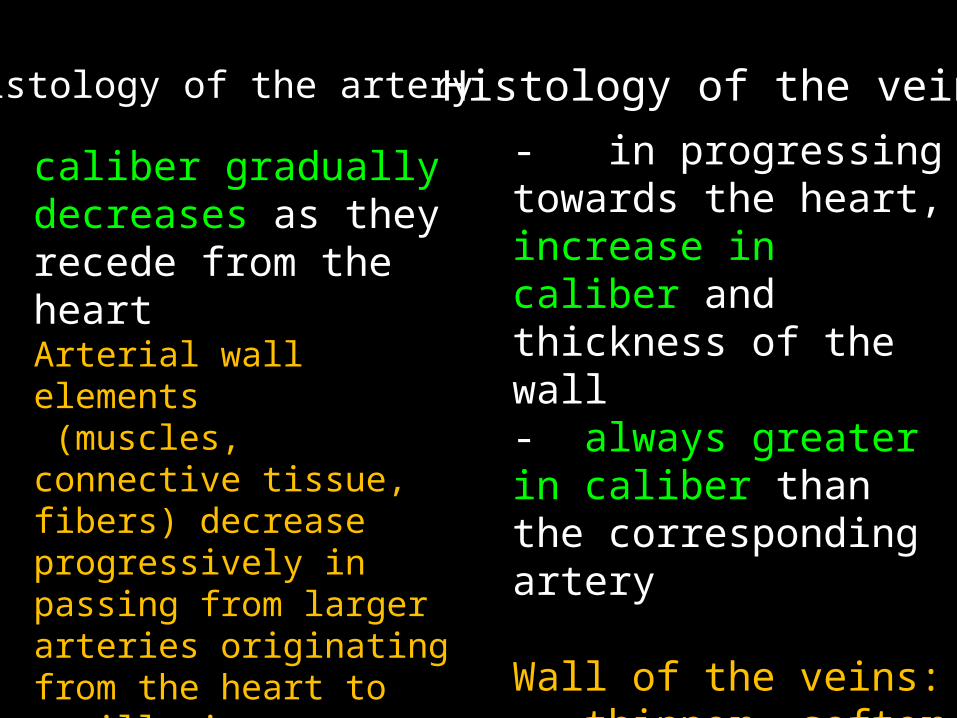
Arterial wall elements (muscles, connective tissue, fibers) decrease progressively in passing from larger arteries originating from the heart to capillaries
Histology of the artery
caliber gradually decreases as they recede from the heart
Histology of the vein- in progressing towards the heart, increase in caliber and thickness of the wall- always greater in caliber than the corresponding artery
Wall of the veins: - thinner, softer and less elastic than arterial wall

Inner lining is the same as found in other blood vessels because it is formed by a continuous layer of endothelial cells Most of the arteries in the body : medium sized and muscular type
The veins if empty are collapsed and the lumen is irregular and slit-like
Artery Vein

- made of inner endothelium, longitudinally directed collagenous and elastic fibers and an elastic membrane
-serves as boundary between intima and media
-thrown into folds-corrugated inner surface
noticeable in medium sized artery
Tunica intima – innercoat
-made of endothelial cells, beneath which is a layer of fine, collagenous and elastic fibers-internal elastic membrane: poorly developed-Larger veins: intima is bounded by network of elastic fibers
Artery Vein

Tunica media –intermediate coat
smooth muscle arranged in layers that encircle the artery
No. of muscle layers depends on caliber of artery
Thin reticular fiber : sheaths of individual muscle cell
Thin elastic fiber :course circularly in the media and continue to the external and internal elastic membrane
Relatively thin and consists of layers of circularly arranged smooth muscle fibers separated by collagenous andelastic tissues- Best developed in veins of the viscera and head- Larger veins media is sometimes absent
Artery Vein

Tunica adventitia/externa
–outer coat - Loose connective tissue - Collagenous and elastic
fibers mostly run parallel to the long axis of the vessel
- These elements merge with surrounding connective tissue that accompanies every blood vessel
- Considerably thicker than the media-Consists of loose connective tissue with longitudinal collagenous fibers and elastic networks-occasionally, longitudinal smooth muscle fibers may be present adjacent to the media
Artery Vein


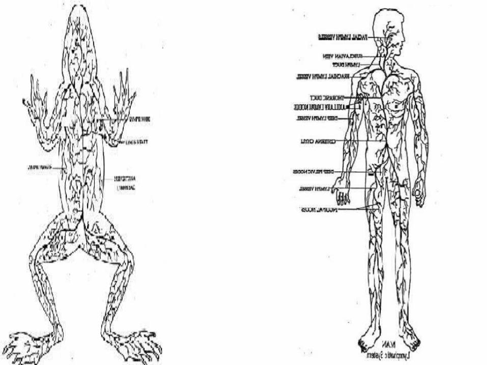
LYMPHATIC SYSTEM MAN FROG

LYMPHATIC SYSTEM
- Begins with small vessels called lymphatic capillaries which are in direct contact with the extra-cellular fluid surrounding tissues - A network of vessels that collects the fluid that is lost by the blood and returns it to the circulatory system

- Drainage of fluid from the tissues and its return to the circulatory system
MAN FROG- Collect and drain fluid that seeps from the bloodstream and accumulates in the extracellular fluid- Return small am. of protein that have left the cells- Transport lipids that was absorbed in the small intestine- Transport foreign particles to lymph nodes

MAN FROG
-possesses lymph nodes
-unique among all vertebrates because of its high rate of lymph production and circulation
- normal rate of lymph production and circulation
house the WBCs
To accommodate rapid exchange of fluid bet. the circulatory and lymphatic systems, frogs have 2 distinct features1.large, interconnecting lymph spaces into which lymph vessels drain2.Presence of lymph heart

• Lymph hearts (LH)• - small organs usually located at the dorsal side of the
animal’s body ( at the entry points of lymph into the veins). - - posterior LH are found 1 of each pair on each side
lying lateral to end of each urostyle - 1 pair in common toad and 2 or more in certain frogs - main function: maintain the directionality of
lymphatic flow and regulate the entry of lymph fluid into the circulation


Lymphatic system of man
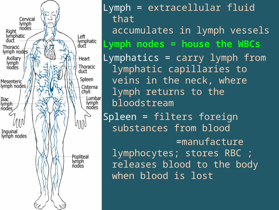
Lymph = extracellular fluid that accumulates in lymph vessels
Lymph nodes = house the WBCs Lymphatics = carry lymph from
lymphatic capillaries to veins in the neck, where lymph returns to the bloodstream
Spleen = filters foreign substances from blood
=manufacture lymphocytes; stores RBC ; releases blood to the body when blood is lost

TRIVIA

• How does temperature affect frog’s heart rate? The cooler the frog the slower the heart rate, the warmer the faster

Lymph heart trivia:- for a short time may stop beating all
together for no apparent reason- has irregular rhythm

• The lymphatic system as we know it today was first described independently by Olaus Rudbeck and Thomas Bartholin.

LASTLY,
If you ever visit Japan, do not forget to try their Frog sashimi and the fresh, still beating heart of a frog. Flushed down with a refreshing glass of lizard sake.

Quiz
• 1. How many chambers do frog’s heart have? And what are those?
• 2. Describe the pulmonary circulation.• right side: left side:• 3. Differentiate arteries, veins and capillaries.• 4. Lymphatic system: what do frog’s have that
we humans don’t’?

• 5. Histology of artery or vein? Caliber gradually decreases as they recede from
the heart6. Amphibian lymphatic system = unique among all
vertebrates because of its high rate of ? production and circulation
7-10. Give any 1 of the 3 layers or coat of the artery and vein
Bonus: Where will I go if I want to eat live frog heart?