Fragment-based screening by X-ray crystallography, MS … · binding to PNMT, showing for the...
16
Biochem. J. (2010) 431, 51–61 (Printed in Great Britain) doi:10.1042/BJ20100651 51 Fragment-based screening by X-ray crystallography, MS and isothermal titration calorimetry to identify PNMT (phenylethanolamine N-methyltransferase) inhibitors Nyssa DRINKWATER*, Hoan VU†, Kimberly M. LOVELL‡, Kevin R. CRISCIONE‡, Brett M. COLLINS*, Thomas E. PRISINZANO‡, Sally-Ann POULSEN†, Michael J. MCLEISH§, Gary L. GRUNEWALD‡ and Jennifer L. MARTIN* 1 *University of Queensland, Institute for Molecular Bioscience, Division of Chemistry and Structural Biology, Brisbane, Queensland 4072, Australia, †Griffith University, Eskitis Institute, Brisbane, Queensland 4111, Australia, ‡Department of Medicinal Chemistry, University of Kansas, Lawrence, KS 66045-7582, U.S.A., and §Department of Chemistry and Chemical Biology, Indiana University–Purdue University Indianapolis (IUPUI), 402 N. Blackford St, LD 326D, IN 46202, U.S.A. CNS (central nervous system) adrenaline (epinephrine) is implicated in a wide range of physiological and pathological conditions. PNMT (phenylethanolamine N-methyltransferase) catalyses the final step in the biosynthesis of adrenaline, the conversion of noradrenaline (norepinephrine) to adrenaline by methylation. To help elucidate the role of CNS adrenaline, and to develop potential drug leads, potent, selective and CNS-active inhibitors are required. The fragment screening approach has advantages over other lead discovery methods including high hit rates, more efficient hits and the ability to sample chemical diversity more easily. In the present study we applied fragment- based screening approaches to the enzyme PNMT. We used crystallography as the primary screen and identified 12 hits from a small commercial library of 384 drug-like fragments. The hits include nine chemicals with two fused rings and three single-ring chemical systems. Eight of the hits come from three chemical classes: benzimidazoles (a known class of PNMT inhibitor), purines and quinolines. Nine of the hits have measurable binding affinities (∼ 5–700 μM) as determined by isothermal titration calorimetry and all nine have ligand efficiencies of 0.39 kcal/mol per heavy atom or better (1 kcal ≈ 4.184 kJ). We synthesized five elaborated benzimidazole compounds and characterized their binding to PNMT, showing for the first time how this class of inhibitors interact with the noradrenaline-binding site. Finally, we performed a pilot study with PNMT for fragment-based screening by MS showing that this approach could be used as a fast and efficient first-pass screening method prior to characterization of binding mode and affinity of hits. Key words: benzimidazole, catecholamine, drug discovery, enzyme inhibition, fragment-based screening, phenylethanol- amine N-methyltransferase (PNMT). INTRODUCTION The identification of suitable starting compounds for development into drugs is a major challenge for drug discovery and development programmes. FBS (fragment-based screening) has been developed as a method for the rapid discovery and subsequent elaboration of hits into quality lead compounds [1,2]. This approach is based on the principle of screening simple low-molecular-mass compounds that make a few high-quality interactions with the target site. Resulting hits tend to bind to the targets efficiently yet with low affinity and, with a practically designed library, are readily tractable for follow up chemistry [3]. These fragment hits are used as ‘anchors’ that can be elaborated to fill additional regions of the target site. This results in potent compounds with minimal chemical complexity that can then be developed into drugs. A major challenge in establishing FBS has been the development of sensitive methods to detect the weak binding of hits (with typically milli- to micro-molar binding affinities) [4]. Methods currently used include ITC (isothermal titration calorimetry), NMR, surface plasmon resonance, MS and X-ray crystallography [1,5–7]. FBS-X (FBS by X-ray crystallography) provides the greatest level of information for the elaboration of hit compounds [8]. However, costly infrastructure for crystallization, crystal handling and data collection is required for screening hundreds of crystals in a high-throughput manner. Although many different FBS methods are now used, there have been few reports comparing hits identified against the same specific target by orthogonal methods. NMR is frequently employed as a first-pass screen before detailed structural characterization of hits by X-ray crystallography, and MS has the potential to be applied in a similar manner [9]. Comparative FBS-X and FBS-MS campaigns have not been reported, as far as we are aware, yet there are potentially significant advantages to a combined MS/X-ray screening approach due to their complementary attributes. Pharmaceutical companies have developed and applied FBS-X with success [10]. An ideal target for FBS-X has an accessible active site, can be crystallized in the apo form and requires only minor conformational changes in the crystal form to accommodate ligand binding. However, many drug targets do not conform to these requirements, including the human enzyme PNMT (phenylethanolamine N-methyltransferase; EC Abbreviations used: AdoHcy, S-adenosyl-L-homocysteine; AdoMet, S-adenosyl-L-methionine; CCD, charge-coupled-device; CNS, central nervous system; ESI, electrospray ionization, FBS, fragment-based screening, FBS-X, FBS by X-ray crystallography; FTMS, Fourier transform MS; HA, heavy atom; HRMS, high-resolution MS; ITC, isothermal titration calorimetry; LE, ligand-binding efficiency; m.p., melting point; MSD, Merck Sharp and Dohme Research Laboratories; PNMT, phenylethanolamine N-methyltransferase; hPNMT, human PNMT. 1 To whom correspondence should be addressed (email [email protected]). The co-ordinates and structure factors for all 17 crystal structures have been deposited with the PDB under accession codes 3KPJ, 3KPU, 3KPV, 3KPW, 3KPY, 3KQM, 3KQS, 3KQT, 3KQV, 3KQW, 3KQO, 3KQP, 3KQQ, 3KQY, 3KR0, 3KR1 and 3KR2. c The Authors Journal compilation c 2010 Biochemical Society
Transcript of Fragment-based screening by X-ray crystallography, MS … · binding to PNMT, showing for the...

Biochem. J. (2010) 431, 51–61 (Printed in Great Britain) doi:10.1042/BJ20100651 51
Fragment-based screening by X-ray crystallography, MS and isothermaltitration calorimetry to identify PNMT (phenylethanolamineN-methyltransferase) inhibitorsNyssa DRINKWATER*, Hoan VU†, Kimberly M. LOVELL‡, Kevin R. CRISCIONE‡, Brett M. COLLINS*, Thomas E. PRISINZANO‡,Sally-Ann POULSEN†, Michael J. MCLEISH§, Gary L. GRUNEWALD‡ and Jennifer L. MARTIN*1
*University of Queensland, Institute for Molecular Bioscience, Division of Chemistry and Structural Biology, Brisbane, Queensland 4072, Australia, †Griffith University, Eskitis Institute,Brisbane, Queensland 4111, Australia, ‡Department of Medicinal Chemistry, University of Kansas, Lawrence, KS 66045-7582, U.S.A., and §Department of Chemistry and ChemicalBiology, Indiana University–Purdue University Indianapolis (IUPUI), 402 N. Blackford St, LD 326D, IN 46202, U.S.A.
CNS (central nervous system) adrenaline (epinephrine) isimplicated in a wide range of physiological and pathologicalconditions. PNMT (phenylethanolamine N-methyltransferase)catalyses the final step in the biosynthesis of adrenaline, theconversion of noradrenaline (norepinephrine) to adrenaline bymethylation. To help elucidate the role of CNS adrenaline, and todevelop potential drug leads, potent, selective and CNS-activeinhibitors are required. The fragment screening approach hasadvantages over other lead discovery methods including highhit rates, more efficient hits and the ability to sample chemicaldiversity more easily. In the present study we applied fragment-based screening approaches to the enzyme PNMT. We usedcrystallography as the primary screen and identified 12 hits froma small commercial library of 384 drug-like fragments. The hitsinclude nine chemicals with two fused rings and three single-ringchemical systems. Eight of the hits come from three chemical
classes: benzimidazoles (a known class of PNMT inhibitor),purines and quinolines. Nine of the hits have measurable bindingaffinities (∼5–700 μM) as determined by isothermal titrationcalorimetry and all nine have ligand efficiencies of 0.39 kcal/molper heavy atom or better (1 kcal ≈ 4.184 kJ). We synthesizedfive elaborated benzimidazole compounds and characterized theirbinding to PNMT, showing for the first time how this class ofinhibitors interact with the noradrenaline-binding site. Finally, weperformed a pilot study with PNMT for fragment-based screeningby MS showing that this approach could be used as a fast andefficient first-pass screening method prior to characterization ofbinding mode and affinity of hits.
Key words: benzimidazole, catecholamine, drug discovery,enzyme inhibition, fragment-based screening, phenylethanol-amine N-methyltransferase (PNMT).
INTRODUCTION
The identification of suitable starting compounds for developmentinto drugs is a major challenge for drug discovery anddevelopment programmes. FBS (fragment-based screening) hasbeen developed as a method for the rapid discovery andsubsequent elaboration of hits into quality lead compounds [1,2].This approach is based on the principle of screening simplelow-molecular-mass compounds that make a few high-qualityinteractions with the target site. Resulting hits tend to bind tothe targets efficiently yet with low affinity and, with a practicallydesigned library, are readily tractable for follow up chemistry [3].These fragment hits are used as ‘anchors’ that can be elaboratedto fill additional regions of the target site. This results in potentcompounds with minimal chemical complexity that can then bedeveloped into drugs. A major challenge in establishing FBS hasbeen the development of sensitive methods to detect the weakbinding of hits (with typically milli- to micro-molar bindingaffinities) [4]. Methods currently used include ITC (isothermaltitration calorimetry), NMR, surface plasmon resonance, MS andX-ray crystallography [1,5–7].
FBS-X (FBS by X-ray crystallography) provides the greatestlevel of information for the elaboration of hit compounds [8].However, costly infrastructure for crystallization, crystal handlingand data collection is required for screening hundreds of crystalsin a high-throughput manner. Although many different FBSmethods are now used, there have been few reports comparing hitsidentified against the same specific target by orthogonal methods.NMR is frequently employed as a first-pass screen before detailedstructural characterization of hits by X-ray crystallography, andMS has the potential to be applied in a similar manner [9].Comparative FBS-X and FBS-MS campaigns have not beenreported, as far as we are aware, yet there are potentially significantadvantages to a combined MS/X-ray screening approach due totheir complementary attributes.
Pharmaceutical companies have developed and appliedFBS-X with success [10]. An ideal target for FBS-X has anaccessible active site, can be crystallized in the apo form andrequires only minor conformational changes in the crystal formto accommodate ligand binding. However, many drug targetsdo not conform to these requirements, including the humanenzyme PNMT (phenylethanolamine N-methyltransferase; EC
Abbreviations used: AdoHcy, S-adenosyl-L-homocysteine; AdoMet, S-adenosyl-L-methionine; CCD, charge-coupled-device; CNS, central nervoussystem; ESI, electrospray ionization, FBS, fragment-based screening, FBS-X, FBS by X-ray crystallography; FTMS, Fourier transform MS; HA, heavy atom;HRMS, high-resolution MS; ITC, isothermal titration calorimetry; LE, ligand-binding efficiency; m.p., melting point; MSD, Merck Sharp and Dohme ResearchLaboratories; PNMT, phenylethanolamine N-methyltransferase; hPNMT, human PNMT.
1 To whom correspondence should be addressed (email [email protected]).The co-ordinates and structure factors for all 17 crystal structures have been deposited with the PDB under accession codes 3KPJ, 3KPU, 3KPV, 3KPW,
3KPY, 3KQM, 3KQS, 3KQT, 3KQV, 3KQW, 3KQO, 3KQP, 3KQQ, 3KQY, 3KR0, 3KR1 and 3KR2.
c© The Authors Journal compilation c© 2010 Biochemical Society

52 N. Drinkwater and others
Figure 1 PNMT catalytic reaction
(A) The reaction catalysed by PNMT involves transfer of a methyl group fromAdoMet to noradrenaline, forming adrenaline and AdoHcy. (B) The crystal structure ofhPNMT–AdoHcy–SK&F29661, shown in dark grey and light grey space-filling representationrespectively, are AdoHcy and SK&F29661; the Cα trace of hPNMT is shown predominantly inlight grey, with the regions covering the AdoHcy- and SK&F29661-binding sites shown in darkgrey. The boxed region shows a close-up of the AdoHcy- and SK&F29661-binding sites.
2.1.1.28; 30.7 kDa), which catalyses the final step in adrenaline(epinephrine) biosynthesis, the methylation of noradrenaline(norepinephrine) to form adrenaline (Figure 1A). PNMThas a potential role in a wide range of disease-relevantprocesses including the central control of blood pressure [11],pituitary hormone secretion [12], ethanol intoxication [13],Parkinson’s disease [14] and the neurodegeneration observedin Alzheimer’s disease [15]. Structures of recombinant hPNMT(human PNMT) in complex with inhibitors and substrates (e.g.PDB code 1HNN) show that hPNMT has an enclosed active site(Figure 1B) suggesting that enzyme conformational changes maybe needed for ligands to bind [16]. Apo crystals are not readilyavailable because we were unable to crystallize the enzyme in theabsence of AdoHcy (S-adenosyl-L-homocysteine), which binds tothe cofactor-binding site. Furthermore, when crystals are grownin the absence of an inhibitor or substrate at the noradrenaline-binding site, this site is occupied by a phosphate molecule [17].However, we were encouraged to attempt FBS-X because wesolved the crystal structure of hPNMT (PDB code 3HCD) incomplex with the physiological substrate R-noradrenaline byusing rapid soaking methods on hPNMT–AdoHcy–PO4 crystals[17]. We therefore proposed to apply this same soaking methodfor FBS-X screening of hPNMT, with the aim of identifying novelchemical frameworks for inhibitor design. We also performed anFBS-MS pilot study to determine whether this approach could beused as a more efficient first-pass screen prior to crystallographicanalysis of selected hits.
Overall, we showed that FBS-X can be successfully imple-mented within an academic environment. Our crystallographicscreening of the non-ideal enzyme target hPNMT identified 12hits, including three benzimidazoles (a known class of PNMTinhibitors), and nine of the hits could be characterized by ITC.Five compounds were synthesized to further characterize the
benzimidazole class. Our pilot study showed that the orthogonalscreening method of FBS-MS could be used as an even moreefficient first step in an FBS campaign.
EXPERIMENTAL
Materials
The FBS library was purchased from ActiveSight (a Rigakucompany), and comprised 384 compounds with a mean molecularmass of 142 Da and of predominantly rigid low-complexity(see the Supplementary Table Library at http://www.BiochemJ.org/bj/431/bj4310051add.htm); all compounds were providedas 200 mM stock solutions in 100% DMSO and as cocktailsof four fragments each at 50 mM in 100 % DMSO. All thecompounds are also available for purchase directly from Sigma–Aldrich. For chemical syntheses, all reagents and solvents usedwere reagent grade and of the highest purity commerciallyavailable. 4-Methoxybenzene-1,2-diamine was sourced from AlfaAesar, all other compounds and reagents were sourced fromSigma–Aldrich.
Crystallization of hPNMT
C-terminally His6-tagged hPNMT was expressed and purifiedas described previously [18]. The protein was concentrated to50 mg/ml and mixed with AdoHcy (to a final concentration of2 mM) taking the final concentration of protein to 40 mg/ml or1.25 mM. The protein mixture was crystallized by hanging dropvapour diffusion using QIAGEN EasyXtal Tool crystallizationtrays for ease of soaking. Drops comprised 1.5 μl of proteinand 1.5 μl of precipitant equilibrated over 400 μl of precipitantsolution {4–8% PEG [poly(ethylene glycol)] 6000, 0.25 M LiCland 0.1 M sodium cacodylate, pH 5.5–6.0}.
FBS by X-ray crystallography
Library screening was conducted blind, in that we made noassumptions about the contents of the library, or what compoundsmight be expected to bind PNMT. Prior to screening, controlexperiments were performed using known hPNMT inhibitors toconfirm that ligands could be soaked into hPNMT–AdoHcy–PO4 crystals and that the solvents used for fragment screeningand cryoprotection did not interfere with binding. Crystals(∼0.25 mm in each dimension) were transferred into 1 μl ofsoaking solution. Crystal soak solutions were prepared by adding1 μl of fragment cocktail to 9 μl of stabilizing solution, takingthe final concentrations to 6.6 % PEG 6000, 0.3 M LiCl, 0.1 Msodium cacodylate, pH 5.7, 4× 5 mM fragments and 10% (v/v)DMSO. Crystals were soaked for 15 min.
Crystals were cryoprotected by transferring directly from thesoak solution into mother liquor supplemented with 25% ethyleneglycol for 10–15 s before being flash-cooled. Data for hPNMT–AdoHcy–PO4 were measured from a crystal transferred directlyinto the cryoprotectant solution without undergoing a soak step.X-ray diffraction data from hPNMT crystals were measured usingthe UQ ROCX Diffraction Facility or the Australian Synchrotron.At the UQ ROCX Diffraction Facility, crystals were irradiatedwith X-rays of wavelength 1.542 Å (1 Å = 0.1 nm) generatedfrom a Rigaku FR-E Superbright copper rotating anode generatoroperating at 45 kV, 45 mA and data were measured usingone of two alternate methods: (i) crystals were flash-cooledby plunging into liquid nitrogen and placed on the camerausing a Rigaku ACTORTM crystal mounting robot, X-rays werefocused with Osmic Vari-Max HF optics and diffraction data
c© The Authors Journal compilation c© 2010 Biochemical Society
Fragment-based screening to identify PNMT inhibitors 53
measured on a Rigaku Saturn 944 CCD (charge-coupled-device)detector; or (ii) crystals were flash-cooled on the camera inthe gaseous nitrogen stream at 100K, X-rays were focusedwith Osmic HiRes2 Confocal Max-FluxTM optics and diffractiondata measured with a Rigaku R-AXIS IV++ imaging plate areadetector. Data collected at UQ ROCX Diffraction Facility wereprocessed using Crystal Clear (Rigaku). Data collected at theAustralian Synchrotron were measured at 100K with X-rays ofwavelength 0.95667 Å on beamline 3BM1 [19] using a MAR165 CCD detector, and processed using HKL2000 [20]. Crystalsdiffracting to better than 2.7 Å resolution were used for datacollection.
Phasing and automatic structure solution were carried outusing MIfit (ActiveSight). The structures were solved bymolecular replacement using the structure of hPNMT–AdoHcy–SK&F29661 (PDB code 1HNN), with the inhibitor andwaters removed, as the starting model. Active-site density wasindividually evaluated and assessed for the presence or absenceof a ligand. If density indicating a bound fragment was present,ligands were modelled manually using COOT [21] and co-ordinates and refinement parameters were generated by PRODRG[22] and PHENIX [23]. Further refinement was carried out usingPHENIX [23]. Cross-validation was done by R-free analysis from5% of the reflections set aside from refinement.
Where density was present at the active site but didnot unambiguously identify the bound ligand from the fourcompounds in the cocktail, a deconvolution step was performed.This involved collecting data from crystals soaked in solutionscontaining the individual compounds (rather than the cocktailof four compounds). In these cases, the final concentration ofthe compounds in the soak solution was increased to 20 mMrather than the 5 mM concentration used in the cocktail screen.The higher concentration generally resulted in improved density.Deconvolution also allowed identification of the binding of morethan one fragment per cocktail.
Structure determination of hPNMT in complex with elaboratedbenzimidazoles
Compounds 13–17 were designed and synthesized (see belowfor details of synthesis) to further characterize the bindingof benzimidazoles to hPNMT. The crystal structures of thesecompounds in complex with hPNMT were determined as follows.Crystals of hPNMT–AdoHcy–PO4 were soaked in a solutionof the synthesized compounds as described above for fragmentscreening. In this case, compounds were solubilized at 100 mMin 50% DMSO/water and used to prepare soak solutionsof final concentrations 6.6% PEG 6000, 0.3 M LiCl, 0.1 Msodium cacodylate, pH 5.7, 5–10 mM compounds and 2.5–5%DMSO.
Crystals were cryoprotected, diffraction data collected andprocessed, and structures solved and refined at the UQ ROCXdiffraction facility using method (ii) described above, with theexception that phasing was carried out using PHENIX [23]and structures were solved by difference Fourier methods. Theprocedure in this case was to model and refine the structure ofthe protein first, followed by addition of water molecules, thenAdoHcy and finally the ligand. The criteria used to include a watermolecule were: the presence of 2Fo−Fc density at 1σ and Fo−Fc
density at 3σ and at least one possible hydrogen bond within3.2 Å. Cross-validation was performed as described above. All ofthe crystal structure electron density maps revealed density indic-ative of AdoHcy at the cofactor-binding site and density consistentwith a benzimidazole bound at the noradrenaline-binding site.
FBS by MS
MS was performed on an APEX® III 4.7 Tesla FTICR massspectrometer (Bruker Daltonics) fitted with an ApolloTM ESI(electrospray ionization) source operated in positive ion mode.XMASS NT V6.1.2 MS software on a PC platform was usedfor data acquisition. Broadband excitation was used to analysea mass range from m/z 50–6000 and each spectrum was anaverage of 64 transients (scans) with 512000 data points acquiredin low-resolution mode with an acquisition time of approx.4 min/sample. Samples were infused into the ESI source at2 μl/min. Nitrogen was used as both the drying gas (125 ◦C) andnebulizing gas. Relevant parameters include a 104-fold pressurereduction between source and analyser regions with an ESIsource pressure (10−7 kPa) and high-vacuum analyser regionpressure (6 × 10⁻¹¹ kPa). Agilent ESI tuning mix (Santa Clara)was used for an external three-point calibration. The hexapole ionaccumulation time was 3 s.
To confirm correct ESI-MS parameter adjustment, para-meters were optimized using a known hPNMT ligand(7-aminosulfonyl-3-ethyl-1,2,3,4-tetrahydroisoquinoline; K i of1.8 μM) [24]. Fragments analysed (Supplementary FigureS1 at http://www.BiochemJ.org/bj/431/bj4310051add.htm) wereselected to include a range of FBS-X-positive and -negative hits.A total of 12 fragments were provided with no indication asto which compounds were hits by FBS-X. A stock solution of31.5 μM hPNMT was prepared in 10 mM ammonium acetate(pH 7); stock solutions of AdoHcy and each of the 12 fragments(representing three of the FBS-X cocktails) were prepared inmethanol. Aliquots of all solutions were used immediately orstored at −30 ◦C. Samples for MS analysis were prepared asfollows: 3 μM hPNMT, 0 or 30 μM AdoHcy and 300 μM(100 equiv.) fragment in a total volume of 100 μl. Samples wereincubated at room temperature (25 ◦C) for 30 min prior to MSanalysis. A total of 24 MS datasets were acquired to analyse the12 fragments (i.e. with and without added AdoHcy cofactor).The protein consumption per 100 μl sample was 0.01 mg,however, this sample volume was prepared for convenience andcould be reduced as the consumed sample volume was 8 μl perMS acquisition (4 min acquisition at 2 μl/min).
If a hPNMT–fragment or hPNMT–AdoHcy– fragment non-covalent complex is formed in solution then the complexwas observed in the ESI mass spectrum. The mass differencebetween the peaks for the unbound hPNMT and the hPNMT–fragment (�m/z) can be multiplied by the charge state to givedirectly the molecular mass of the binding fragment i.e. Mr
fragment =�m/z × z to provide confirmation of the identity of thehit fragment. Complexes with AdoHcy were similarly treated toidentify and confirm the fragment hits. ESI-FT (Fourier transform)MS analysis of a solution containing 3 μM hPNMT yielded theESI positive ion mass spectrum shown in Supplementary FigureS2(A) at http://www.BiochemJ.org/bj/431/bj4310051add.htm.Peaks corresponding to the 13+ to 10+ charge states of hPNMTwere observed. The ESI-FTMS spectrum of 3 μM hPNMT withadded 30 μM AdoHcy yielded the ESI positive ion mass spectrumshown in Supplementary Figure S2(B) and, as an example, theESI-FTMS mass spectrum of a hPNMT–AdoHcy–hit (compound6) solution is shown in Supplementary Figure S2(C). In thisspectrum each charge state consists of a peak correspondingto hPNMT–AdoHcy and a peak corresponding to the hPNMT–AdoHcy–fragment non-covalent complex. Actual mass valuesof the peaks observed in Supplementary Figure S2(C) andcalculation of the mass of the fragment hit with compound 6are given in Supplementary Table S1 at http://www.BiochemJ.org/bj/431/bj4310051add.htm.
c© The Authors Journal compilation c© 2010 Biochemical Society

54 N. Drinkwater and others
ITC
Compounds found to bind hPNMT by FBS-X or FBS-MS weresubsequently analysed by ITC. ITC was carried out using a high-sensitivity MicroCal (GE Healthcare) iTC200 system at 25 ◦C.Purified protein at a concentration of 200 μM plus 600 μMAdoMet (S-adenosyl-L-methionine) in 20 mM Tris/HCl, pH 7.2,1 mM EDTA and 4.3% DMSO were loaded into the samplecell (Vcell, reaction cell volume of ≈ 200 μl). DMSO in thesample was calibrated to match the residual DMSO after fragmentdilutions. The fragment hit at 1.5–3 mM in the same buffer wasthen titrated into the sample cell with 20 × 2.5 μl injections. Theheat released was measured and integrated using MicroCal Origin7.0 software. The association constant (Ka; 1/Kd), enthalpy (�H)and stoichiometry (N) were calculated using a single-site bindingmodel. Measurements were repeated 2–4 times for each ligandand errors calculated as the S.D. from replicate experiments. Ineach case the protein concentration was fixed as a known quantityand the apparent ligand concentration was adjusted slightly duringcurve fitting so that the binding stoichiometry was approx. 1 (i.e.an assumption of 1:1 binding was made).
The energies of interaction were calculated from Ka valuesusing the Gibbs free energy equation: �G =−RT(ln Ka), whereR is the gas constant (1.9872 kcal/mol; 1 kcal ≈ 4.184 kJ), T is thetemperature of the experiment (298 K) and Ka is the associationconstant for the compound. LE (ligand-binding efficiency) valueswere calculated according to LE = −�G/HA, where �G is thefree energy of binding (kcal/mol) and HA is the number of heavyatoms (non-hydrogen atoms) in the compound.
Compound synthesis
Benzimidazoles (compounds 13–17, see Figure 5) weresynthesized using procedures published previously with minormodifications as indicated (see the Supplementary Experimentalsection at http://www.BiochemJ.org/bj/431/bj4310051add.htm).Generally, an appropriately substituted nitroaniline was reducedby palladium-catalysed hydrogenation to the correspondingphenylenediamine. The phenylenediamine derivative was con-densed with cyanogen bromide to form the desired 2-aminobenzi-midazole as the hydrobromide salt. Methoxy-substituted 2-aminobenzimidazoles were converted to their correspondingphenol derivatives using 48 % hydrogen bromide. Compounds13 and 17 have not been reported previously and details oftheir synthesis are reported below. Compounds 14–16 are knowncompounds and the details of our syntheses of them are given inthe Supplementary Experimental section.
The m.p. (melting point) was determined in open capillarytubes on a Thomas–Hoover melting point apparatus calibratedwith known compounds, but are uncorrected. Proton (1H-NMR)and carbon (13C-NMR) NMR spectra were taken on a BrukerDRX-500 spectrophotometer. Proton chemical shifts are reportedin p.p.m. relative to TMS (tetramethylsilane; 0.00 p.p.m.) or tomethanol-d4 (3.31 p.p.m.). Carbon chemical shifts are reportedin p.p.m. relative to C2HCl3 (77.2 p.p.m.) or methanol-d4
(49.1 p.p.m.). HRMS (high-resolution MS) was performed ona Ribermag R 10-10 mass spectrophotometer. Hexane refers tothe mixture of hexane isomers (boiling point of 68–70 ◦C).
2-Amino-4(7)-hydroxybenzimidazole · hydrobromide (compound 13 · HBr)
Ether-mediated cleavage [25] of 2-amino-4(7)-methoxy-benzimidazole [25] (compound 18) was performed by dissolvingcompound 18 (0.40 g, 2.4 mmol) in 20 ml of 48% hydrogen
bromide. The solution was refluxed at 100 ◦C for 16 h. Thereaction was washed with diethyl ether and evaporated underreduced pressure to yield the crude compound 13 · HBr as a brownsolid. Trituration from 2-propanol/hexane yielded a viscous brownliquid that was dried in vacuo to yield 13 · HBr as a brown solid(0.24 g, 66%), m.p. >250 ◦C: 1H-NMR (500 MHz, MeOD) δ 7.07(t, J = 8.1, 1H), 6.84 (d, J = 7.5, 1H), 6.70 (dd, J = 0.6, 8.1, 1H);13C-NMR (126 MHz, MeOD) δ 151.8, 145.0, 132.7, 125.9, 119.7,110.8, 103.6; HRMS (m/z): [M+H] calculated for C7H7N3OBr,227.9772, found 227.9775.
2-Amino-5(6)-chloro-7(4)-hydroxybenzimidazole · hydrobromide (compound17 · HBr)
Reduction [26] of 4-chloro-2-methoxy-6-nitroaniline [27](compound 19) was performed by dissolving compound 19(0.30 g, 1.8 mmol) in methanol (40 ml) and adding 30 mg of10% palladium on carbon. The mixture was stirred underhydrogen for 16 h. The palladium was removed by filtration overCelite®. The Celite® was washed with methanol and ethyl acetate.The combined organic washes were concentrated under reducedpressure to a viscous brown liquid. Column chromatography(silica gel, ethyl acetate/hexanes of 3:7) yielded 5-chloro-3-methoxybenzene-1,2-diamine (compound 20) as a brown solid(0.18 g, 58%) that was used for the next synthetic step withoutfurther purification: m.p., 97–99 ◦C; 1H-NMR (500 MHz, CDCl3)δ 6.39 (dd, J = 2.1, 4.4, 2H), 3.82 (s, 3H), 3.42 (s, 4H); 13C-NMR (126 MHz, C2HCl3) δ 149.0, 136.4, 124.5, 122.0, 109.7,103.3, 56.1; HRMS (m/z): [M+H] calculated for C7H10ClN2O,173.0482; found 173.0373.
Diamine compound 20 (0.18 g, 1.0 mmol) was dissolvedin acetonitrile (20 ml) and water (5 ml) and cooled to 0 ◦C.Cyanogen bromide in acetonitrile (5M, 0.20 ml, 1.0 mmol)was added dropwise over 5 min. The reaction mixture waswarmed to room temperature and stirred overnight. The solventswere evaporated under reduced pressure to give 2-amino-5(6)-chloro-7(4)-methoxybenzimidazole · hydrobromide (compound21 · HBr) as a brown solid (0.30 g), which was used for the nextsynthetic step without further purification, m.p., 68–70 ◦C: 1H-NMR (500 MHz, MeOD) δ 7.01 (d, J = 1.6, 1H), 6.94 (d, J = 1.6,1H), 3.98 (s, 3H); 13C-NMR (126 MHz, MeOD) δ 152.6, 147.5,132.8, 131.2, 119.4, 107.9, 105.6, 57.1; HRMS (m/z): [M+H]calculated for C8H9ClN3O, 198.0434; found 198.0383.
Ether cleavage of compound 21 · HBr was carried out using theprocedure given for the synthesis of compound 13 · HBr above,yielding compound 17 · HBr as a brown solid (0.10 g, 36 %),m.p. >250 ◦C: 1H-NMR (500 MHz, MeOD) δ 6.88 (d, J = 1.7,1H), 6.72 (d, J = 1.7, 1H); 13C-NMR (126 MHz, MeOD) δ 152.3,145.5, 133.1, 130.7, 118.8, 111.2, 104.0; HRMS (m/z): [M+H]calculated for C7H7N3OCl, 184.0278; found 184.0266.
RESULTS
FBS-X
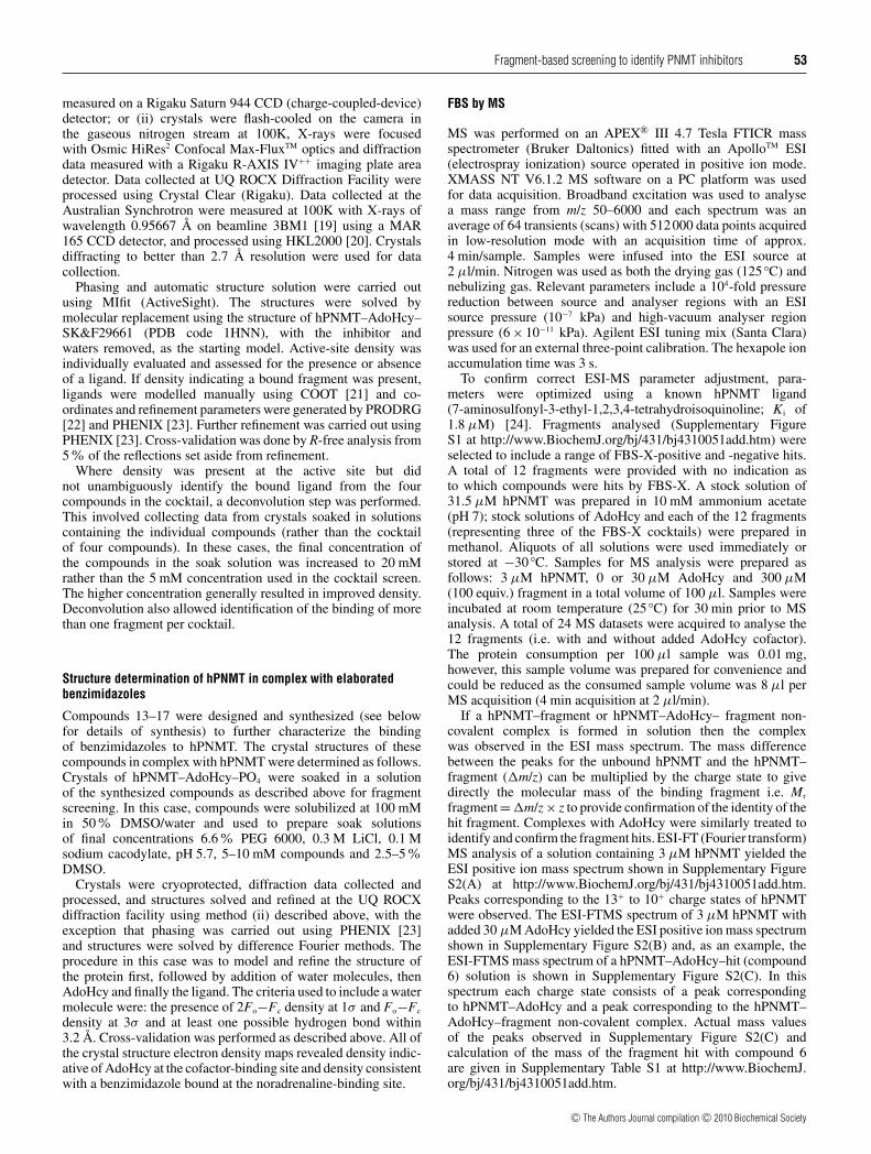
For the FBS-X experiments, we used crystals of hPNMT grownin the absence of inhibitor or ligand for the noradrenaline-binding site. The crystal structure resulting from such crystalsreveals a phosphate molecule bound in the noradrenaline-bindingsite (Figure 2); the phosphate apparently co-purifies with theenzyme during an affinity purification step. The phosphate formsfavourable interactions with the side chains of Lys57 and Asn39.For a hit to be detected in the FBS-X experiments, one ormore of the four fragments of the cocktail must displace the
c© The Authors Journal compilation c© 2010 Biochemical Society
Fragment-based screening to identify PNMT inhibitors 55
Figure 2 hPNMT noradrenaline-binding site
The hPNMT target binding site for inhibitors is shown for the enzyme crystallized in the absenceof inhibitor/substrate. This crystal form was used for FBS-X screening. The phosphate moleculein the noradrenaline site must be displaced by a soaked fragment for a hit to be detected.
Table 1 Statistics for FBS-X screening on hPNMT
The hit rate is calculated on the basis of 12 hits from 376 screened compounds.
Parameter Results
Number of compounds in library/number screened 384/376Number of datasets collected in first round screen 94Number of datasets collected in deconvolution steps 51Average dataset resolution 2.5 AAverage Rwork/R free 0.232/0.298Number of hits 12Hit rate 3.2%Average resolution of hit structures 2.4 AAverage Rwork/R free of hit structures 0.255/0.281
phosphate during the 15 min soaking step. Data collection andrefinement statistics for a representative crystal structure ofhPNMT–AdoHcy–PO4 are included in Supplementary Table S2at http://www.BiochemJ.org/bj/431/bj4310051add.htm.
Of the 96 cocktails containing four compounds (384compounds in total) in the library, X-ray diffraction data werecollected for 94 cocktails (Table 1). Two of the 96 cocktails causedcrystal cracking during the soak, preventing high-resolutiondiffraction data measurement. Owing to the high-throughputnature of the screen, these two cocktails were not consideredfurther. Evaluation of the eight compounds in these two cocktailswill be investigated in another study. A total of 16 of the 94cocktail soaks gave electron density suggestive of hits, i.e. bindingof a chemical to the noradrenaline-binding site. The chemicalidentity of two of the hits was obvious from the initial X-raydata. An additional 51 datasets were measured to deconvolutehits where it was not clear which compound of the cocktailhad bound or to measure higher resolution data for confirmedhits. During this process, four of the initially identified hitswere found to be false positives. In summary, a total of 12compounds (out of a possible 376) were identified as bindingin the target site of hPNMT, giving a hit rate of 3.2% for theFBS-X process. The 2Fo−Fc maps showing the ligand densityfor these 12 hits are provided as Supplementary Figure S3 athttp://www.BiochemJ.org/bj/431/bj4310051add.htm. Summarystatistics for the X-ray data are given in Table 1 and data collectionand refinement statistics for the 12 hit crystal structures are
included as Supplementary Table S2. The structures of the 12hits and their binding mode to hPNMT are shown in Figure 3.
Analysis of fragment hits
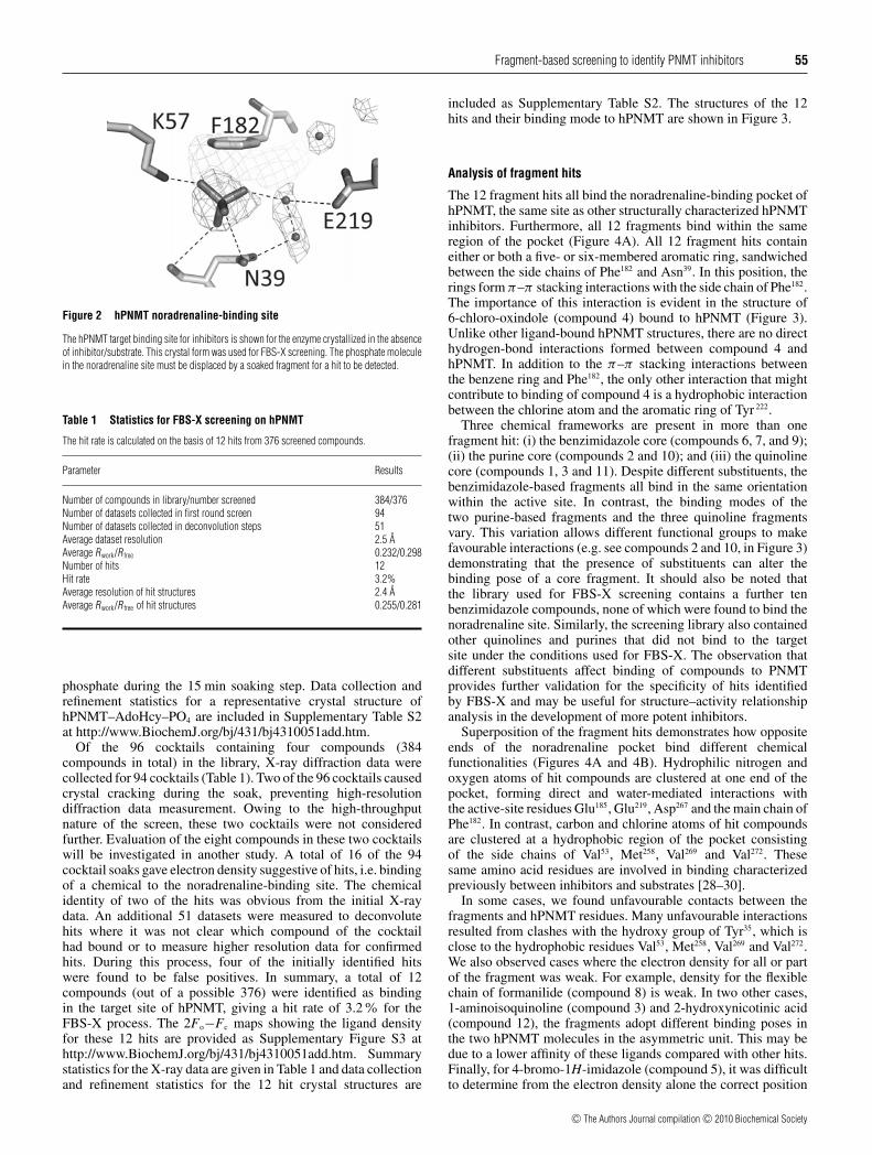
The 12 fragment hits all bind the noradrenaline-binding pocket ofhPNMT, the same site as other structurally characterized hPNMTinhibitors. Furthermore, all 12 fragments bind within the sameregion of the pocket (Figure 4A). All 12 fragment hits containeither or both a five- or six-membered aromatic ring, sandwichedbetween the side chains of Phe182 and Asn39. In this position, therings form π–π stacking interactions with the side chain of Phe182.The importance of this interaction is evident in the structure of6-chloro-oxindole (compound 4) bound to hPNMT (Figure 3).Unlike other ligand-bound hPNMT structures, there are no directhydrogen-bond interactions formed between compound 4 andhPNMT. In addition to the π–π stacking interactions betweenthe benzene ring and Phe182, the only other interaction that mightcontribute to binding of compound 4 is a hydrophobic interactionbetween the chlorine atom and the aromatic ring of Tyr 222.
Three chemical frameworks are present in more than onefragment hit: (i) the benzimidazole core (compounds 6, 7, and 9);(ii) the purine core (compounds 2 and 10); and (iii) the quinolinecore (compounds 1, 3 and 11). Despite different substituents, thebenzimidazole-based fragments all bind in the same orientationwithin the active site. In contrast, the binding modes of thetwo purine-based fragments and the three quinoline fragmentsvary. This variation allows different functional groups to makefavourable interactions (e.g. see compounds 2 and 10, in Figure 3)demonstrating that the presence of substituents can alter thebinding pose of a core fragment. It should also be noted thatthe library used for FBS-X screening contains a further tenbenzimidazole compounds, none of which were found to bind thenoradrenaline site. Similarly, the screening library also containedother quinolines and purines that did not bind to the targetsite under the conditions used for FBS-X. The observation thatdifferent substituents affect binding of compounds to PNMTprovides further validation for the specificity of hits identifiedby FBS-X and may be useful for structure–activity relationshipanalysis in the development of more potent inhibitors.
Superposition of the fragment hits demonstrates how oppositeends of the noradrenaline pocket bind different chemicalfunctionalities (Figures 4A and 4B). Hydrophilic nitrogen andoxygen atoms of hit compounds are clustered at one end of thepocket, forming direct and water-mediated interactions withthe active-site residues Glu185, Glu219, Asp267 and the main chain ofPhe182. In contrast, carbon and chlorine atoms of hit compoundsare clustered at a hydrophobic region of the pocket consistingof the side chains of Val53, Met258, Val269 and Val272. Thesesame amino acid residues are involved in binding characterizedpreviously between inhibitors and substrates [28–30].
In some cases, we found unfavourable contacts between thefragments and hPNMT residues. Many unfavourable interactionsresulted from clashes with the hydroxy group of Tyr35, which isclose to the hydrophobic residues Val53, Met258, Val269 and Val272.We also observed cases where the electron density for all or partof the fragment was weak. For example, density for the flexiblechain of formanilide (compound 8) is weak. In two other cases,1-aminoisoquinoline (compound 3) and 2-hydroxynicotinic acid(compound 12), the fragments adopt different binding poses inthe two hPNMT molecules in the asymmetric unit. This may bedue to a lower affinity of these ligands compared with other hits.Finally, for 4-bromo-1H-imidazole (compound 5), it was difficultto determine from the electron density alone the correct position
c© The Authors Journal compilation c© 2010 Biochemical Society
56 N. Drinkwater and others
Figure 3 Summary of the results of FBS-X targeting hPNMT
The 12 fragment hits were identified, and the chemical structures and binding poses of each are shown. Hydrogen bond interactions are shown as black and hydrophobic interactions are shown asorange broken lines. Binding affinities determined by ITC (K d in μM +− S.D.) and calculated LE values (in kcal/mol per HA) are given for each hit.
of the ring nitrogen atoms because the compound fits the densityequally well in two orientations, rotated 180◦ around the longaxis. Compound 5 was modelled in one orientation on the basisof predicted favourable interactions (see Figure 3).
Water molecules in the active site are important becausethey can indicate potential areas for favourable elaboration offragments. We found that many water molecules are conservedacross the 12 enzyme–fragment structures (Figure 4C). Thereare five major water-binding sites, four of which are located inthe hydrophilic region of the binding pocket described above.These four sites, show the greatest level of conservation acrossthe 12 structures, and mediate interactions with Glu185, Glu219 andAsp267. The presence of defined water-binding pockets suggests
that these regions favour ligand interactions and would probablybe amenable to interactions with elaborated inhibitors.
FBS by MS
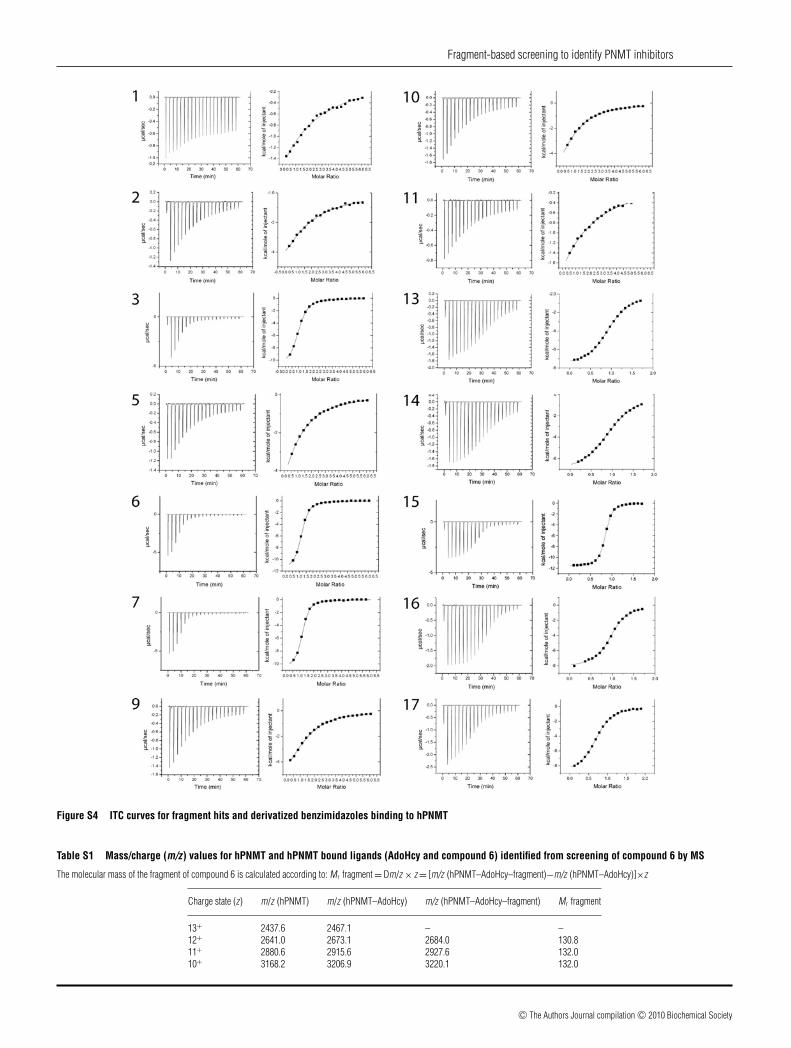
MS has high sensitivity and a wide dynamic range and can beused to detect non-covalent complexes with a Kd in the rangefrom 10 nM to 1 mM [9]. We were interested in using MS asa first-pass FBS method, so we performed a pilot study onhPNMT to ascertain whether MS could identify the same hitsas FBS-X. For proof-of-concept, an ESI-FTMS analysis wasconducted on a subset of 12 of the 384 fragments from the library.
c© The Authors Journal compilation c© 2010 Biochemical Society

Fragment-based screening to identify PNMT inhibitors 57
Figure 4 Solvent accessible surface of the hPNMT active site showing theoverlaid structures of the 12 FBS-X hits
(A and B) Binding positions of the FBS-X hits in the hPNMT target site, shown in two differentorientations. The 12 fragment hits are shown superimposed in space-filling representation(oxygen atoms in red, nitrogen atoms in blue, carbon atoms in white and chlorine atomsin green). The positions of residues discussed in the text are indicated, although for claritythe structures of these residues are not shown. (C) Water molecules are bound in five majorsub-sites represented by green, blue, purple, orange or red spheres. Fragment hits are shownin transparent space filling representation. This panel is shown in the same orientation as (B).
The 12 fragments were intentionally chosen to include severalFBS-X hits. ESI-FTMS screening was conducted blind in that theresearcher collecting and analysing the data did not know whichof the 12 compounds were FBS-X hits. Of the 12 fragments tested,FBS-X identified four fragments as hits (compounds 1, 6, 11 and12; Figure 3), three of which were detected by ESI-FTMS onfirst inspection of the data (compounds 1, 6 and 12). The fourthFBS-X hit, compound 11, was detected upon re-evaluation of theESI-FTMS spectra, although it was identified as a weak complex.In addition, ESI-FTMS identified two other hits (SupplementaryFigure S3); these two fragments were not identified by FBS-X.
ITC evaluation of fragment hits
To confirm and characterize further the 14 fragment hits (12identified by FBS-X and the two identified by ESI-FTMS),and to prioritize the fragment hits for follow up chemistryand optimization, ITC was used to determine their dissociation
constants (Kd) for hPNMT, and thermodynamic parametersof binding. All of the nine fragment hits detected by ITCdemonstrated favourable enthalpic contributions (Figure 3 andTable 2). Of the 12 FBS-X hits, the measured binding affinitiesfor nine fragments ranged from approx. 5 to 700 μM. Of these,two fragments (compounds 1 and 11) show low-affinity binding(below 200 μM) that results in sub-optimal Wiseman c-values(<1) for accurate affinity determination by ITC and this isreflected in the relatively large errors [31]. Nonetheless thedata do allow prioritization of these compounds for follow-up. Binding of the remaining three FBS-X hits (compounds 4,8 and 12) could not be detected by ITC. The two additionalhits identified by ESI-FTMS were also not detected by ITC.Furthermore, whereas the binding of 4-quinolinol (compound1) was detected in ITC experiments, and a Kd of 0.7 mMwas calculated from the ITC measurements, the signal wasweak. The two tightest binding fragments with Kd valuesof approx. 5 μM (2-aminobenzimidazole, compound 6, and2-amino-1-methybenzimidazole, compound 7), are both basedon the benzimidazole core. As discussed above, benzimidazoleshave been shown previously to inhibit PNMT [32]. The thirdfragment hit with a benzimidazole core (5-chlorobenzimidazole,compound 9), had a 20-fold lower binding affinity (Kd of 97 μM),suggesting that direct and water-mediated hydrogen bonds to the2-amino groups of compounds 6 and 7 contribute considerablyto ligand affinity for hPNMT. The dissociation constants wereused to calculate their LE values (Figure 3 and Table 2). The LEvalues ranged from 0.39 kcal/mol per HA for 4-quinolinol to0.86 kcal/mol per HA for 4-bromo-1H-imidazole (compound5). The two tightest binding benzimidazole-based fragments,compounds 6 and 7, had high LEs of ∼0.7 kcal/mol per HA.
Elaborated benzimidazole compounds
Three of the identified hits (compounds 6, 7 and 9) contain abenzimidazole ring system. The electron density for these threehits was excellent, and ITC measurements confirmed that allthree bind to hPNMT, two of them with very high LE values.Benzimidazoles have been investigated previously as inhibitorsof PNMT, although the earlier work was performed on thebovine enzyme [32]. To further characterize the interaction ofbenzimidazole-based fragments with the human enzyme, a seriesof compounds (compounds 13–17; Figure 5) closely related tothe FBS-X benzimidazole hits were synthesized. The 2-aminogroup was retained in this series because in compounds 6 and7 it forms direct and water-mediated hydrogen bonds to theenzyme. The binding of compounds 13–17 to hPNMT wasevaluated by ITC and X-ray crystallography. The results of theseanalyses are shown in Figure 5 and data collection and refinementstatistics for the structures are included as SupplementaryTable S2. ITC curves are shown in Supplementary Figure S4at http://www.BiochemJ.org/bj/431/bj4310051add.htm, and theITC-derived thermodynamic parameters of the 12 fragment hitsand five elaborated compounds are given in Table 2.
Hydroxy-substituted 2-aminobenzimidazole compounds(compounds 13 and 14)
Superposition of the benzimidazole-based fragments with thefragment hit 4-quinolinol (compound 1), suggested that ahydroxy group added to the 7- or 6- position of compound 6would interact with Lys57 and potentially with Tyr40. To testthis, compounds 13 and 14 were synthesized and their bindingcharacterized (Figure 5 and Table 2). The crystal structures of
c© The Authors Journal compilation c© 2010 Biochemical Society
58 N. Drinkwater and others
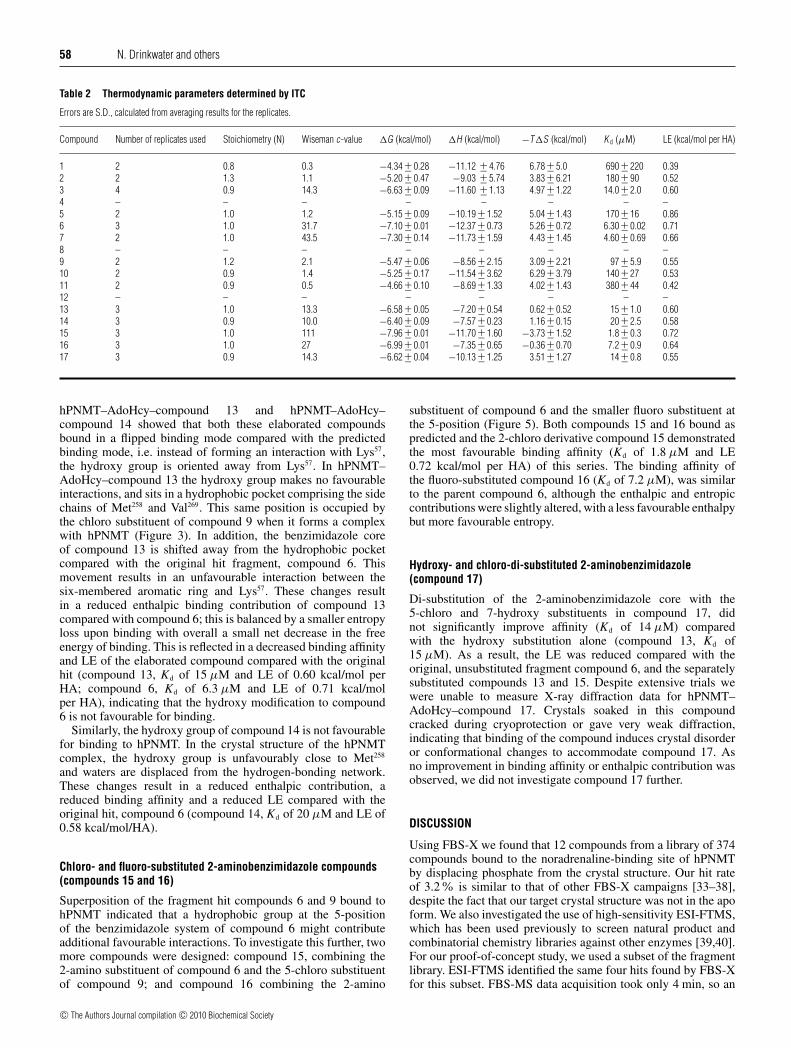
Table 2 Thermodynamic parameters determined by ITC
Errors are S.D., calculated from averaging results for the replicates.
Compound Number of replicates used Stoichiometry (N) Wiseman c-value �G (kcal/mol) �H (kcal/mol) −T�S (kcal/mol) K d (μM) LE (kcal/mol per HA)
1 2 0.8 0.3 −4.34 +− 0.28 −11.12 +− 4.76 6.78 +− 5.0 690 +− 220 0.392 2 1.3 1.1 −5.20 +− 0.47 −9.03 +− 5.74 3.83 +− 6.21 180 +− 90 0.523 4 0.9 14.3 −6.63 +− 0.09 −11.60 +− 1.13 4.97 +− 1.22 14.0 +− 2.0 0.604 – – – – – – – –5 2 1.0 1.2 −5.15 +− 0.09 −10.19 +− 1.52 5.04 +− 1.43 170 +− 16 0.866 3 1.0 31.7 −7.10 +− 0.01 −12.37 +− 0.73 5.26 +− 0.72 6.30 +− 0.02 0.717 2 1.0 43.5 −7.30 +− 0.14 −11.73 +− 1.59 4.43 +− 1.45 4.60 +− 0.69 0.668 – – – – – – – –9 2 1.2 2.1 −5.47 +− 0.06 −8.56 +− 2.15 3.09 +− 2.21 97 +− 5.9 0.5510 2 0.9 1.4 −5.25 +− 0.17 −11.54 +− 3.62 6.29 +− 3.79 140 +− 27 0.5311 2 0.9 0.5 −4.66 +− 0.10 −8.69 +− 1.33 4.02 +− 1.43 380 +− 44 0.4212 – – – – – – – –13 3 1.0 13.3 −6.58 +− 0.05 −7.20 +− 0.54 0.62 +− 0.52 15 +− 1.0 0.6014 3 0.9 10.0 −6.40 +− 0.09 −7.57 +− 0.23 1.16 +− 0.15 20 +− 2.5 0.5815 3 1.0 111 −7.96 +− 0.01 −11.70 +− 1.60 −3.73 +− 1.52 1.8 +− 0.3 0.7216 3 1.0 27 −6.99 +− 0.01 −7.35 +− 0.65 −0.36 +− 0.70 7.2 +− 0.9 0.6417 3 0.9 14.3 −6.62 +− 0.04 −10.13 +− 1.25 3.51 +− 1.27 14 +− 0.8 0.55
hPNMT–AdoHcy–compound 13 and hPNMT–AdoHcy–compound 14 showed that both these elaborated compoundsbound in a flipped binding mode compared with the predictedbinding mode, i.e. instead of forming an interaction with Lys57,the hydroxy group is oriented away from Lys57. In hPNMT–AdoHcy–compound 13 the hydroxy group makes no favourableinteractions, and sits in a hydrophobic pocket comprising the sidechains of Met258 and Val269. This same position is occupied bythe chloro substituent of compound 9 when it forms a complexwith hPNMT (Figure 3). In addition, the benzimidazole coreof compound 13 is shifted away from the hydrophobic pocketcompared with the original hit fragment, compound 6. Thismovement results in an unfavourable interaction between thesix-membered aromatic ring and Lys57. These changes resultin a reduced enthalpic binding contribution of compound 13compared with compound 6; this is balanced by a smaller entropyloss upon binding with overall a small net decrease in the freeenergy of binding. This is reflected in a decreased binding affinityand LE of the elaborated compound compared with the originalhit (compound 13, Kd of 15 μM and LE of 0.60 kcal/mol perHA; compound 6, Kd of 6.3 μM and LE of 0.71 kcal/molper HA), indicating that the hydroxy modification to compound6 is not favourable for binding.
Similarly, the hydroxy group of compound 14 is not favourablefor binding to hPNMT. In the crystal structure of the hPNMTcomplex, the hydroxy group is unfavourably close to Met258
and waters are displaced from the hydrogen-bonding network.These changes result in a reduced enthalpic contribution, areduced binding affinity and a reduced LE compared with theoriginal hit, compound 6 (compound 14, Kd of 20 μM and LE of0.58 kcal/mol/HA).
Chloro- and fluoro-substituted 2-aminobenzimidazole compounds(compounds 15 and 16)
Superposition of the fragment hit compounds 6 and 9 bound tohPNMT indicated that a hydrophobic group at the 5-positionof the benzimidazole system of compound 6 might contributeadditional favourable interactions. To investigate this further, twomore compounds were designed: compound 15, combining the2-amino substituent of compound 6 and the 5-chloro substituentof compound 9; and compound 16 combining the 2-amino
substituent of compound 6 and the smaller fluoro substituent atthe 5-position (Figure 5). Both compounds 15 and 16 bound aspredicted and the 2-chloro derivative compound 15 demonstratedthe most favourable binding affinity (Kd of 1.8 μM and LE0.72 kcal/mol per HA) of this series. The binding affinity ofthe fluoro-substituted compound 16 (Kd of 7.2 μM), was similarto the parent compound 6, although the enthalpic and entropiccontributions were slightly altered, with a less favourable enthalpybut more favourable entropy.
Hydroxy- and chloro-di-substituted 2-aminobenzimidazole(compound 17)
Di-substitution of the 2-aminobenzimidazole core with the5-chloro and 7-hydroxy substituents in compound 17, didnot significantly improve affinity (Kd of 14 μM) comparedwith the hydroxy substitution alone (compound 13, Kd of15 μM). As a result, the LE was reduced compared with theoriginal, unsubstituted fragment compound 6, and the separatelysubstituted compounds 13 and 15. Despite extensive trials wewere unable to measure X-ray diffraction data for hPNMT–AdoHcy–compound 17. Crystals soaked in this compoundcracked during cryoprotection or gave very weak diffraction,indicating that binding of the compound induces crystal disorderor conformational changes to accommodate compound 17. Asno improvement in binding affinity or enthalpic contribution wasobserved, we did not investigate compound 17 further.
DISCUSSION
Using FBS-X we found that 12 compounds from a library of 374compounds bound to the noradrenaline-binding site of hPNMTby displacing phosphate from the crystal structure. Our hit rateof 3.2% is similar to that of other FBS-X campaigns [33–38],despite the fact that our target crystal structure was not in the apoform. We also investigated the use of high-sensitivity ESI-FTMS,which has been used previously to screen natural product andcombinatorial chemistry libraries against other enzymes [39,40].For our proof-of-concept study, we used a subset of the fragmentlibrary. ESI-FTMS identified the same four hits found by FBS-Xfor this subset. FBS-MS data acquisition took only 4 min, so an
c© The Authors Journal compilation c© 2010 Biochemical Society

Fragment-based screening to identify PNMT inhibitors 59
Figure 5 Enzyme–ligand structures and binding information of elaboratedbenzimidazoles
Chemical structures and binding poses for the elaborated benzimidazole compounds are given(the binding pose for compound 17 is not provided because crystallographic data could not bemeasured for that complex with hPNMT). Hydrogen bonding interactions are shown in black,hydrophobic interactions in orange and unfavourable interactions in cyan broken lines. Bindingaffinities determined by ITC (K d in μM +− S.D.) and calculated LE values (in kcal/mol per HA)are also indicated.
automated approach could process a 384 compound libraryin just over 24 h using 0.4 mg of protein. By comparison, oursuccessful FBS-X campaign took 2–3 months and used ∼20 mgof protein. These results suggest that ESI-FTMS could be used asa filter to generate a short-list for subsequent characterization by
X-ray crystallography. This could in theory reduce the numberof crystal structures to be measured from ∼150 to ∼20, therebysaving time, human resources and protein, and making thetechnique accessible to many more academic laboratories.
Using ITC we determined the dissociation constants of nine ofthe 12 FBS-X fragment hits. The results showed that the fragmentsbind with affinities in the high to low micromolar range and allnine hits for which affinity data were measured had LE values� 0.4 kcal/mol per HA. Generally, LE values above 0.4 kcal/molper HA are desirable through the process of optimization [38,41].Many of our hits had considerably higher LE values, emphasizingtheir high quality.
Several of the FBS-X hits are benzimidazoles, a knownclass of PNMT inhibitor. In 1970, researchers at the MSD(Merck Sharp and Dohme Research Laboratories) showed thatbenzimidazoles (including 2-aminobenzimidazole, compound 6,and 5-chlorobenzimidazole, compound 9) inhibit bovine PNMT.Thus at 28 μg/ml compound 6 inhibits binding of 0.57 mM DL-normetanephrine to bovine PNMT by 30% and compound 9inhibits binding by 57% [32]. Our FBS-X results identified thatthese compounds also interact with human PNMT and show forthe first time how they bind in the noradrenaline pocket. The MSDstudy also showed that unsubstituted benzimidazole is an inhibitorof bovine PNMT (28% inhibition under the conditions describedabove) [32]. Benzimidazole is one of the 376 compounds in thelibrary used in the present study, but was not identified as a hitby FBS-X. This finding could be explained by one or more of thefollowing: (i) benzimidazole does not inhibit PNMT by bindingthe noradrenaline-binding site; (ii) the noradrenaline-binding siteof the human and bovine enzymes differ sufficiently that theyrecognize benzimidazole differently; or (iii) the screening methodwe employed is less sensitive than the assay used to detectinhibition in the bovine enzyme.
Several PNMT inhibitors have been shown to be competitive fornoradrenaline binding [42–44], although it has been shown thata dichloro-substituted benzimidazole PNMT inhibitor was non-competitive with noradrenaline and uncompetitive with AdoMet[45]. These early results indicate the possibility of an additionalbinding site for benzimidazoles. Our data clearly show that thethree benzimidazole hits and the elaborated compounds bind atthe noradrenaline site. However, the hPNMT crystals used for ourstudies have AdoHcy (as a cofactor product) bound at the AdoMetsite. Furthermore, ITC analysis was performed in the presence ofexcess AdoHcy. Consequently, if the benzimidazole compoundsbind to the hPNMT AdoMet site this would not be identified underthe conditions of our experiments.
The improved binding affinity of the designed benzimidazolecompound 15 compared with the original compound 6, islargely due to an increased entropic contribution to binding(Table 2). Enthalpic optimization is more desirable than entropicopimization in progressing from hit to lead [46]. Typically,enthalpically optimized leads more closely mimic the thermo-dynamic characteristics of natural protein–ligand interactions, aremore soluble and often represent best-in-class compounds in drugdiscovery programmes [46,47]. Our results therefore indicate thatalthough compound 15 shows higher affinity than compound 6 thisimprovement may not necessarily correlate with an improved druglead.
In summary, we have shown that FBS-X can be implementedsuccessfully within an academic environment on a non-idealprotein target. Using FBS-X we identified 12 hits that bindhPNMT at the noradrenaline site. Our pilot study suggests thescreening process could be streamlined further by performingFBS-MS first. Three of the FBS-X hits are from a chemicalclass of known PNMT inhibitors that provide an unanticipated
c© The Authors Journal compilation c© 2010 Biochemical Society

60 N. Drinkwater and others
internal control for the screen. Overall, we structurally andthermodynamically characterized the binding of 17 small mol-ecules to hPNMT, including the first such characterization of thebenzimidazole class of compounds bound to the noradrenaline-binding site.
AUTHOR CONTRIBUTION
Nyssa Drinkwater and Jennifer Martin conceived the idea. Nyssa Drinkwater performed themajority of the research and Jennifer Martin co-ordinated the study. Hoan Vu and Sally-Ann Poulsen designed and performed the MS experiments. Kimberly Lovell, ThomasPrisinzano, Kevin Criscione and Gary Grunewald designed and supervized compoundsynthesis and contributed to compound characterization. Michael McLeish provided theplasmid for the enzyme and contributed to data interpretation. Brett Collins assisted inmeasurement and analysis of ITC data. Nyssa Drinkwater prepared the first draft of thepaper. All authors contributed to discussions on the study and provided intellectual inputto the manuscript.
ACKNOWLEDGEMENTS
We are grateful for access to the UQ ROCX Diffraction Facility and thank Karl Byriel andGordon King for their assistance. We thank staff at the MX1 beamline of the AustralianSynchrotron for their invaluable advice. The MS data could not have been measuredwithout access to the Eskitis Institute FTMS facility. We thank Dr F. Anthony Romero forhelpful preliminary discussions and Duncan McRee for advice and information regardingthe ActiveSight library.
FUNDING
This work was supported by an University of Queensland Graduate School ResearchTravel Grant and an Australian Postgraduate Award (to N.D.); an Australian ResearchCouncil Discovery Project award [grant number DP0664564 (to J.L.M.)]; an AustralianNational Health and Medical Research Council Biomedical RD Wright Career DevelopmentAward (to B.M.C.) and Senior Research Fellowship [grant number 455829 (to J.L.M.)]; anAustralian Research Council Linkage Equipment and Infrastructure Award [grant numbersLE0668382 (to J.L.M.), LE237908 (to S.-A.P.)]; and by the National Institutes of Health[grant number NIH HL034193 (to G.L.G.)].
REFERENCES
1 Hajduk, P. J. and Greer, J. (2007) A decade of fragment-based drug design: strategicadvances and lessons learned. Nat. Rev. Drug Discov. 6, 211–219
2 Murray, C. W. and Rees, D. C. (2009) The rise of fragment-based drug discovery. Nat.Chem. 1, 187–192
3 Congreve, M., Carr, R., Murray, C. and Jhoti, H. (2003) A rule of three for fragment-basedlead discovery? Drug Discov. Today 8, 876–877
4 Rees, D. C., Congreve, M., Murray, C. W. and Carr, R. (2004) Fragment-based leaddiscovery. Nat. Rev. Drug Discov. 3, 660–672
5 Erlanson, D. A. (2006) Fragment-based lead discovery: a chemical update. Curr. Opin.Biotech. 17, 643–652
6 Neumann, T., Junker, H. D., Schmidt, K. and Sekul, R. (2007) SPR-based fragmentscreening: advantages and applications. Curr. Top. Med. Chem. 7, 1630–1642
7 Ciulli, A., Williams, G., Smith, A. G., Blundell, T. L. and Abell, C. (2006) Probing hotspots at protein-ligand-binding sites: a fragment-based approach using biophysicalmethods. J. Med. Chem. 49, 4992–5000
8 Orita, M., Warizaya, M., Amano, Y., Ohno, K. and Niimi, T. (2009) Advances infragment-based drug discovery platforms. Exp. Opin. Drug Discov. 4, 1125–1144
9 Hofstadler, S. A. and Sannes-Lowery, K. A. (2006) Applications of ESI-MS in drugdiscovery: interrogation of noncovalent complexes. Nat. Rev. Drug Discov. 5, 585–595
10 Schulz, M. N. and Hubbard, R. E. (2009) Recent progress in fragment-based leaddiscovery. Curr. Opin. Pharmacol. 9, 615–621
11 Fuller, R. W. (1982) Pharmacology of brain epinephrine neurons. Ann. Rev. Pharmacol.Toxicol. 22, 31–55
12 Crowley, W. R., Terry, L. C. and Johnson, M. D. (1982) Evidence for the involvement ofcentral epinephrine systems in the regulation of luteinizing-hormone, prolactin, andgrowth-hormone release in female rats. Endocrinology 110, 1102–1107
13 Mefford, I. N., Lister, R. G., Ota, M. and Linnoila, M. (1990) Antagonism of ethanolintoxication in rats by inhibitors of phenylethanolamine N-methyltransferase. AlcoholClin. Exper. Res. 14, 53–57
14 Gearhart, D. A., Neafsey, E. J. and Collins, M. A. (2002) PhenylethanolamineN-methyltransferase has β-carboline 2N-methyltransferase activity: hypotheticalrelevance to Parkinson’s disease. Neurochem. Int. 40, 611–620
15 Kennedy, B. P., Bottiglieri, T., Arning, E., Ziegler, M. G., Hansen, L. A. and Masliah, E.(2004) Elevated S-adenosylhomocysteine in Alzheimer brain: influence onmethyltransferases and cognitive function. J. Neural Transm. 111, 547–567
16 Martin, J. L., Begun, J., McLeish, M. J., Caine, J. M. and Grunewald, G. L. (2001) Gettingthe adrenaline going: crystal structure of the adrenaline-synthesizing enzyme PNMT.Structure 9, 977–985
17 Drinkwater, N., Gee, C. L., Puri, M., Criscione, K. R., McLeish, M. J., Grunewald, G. L.and Martin, J. L. (2009) Molecular recognition of physiological substrate noradrenalineby the adrenaline-synthesizing enzyme PNMT and factors influencing itsmethyltransferase activity. Biochem. J. 422, 463–471
18 Gee, C. L., Tyndall, J. D., Grunewald, G. L., Wu, Q., McLeish, M. J. and Martin, J. L.(2005) Mode of binding of methyl acceptor substrates to the adrenaline-synthesizingenzyme phenylethanolamine N-methyltransferase: implications for catalysis.Biochemistry 44, 16875–16885
19 McPhillips, T. M., McPhillips, S. E., Chiu, H. J., Cohen, A. E., Deacon, A. M., Ellis, P. J.,Garman, E., Gonzalez, A., Sauter, N. K., Phizackerley, R. P. et al. (2002) Blu-Ice and thedistributed control system: software for data acquisition and instrument control atmacromolecular crystallography beamlines. J. Synchrotron Radiat. 9, 401–406
20 Otwinowski, Z. and Minor, W. (1997) Processing of X-ray diffraction data collected inoscillation mode. Methods Enzymol. 276, p.307–326
21 Emsley, P. and Cowtan, K. (2004) COOT: model-building tools for molecular graphics.Acta Crystallogr. Sect. D Biol. Crystallogr. 60, 2126–2132
22 Schuettelkopf, A. W. and van Aalten, D. M. F. (2004) PRODRG – a tool for high-throughput crystallography of protein–ligand complexes. Acta Crystallogr. Sect. D Biol.Crystallogr. 60, 1355–1363
23 Adams, P. D., Afonine, P. V., Bunkoczi, G., Chen, V. B., Davis, I. W., Echols, N., Headd,J. J., Hung, L. W., Kapral, G. J., Grosse-Kunstleve, R. W. et al. (2010) PHENIX: acomprehensive Python-based system for macromolecular structure solution. ActaCrystallogr. Sect. D Biol. Crystallogr. 66, 213–221
24 Grunewald, G. L., Romero, F. A., Chieu, A. D., Fincham, K. J., Bhat, S. R. and Criscione,K. R. (2005) Exploring the active site of phenylethanolamine N-methyltransferase:3-alkyl-7-substituted-1,2,3,4-tetrahydroisoquinoline inhibitors. Bioorg. Med. Chem. 13,1261–1273
25 Morningstar, M. L., Roth, T., Farnsworth, D. W., Smith, M. K., Watson, K., Buckheit, R. W.,Das, K., Zhang, W. Y., Arnold, E., Julias, J. G. et al. (2007) Synthesis, biological activity,and crystal structure of potent nonnucleoside inhibitors of HIV-1 reverse transcriptasethat retain activity against mutant forms of the enzyme. J. Med. Chem. 50, 4003–4015
26 Galan, A. A., Chen, J., Du, H., Forsyth, T., Huynh, T. P., Johnson, H. W. B., Kearney, P.,Leahy, J. W., Lee, M. S., Mann, G. et al. (2008) Preparation of 1H-imidazole-4,5-dicarboxamides as JAK-2 modulators. European Patent Foundation no.WO2008042282-A2
27 Parmee, E. R., Kim, R. M., Rouse, E. A., Schmidt, D. R., Sinz, C. J. and Chang, J. (2005)Preparation of cyclic guanidines as glucagon receptor antagonists for the treatment oftype 2 diabetes. PCT Int. Appl., no. WO 2005065680-A1 20050721
28 Gee, C. L., Drinkwater, N., Tyndall, J. D. A., Grunewald, G. L., Wu, Q., McLeish, M. J. andMartin, J. L. (2007) Enzyme adaptation to inhibitor binding: a cryptic binding site inphenylethanolamine N-methyltransferase. J. Med. Chem. 50, 4845–4853
29 McMillan, F. M., Archbold, J., McLeish, M. J., Caine, J. M., Criscione, K. R., Grunewald,G. L. and Martin, J. L. (2004) Molecular recognition of sub-micromolar inhibitors by theepinephrine-synthesizing enzyme phenylethanolamine N-methyltransferase. J. Med.Chem. 47, 37–44
30 Wu, Q., Gee, C. L., Lin, F., Tyndall, J. D., Martin, J. L., Grunewald, G. L. and McLeish,M. J. (2005) Structural, mutagenic, and kinetic analysis of the binding of substrates andinhibitors of human phenylethanolamine N-methyltransferase. J. Med. Chem. 48,7243–7252
31 Turnbull, W. B. and Daranas, A. H. (2003) On the value of c: can low affinity systems bestudied by isothermal titration calorimetry? J. Am. Chem. Soc. 125, 14859–14866
32 Mandel, L. R., Porter, C. C., Kuehl, F. A., Jensen, N. P., Schmitt, S. M., Windholz, T. B.,Beattie, T. R., Carty, J. A., Christen, B. G. and Shen, T. Y. (1970) Inhibition of adrenalphenethanolamine N-methyltransferase by substituted benzimidazoles. J. Med. Chem.13, 1043–1047
33 Antonysamy, S. S., Aubol, B., Blaney, J., Browner, M. F., Giannetti, A. M., Harris, S. F.,Hebert, N., Hendle, J., Hopkins, S., Jefferson, E. et al. (2008) Fragment-based discoveryof hepatitis C virus NS5b RNA polymerase inhibitors. Bioorg. Med. Chem. Lett. 18,2990–2995
34 Bosch, J., Robien, M. A., Mehlin, C., Boni, E., Riechers, A., Buckner, F. S., Van Voorhis,W. C., Myler, P. J., Worthey, E. A., DeTitta, G. et al. (2006) Using fragment cocktailcrystallography to assist inhibitor design of Trypanosoma brucei nucleoside2-deoxyribosyltransferase. J. Med. Chem. 49, 5939–5946
c© The Authors Journal compilation c© 2010 Biochemical Society

Fragment-based screening to identify PNMT inhibitors 61
35 Lesuisse, D., Lange, G., Deprez, P., Benard, D., Schoot, B., Delettre, G., Marquette, J. P.,Broto, P., Jean-Baptiste, V., Bichet, P. et al. (2002) SAR and X-ray. A new approachcombining fragment-based screening and rational drug design: application to thediscovery of nanomolar inhibitors of Src SH2. J. Med. Chem. 45, 2379–2387
36 Nienaber, V. L., Richardson, P. L., Klighofer, V., Bouska, J. J., Giranda, V. L. and Greer, J.(2000) Discovering novel ligands for macromolecules using X-ray crystallographicscreening. Nat. Biotech. 18, 1105–1108
37 Schuffenhauer, A., Ruedisser, S., Marzinzik, A. L., Jahnke, W., Blommers, M., Selzer, P.and Jacoby, E. (2005) Library design for fragment based screening. Curr. Top. Med.Chem. 5, 751–762
38 Wyatt, P. G., Woodhead, A. J., Berdini, V., Boulstridge, J. A., Carr, M. G., Cross, D. M.,Davis, D. J., Devine, L. A., Early, T. R., Feltell, R. E. et al. (2008) Identification ofN-(4-piperidinyl)-4-(2,6-dichlorobenzoylamino)-1H-pyrazole-3-carboxamide (AT7519),a novel cyclin dependent kinase inhibitor using fragment-based X-ray crystallographyand structure based drug design. J. Med. Chem. 51, 4986–4999
39 Poulsen, S. A. (2006) Direct screening of a dynamic combinatorial library using massspectrometry. J. Am. Soc. Mass Spec. 17, 1074–1080
40 Vu, H., Pharm, N. B. and Quinn, R. J. (2008) Direct screening of natural product extractsusing mass spectrometry. J. Biomol. Screen. 13, 265–275
41 Bembenek, S. D., Tounge, B. A. and Reynolds, C. H. (2009) Ligand efficiency andfragment-based drug discovery. Drug Discov. Today 14, 278–283
42 Fuller, R. W., Hemrick-Luecke, S. K. and Perry, K. W. (1983) Inhibition of brainepinephrine synthesis by 3,4-dichlorophenylethanolamine, a competitivesubstrate for norepinephrine N-methyltransferase. Biochem. Pharmacol. 32,215–220
43 Fuller, R. W., Roush, B. W., Snoddy, H. D., Day, W. A. and Molloy, B. B. (1975)Norepinephrine N-methyltransferase inhibition by benzamidines, phenylacetamidines,benzylguanidines, and phenylethylguanidines. J. Med. Chem. 18, 304–307
44 Fuller, R. W., Roush, B. W., Snoddy, H. D. and Molloy, B. B. (1973) Inhibition ofphenylethanolamine N-methyltransferase by benzylamines. 2. In-vivo studies with2,3-dichloro-α-methylbenzylamine. J. Med. Chem. 16, 106–109
45 Pendleton, R. G., Green, H., Snow, I. B., Wang, J. and Kaiser, C. (1972) Studies on themechanism of phenylethanolamine-N-methyl-transferase inhibition by adichloro-substituted benzimidazole. Biochem. Pharmacol. 21, 2967–2975
46 Ladbury, J. E., Klebe, G. and Freire, E. (2010) Adding calorimetric data to decision makingin lead discovery: a hot tip. Nat. Rev. Drug Discov. 9, 23–27
47 Freire, E. (2008) Do enthalpy and entropy distinguish first in class from best in class?Drug Discov. Today 13, 869–874
Received 27 April 2010/29 June 2010; accepted 20 July 2010Published as BJ Immediate Publication 20 July 2010, doi:10.1042/BJ20100651
c© The Authors Journal compilation c© 2010 Biochemical Society
Biochem. J. (2010) 431, 51–61 (Printed in Great Britain) doi:10.1042/BJ20100651
SUPPLEMENTARY ONLINE DATAFragment-based screening by X-ray crystallography, MS and isothermaltitration calorimetry to identify PNMT (phenylethanolamineN-methyltransferase) inhibitorsNyssa DRINKWATER*, Hoan VU†, Kimberly M. LOVELL‡, Kevin R. CRISCIONE‡, Brett M. COLLINS*, Thomas E. PRISINZANO‡,Sally-Ann POULSEN†, Michael J. MCLEISH§, Gary L. GRUNEWALD‡ and Jennifer L. MARTIN*1
*University of Queensland, Institute for Molecular Bioscience, Division of Chemistry and Structural Biology, Brisbane, Queensland 4072, Australia, †Griffith University, Eskitis Institute,Brisbane, Queensland 4111, Australia, ‡Department of Medicinal Chemistry, University of Kansas, Lawrence, KS 66045-7582, U.S.A., and §Department of Chemistry and ChemicalBiology, Indiana University–Purdue University Indianapolis (IUPUI), 402 N. Blackford St, LD 326D, IN 46202, U.S.A.
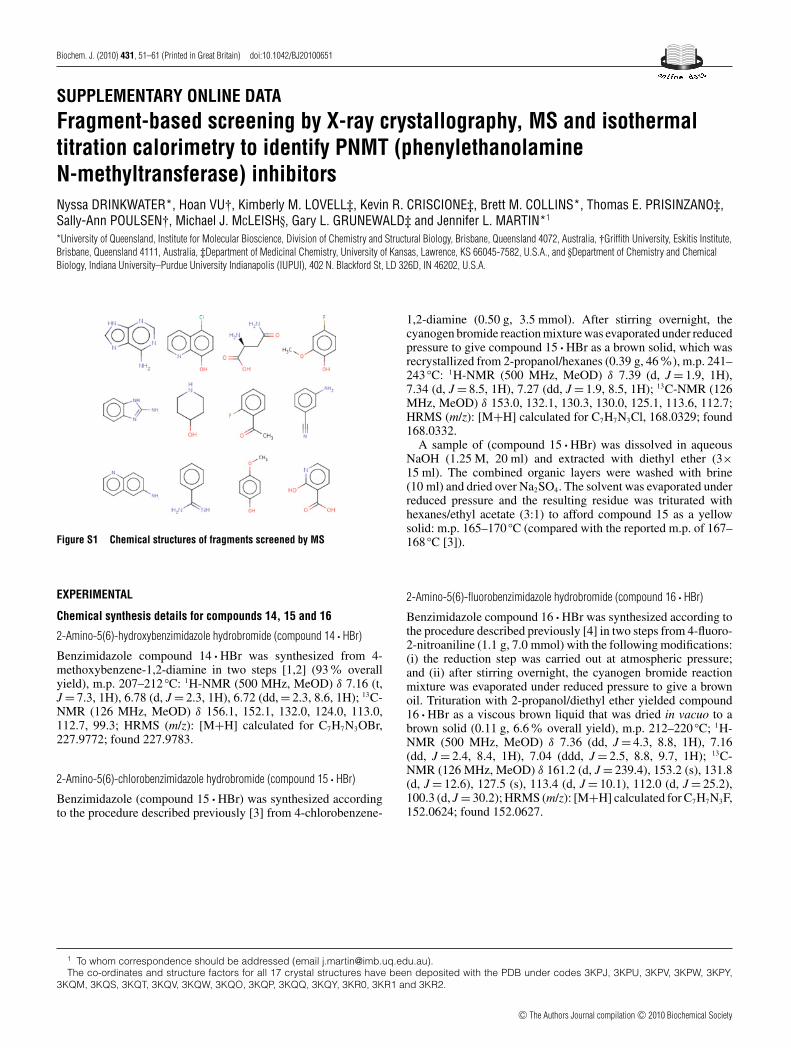
Figure S1 Chemical structures of fragments screened by MS
EXPERIMENTAL
Chemical synthesis details for compounds 14, 15 and 16
2-Amino-5(6)-hydroxybenzimidazole hydrobromide (compound 14 · HBr)
Benzimidazole compound 14 · HBr was synthesized from 4-methoxybenzene-1,2-diamine in two steps [1,2] (93% overallyield), m.p. 207–212 ◦C: 1H-NMR (500 MHz, MeOD) δ 7.16 (t,J = 7.3, 1H), 6.78 (d, J = 2.3, 1H), 6.72 (dd, = 2.3, 8.6, 1H); 13C-NMR (126 MHz, MeOD) δ 156.1, 152.1, 132.0, 124.0, 113.0,112.7, 99.3; HRMS (m/z): [M+H] calculated for C7H7N3OBr,227.9772; found 227.9783.
2-Amino-5(6)-chlorobenzimidazole hydrobromide (compound 15 · HBr)
Benzimidazole (compound 15 · HBr) was synthesized accordingto the procedure described previously [3] from 4-chlorobenzene-
1,2-diamine (0.50 g, 3.5 mmol). After stirring overnight, thecyanogen bromide reaction mixture was evaporated under reducedpressure to give compound 15 · HBr as a brown solid, which wasrecrystallized from 2-propanol/hexanes (0.39 g, 46%), m.p. 241–243 ◦C: 1H-NMR (500 MHz, MeOD) δ 7.39 (d, J = 1.9, 1H),7.34 (d, J = 8.5, 1H), 7.27 (dd, J = 1.9, 8.5, 1H); 13C-NMR (126MHz, MeOD) δ 153.0, 132.1, 130.3, 130.0, 125.1, 113.6, 112.7;HRMS (m/z): [M+H] calculated for C7H7N3Cl, 168.0329; found168.0332.
A sample of (compound 15 · HBr) was dissolved in aqueousNaOH (1.25 M, 20 ml) and extracted with diethyl ether (3×15 ml). The combined organic layers were washed with brine(10 ml) and dried over Na2SO4. The solvent was evaporated underreduced pressure and the resulting residue was triturated withhexanes/ethyl acetate (3:1) to afford compound 15 as a yellowsolid: m.p. 165–170 ◦C (compared with the reported m.p. of 167–168 ◦C [3]).
2-Amino-5(6)-fluorobenzimidazole hydrobromide (compound 16 · HBr)
Benzimidazole compound 16 · HBr was synthesized according tothe procedure described previously [4] in two steps from 4-fluoro-2-nitroaniline (1.1 g, 7.0 mmol) with the following modifications:(i) the reduction step was carried out at atmospheric pressure;and (ii) after stirring overnight, the cyanogen bromide reactionmixture was evaporated under reduced pressure to give a brownoil. Trituration with 2-propanol/diethyl ether yielded compound16 · HBr as a viscous brown liquid that was dried in vacuo to abrown solid (0.11 g, 6.6% overall yield), m.p. 212–220 ◦C; 1H-NMR (500 MHz, MeOD) δ 7.36 (dd, J = 4.3, 8.8, 1H), 7.16(dd, J = 2.4, 8.4, 1H), 7.04 (ddd, J = 2.5, 8.8, 9.7, 1H); 13C-NMR (126 MHz, MeOD) δ 161.2 (d, J = 239.4), 153.2 (s), 131.8(d, J = 12.6), 127.5 (s), 113.4 (d, J = 10.1), 112.0 (d, J = 25.2),100.3 (d, J = 30.2); HRMS (m/z): [M+H] calculated for C7H7N3F,152.0624; found 152.0627.
1 To whom correspondence should be addressed (email [email protected]).The co-ordinates and structure factors for all 17 crystal structures have been deposited with the PDB under codes 3KPJ, 3KPU, 3KPV, 3KPW, 3KPY,
3KQM, 3KQS, 3KQT, 3KQV, 3KQW, 3KQO, 3KQP, 3KQQ, 3KQY, 3KR0, 3KR1 and 3KR2.
c© The Authors Journal compilation c© 2010 Biochemical Society

N. Drinkwater and others
Figure S2 ESI-FTMS screening
(A) ESI-FTMS positive ion mass spectrum of hPNMT (3 μM) in 10 mM ammonium acetatesolution. Peaks corresponding to hPNMT in complex with AdoHcy are also observed indicatinga small amount of AdoHcy co-purifies with the enzyme. (B) ESI-FTMS positive ion massspectrum of a mixture of hPNMT (3 μM) and AdoHcy (30 μM) in 10 mM ammonium acetateand 5 % methanol. (C) ESI-FTMS positive ion mass spectrum of a mixture of hPNMT (3 μM),AdoHcy (30 μM) and compound 6 (300 μM) in 10 mM ammonium acetate and 5 % methanol.(D) Structures of the two additional fragments detected by ESI-FTMS, but not detected by FBS-Xor ITC.
Figure S3 Electron density (2Fo−Fc shown at 1σ ) for ligands bound tohPNMT
(A–P) represent density for compounds 1–17.
c© The Authors Journal compilation c© 2010 Biochemical Society
Fragment-based screening to identify PNMT inhibitors
Figure S4 ITC curves for fragment hits and derivatized benzimidazoles binding to hPNMT
Table S1 Mass/charge (m/z) values for hPNMT and hPNMT bound ligands (AdoHcy and compound 6) identified from screening of compound 6 by MS
The molecular mass of the fragment of compound 6 is calculated according to: Mr fragment = Dm/z × z = [m/z (hPNMT–AdoHcy–fragment)−m/z (hPNMT–AdoHcy)]×z
Charge state (z) m/z (hPNMT) m/z (hPNMT–AdoHcy) m/z (hPNMT–AdoHcy–fragment) Mr fragment
13+ 2437.6 2467.1 – –12+ 2641.0 2673.1 2684.0 130.811+ 2880.6 2915.6 2927.6 132.010+ 3168.2 3206.9 3220.1 132.0
c© The Authors Journal compilation c© 2010 Biochemical Society

N.Drinkwaterandothers
Table S2 Crystallographic data and refinement statistics for structures of fragment hits in complex with hPNMT–AdoHcy
The data for fragment hit compounds 1–5 were collected in house using the UQ ROCX Diffraction Facility, whereas the data for fragment hit compounds 6–12 were collected at the Australian Synchrotron (3BM1). The completeness is number of measured uniquereflections divided by the number of theoretical reflections, expressed as a percentage. Rmerge = �|Iobs−Iav|/�Iav, over all symmetry-related observations. Rcryst = �|F obs−F calc|/�|F obs|, over all reflections. R free is calculated as for Rcryst from 5–10 % of the dataexcluded from refinement. Values in parentheses are for the highest resolution shell of data.
Fragment hit
Parameter – 1 2 3 4 5 6 7 8 9 10 11 12
Space group P43212 P43212 P43212 P43212 P43212 P43212 P43212 P43212 P43212 P43212 P43212 P43212 P43212Unit cell
a,b (A) 94.4 94.4 94.2 94.3 93.9 93.8 94.7 93.9 94.4 94.4 94.3 93.6 94.0c (A) 186.9 188.5 188.8 188.6 188.6 188.7 189.2 188.5 189.4 189.5 189.0 188.8 188.9α,β ,γ (◦) 90 90 90 90 90 90 90 90 90 90 90 90 90
No. observations 112 323 150 000 196 450 179 390 149 936 228 299 510 457 317 982 325 385 211 596 171 507 151 324 147 430No. unique reflections 29790 31980 28540 33323 30511 30146 58278 33746 38736 28974 33145 30322 26826Resolution (A) 33.38–2.50
(2.59– 2.50)35.03–2.40(2.49–2.40)
45.69–2.40(2.49–2.40)
37.73–2.40(2.49–2.40)
45.56–2.40(2.49–2.40)
45.63–2.40(2.49–2.40)
50.00–2.00(2.01–2.00)
50.00–2.40(2.49–2.40)
50.00–2.30(2.38–2.30)
50.0–2.50(2.59–2.50)
47.13–2.40(2.49–2.40)
45.41– 2.40(2.49–2.40)
38.40–2.50(2.59–2.50)
Redundancy 3.8 (3.8) 4.7 (3.2) 6.9 (7.1) 5.4 (2.6) 4.9 (3.9) 7.6 (6.1) 8.8 (6.3) 9.4 (8.3) 8.4 (7.8) 7.3 (3.5) 5.2 (3.2) 5.0 (3.5) 5.5 (4.8)I/σ I 11.7 (2.2) 5.3 (2.2) 10.5 (6.4) 12.1 (1.6) 8.6 (3.8) 6.8 (3.0) 8.7 (1.6) 9.1 (2.7) 12.4 (5.1) 6.5 (0.9) 6.7 (2.1) 6.2 (2.9) 7.3 (2.6)Completeness (%) 99.1 (99.9) 93.8 (88.3) 83.8 (54.3) 97.7 (90.1) 90.2 (75.0) 89.3 (68.3) 99.6 (98.5) 99.7 (99.8) 99.6 (99.7) 93.6 (62.5) 97.1 (89.3) 90.3 (82.9) 89.2 (82.4)Rmerge (%) 5.0 (52.5) 15.1 (43.6) 10.0 (24.6) 7.1 (43.9) 9.6 (27.6) 15.3 (46.2) 7.5 (5.6) 7.7 (54.6) 5.5 (28.1) 14.3 (58.5) 12.3 (43.3) 13.8 (33.8) 11.7 (42.9)Refinement
Rcryst/R free (%) 21.1/26.7 22.7/29.1 21.4/27.2 21.2/28.4 20.6/27.1 22.7/29.2 18.8/21.7 18.1/23.4 18.4/23.0 19.4/26.2 21.9/27.9 23.1/30.2 22.5/30.6
c ©TheAuthorsJournalcom
pilationc ©
2010Biochem
icalSociety
Fragment-based screening to identify PNMT inhibitors
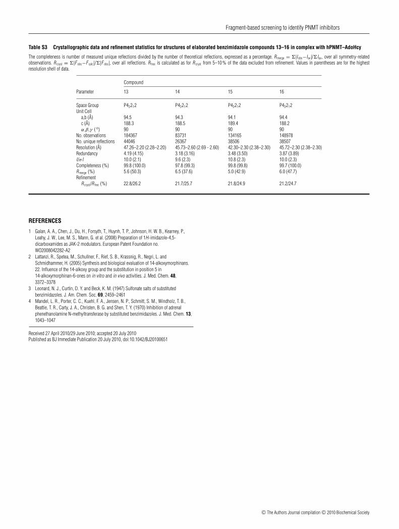
Table S3 Crystallographic data and refinement statistics for structures of elaborated benzimidazole compounds 13–16 in complex with hPNMT–AdoHcy
The completeness is number of measured unique reflections divided by the number of theoretical reflections, expressed as a percentage. Rmerge = �|Iobs−Iav|/�Iav, over all symmetry-relatedobservations. Rcryst = �|F obs−F calc|/�|F obs|, over all reflections. R free is calculated as for Rcryst from 5–10 % of the data excluded from refinement. Values in parentheses are for the highestresolution shell of data.
Compound
Parameter 13 14 15 16
Space Group P43212 P43212 P43212 P43212Unit Cell
a,b (A) 94.5 94.3 94.1 94.4c (A) 188.3 188.5 189.4 188.2α,β ,γ (◦) 90 90 90 90
No. observations 184367 83731 134165 148978No. unique reflections 44046 26367 38506 38507Resolution (A) 47.26–2.20 (2.28–2.20) 45.73–2.60 (2.69 - 2.60) 42.30–2.30 (2.38–2.30) 45.72–2.30 (2.38–2.30)Redundancy 4.19 (4.15) 3.18 (3.16) 3.48 (3.50) 3.87 (3.89)I/σ I 10.0 (2.1) 9.6 (2.3) 10.8 (2.3) 10.0 (2.3)Completeness (%) 99.8 (100.0) 97.8 (99.3) 99.8 (99.8) 99.7 (100.0)Rmerge (%) 5.6 (50.3) 6.5 (37.6) 5.0 (42.9) 6.0 (47.7)Refinement
Rcryst/R free (%) 22.8/26.2 21.7/25.7 21.8/24.9 21.2/24.7
REFERENCES
1 Galan, A. A., Chen, J., Du, H., Forsyth, T., Huynh, T. P., Johnson, H. W. B., Kearney, P.,Leahy, J. W., Lee, M. S., Mann, G. et al. (2008) Preparation of 1H-imidazole-4,5-dicarboxamides as JAK-2 modulators. European Patent Foundation no.WO2008042282-A2
2 Lattanzi, R., Spetea, M., Schullner, F., Rief, S. B., Krassnig, R., Negri, L. andSchmidhammer, H. (2005) Synthesis and biological evaluation of 14-alkoxymorphinans.22. Influence of the 14-alkoxy group and the substitution in position 5 in14-alkoxymorphinan-6-ones on in vitro and in vivo activities. J. Med. Chem. 48,3372–3378
3 Leonard, N. J., Curtin, D. Y. and Beck, K. M. (1947) Sulfonate salts of substitutedbenzimidazoles. J. Am. Chem. Soc. 69, 2459–2461
4 Mandel, L. R., Porter, C. C., Kuehl, F. A., Jensen, N. P., Schmitt, S. M., Windholz, T. B.,Beattie, T. R., Carty, J. A., Christen, B. G. and Shen, T. Y. (1970) Inhibition of adrenalphenethanolamine N-methyltransferase by substituted benzimidazoles. J. Med. Chem. 13,1043–1047
Received 27 April 2010/29 June 2010; accepted 20 July 2010Published as BJ Immediate Publication 20 July 2010, doi:10.1042/BJ20100651
c© The Authors Journal compilation c© 2010 Biochemical Society