Fractures of the Spine in Children of the Spine in Children.pdfC Spine Evaluation in Children •...
48
Fractures of the Spine in Children Shari Cui MD & John France MD February 2016 Original: Steven Frick, MD; March 2004 Past Revised: Steven Frick, MD; August 2006 Timothy Moore, MD; November 2011
Transcript of Fractures of the Spine in Children of the Spine in Children.pdfC Spine Evaluation in Children •...

Fractures of the Spine in Children
Shari Cui MD & John France MD February 2016
Original: Steven Frick, MD; March 2004 Past Revised: Steven Frick, MD; August 2006 Timothy Moore, MD; November 2011

Important Pediatric Differences • Not just “little adults” • Anatomic / Radiographic differences/variants • Flexible • Large heads relative to body • Physeal/synchondrosis/periosteal tube
fractures - apparent dislocations • Surgery rarely indicated • Immobilization well tolerated

Epidemiology
• Incidence – 108 per million
• 3M:2F • >15 yr highest risk • Etiology:
– MVC – Falls – Sports – Non-accidental Trauma
Mendoza et al. Pediatric Spine Trauma in the United States. Iowa Orthopaedic Journal, 2015. Akbarnia (Ed) (2011). The Growing Spine. 1st Ed. Springer, 2011.
2% 4%
12%
82%
< 5 yrs 5-9 yrs 10-14 yrs 15-19 yrs

Epidemiology
Mendoza et al. Pediatric Spine Trauma in the United States. Iowa Orthopaedic Journal, 2015.
• Injury Distribution
Cervical 52%
Thoracic 27%
Lumbar 21%
0-4 yrs
Cervical 34%
Thoracic 24%
Lumbar 42%
5-9 yrs
Cervical 28%
Thoracic 29%
Lumbar 43%
10-14 yrs
Cervical 33%
Thoracic 26%
Lumbar 41%
15-20 yrs
•Overall Neurologic Injury 15% > 50% cervical origin
0-4 yr Cervical Spine 5-20 yr Lumbar Spine

Epidemiology • Patterns vary
– Age (Adolescents predominate) – Race
• Ie. African American – 24% firearm, caucasian – 1% firearm
– Economic Status
• Young children <9yo – Ligamentous injury > Bony Injury – SCIWORA
Knox et al. Spine Trauma in Very Young Children. JPedsOrthop, 2014. Piatt et al. Pediatric Spinal Injry in the US: Epidemiology and Disparities. JNeurosurgPediatr 2015.

Cervical Spine Injuries
• Rare - < 1% of children’s fractures • Neurologic Injury – “rare” to 44% Mortality in ≤ 9yrs
• Age ≤ 7 yrs
– Majority upper cervical, esp. craniocervical junction – Larger Head:Torso ratio
• Age > 7 yrs – Lower cervical injuries predominate
Jones. Pediatric cervical spine trauma. J Am Acad Orthop Surg. 2011;19:600
Shin. Pediatric C Spine and SCI: A National Database Study. Spine. 2015;Epub Ahead of print.

Cervical Spine Injuries
• Upper cervical anatomy – Occiput-C1 articulation
horizontally based – Child: large head/body ratio – Prone to occiput-C1 injury

Anatomy – C1
• Birth: 3 ossification centers – Body & 2x neurocentral
arches
• 7 yrs: Neurocentral synchondroses fuse
Copley. Cervical spine disorders in infants and children. J Am Acad Orthop Surg. 1998;6:204.
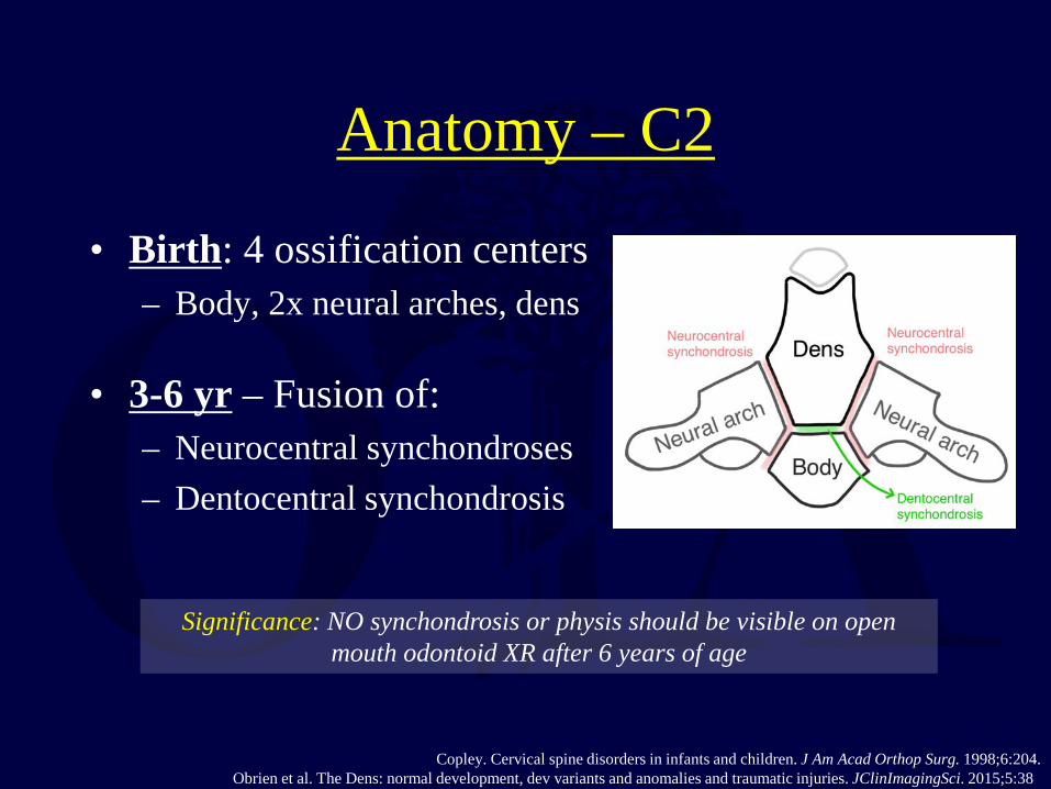
Anatomy – C2
• Birth: 4 ossification centers – Body, 2x neural arches, dens
• 3-6 yr – Fusion of: – Neurocentral synchondroses – Dentocentral synchondrosis
Copley. Cervical spine disorders in infants and children. J Am Acad Orthop Surg. 1998;6:204. Obrien et al. The Dens: normal development, dev variants and anomalies and traumatic injuries. JClinImagingSci. 2015;5:38
Significance: NO synchondrosis or physis should be visible on open mouth odontoid XR after 6 years of age

Anatomy – C2
• Summit ossification center – Appears at 3 – 6 yrs – Fuses ~ 12yrs
Do not confuse with os
odontoideum. Creates confusion with studies
Copley. Cervical spine disorders in infants and children. J Am Acad Orthop Surg. 1998;6:204. Obrien et al. The Dens: normal development, dev variants and anomalies and traumatic injuries. JClinImagingSci. 2015;5:38

• Origin hypotheses: – Congenital – Traumatic (favored)
• Potential C1-C2 instability • Usually asymptomatic • Debate about participation
in contact sports
Fielding. Os odontoideum. J Bone Joint Surg Am 1980;62:376. Arvin et al. Os Odontoideum: Etiology & surg Management, Neurosurgery, 2009
Os Odontoideum Anatomy – C2

Subaxial Cervical Anatomy C3 – C7
• 3-6 yrs: Neurocentral synchondroses fuse
• Vertebral bodies wedge shaped until 7yo bodies square out
• Superior and inferior cartilage endplates firmly attached to disc
Copley. Cervical spine disorders in infants and children. J Am Acad Orthop Surg. 1998;6:204.

Mechanism of Injury • Young child C-spine susceptible to injury:
– Very mobile – ligamentous laxity & shallow angle of facet joints
– Relatively larger head – Delayed ossification of uncinate processes – Anterior vertebral body wedging – Underdeveloped para-spinal muscles
• Combination leads to upper cervical injuries • Most Common Etiologies: MVC & Falls

C-Spine Fracture Pattern
• Junction b/w cartilage endplate and bony vertebral body
• Fractures split the endplate b/w columnar growth cartilage and calcified cartilage
• Does not typically occur by fracture through the endplate – disc junction
Jones. Pediatric cervical spine trauma. J Am Acad Orthop Surg. 2011;19:600.

Transport & the Pediatric C Spine
• Large head! – Standard backboard
increased flexion of C spine
• Remedy: – Pediatric backboard w/
cut-out for head (A) – Elevate trunk relative to
head (w blankets) (B)
Herzenberg et al. Emergency Transport and positioning of young children who have an injury of the cervical spine, JBJS. 1989;71:!5-22
A B

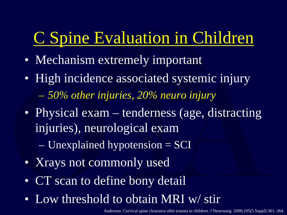
C Spine Evaluation in Children • Mechanism extremely important • High incidence associated systemic injury
– 50% other injuries, 20% neuro injury • Physical exam – tenderness (age, distracting
injuries), neurological exam – Unexplained hypotension = SCI
• Xrays not commonly used • CT scan to define bony detail • Low threshold to obtain MRI w/ stir
Anderson. Cervical spine clearance after trauma in children. J Neurosurg. 2006;105(5 Suppl):361–364.

ED C Spine Evaluation
ADI
PR
Anderson. Cervical spine clearance after trauma in children. J Neurosurg. 2006;105(5 Suppl):361–364.
PR – Powers Ratio. ADI – Atlanto-dens interval. & Others (see reference)

Swichuk’s Line
Anderson. Cervical spine clearance after trauma in children. J Neurosurg. 2006;105(5 Suppl):361–364.
Spinolaminar line drawn from C1 to C3 Distinguishes normal variant from Hangman’s fracture

C Spine XR Evaluation in Children
• Be aware of normal ossification centers and physes
• C2/3 pseudosubluxation common in children < 8yrs (Check spinolaminar line of Swischuk)
• Evaluation of anterior soft tissues unreliable in crying child
• In uninjured normal patients <8yrs, 20% can demonstrate ADI 3-5mm (Adult ADI normal ≤ 3mm)
-Eubanks. Clearing the pediatric cervical spine following injury. J Am Acad Orthop Surg 2006;14:552. -Shaw. Pseudosubluxation of C2 on C3 in polytraumatized children: Prevalence and significance. Clin Radiol 1999;54: 377.

Normal Radiographic Findings • Ossiculum terminale • C1 override C2 (20%) • Multiple 2ndary ossification centers • Normal synchondrosis • Odontoid angulation (4%) • Basilar subdental synchondrosis (>7ys) • Pseudosubluxation (<9yrs) • ADI < 5mm (why? ligamentous laxity & cartilage components in kids)
• RSTS • Normal anterior body wedging <7yrs • Horizontal facets as pillar fxs • Single-level kyphosis (16%)

C2-3 Pseudosubluxation
• Anatomic variant - C2 pseudosubluxing on C3 (occasionally C3 on 4) – Swichuk intact
• Differentiate from true injury (which is uncommon): – Presence of prevertebral soft
tissue swelling – Break in the spinolaminar line
of Swischuk
Shaw. Pseudosubluxation of C2 on C3 in polytraumatized children: Prevalence and significance. Clin Radiol 1999;54: 377.

Traumatic Spinal Cord Injury
• Rare in children • Better prognosis for recovery than adults • Treat aggressively with immobilization +/-
decompression • Late sequelae = paralytic scoliosis (affects
almost all quadriplegic children if injured when < 10 yrs old)
Parent. Spinal cord injury in the pediatric population: a systematic review of the literature. J. Neurotrauma. 2011;28:1515.

SCIWORA Spinal Cord Injury W/o Radiographic Abnormality
• Distraction Mechanism - Spinal column more flexible than Spinal Cord
• Cord traction injury w/ normal XRs • Usually upper C spine and <8yrs • MRI – diagnose cord injury & eval
posterior soft tissues • SCIWORA & dislocations w/ age
– 16.99% toddlers (w C spine injuries) – 5.04% young adults (w C spine injuries)
High Suspicion - GCS 3 w/ normal CT head
may be upper cervical spinal cord injury! Occiput –C1 SCIWORA Parent. Spinal cord injury in the pediatric population: a systematic review of the literature. J. Neurotrauma. 2011;28:1515.
Shin. Pediatric C Spine and SCI: A National Database Study. Spine. 2015;Epub Ahead of print.

SCIWORA
• Stretch skeleton > Stretch cord
• Stretch Capacity
– Spinal Column 2” > Spinal Cord ¼”
• Cord restricted by horizontal cervical roots, foramen magnum
Leventhal, JPedsOrthop 1960

C-Spine Clearance & Evaluation
• 3 view plain film series still used • Low threshold for further imaging • CT scan upper C-spine (O-C2) • Consider MRI if intubated or obtunded
Sharma. Assessment for additional spinal trauma in patients with cervical spine injury. Am Surg. 2007;73:70.

• CT scan – Advantages – Fast, No sedation or anesthesia – Disadvantages – radiation, Limited evaluation soft
tissues & cartilage
• Assess alignment & bony injury
Sharma. Assessment for additional spinal trauma in patients with cervical spine injury. Am Surg. 2007;73:70.
C-Spine Clearance & Evaluation Not “Cleared” by Plain Radiographs

Not “Cleared” • MRI scan – currently favored • Rapid sequence/image
acquisition algorithms – gradient echo
• Evaluate non osseous tissues and spinal cord
• MRI scan should be considered in critically injured child for whom adequate plain films cannot be obtained to rule out spinal injury
Sharma. Assessment for additional spinal trauma in patients with cervical spine injury. Am Surg. 2007;73:70.

If not “Cleared” within 12 Hours
• Switch to pediatric Aspen or Miami J collar • Consider CT or MRI
McCall. Cervical spine trauma in children: a review. Neurosurg Focus. 2006;20(2):E5.

Child in C-spine collar
Meets NEXUS criteria: 1. Absence of midline cervical tenderness 2. No evidence of intoxication 3. Normal level of alertness 4. Normal neurological exam 5. Absence of a painful, distracting injury
C-SPINE CLEAR
YES
Trauma evaluation and Cervical spine radiographs:
AP/lateral/odontoid for age > 5 yr AP/lateral only for age ≤ 5 yr
ABNORMAL RADIOGRAPH
S Spine Service
Consult
YES
Communicative child ≥ 3 years
Spine Service Consult
NO
NO
NORMAL
Normal neurological exam Spine Service
Consult NO
YES
Flexion/Extension C-spine x-rays
Spine Service Consult
ABNORMAL C-SPINE CLEAR
NORMAL
Leave in collar; refer to neurosurgery clinic in 1-2 weeks
INADEQUATE
Anderson. Cervical spine clearance after trauma in children. J Neurosurg. 2006;105(5 Suppl):361.
Clearance Protocol

If You See a Spine Fracture in a Child
• Look hard for another one
“The most commonly missed spinal fracture is the second one”. -J. Dormans
• High incidence of noncontiguous spine fractures in children
Firth. Pediatric Non-Contiguous Spinal Injuries: The 15 year Experience at One Pediatric Trauma Centre. Spine. 2011 Nov. 14 (Ahead of Print)

Thoracic Spine Fractures
• Less common spinal fracture in children than in more mobile regions
• Rib cage offers some support / protection • Motor vehicle crashes, falls from heights • Child abuse in very young • Compression fractures in severely osteopenic
conditions (OI, chemotherapy) • Multiple contiguous – hyperflexion neck/chest
injury (motorcross)
Slotkin. Thoracolumbar spinal trauma in children. Neurosurg. Clin. N. Am. 2007;18:621.

• 11M, motorcross, flew over handlebars

Thoracic Spine Fracture Dislocations
• High energy mechanisms • Often spinal cord injury, can be transected • Prognosis for recovery most dependent on
initial exam – complete deficits unlikely to have recovery
• Infarction of cord (artery of Adamkiewicz) may play some role –especially in delayed paraplegia
Slotkin. Thoracolumbar spinal trauma in children. Neurosurg. Clin. N. Am. 2007;18:621.
Thoracolumbar Junction Injuries T11-L2
• Classically lap-belt flexion-distraction injuries
• Chance fractures and variants • High association with intraabdominal injury
(50-90%) • Neurologic injury infrequent but can occur
Arkader. Pediatric chance fractures: a multicenter perspective. J Pediatr Orthop. 2011;31:741.

Chance Fractures and Variants
• Flexion over fulcrum • Posterior elements fail in tension, anterior
elements in compression – Can occur through bone, soft tissue or combination
• Treatment – Pure bony injuries can be treated with immobilization
in extension – Partial or whole ligamentous injuries may be best
treated with surgical stabilization
Arkader. Pediatric chance fractures: a multicenter perspective. J Pediatr Orthop. 2011;31:741.

Seatbelt/Flexion-Distraction Injury Classification
Rumball. Seat-belt injuries of the spine in young children. J Bone Joint Surg Br. 1992;74:571.
A C
B D

Bony Flexion Distraction Injury
Cast 3 month

Ligamentous Flex/Dist Injury 1
• 5yF, MVC, bowel perforations
Tx: L 2-3 open short-segment fixation/fusion

Ligamentous Flex/Dist Injury 2 12yM, MVC, initial missed injury Upright lateral + Flexion/Extension XR
Tx: L3-4 Percutaneous fusionless fixation w/ removal @ 6mo
30 months post op

Combined (Bony+Ligamentous) Flexion Distraction Injury
16yM, MVC, bowel injury
Tx: Closed reduction, perc fixation
Postop
3 yrs postop Healed Broken screws (disc motion) - removed

Lap Belt Sign
• High association with intraabdominal injury and lumbar spine fracture
• Lumbar spine films mandatory
Arkader. Pediatric chance fractures: a multicenter perspective. J Pediatr Orthop. 2011;31:741.
Lumbar Spine Fractures L3-L5
• Infrequent until late adolescence – Can be associated with lap belt injuries
• Usually compression fractures that are stable injuries
• Burst fractures – May progress to kyphosis
• Lumbar apophyseal injuries – Posterior displacement can cause stenosis, may need
surgical excision
Slotkin. Thoracolumbar spinal trauma in children. Neurosurg. Clin. N. Am. 2007;18:621.

Lumbar Apophyseal Injuries Slipped Apophysis
• Compression-shear injuries • Same age group as SCFE • Typically adolescent males, inferior
endplates of L4 or L5 • Traumatic displacement of vertebral ring
apophysis and disc into spinal canal • If causes significant compression of cauda
equina, treatment is surgical excision
Chang. Clinical significance of ring apophysis fracture in adolescent lumbar disc herniation. Spine. 2008;33:1750.

Burst Fractures
• Usually in older adolescents • Treatment similar to adults • May not need surgery in neurologically
intact patient • Injuries at thoracolumbar junction higher
risk for progressive kyphosis
Slotkin. Thoracolumbar spinal trauma in children. Neurosurg. Clin. N. Am. 2007;18:621.

Bibliography • Special Thanks - Additional Cases and imaging from:
– Dr. John C France (West Virginia University, Ruby Memorial) – Dr. Aki S Puryear (St Louis University, Cardinal Glennon Children’s Hospital)
• Anderson RCE, Scaife ER, Fenton SJ, Kan P, Hansen KW, Brockmeyer DL. Cervical spine clearance after trauma
in children. J Neurosurg. 2006 Nov.;105(5 Suppl):361–364. • Arkader A, Warner WC, Tolo VT, Sponseller PD, Skaggs DL. Pediatric chance fractures: a multicenter perspective.
J Pediatr Orthop. 2011 Sep.;31(7):741–744. • Arvin et al. Os Odontoideum: Etiology & surg Management, Neurosurgery, 2009 • Chang C-H, Lee Z-L, Chen W-J, Tan C-F, Chen L-H. Clinical significance of ring apophysis fracture in adolescent
lumbar disc herniation. Spine. 2008 Jul. 15;33(16):1750–1754. • Copley LA, Dormans JP. Cervical spine d/o’s in infants & children. JAmAcadOrthopSurg. 1998 Jun.;6(4):204–214. • Cui S, Busel G, Puryear AS. Percutaneous Pedicle Screw Stabilization without Fusion of Adolescent
Thoracolumbar Spine Fractures. JPedsOrthop, 2015. (Ahead of Print) • Eubanks JD, Gilmore A, Bess S, Cooperman DR: Clearing the pediatric cervical spine following injury. J Am Acad
Orthop Surg 2006;14(9):552-564. • Fielding JWHensinger RN, Hawkins RJ: Os odontoideum. J Bone Joint Surg Am 1980;62:376-383. • Firth GB, Kingwell S, Moroz P. Pediatric Non-Contiguous Spinal Injuries: The 15 year Experience at One Pediatric
Trauma Centre. Spine. 2011 Nov. 14 (Ahead of Print) • Herzenberg et al. Emergency Transport and positioning of young children who have an injury of the cervical spine,
JBJS. 1989;71:!5-22 • Jones TM, Anderson PA, Noonan KJ. Pediatric c spine trauma. JAmAcadOrthopSurg. 2011 Oct.;19(10):600–611. • Knox et al. Spine Trauma in Very Young Children. JPedsOrthop, 2014. • Leventhal, JPedsOrthop 1960
Bibliography • McCall T, Fassett D, Brockmeyer D. Cervical spine trauma in children: a review. Neurosurg Focus. 2006;20(2):E5. • Mendoza et al. Pediatric Spine Trauma in the United States. Iowa Orthopaedic Journal, 2015. • Obrien et al. The Dens: normal development, dev variants and anomalies and traumatic injuries. JClinImagingSci.
2015;5:38 • Parent S, Mac-Thiong J-M, Roy-Beaudry M, Sosa JF, Labelle H. Spinal cord injury in the pediatric population: a
systematic review of the literature. J. Neurotrauma. 2011 Aug.;28(8):1515–1524. • Piatt et al. Pediatric Spinal Injry in the US: Epidemiology and Disparities. JNeurosurgPediatr 2015. • Rumball K, Jarvis J. Seat-belt injuries of the spine in young children. J Bone Joint Surg Br. 1992 Jul.;74(4):571–
574. • Sharma OP, Oswanski MF, Yazdi JS, Jindal S, Taylor M. Assessment for additional spinal trauma in patients with
cervical spine injury. Am Surg. 2007 Jan.;73(1):70–74. • Shaw M, Burnett H, Wilson A, Chan O: Pseudosubluxation of C2 on C3 in polytraumatized children: Prevalence
and significance. Clin Radiol 1999;54(6): 377-380. • Shin. Pediatric C Spine and SCI: A National Database Study. Spine. 2015;Epub Ahead of print. • Slotkin JR, Lu Y, Wood KB. Thoracolumbar spinal trauma in children. Neurosurg. Clin. N. Am. 2007
Oct.;18(4):621–630. • Tarr RW, Drolshagen LF, Kerner TC, Allen JH, Partain CL, James AE. MR imaging of recent spinal trauma. J
Comput Assist Tomogr. 1987 Apr.;11(3):412–417.
If you would like to volunteer as an author for the Resident Slide Project or recommend updates to any of the following slides, please send an e-mail to [email protected]
Return to Pediatrics
Index

• For questions or comments, please send to [email protected]