FRACTURES OF MAXILLA AND MANDIBLE
39
FRACTURES OF MAXILLA AND MANDIBLE By DR.CHAMPA SUSHEL MBBS- FCPS ASSISTANT PROFESSOR SURGICAL UNIT -4
description
FRACTURES OF MAXILLA AND MANDIBLE. By DR.CHAMPA SUSHEL MBBS- FCPS ASSISTANT PROFESSOR SURGICAL UNIT -4. Etiology. Maxillofacial fractures result from either blunt or penetrating trauma. - PowerPoint PPT Presentation
Transcript of FRACTURES OF MAXILLA AND MANDIBLE

FRACTURES OF MAXILLA AND MANDIBLE
By DR.CHAMPA SUSHEL
MBBS- FCPS
ASSISTANT PROFESSOR SURGICAL UNIT -4

Etiology
• Maxillofacial fractures result from either blunt or penetrating trauma.
• @60% of patients with severe facial trauma have multisystem trauma and the potential for airway compromise.– 20-50% concurrent brain injury.– 1-4% cervical spine injuries.– Blindness occurs in 0.5-3%

Etiology
• 25% of women with facial trauma are victims of domestic violence.
• 25% of patients with severe facial trauma will develop Post Traumatic Stress Disorder

Anatomy

Anatomy

Emergency ManagementAirway Control
• Control airway:– Chin lift.– Jaw thrust.– Oropharyngeal suctioning.– Manually move the tongue forward.– Maintain cervical immobilization

Emergency ManagementIntubation Considerations
• Avoid nasotracheal intubation• Consider fiberoptic intubation if available. • Alternatives include percutaneous
transtracheal ventilation and retrograde intubation.
• Be prepared for cricothyroidotomy.


Emergency ManagementHemorrhage Control
• Maxillofacial bleeding:– Direct pressure.– Avoid blind clamping in wounds.
• Nasal bleeding:– Direct pressure.– Anterior and posterior packing.
• Pharyngeal bleeding:– Packing of the pharynx around ET tube.
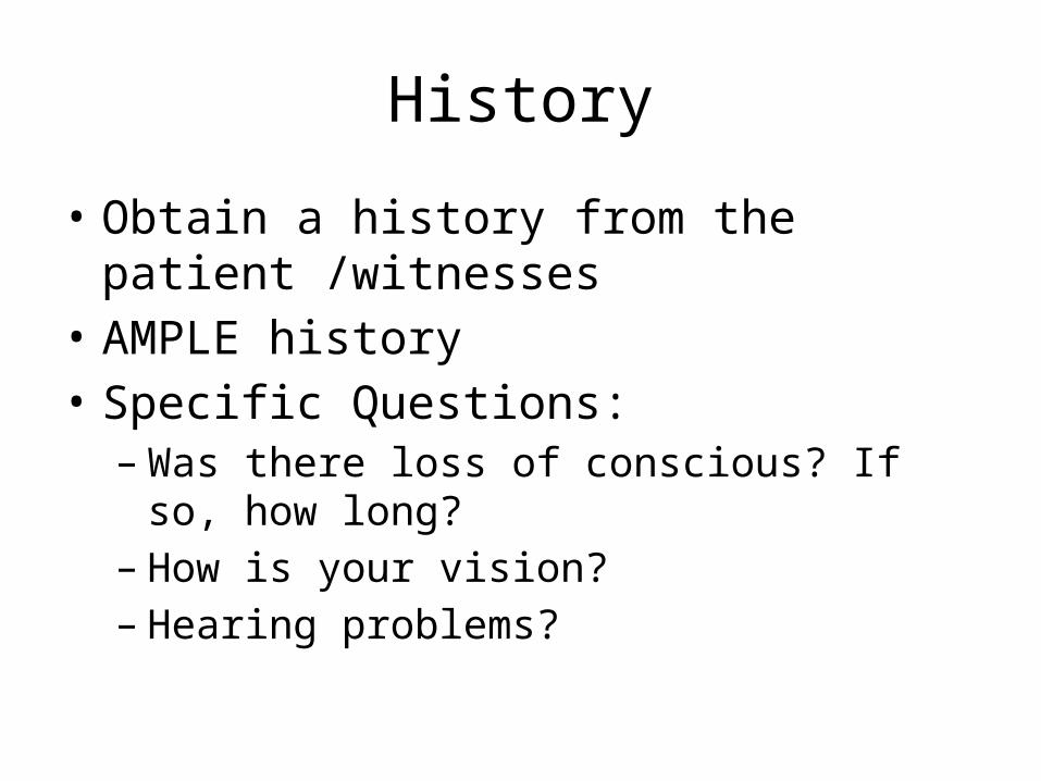
History
• Obtain a history from the patient /witnesses • AMPLE history• Specific Questions:– Was there loss of conscious? If so, how long?– How is your vision?– Hearing problems?

History
• Specific Questions:– Is there pain with eye movement?– Are there areas of numbness or tingling on your
face?– Is the patient able to bite down without any pain?– Is there pain with moving the jaw?
Physical Examination
• Inspection of the face for asymmetry.• Inspect open wounds for foreign bodies.• Palpate the entire face.– Supraorbital and Infraorbital rim– Zygomatic-frontal suture– Zygomatic arches

Physical Examination• Inspect the nose for asymmetry, telecanthus,
widening of the nasal bridge.• Inspect nasal septum for septal hematoma, CSF or
blood.• Palpate nose for crepitus, deformity and
subcutaneous air.• Palpate the zygoma along its arch and its
articulations with the maxilla, frontal and temporal bone.

Physical Examination• Check facial stability.• Inspect the teeth for malocclusions, bleeding and
step-off.• Intraoral examination: – Manipulation of each tooth.– Check for lacerations.– Stress the mandible.– Tongue blade test.
• Palpate the mandible for tenderness, swelling and step-off.

Physical Examination
• Check visual acuity.• Check pupils for roundness and reactivity.• Examine the eyelids for lacerations.• Test extra ocular muscles.• Palpate around the entire orbits..

Physical Examination
• Examine the cornea for abrasions and lacerations.
• Examine the anterior chamber for blood or hyphema.
• Perform fundoscopic exam and examine the posterior chamber and the retina.

Physical Examination
• Examine and palpate the exterior ears.• Examine the ear canals.• Check nuero distributions of the supraorbital,
infraorbital, inferior alveolar and mental nerves.

Maxillary Fractures
• High energy injuries.• Impact 100 times the force of gravity is
required .• Patients often have significant multisystem
trauma.• Classified as LeFort fractures.

Maxillary FracturesLeFort I
• Definition:– Horizontal fracture of
the maxilla at the level of the nasal fossa.
– Allows motion of the maxilla while the nasal bridge remains stable.

Maxillary FracturesLeFort I
• Clinical findings:– Facial edema– Malocclusion of the
teeth– Motion of the maxilla
while the nasal bridge remains stable

Maxillary FracturesLeFort I
• Radiographic findings:– Fracture line which
involves• Nasal aperture• Inferior maxilla• Lateral wall of maxilla
• CT of the face and head – coronal cuts– 3-D reconstruction

Maxillary FracturesLeFort II
• Definition:– Pyramidal fracture
• Maxilla• Nasal bones • Medial aspect of the
orbits

Maxillary FracturesLeFort II
• Clinical findings:– Marked facial edema– Nasal flattening– Traumatic telecanthus– Epistaxis or CSF
rhinorrhea – Movement of the
upper jaw and the nose.

Maxillary FracturesLeFort II
• Radiographic imaging:– Fracture involves:
• Nasal bones• Medial orbit• Maxillary sinus• Frontal process of the
maxilla
• CT of the face and head

Maxillary FracturesLeFort III
• Definition:– Fractures through:
• Maxilla• Zygoma• Nasal bones• Ethmoid bones• Base of the skull

Maxillary FracturesLeFort III
• Clinical findings:– Dish faced deformity– Epistaxis and CSF
rhinorrhea – Motion of the maxilla,
nasal bones and zygoma
– Severe airway obstruction

Maxillary FracturesLeFort III
• Radiographic imaging:– Fractures through:
• Zygomaticfrontal suture• Zygoma• Medial orbital wall• Nasal bone
• CT Face and the Head

Maxillary FracturesTreatment
• Secure airway• Control Bleeding• Head elevation 40-60 degrees• Consult with maxillofacial surgeon• Consider antibiotics• Admission
Mandible FracturesPathophysiology
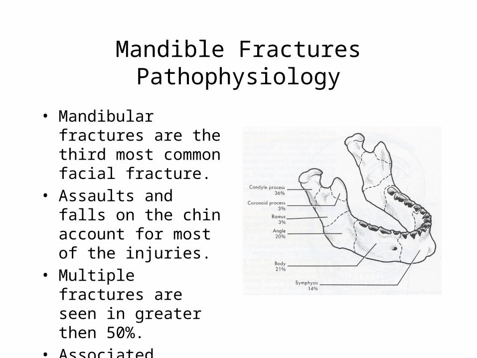
• Mandibular fractures are the third most common facial fracture.
• Assaults and falls on the chin account for most of the injuries.
• Multiple fractures are seen in greater then 50%.
• Associated Cervical spine injuries – 0.2-6%.

Mandible FracturesClinical findings
• Mandibular pain.• Malocclusion of the teeth• Separation of teeth with
intraoral bleeding• Inability to fully open
mouth.• Preauricular pain with
biting. • Positive tongue blade test.

Mandible Fractures
• Radiographs:– Panoramic view– Plain view: PA and Lateral view

Mandibular FracturesTreatment
• Nondisplaced fractures:– Analgesics– Soft diet– oral surgery referral in 1-2 days
• Displaced fractures, open fractures and fractures with associated dental trauma– Urgent oral surgery consultation
• All fractures should be treated with antibiotics and tetanus prophylaxis.
Mandibular Dislocation
• Causes of mandibular dislocation are:– Blunt trauma– Excessive mouth opening
• Risk factors:– Weakness of the temporal mandibular ligament– Over stretched joint capsule – Shallow articular eminence– Neurologic diseases

Mandibular Dislocation
• The mandible can be dislocated:– Anterior 70%– Posterior– Lateral– Superior
• Dislocations are mostly bilateral.

Mandibular Dislocation
• Clinical features:– Inability to close
mouth– Pain– Facial swelling
• Physical exam:– Palpable depression– Jaw will deviate away– Jaw displaced anterior
Mandibular Dislocation
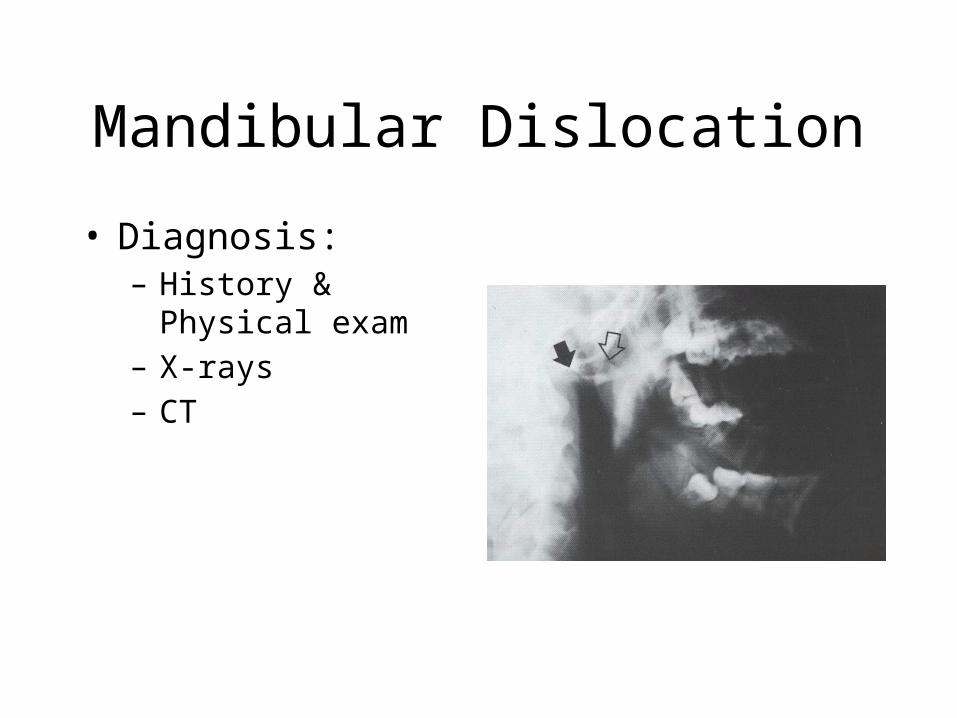
• Diagnosis:– History & Physical
exam– X-rays– CT

Mandibular Dislocation
• Treatment:– Muscle relaxant– Analgesic– Closed reduction in the
emergency room

Mandibular Dislocation
• Treatment:– Oral surgeon consultation:• Open dislocations• Superior, posterior or lateral dislocations• Non-reducible dislocations• Dislocations associated with fractures

THANK YOU