Fourier Domain Optical Coherence Tomography (FD-OCT)
26
1 Fourier Domain Optical Coherence Tomography (FD-OCT) PAYMAN RAJAI 1 References: •Fercher 1995, Measurement of intraocular distances by backscattering spectral Interferometry, Optics Communications 117 (1995) 43-48 •Wolf 1969, Three dimensional structure determination of semi transparent objects from holography data, Optics Communications, 1,4,153-156 •Born and Wolf, Principles of Optics, Seventh Edition •Izatt, Theory of Optical Coherence Tomography
-
Upload
payman-rajai -
Category
Science
-
view
46 -
download
5
Transcript of Fourier Domain Optical Coherence Tomography (FD-OCT)

1
Fourier Domain Optical
Coherence Tomography (FD-OCT)
PAYMAN RAJAI
1
References:
•Fercher 1995, Measurement of intraocular distances by
backscattering spectral Interferometry, Optics Communications 117
(1995) 43-48
•Wolf 1969, Three dimensional structure determination of semi
transparent objects from holography data, Optics Communications,
1,4,153-156
•Born and Wolf, Principles of Optics, Seventh Edition
•Izatt, Theory of Optical Coherence Tomography

2
• FD-OCT Overview
• Born Approximation
• Back Scattered light
• Measurement by Backscattering spectral
Interferometry
• Sample Calculation for δ-like Scattering Potential
• Removing Noises from FDOCT by Phase Shifting
2

3
3

4
Born approximation
4

5
5
Helmholtz eq.
Scattering Potential

6
Green’s function for
Helmholtz equation
6
Subtract two eqs and use Green’s Theorem.
Integral over r’ bounded by a large sphere, when R → infinity
Free space green’s function

7
7
First Born approximation makes it easy to compute. For weakly scattering sites,
n≈1 and F(r) would be very small. Hence U(r’) inside the integral is the same as
U(incident).
Scattering Amplitude

8
Back Scattered light
8
9
9
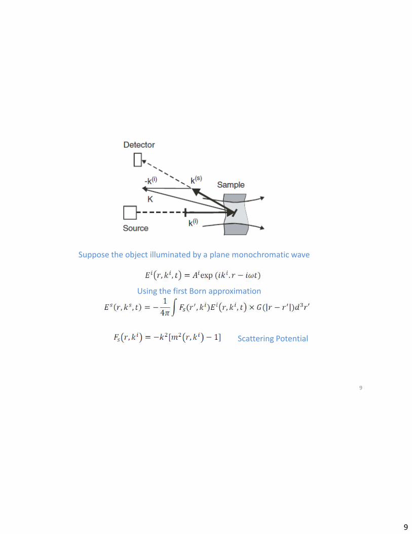
Suppose the object illuminated by a plane monochromatic wave
Using the first Born approximation
Scattering Potential

10
10
Origin of x,y,z
“P(r)” observation point
located on the z-axis, a
distance D outside of the
object
PZ axis
Thickness of the sample
If D ›› T then r-r’ ≈ D

11
11
Integration over x’ and y’ can be replaced by a constant W chosen proportional to the
cross section of the beam waist.
When the detector is placed at θ=π
three dimensional Fourier transform is replaced the by a one dimensional.

12
12
Back Scattered light at the point “P” located on the z-axis in D
distance from the the object
It can be done if the phase and amplitude of the scattered field are known for a
range of k-values
These describe why we have to use a multi wavelength source of illumination.

13
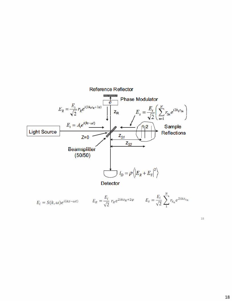
Measurement by Backscattering
spectral Interferometry
13

14
14
We have obtained auto correlation function of the scattering potential not the potential itself !

15
How to obtain F(z)
• 1) if we interfere an additional singular light at the distance L
from the object (Reference Mirror)
• 2) if the object itself contains one interface with large
reflectivity to act as a reference mirror
15
Photodiode ArrayBroadband light source
Spectrometer
Diffraction
Grating

16
16
Actual object potential
auto correlation
function of the
sample
structure
centered at Z=0
complex
conjugate of
sample potential
centered at Z=zR
true
reconstruction of
the potential
centered at Z=-zR .
additional peak at
the origin of the
reconstructed
sample space
Intense light
background noise at
center caused by the
mirror
Weak background noise
at center caused by
different objects sites
Symmetric mirror images

17
Sample Calculation for δ-like Scattering
Potential
17
18
18

19
19

20
20

21
Sample Data Looks like:
21

22
Removing Noises from FDOCT by
Phase Shifting
22

23
23
DC Terms removed
To eliminate the mirror image, we need to acquire interferogram with 2φ differs
from π, for example 3π/2 and π/2. In this case the subtracting interferogram results
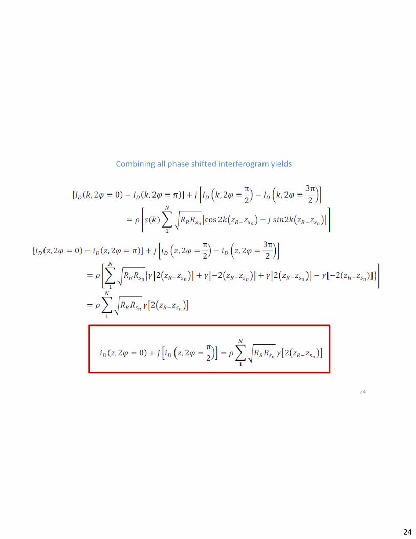
24
Combining all phase shifted interferogram yields
24

25
25
Standard FD-OCT images Mirror image and DC
terms removed images
the anterior chamber
of the human eye
a three-day-old
chicken embryo.
http://spie.org

26
26
Standard FD-OCT images Mirror image and DC
terms removed images
the palm skin
finger nail near the
nail fold region of a
human
http://spie.org