Four cases of abdominal pain Case one -
54
1 Four cases of abdominal pain Case one A nearly 8 year old female came to the ED with a chief complaint of stomach aches for one week. These pains would come and go and were best characterized as crampy in nature with no particular location. She also complained of back aches since her stretching exercises yesterday. There was no history of fever, nausea, vomiting, diarrhea, or respiratory symptoms. Her bowel movements were regular and soft. Exam: VS T36.8, P98, R24, BP 114/88. She was alert, cooperative, and somewhat anxious. HEENT exam was unremarkable. Neck supple without adenopathy. Heart regular without murmurs. Lungs clear. Abdomen was soft, flat, and non-tender. There was no rebound. Bowel sounds were active. No masses or hepatosplenomegaly were appreciated. A rectal exam revealed no stool in the rectum and no masses. She was observed to have more pain when standing or when sitting up. There was no CVA tenderness. Left flank pain and epigastric pain were elicited on straight leg raising. An abdominal series was obtained. Investigations: This radiograph was initially read as showing non-specific findings. Other laboratory results: CBC WBC 2.9, 32 segs, 64 lymphs, 4 monos, Hgb 12.4, Hct 36.5, platelets adequate. Amylase 123, SGOT 24. Her pain persisted. Radiological findings by a radiologist: Review of her radiographs by a radiologist revealed subtle compression fractures of the vertebral bodies. Follow-up radiographs were obtained. Final diagnosis This follow-up radiograph showed progressive demineralization and multiple compression fractures of the thoracic and lumbar vertebral bodies. This is not obvious initially if your attention is directed at the abdominal soft tissue. Upon close inspection, you can appreciate multiple vertebral compression fractures of her thoracic and lumbar vertebrae. Note that the vertebral bodies appear to be flatter than normal. It is remarkable that her clinical symptoms pointed to the abdomen rather than her spine. Some hepatomegaly is also noted on one of the views. These vertebral fractures were felt to be most consistent with acute leukemia. Bone marrow studies confirmed the diagnosis of acute lymphocytic leukemia. A lateral view of her lumbar spine makes these fractures easier to appreciate. The vertebral bodies are obviously flatter than they should be on this view. Teaching Points: Abdominal radiographs are generally non-diagnostic for the vast majority of cases. However, when they reveal significant findings, they are often difficult to appreciate. Abnormalities of the bony structures include vertebral fractures, pelvic fractures, rib fractures, congenital dislocated hips, other hip injuries, etc. If one is not paying careful attention to the bony structures, these findings can be easily overlooked, although they may appear obvious once the abnormalities are identified. Soft tissue findings include fecaliths, intussusception, pneumoperitoneum, subtle obstructions, volvulus, mass effects, etc. Abdominal pain is a non-specific presentation for many serious diagnoses, but abdominal pain is most often the result of a benign cause. It is often useful to observe the patient ambulating since this can provide significant clues to the patient's severity. Patients who cannot walk upright easily should be taken more seriously than those who can ambulate normally. Coughing and jumping are useful peritoneal signs for children since this tends to distract them away from the abdomen. When the patient is walking, ask the child to jump and challenge them to jump higher if possible. Then ask them if jumping hurt their tummy. Ask the child to cough and then ask them if the cough hurt their tummy. Negative findings on jumping and coughing make the likelihood of peritoneal irritation extremely remote. In this case, if the examiners had observed the patient ambulating, they may have noted some difficulty since she did complain of pain with standing. If the examiners asked her to jump, it is likely that she would have complained of pain in her back, though in this instance, such a maneuver may have worsened her compression fractures. Hopefully, the patient would be
Transcript of Four cases of abdominal pain Case one -

1
Four cases of abdominal pain Case one
A nearly 8 year old female came to the ED with a chief complaint of stomach aches for one week. These pains would come and go and were best characterized as crampy in nature with no particular location. She also complained of back aches since her stretching exercises yesterday. There was no history of fever, nausea, vomiting, diarrhea, or respiratory symptoms. Her bowel movements were regular and soft. Exam: VS T36.8, P98, R24, BP 114/88. She was alert, cooperative, and somewhat anxious. HEENT exam was unremarkable. Neck supple without adenopathy. Heart regular without murmurs. Lungs clear. Abdomen was soft, flat, and non-tender. There was no rebound. Bowel sounds were active. No masses or hepatosplenomegaly were appreciated. A rectal exam revealed no stool in the rectum and no masses. She was observed to have more pain when standing or when sitting up. There was no CVA tenderness. Left flank pain and epigastric pain were elicited on straight leg raising. An abdominal series was obtained.
Investigations:
This radiograph was initially read as showing non-specific findings. Other laboratory results: CBC WBC 2.9, 32 segs, 64 lymphs, 4 monos, Hgb 12.4, Hct 36.5, platelets adequate. Amylase 123, SGOT 24. Her pain persisted. Radiological findings by a radiologist: Review of her radiographs by a radiologist revealed subtle compression fractures of the vertebral bodies. Follow-up radiographs were obtained. Final diagnosis
This follow-up radiograph showed progressive demineralization and multiple compression fractures of the thoracic and lumbar vertebral bodies. This is not obvious initially if your attention is directed at the abdominal soft tissue. Upon close inspection, you can appreciate multiple vertebral compression fractures of her thoracic and lumbar vertebrae. Note that the vertebral bodies appear to be flatter than normal. It is remarkable that her clinical symptoms pointed to the abdomen rather than her spine. Some hepatomegaly is also noted on one of the views. These vertebral fractures were felt to be most consistent with acute leukemia. Bone marrow studies confirmed the diagnosis of acute lymphocytic leukemia. A lateral view of her lumbar spine makes these fractures easier to appreciate. The vertebral bodies are obviously flatter than they should be on this view.
Teaching Points:
Abdominal radiographs are generally non-diagnostic for the vast majority of cases. However, when they reveal significant findings, they are often difficult to appreciate. Abnormalities of the bony structures include vertebral fractures, pelvic fractures, rib fractures, congenital dislocated hips, other hip injuries, etc. If one is not paying careful attention to the bony structures, these findings can be easily overlooked, although they may appear obvious once the abnormalities are identified. Soft tissue findings include fecaliths, intussusception, pneumoperitoneum, subtle obstructions, volvulus, mass effects, etc. Abdominal pain is a non-specific presentation for many serious diagnoses, but abdominal pain is most often the result of a benign cause. It is often useful to observe the patient ambulating since this can provide significant clues to the patient's severity. Patients who cannot walk upright easily should be taken more seriously than those who can ambulate normally. Coughing and jumping are useful peritoneal signs for children since this tends to distract them away from the abdomen. When the patient is walking, ask the child to jump and challenge them to jump higher if possible. Then ask them if jumping hurt their tummy. Ask the child to cough and then ask them if the cough hurt their tummy. Negative findings on jumping and coughing make the likelihood of peritoneal irritation extremely remote. In this case, if the examiners had observed the patient ambulating, they may have noted some difficulty since she did complain of pain with standing. If the examiners asked her to jump, it is likely that she would have complained of pain in her back, though in this instance, such a maneuver may have worsened her compression fractures. Hopefully, the patient would be
2
able to appreciate this and refuse to jump. It is difficult to conceive that this patient with so many vertebral compression fractures could tolerate ballet practice the previous day and that she presented with such non-specific findings. Often children can be extraordinarily stoic despite being in substantial pain. This can be deceiving for the examiners. A good practice is to palpate all parts of the abdomen and all parts of the back in patients with abdominal pain.
Case two
A 6 year old male presents to the ED with a chief complaint of fever and stomach pain since last night. It is now 11:00 a.m. The temperature was not measured at home but he felt warm. He was given an 7.5 ml of paracetamol at 4:00 a.m. There was no history of nausea, vomiting, or diarrhea. His last bowel movement was three days ago. He pointed to his epigastrium as the location of most of his pain. Exam: VS T38, P136, R24, BP 113/61. He was noted to be small for age (15.3 kg), alert, active, in no distress. He did not appear to be uncomfortable at all. HEENT exam was unremarkable. Neck supple without adenopathy. Heart regular without murmurs. Lungs clear. Abdominal exam was positive for mild tenderness in the epigastrium. Bowel sounds were active. No tenderness in the right lower quadrant. No rebound tenderness. No hepatosplenomegaly or masses were appreciated. Testes were normal. A rectal exam revealed normal sphincter tone, no masses, and no right lower quadrant tenderness. The stool tested negative for occult blood. An abdominal series was ordered. An AP view of the chest was also ordered as part of the abdominal series.
The radiographs were interpreted as showing non-specific findings. Because the cause of the abdominal pain was suspected to be constipation, the patient was given an enema. Following this, he passed a large amount of stool and felt much better. His abdominal exam continued to be benign. He was discharged from the ED. Overnight, the patient continued to experience fever at home and some abdominal pain though the degree of abdominal pain was improved.
Radiological findings
A review of his radiographs the following morning revealed an alternative diagnosis for his symptoms.
This represents a pulmonary infiltrate in the medial aspect of the left lower lobe. The top of it is cut off in the flat (supine) view of the abdomen. It is almost impossible to appreciate this density on the upright view because most of it is cut off. The chest radiograph was taken using a different degree of penetration to view the lungs better. Because of this, it is even more difficult to appreciate the infiltrate behind the heart. Upon close inspection, you should be able to appreciate the triangular density superimposed on the heart on the chest radiograph view. A lateral view of the chest was not taken in this case since the chest view was part of an abdominal series that was ordered. Further management: The patient was placed on antibiotics and his fever promptly improved by the next day. His abdominal pain and his other symptoms gradually improved. Discussion and Teaching Points:
Pneumonia is a known cause of abdominal pain. This diagnosis is often not considered because the abdominal pain is the chief complaint. The pain can be very severe at times. This can easily mislead a clinician to limit the area of investigation to the abdomen. This pitfall should be avoided. Causes of abdominal pain that are not related to the abdomen include pneumonia, pneumothorax, pneumomediastinum, pericarditis, zoster, vertebral conditions (eg., osteomyelitis, discitis), diabetic include myocardial ischemia and aortic dissection.Pulmonary conditions should be considered in with respiratory symptoms, tachypnea, or a borderline oxygen saturation. Documentation of these findings should be routine in patients with abdominal pain. The history should include the presence of and the severity of respiratory symptoms. The vital signs should include a respiratory rate and a pulse oximetry reading. The examination should include notes describing the presence or absence of any observed tachypnea, the degree of coughing observed, the characteristics of

3
the cough (eg., moist, productive, bronchospastic, dry, etc.), and the standard pulmonary auscultation and percussion findings. If any of these findings suggest the possibility of pneumonia, PA and lateral chest radiographs should be ordered, or alternatively, treatment prescribed for a clinical diagnosis of a respiratory infection. Although the likelihood of aortic dissection is low (especially in children), this condition is associated with a substantial likelihood of death which may be preventable if the diagnosis is suspected early. While aortic contrast studies by CT or aortography are not routine, one suggestion has been to document the presence and character of peripheral pulses in all patients presenting with abdominal pain.Although the appendix is often the focus of clinical examination in patients with abdominal pain, there are other serious causes of abdominal pain that should be considered as well, such as intussusception, volvulus, pancreatitis, ovarian torsion, testicular torsion, acute cholecystitis, etc. The radiographic findings in intussusception may range from normal to various indirect signs of intussusception (refer to Case 2 which describes the radiographic findings in intussusception). A volvulus is usually associated with a true bowel obstruction, but the presentation clinically and radiographically can occasionally be subtle.Ovarian torsion may be a difficult diagnosis to make. Even the use of color flow doppler ultrasound used to assess blood flow to the ovaries is not able to totally rule out this diagnosis since, early in its presentation, some blood flow may still be preserved.Testicular torsion is usually suspected on clinical grounds, but occasionally the testes are not examined in some patients because their pants and underwear (or diapers) are not removed. Younger patients may fail to point to their testes as the location of the pain. Some may complain of non-specific abdominal pain because of failure to appreciate the source of the pain, or because of modesty.
In summary, the causes of abdominal pain are extensive. In the acute care setting, it is most important to rule out diagnoses that must be made early to result in the best possible outcome for the patient. Some of these diagnoses have been mentioned, but there are others.
Case three
An 11 month old male with history of "stomach flu" symptoms two weeks ago that had resolved, now presents to the ED with emesis five times the night prior, without blood or bilious material. In the morning he had three loose stools with blood but no mucous. There are no URI symptoms, and no history of fever. He cries intermittently in cycles of 10 to 20 minutes. His past medical history is unremarkable. Exam: Vital signs T36.5Ax, P118, RR40, Wt 50%ile. He is alert, smiling, and not toxic appearing. Skin exam shows good perfusion (capillary refill time 2 seconds). Pupils reactive. Tympanic membranes no erythema. Oral mucosa moist. Heart regular, no murmur. Lungs clear breath sounds, good aeration. Abdomen soft, flat, active bowel sounds, no mass palpated. Testes descended bilaterally, nontender. No anal fissure, stool heme-positive. Pulses were good. A stool culture was sent and an abdominal series was obtained.
Radiological findings:
There is a suspicion of a soft-tissue mass in the right upper quadrant. There is some distention of a single loop of small bowel in the mid-abdomen and gaseous distention of the transverse colon and proximal left colon. No peritoneal free air. The liver edge is not easily identified in these views (the absent liver edge sign). There is a paucity of bowel gas. A barium enema demonstrated an intussusception at the hepatic flexure which was successfully reduced.
Teaching points and Discussion: 1. Intussusception is a common abdominal emergency in young children. A delay in
establishing the diagnosis leads to a delay in treatment, bowel ischemia, and bowel infarction. An early diagnosis is essential.
2. The most common is ileocolic, with the lead point proximal to the ileocecal valve. Bloody mucousy stool (currant-jelly stool) is a late sign, resulting from engorgement of
4
the intestine, edema, and then bleeding from the mucosa. Although this finding is known as currant jelly stools, it can resemble blood mixed with stool as in dysentery. This can easily be dismissed as being caused by gastroenteritis due to shigella or salmonella. This pitfall can be avoided by considering the diagnosis of intussusception in all cases of bloody diarrhea and bloody stools.
3. Males outnumber females 2:1. The 3 - 12 month old age group is the most common. 4. The triad of symptoms: a) intermittent crampy abdominal pain (episodic pain, child may
appear comfortable in between episodes); b) emesis, and c) passage of bloody, mucousy stools. Most patients with intussuception do not present with this triad, therefore it is not useful to use this set of findings to rule out intussusception.
5. An abdominal mass is not part of the triad, but this finding, that represents the leading head of the intussusception, may be helpful in establishing the diagnosis. The mass may be present in any part of the abdomen depending on where the intussuception originates and where it ends. This mass is usually palpated in the right abdomen, but in severe cases, it may be present in the left abdomen if the intussusception has passed the splenic flexure and has entered the descending colon.
6. Plain abdominal films may be normal. There may be evidence of bowel obstruction after 6-12 hours of symptoms. Thus, plain abdominal films cannot be used to rule out intussusception. However, plain films may be used to add to the body of clinical evidence prompting one to do a barium enema.
7. Radiographic signs on plain abdominal films include the target sign, the crescent sign, the absent liver edge sign, and other signs that are less specific for intussusception. Target sign: Two approximately concentric circles of fat density to the right of the spine, due to layers of peritoneal fat surrounding and within the intussusceptum alternating with layers of mucosa and muscle. This sign resembles a very faint target, or bull's eye, or doughnut appearance.
8. Barium enema is the gold standard of diagnosis. It often results in a successful reduction of the intussusception as well. Ultrasound and air contrast enemas have also been used to diagnose intussusception. The two contraindications to performing a barium enema include shock and/or radiographic or clinical evidence of bowel perforation. Patients with hypovolemic shock should first have their intravascular volume restored before undergoing a barium enema. Any patient with evidence of bowel perforation should be taken immediately to surgery.
9. Vomiting is a common reason to seek emergency or acute care. It is usually the result of a benign cause. However, it may be difficult to distinguish serious causes from benign causes if the evaluation is superficial. Whenever the chief complaint is vomiting, the diagnosis of intussusception should be considered. The history and examination should be directed at determining whether intussusception is possible based on clinical grounds. The chart should include comments in the history regarding the frequency of vomiting, the color of the emesis, the presence or absence of abdominal pain, the frequency of abdominal pain, and the activity level of the child. Intussusception is more likely if the emesis is bilious and/or frequent. Intussusception is more likely if the pattern of the pain is colicky in nature (intermittent and severe in regular cycles 5-20 minutes apart). Intussusception is more likely if the child exhibits lethargy. The absence of these symptoms does not rule out intussusception. Patients with intussusception may have all, some, or none of these symptoms. The physical exam portion of the chart should document the presence or absence of lethargy and an abdominal mass. The exam should include the testes (in males) and the inguinal region looking for incarcerated hernias. The rectal exam and stool guaiac results should also be recorded. Ideally, the chart should comment on whether the examiner has noted a colicky abdominal pain pattern observed during the evaluation period. Infants presenting purely with lethargy (no vomiting) have often been evaluated for possible sepsis. However, lethargy is a common presentation for intussusception despite the absence of all the other signs of intussusception.

5
Case four
A 7 year old female is brought to the ED with a chief complaint of abdominal pain. She vomited once and feels weak. Emesis occurred about 1 hour after eating Lakhamari (a local fod) from her friend's tiffin box. The pain is worse in the periumbilical region described as painful and somewhat intermittent. Her mother stated that this happened to her in the past too. During previous episode, she was given IV fluids and her symptoms largely resolved. Her mother didn't want her to suffer as much as she did the last time, so she was brought in early this time, despite only vomiting once. Exam VS T36.7, HR 91, RR 24, BP 140/81. She is uncomfortable, but in no respiratory distress. She is alert and cooperative. Her oral mucosa is moist and her eyes are not sunken. Neck supple. Heart regular without murmurs. Lungs clear. Abdomen flat, soft, and non-tender. Bowel sounds are active. No masses are felt. No hernias and no CVA tenderness. Laboratory studies were drawn and an IV infusion of Ringer's Lactate was started because this was indirectly requested by her mother in the description of her past experience. Additionally, the patient seemed so disproportionately uncomfortable despite her benign exam findings and a history suggestive of food poisoning. Lab results CBC Hgb 15, Hct 45, WBC 14,000 without a left shift. Na 144, K 3.2, Cl 110, Bicarb 22, glucose 169. The patient received a total of 400cc of Ringer's Lactate and a Buscopan inj. while in the E.D. At this time, her abdominal pain resolved. There was no further vomiting since her initial episode of emesis prior to arrival. She was not retching and she was feeling much better. She was sleeping and had to be awakened to go home. She ambulated briefly but became grumpy after awakening and wanted her mother to carry her. Her abdomen was non-tender. She was discharged with a diagnosis of "Food Poisoning" with the usual vomiting instructions. She was instructed to return if worse. You might wonder why a patient who is ill for only an hour had blood tests and IV fluids. Call it overkill or instinct. Six hours after discharge from the E.D. the patient returns because she it still vomiting, has pain, and feels her abdomen is distended. She has not had a bowel movement since a small one early in the morning before the onset of symptoms.. Exam VS T37.0, HR 166, RR 48, BP 88/57. Her exam showed a distended abdomen, diffuse tenderness (more so periumbilical without rebound), no stool and no tenderness on rectal exam.. A repeat of her labs was done. CBC WBC 21,500, 65 segs, 20 bands, Hgb 12.4, Hct 36.4. Na 142, K 3.1, Bicarb 17. Shortly after arrival she vomited 800cc of yellow fluid. An abdominal series was ordered. Radiological findings This series of radiographs shows a large distended loop in the RUQ. There are other less dilated loops in the RLQ. The remainder of the abdomen is relatively gasless. The lateral decubitus view shows only a few small air fluid levels and the same distended loops. A surgical consultation was sought. Final diagnosis The patient received Ringers 500cc and was admitted to the hospital. She was observed and continued to receive fluid support but became progressively worse. She developed a fever and dropped her hemoglobin to 5.3. At surgery, approximately 24 hours after her initial presentation, she was found to have malrotation with a midgut volvulus. The small bowel was infarcted and necrotic and required removal of her entire small intestine. In reviewing this case we see the initial presentation is entirely nonspecific. However, the rapidity of change in the patient's vital signs, labs, and requirement for very aggressive fluid management point to the evolution of a serious problem. The abdominal radiographs provided important information that could (should?) have been acted on sooner. The distended loops and absence of gas in the other areas of the abdomen in conjunction with the clinical findings of abdominal distention and bilious vomiting should raise the suspicion of a bowel obstruction. Unfortunately in pediatrics, the radiographic diagnosis of a bowel obstruction may not be very obvious. The aim (aaiimm) of this case is to consider the
6
following in the differential diagnosis of a bowel obstruction using the mnemonic A-A-I-I-M-M: Adhesions Appendicitis Intussusception Inguinal hernia Malrotation Miscellaneous (Meckel's, tumor, duplication, etc.)
Sunday’s problem solving teaching/learning activities. Learning objectives:
List the causes for the problem given in the trigger (presenting problem). List the important positive and negetive points that will identify the specific problem Identify key points ( symptoms, risk factors etc) given in the trigger (history). Prioritize the causes of the problem listed earlier. Identify key points (eg signs) that will lead to the most possible diagnosis given in the trigger (examination findings). Prepare the working diagnosis and differential diagnosis List appropitate investigations which will lead to the diagnosis. Identify those investigations findings that will lead to the diagnosis in the trigger (investiagations findings). Make a final diagnosis Suggest the plan of management
Teacher’s guide:
Give threee problems on Wednesday to the group. Three problems should have similar presenting complaints. These problems should be real ( as presented in the Kanti Children’s Hospital either in OPD or ER). The subsequent triggers should lead to the different working diagnosis. Tell the group that one will be moderator and others will take active part in the discussioin. Any body from the group can be asked to present his thoughts in relation to the triggers. Provide triggers in steps as mentioned in the learning objectives. Supply real x-rays and other investigations. At the end summarize the important points in relation to the informations provided in the triggers to reach the diagnosis.

7
Some of the examples of cases for the Sunday’s Teaching/Learning acitvites (problem solving approach). A. Cough and difficult breathing: Bronchopneumonia, tuberculosis, asthma. Pericardial effusion, rheumatic valvular disease, congenital heart disease. B. Fever with rash: Measles, chicken pox, meningococcaemia. C. Convulsion: Febrile convulsion, meningitis (tubercular,pyogenic and meningo-encephalitis) tuberculoma. D. Oedema of bilateral feet: Protein energy malnutrition, congestive cardiac failure, nephrotic syndrome E. Jaundice: ABO incompatibility, acute infective hepatitis, drug induced. F. Diarrhoea: Acute watery diarrhoea, persistent diarrhoea, dysentery. G. Pallor: Hypoplastic anaemia, leukaemia, nutritional anaemia H. Hepatosplenomegaly:
Amoebic liver abscess, kalazar, portal hypertension. I. Purpura:
Henoch Schonlein purpra, ITP, septicaemia M. Fever:
Typhoid, UTI, non specific enteroviral infection G. Cervical lymphadenopathy:
Tubercular, Hodgkin’s lymphoma, Pyogenic H. Joint swelling:
Rheumatoid, Rheumatic, Pyogenic.

8
At the end of the session students should:
a. list four most likely differential diagnosis. b. write the positive or negative findings on history, clinical examination and investigations for each of the differential diagnosis.
c. make the working diagnosis. d. list the drugs that will be used in the management. At the end summarize the each case by :
a. praising the students for their work. b. the differential diagnosis that should not have been should have been listed if any.
c. emphasize the key symptoms and signs of the disease. d. importance of the investigations which are necessary. e. principles of management. Example: Three children with respiratory problem
Case one First trigger to be provided on Wednesday: Kosheli Rai, three-year-old female, was brought in the OPD with the presenting complaints of fever and cough for 7 days and difficulty in breathing for 2 days. Second trigger to be provided on the day of discussion History: Kosheli was well till 7 days back. She developed mild fever and cough on the evening after returning from a marriage party. She was upset for the whole night and vomited two times after cough. Mother gave paracetamol one teaspoon in the midnight and she went to sleep. Kosheli continued to develop cough and fever next 5 days. During these 5 days a medical shopkeeper gave her paracetamol and a cough mixture. For last two days mother had noticed increasing temperature and difficulty in breathing. There was no history of swelling of feet and puffiness of face. These coughing episodes were not associated with convulsion, or noisy breathing. Kosheli was tolerating oral feeding. There was no history of redeye, rash, and ear discharge. Kosheli was born at term in hospital by normal vaginal delivery with the birth weight of 2200 grams. Her perinatal period was uneventful. She was exclusively breast fed for one month and thereafter cow’s milk was given in bottle. Mother introduced lito from third month. Mother used to dilute cow’s milk with half water. She gives thin lito two times daily and had stopped giving haluwa because of recurrent attack of cough. During the illness, Kosheli was having thin jaulo two times daily and few biscuits. Mother had stopped giving cow’s milk, because she thinks that cow’s milk is bad for cough.

9
Kosheli was fully immunized with primary vaccinations. At present she was runn well and communicate well with parent. Mother was confident about her hearing and vision. She had received deworming medicine and vitamin A last month. Kosheli used to have diarrhoea off and on since infancy. These diarrhoeal episodes used to get better with Jeevan Jal. She was admitted in hospital at the age of 2 years for cough and difficulty in breathing and got better. At present there was no diarrhoea and ear problem. Her mother worked in the carpet factory, her father was a lorry driver and both of them were smokers. Kosheli had an elder brother aged 5 years and was well. They lived in one rented room. Mother prepareed food in kerosene stove. Father was treated for tuberculosis two years back and completed the course. They had not visited any place outside Kathmandu within past one year. Third trigger Examination findings: Kosheli was irritable. Temperature was 102°F. Pulse was 180/minute. Respiratory rate was 65/minute. Cyanosis was absent. She had palmar pallor. Weight was 10 Kg. There was flaring of alae nasi. Chest indrawing was present. She was not grunting. Bilateral crackles and bronchial breath sound was present in the right mammary region. There was no neck stiffness. Ear, nose, pharynx and eyes were normal. There were no skin rashes. Fourth trigger Investigations: Blood: TLC: 18,200/cmm. P: 85%, L: 15%. Neutrophil: shift to left. ESR: 47mm. Culture: sterile. AFB in sputum: -ve. Mantaux: 0 mm. Urine: Pus cells: 6-8/hpf. Culture: sterile. CXR: supplied. Case Two First trigger Hari Bahadur, aged eight years presented in the OPD with the presenting complaints of cough and fever for 7days. Second trigger History: Hari Bahadur was well till 10 days back. He developed mild fever, cough and runny nose after returning from the school. He often felt pain in the left side of chest, which was exaggerated by deep breathing. It was not associated with shortness of breath, blood stained sputum, vomiting or palpitation. There was no history of trauma. His appetite gradually diminished and temperature gradually increased. He was seen in a clinic on third day of this illness and was given paracetamol with oral cephalosporin. He continued to develop fever, cough and chest pain even after three days of treatment. He was seen by a medical practitioner and was prescribed amoxycilin for three days but temperature still persisted. Hari had completed the primary immunization. He was studying in class two and was average in his class. He had one younger sister aged four years and she was well. His past medical history was uneventful. His parents were service holders, working as teacher in university.
10
Third trigger Examination: Hari was alert. Temperature 100°F. Pulse 98/minurte. Respiratory rate 36/minute. Weight 20 kilogram. Nose: mild congestion of the nasal mucosa, with nasal septal deviation. Throat: Enlarged tonsils without exudation. Ear: Bilateral wax. No mastoid tenderness. Chest: Bilaterally symmetrical. Expansion was diminished in the left side. Auscultation revealed bilateral few ronchi with crackles. Air entry was diminished on the left scapular and subscapular region. Lung fields were resonant except over the left lower scapular region dullness was noticed. Apex beat was in the left 4th intercostal space inside the mid clavicular line. Other system examinations were normal. Fourth trigger Investigations: Blood: TLC 6500/cmm; P 60%, L 40%; ESR 25 mm; CXR: supplied. Mantaux: negative. Case three First rigger Tashi aged 10 years presented in the OPD with the presenting complaints of fever and cough for 2 weeks. Second trigger History Tashi was well till one-month back. He gradually felt weakness and began to develop cough. The cough was not productive. He first felt fever two weeks back and it was mild. Fever was not associated with chills and rigor. He had not visited any place outside Kathmandu within last six months. He sweat a lot in the night and temperature used to subside in the morning. A local medical practitioner treated him with oral ciprofloxacin 10 days back for one week. His symptoms persisted and began to develop mild chest pain while coughing. He felt that he was loosing weight. Tashi was studying in a Gumba. He was sleeping in the dormitory of the Gumba with other students, 20 in one room. His father owns a small restaurant. Tashi’s mother used to help her husband in the restaurant. Both parents were smoker. They had migrated from Solu three years back. Tashi had one elder sister and one younger brother aged 16 years and 4 years. Both of them were well. Tashi was fully immunized with the primary vaccinations. He was admitted in the hospital one year back for pneumonia for 7 days. He had developed generalized rash with fever two months back, which got better after treatment from a Nursing Home. Third trigger Examination finding: Active, and alert. Weight: 25 Kg; height:125 cms. Temperature: 100°F. Pulse: 93/minute. Resp: 29/min. B.P: 115/70 mm of Hg.
11
Pallor: present. Oedema: absent. There were palpable multiple non-tender and discrete lymphnodes in the neck having the size of less than 0.5cms. BCG scar was absent. Ear, nose and throat were normal. Apical impulse in the left 5th intercostal space in the mid-clavicular line. Auscultation revealed bilateral crepitations. Fourth trigger Investigations: Blood: TLC 9800/cmm; P 64% L 46%. ESR: 64 mm. Mantaux: 18 mm +ve. Chest XRAY supplied. Three consecutive sputum specimen microscopic examination: normal.
Schedule for the Problem solving approach seminar Sunday Week
Topics to cover Comments
Third Three child with diarrhoea (acute watery, persistent and dysentery)
Fourth Three child with respiratory problem (pneumonia, tuberculosis, asthma)
Fifth Three child with malnutrition (wasting, stunting, complication hypoglycaemia)
Sixth Three child with oedema (nephrotic syndrome, nephritis, kwashiorkor)
Seventh Three child with fever and rash (measles, rubella, meningococcemia)
Eighth Three child with purpura (Henoch shonlein, leukaemia, ITP)
Ninth Three child with cervical lymphadenopathy ( tubercular, hodjkins and infective)
Tenth Three child with joint swelling (rheumatic, rheumatoid, septic)
Eleven Three child with convulsion (febrile, tuberculoma, epilepsy)
Twelve Three neonate with jaundice (physiological, blood group incompatibility, sepsis)
12
Child with dirrhoea
Trigger 1
1. Harish, a two-year-old male child, presented in the OPD with the presenting complaints of recurrent episodes of loose motions since early infancy.

13
Trigger 2 Harish was born at term by cesarean section in Patan Hospital with the birth weight of 2.9 kg. The indication for CS was cephalopelvic disproportion. His perinatal period was uneventful. He passed urine within 24 hours of birth and passed meconium on the third day. He was bottle fed for first two days of his life and thereafter switched over to breast milk. He received one vaccination on second day of birth. He has received subsequent vaccinations on 2, 3, 4 and 9 months. Mother started feeding him lito at 6 months after the rice eating ceremony. With the introduction of lito mother noticed abnormality in defecation and excessive crying off and on. He used to be constipated for 2-5 days and the abdomen gets distended. This distention of abdomen is relieved by the passage of foul smelling liquid stool. This process of constipation, distention of abdomen and passage of loose stool is going on since then. Various practitioners treated Harish during these two years. The prescriptions revealed the following medicines, which was prescribed on various occasions: metornidazole, pyrental palmoate, nalidixic acid, metochlopropamide, aluminum hydroxide, enzymes, and cotrimoxazole. The stool never contained blood. His appetite is normal and he has never vomited. He is passing urine normally. He has two episodes of cough and cold in the past and was treated at home with honey and water. Harish runs well, can name 4 toys and is dry by day. He is the only son of parent. Mother is a housewife and she is 23 year. Father who is 26 year old runs a cold store. Their earning is satisfactory.

14
Trigger 3 Examination: Temperature: 36°C in the axilla. Respiratory rate: 30/minute. Pulse rate: 98/minute. Some palmar pallor was present. Oedema absent. Weight: 9.5 kg. Height: 80 cms. Sitting comfortably in the bed with mother. He is alert. Eyes are normal. Mouth and tongue are moist and normal. Skin pinch goes back quickly. Fontanel is closed. Hairs are normal. Two cervical lymph nodes over the apex of right posterior triangle of neck: non-tender, discrete, firm measuring < .5cm. Chest examination was normal. Abdomen was soft but protuberant. Liver: 1cm below the costal margin on the mid-clavivular line, smooth, soft and nontender. Spleen not palpable. All the area of abdomen was resonant. Large fecal mass was palpable in the left lower abdomen.

15
Trigger 4 Barium enema showed confirmatory findings.
Biopsy of the rectum s howed diagnostic findings.

16
Trigger 1
2. Kamala, a one and half year old female child was seen in the
emergency in the month of Poush for acute onset of vomiting and diarrhoea of two days duration.

17
18
Trigger 2 Kamala was well till two days back when she developed mild cough and vomiting. The vomiting was often associated with bouts of cough in the beginning. She continued to vomit thereafter more often without preceding cough. On the second day of the illness she got loose motions since then she had loose motions every 1-2 hours and vomiting every 3-4 hours. She has mild fever. She does not have rash. The stool is watery and does not contain blood. The colour is yellowish green and has fishy smell. She is passing scanty urine every 6-8 hours. Her mother has observed that Kamala’s bottom is red and she thinks this is due to repeated wiping of stool by napkin. Kamala was born in Teaching Hospital by normal vaginal delivery with the birth weight of 2700 gms. She developed jaundice on the third day but got better without treatment. She was bottle fed from the beginning. Mother introduced lito at the age of four months. Kamala has received all primary immunizations. She has also received one vaccination last week. Kamala has one brother who is now 4 years old and is healthy. Her mother is a doctor and father is an engineer.
Trigger 1
3. Harish, a six year old male child presented in the OPD with the
presenting complaints of acute abdominal pain and blood in the stool of three days duration.
19
Trigger 2 Harish was well till one week back when he developed mild cough and fever. He was feeding well and there were no rashes. He was treated by a local medical practitioner with oral amoxycilin for about 5 days and got better. Three days back he returned from school with severe abdominal pain. He passed two loose motions on that day containing blood. The abdominal pain was off and on. It was very severe. There was no fever. The severe abdominal pain sometimes is associated with vomiting. Since last three days he is complaining of severe abdominal pain and blood in the stool. The stool is not watery and the frequency is 2-4 per day. The past medical history is not significant. Family and socio-economic history is also not remarkable.
20
Trigger 3 Examination: Crying because of severe abdominal pain and is restless. Apyrexial. Blood pressure: 105/75 mm of Hg. Pallor and oedema is absent. Not in respiratory distress. Head and neck normal. Chest: normal. Abdomen: soft generalized tenderness, no Organomegaly. Limbs: symmetrical few purpuric rashes over the lower and upper limbs.

21
Trigger 4 Investigations: Blood: TLC: 12,300/cmm; P: 52%, L: 40%, E: 8%; Hb: 11.2 g%, ESR: 30 mm in first hour. Stool: Semisolid, brown, pus cells: 2-4/hpf. RBC: plenty. OPC: absent.
Urine: Yellow, RBC: 10-20/hpf, protein: trace and cast absent.

22
Child with bilateral swollen feet: 1. Ningma Tamang, a three year old female child presented in the OPD with the presenting complaint of bilateral swelling of feet for the last one week. Ningma was well till three months back when she developed diarrhoea. This
episode of diarrhoea lasted for one week and got better without any medical treatment. In this episode mother did not notice any blood in the stool. She was passing urine normally and the frequency was 4-8 per day. After this episode mother noticed decreased appetite in her. Ningma again developed loose motion after about one week of the first episode of diarrhoea and continues to pass small amount of lequid stool 3-6 times per day. There is no blood in the stool and she is passing normal coloured urine off and on. She is lethargic but cries very frequently. The swollen feet was first noticed by a relative. It is getting more worse from last one week.
She had two episodes of cough and runny nose in the past one year but got better
with local home treatment. At present also she has coughShe used to get small skin wounds in her leg and face. Mother treats this with an ointment given by a local medical shopkeeper.
Ningma has a one year old brother who is well. Her father is a farmer and grows
sufficient amount of food for the family. Both parents are smoker and well. Ningma was delivered at home by the local Sudeni. She was absolutely breast fed
for first 5 months of her life. At this age mother introduced lito and was growing well. Prior to this illness Ningma used to take cows milk, dal bhat, eggs and fruits normally. She is fully immunized. At present she can jump on both feet, tell her name and play with small toys.
Examination: Comfortably sitting in the bed with two apples in her hand. Resp. rate: 34/min. Temp: 36.5°C in the axilla. Pulse: 98/minute, regular with
normal volume. Height: 86 cms. Weight: 9.5 kg. Palmar pallor present. Gross oedema of both legs and feet.
Hair: lack of lusture and dyspigmintation. Lips: angular stomatitis. Visible muscle wasting. On auscultation of the chest crepitations were present bilaterally. Investigations: Chest x-ray: supplied (milliary mottling).
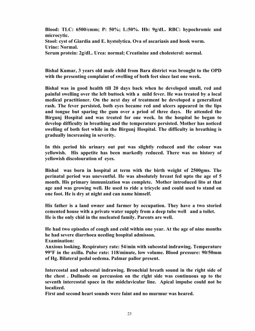
23
Blood: TLC: 6500/cmm; P: 50%; L:50%. Hb: 9g/dL. RBC: hypochromic and microcytic.
Stool: cyst of Giardia and E. hystolytica. Ova of ascariasis and hook worm. Urine: Normal. Serum protein: 2g/dL. Urea: normal; Creatinine and cholesterol: normal. Bishal Kumar, 3 years old male child from Bara district was brought to the OPD
with the presenting complaint of swelling of both feet since last one week. Bishal was in good health till 20 days back when he developed small, red and
painful swelling over the left buttock with a mild fever. He was treated by a local medical practitioner. On the next day of treatment he developed a generalized rash. The fever persisted, both eyes became red and ulcers appeared in the lips and tongue but sparing the gum over a priod of three days. He attended the Birgunj Hospital and was treated for one week. In the hospital he began to develop difficulty in breathing and the temperature persisted. Mother has noticed swelling of both feet while in the Birgunj Hospital. The difficulty in breathing is gradually incsreasing in severity.
In this period his urinary out put was slightly reduced and the colour was
yellowish. His appetite has been markedly reduced. There was no history of yellowish discolouration of eyes.
Bishal was born in hospital at term with the birth weight of 2500gms. The
perinatal period was uneventful. He was absolutely breast fed upto the age of 5 month. His primary immunization was complete. Mother introduced lito at that age and was growing well. He used to ride a tricycle and could used to stand on one foot. He is dry at night and can name himself.
His father is a land owner and farmer by occupation. They have a two storied
cemented house with a private water supply from a deep tube well and a toilet. He is the only chid in the nucleated family. Parents are well. He had two episodes of cough and cold within one year. At the age of nine months
he had severe diarrhoea needing hospital admisson. Examination: Anxious looking. Respiratory rate: 54/min with subcostal indrawing. Temperature
99°F in the axilla. Pulse rate: 118/minute, low volume. Blood pressure: 90/50mm of Hg. Bilateral pedal oedema. Palmar pallor present.
Intercostal and subcostal indrawing. Bronchial breath sound in the right side of
the chest . Dullnode on percussion on the right side was continuous up to the seventh intercostal space in the midclavicular line. Apical impulse could not be localized.
First and second heart sounds were faint and no murmur was heared.

24
Liver was 3 cms bellow the costal margin on the right side, soft and tender. Hepatojugalar reflux was present.
Investigation: Chest x-ray: homogenous opacity all over the right lung field with clear
costophrenic angle. Increased cardiac shadow with globular pattern. Blood: Ultrasound guided pericardial tapping revealed thick pus. 120 ml was aspirated.
Repeat x-ray showed marked improvement. Kamala, a three year old healthy looking female child from Baneshwar was brought in the OPD with the presenting complaints of swelling of both feet lasting for three days. This child was well till two weeks back when she developed mild cough and cold with slight rise of temperature. Her was treated by a physician and got better within a few days. Her mother noticed lethargy and loss of appetite after 10 days of the illness. She developed swelling of the both feet three days ago and this swelling is gradually increasing. Mother thinks her child's face looked puffy specially in the morning. Se has had two episodes of cough and cold and one episode of diarrhoea without any hcomplications in the past. Shee was born in Prasuti Griha at term with the birth weight of 2.7 kg. The Perinaatal period was uneventful and was on absolute breast feeding for the first four month of life. She is fully immunized with primary immunization. At present she can ride a tricycle and can tell her name. Her father is a officer in army and mother is a primary school teacher. They have their own three storied house at Baneshwar. She is the only daughter of the parent. Examination: Pulse: 96/minute. Respiratory rate: 28/min. Temperature:37°C. Blood pressure: 100/65 mm of Hg. Height 95cms. Weight:16 kg. Bilateral pedal oedema present. Palmar pallor present. Chest: no abnormal clinical signs. Abdomen: Non tender. Liver and spleen not palpable. Shifting dullness present. Child with convulsion: Do not write or put any mark on this paper. Please return these case notes. 1. A 9 month old male child was brought in the emergency by his mother with the presenting complaints of sudden loss of consciousness this morning.

25
This child was sleeping with his mother this afternoon. His mother suddenly noticed stiffening of the child's body. She also noticed saliva drooling from the angle of the mouth and a staring look. There was no jerky movement of limbs or body. He did not pass urine or feces in the nappy. She thinks the duration of this phenomenon lasted nearly for 3 minutes. The mother had an uneventful pregnancy. He was born at Prasuti Griha at term by normal vaginal delivery with the birth weight of 2570 gram. He had jaundice on the 3rd day of life but was discharged home on the fourth day. He was breast and bottle fed from the beginning. The mother started feeding him lito at the age of 4 months. He is not immunized with any vaccination because on the due date of vaccination, he used to develop cough and cold or have loose motions. He had not had any such episodes in the past. He sits without support and tries to communicate with his mother. His mother had episodes of convulsions till the age of 5 years. She was not treated with any anticonvulscant drugs. Examination findings: He was breast feeding. Respiratory rate was 50/minute. Temperature was 102°F in the axilla. Runny nose was present. Fontanel was not raised. No rashes were noticed. Chest indrawing was absent. Pharyngeal faces were mildly congested. Tonsils were enlarged but not congested. Cervical glands were not palpable. Auscultation was performed during breast feeding and no abnormal sounds were heard. Abdominal examination revealed: liver one cm bellow the R. midclavicular line. It was smooth and margin was sharp. No other abnormality noted. Investigations: 1.Blood. TLC: 6,900/cmm; P:46%, L:54% 2. Xray chest. Within normal limits. 3. CSF. TLC: 2/cmm; Protein: 40mg/dL; Glucose: 80mg/dL. 2. Sarala, 9 year old child was brought in the emergency by her father with the history of sudden onset unconsciousness lasting 35 minutes.

26
Sarala's father noticed sudden onset of generalized jerky movement this evening while she was lying in bed. She returned from school earlier today because of headache. She was complaining of headache and fever off and on since the last few weeks. Her headache was often associated with vomitting. During this episode of convulsion she remained unconscious for nearly one hour. The unconsciousness was associated with repeated episodes of jerky movement of whole body which lasted nearly for 10 minutes. It subsided on way to the hospital. Her father did not notice any change in her behavior during this illness. Six months back she was treated in the hospital for cough and fever with oral antibiotics. Her cough persisted and she was treated with herbal medicines. She felt better for few weeks but began to complain of lethargy after returning from school. Her appetite decreased gradually. Her father believes she has lost weight although she eats meat and other family foods frequently. She is studying in class 3 and is average in her class. She has one younger brother who is well. Mother died one year ago because of chronic cough for one year, she used to work in a carpet factory. Father also works as a peon in the same carpet factory. On examination: Unconscious, responds only to painful stimuli. Resp.rate: 28/ min. Pulse: 64/min. B.P. 110/85 mm of Hg. Temp: 37°F (axillary). Auscultatory findings of lungs and heart were normal. Planter reflex were equivocal. Jerks not exaggerated. Investigations: 1. Blood a. TLC: 9200/cmm; P:62%, L:32%, M:1%, E:5%. b. Glucose: 6.3 mmol/L; Urea: 6 mmol/L; Sodium: 136 meq/L; Potassium: 3.5 meq/L. c. CSF: TLC: 125/cmm; P:20%, L:80%, Protein:>100mg/dL; Glucose:46mg/dL. 3. A 12 year old male child was brought in the emergency with the history of sudden onset of jerky movement limited to the left lower limb, lasting for 15 minutes. Hari was well till this morning when he developed uncontrolled jerky movement of the left lower limb. He was conscious during this episode. His

27
temperature was not elevated. There were no rashes. This abnormal movement lasted for nearly 10 minutes. In the afternoon he again developed similar episodes lasting for nearly five minutes. After this episode he is feeling weakness in that leg. There is no such history in the past. He had not visited any areas outside this valley within one year. He is studying in class 8 at Jorpati High School and is average in class. His bowel and bladder functions are normal. He is a non vegetarian and has good appetite. He is immunized fully with primary immunizations only. He never suffered from any major illness in the past. All the family members are well. On examination: He is conscious and all the vital signs are normal. Only positive signs are: Power in the left lower limb is grade 4. Investigation: Blood: TLC: 11000/cmm; P:45%, L:43%, E:12%. ESR: 18mm in first hour. Sugar: 6.5 mmol/L; Calcium: 2.4 mmol/L. CSF: TLC: 2/cmm; Protein: 40mg/dL; Sugar: 60mg/dL. Mantaux test: 2mm. CTScan of brain (reported as): Ring shaped lesion with oedema sorrounding the area in the right parietal region. A 4 month old Bina presented in the OPD with the history of not moving her limbs since one week. Bina was born to a married housewife aged 19 years with normal vaginal delivary in Bhaktapur hospital. Her birth weight was 2300 gms. Her mother never attended the antenatal clinic. Her perinatal period was uneventful. She is absolutely breast fed. Her father is a truck driver and earns enough to maintain their daily life. They live in a two room apartment, one room is used for cooking and the other as a bed room. Bina received only one vaccination on the second day of her birth. Her mother first noticed excessive crying while moving her right upper limb two weeks back for which she attended a local medical shop. Examination findings: weight: 4.2 kg. Length: 59 cms. Temperature: 37.2°C. Respiratory rate: 46/min. Heart rate: 112/min. Jaundice: present over the sclera. Pallor: present. Cyanosis: absent. Nose: snuffles. Abdomen: hepatosplenomegaly. All limbs in paralytic position and cries during passive movement. Investigations:
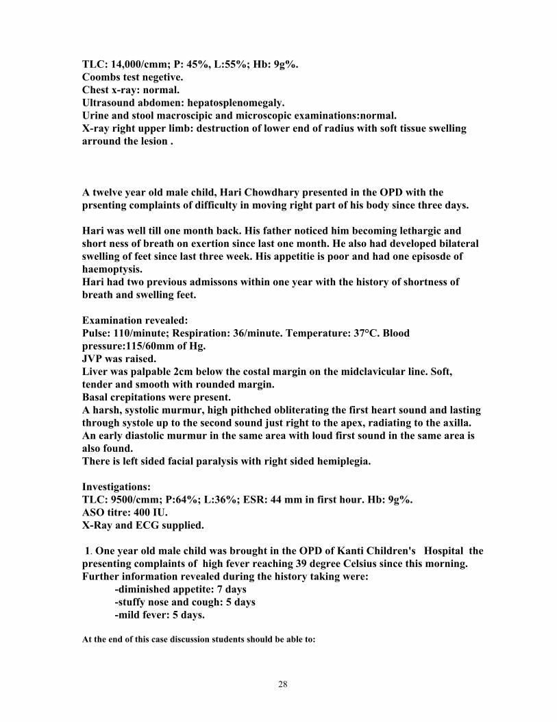
28
TLC: 14,000/cmm; P: 45%, L:55%; Hb: 9g%. Coombs test negetive. Chest x-ray: normal. Ultrasound abdomen: hepatosplenomegaly. Urine and stool macroscipic and microscopic examinations:normal. X-ray right upper limb: destruction of lower end of radius with soft tissue swelling arround the lesion . A twelve year old male child, Hari Chowdhary presented in the OPD with the prsenting complaints of difficulty in moving right part of his body since three days. Hari was well till one month back. His father noticed him becoming lethargic and short ness of breath on exertion since last one month. He also had developed bilateral swelling of feet since last three week. His appetitie is poor and had one episosde of haemoptysis. Hari had two previous admissons within one year with the history of shortness of breath and swelling feet. Examination revealed: Pulse: 110/minute; Respiration: 36/minute. Temperature: 37°C. Blood pressure:115/60mm of Hg. JVP was raised. Liver was palpable 2cm below the costal margin on the midclavicular line. Soft, tender and smooth with rounded margin. Basal crepitations were present. A harsh, systolic murmur, high pithched obliterating the first heart sound and lasting through systole up to the second sound just right to the apex, radiating to the axilla. An early diastolic murmur in the same area with loud first sound in the same area is also found. There is left sided facial paralysis with right sided hemiplegia. Investigations: TLC: 9500/cmm; P:64%; L:36%; ESR: 44 mm in first hour. Hb: 9g%. ASO titre: 400 IU. X-Ray and ECG supplied. 1. One year old male child was brought in the OPD of Kanti Children's Hospital the presenting complaints of high fever reaching 39 degree Celsius since this morning. Further information revealed during the history taking were: -diminished appetite: 7 days -stuffy nose and cough: 5 days -mild fever: 5 days. At the end of this case discussion students should be able to:

29
a- list the common causes of fever with cough. b- list the important signs of pneumonia; common cold, pharyngitis, laryngitis, laryngotracheobronchitis; epiglottitis. c- list the investigations for a child with a cough and fever (as listed in b). d- list the drugs and their doses used in a child with cough and fever. e- list the important points in counselling the mother of a child with cough and fever. 2. A 9 year old boy attended emergency complaining vomiting of blood. 8 weeks previously he had developed a mild, diffuse abdominal discomfort which was relieved by taking thick whitish liquid medicine as given by a medical shopkeeper. This patient was admitted in Nepalgunj Hospital one year back because of sudden profuse bleeding per mouth and black stool. During this episode he received one unit of whole blood. No further investigation reports of that episode were available. The second episode of bleeding per mouth also occurred 6 months back in Nepalgunj for which he was treated with anti tubercular drugs. Parent felt he was better and was taken home where he continued the anti tubercular drugs. The child was born to 28 year old women by spontaneous vaginal home delivery. Perinatal period was uneventful. The birth weight was comparable to other siblings. The child's early growth and development were normal. The more information on physical examination and investigations will be supplied during the discussion. Physical examination: Height : 102 cm. Weight : 14.3 kg. Temperature: 98º F orally. Pulse: 130, regular and of low volume. BP: 90/50 mm of Hg. Pallor: present. No rashes were noticed. Neck: three submandibular lymph nodes on both sides, non tender, mobile, less than ½ cm size. Abdomen: protuberant, everted umbilicus, prominent superficial veins. Liver: 1 cm bellow the MCL, non tender, firm and smooth. Spleen:2 cm bellow the left costal margin in the long axis, firm, smooth,and nontender. Shifting dullness: positive. Immediate investigations performed: Haematocrit: 13%, Haemoglobin 4.6 g%, WBC: 8700/cmm N: 33%, L: 56%. ESR: 40 mm/hr. Blood group: B +. Differential diagnosis: Bleeding from: -Oesophageal varices. -Peptic ulcer/gastritis. -Oesophagitis. -Haemangioma, arteriovenous malformations. Immediate management in the emergency room: -oxygen by nasal pronge. -iv normal saline/crystalloids through a canula. -arrange blood transfusion. One unit of whole blood, 52 ml/hour. -monitor blood pressure ½ hourly. -arrange for endoscopy.

30
Other investigations performed subsequently: Repeat haematocrit and haemoglobin. Liver function test: Alkaline phosphatase 40 U/L. Albumin 52g/L Bilirubin (total) 18 µ mol/L. Prothrombin time 2 seconds of control. HBsAg : negative. Chest x-ray ; supplied. Splenic venography: supplied. Ultra sound abdomen: free fluid in the peritoneal cavity, portal vein is dilated, hepatic vein is normal. Liver is normal and spleen is enlarged. Endoscopic examination: grade 3 oesophageal varices with congestive gastropathy. Mantaux test: negative. Diagnosis: Bleeding oesophageal varices secondary to extra hepatic portal hypertension. Management: 1. Immediate management: Nasogastric aspiration Blood transfusion. Ranitidine: 20 mg BD i.v. (1-3mg/kg) Vasopressin: 4.6 U over 20 minutes (0.33 U/kg). Continuous iv infusion at the rate of 0.2 U/1.73m²/min. Watch for the side effects of vasoconstriction. Endoscopic sclerosis. Sengstaken Blakemore tube compression of the oesophagus. 2. Prophylaxis for subsequent bleeding: Surgical prodedures: -endoscopic sclerosis. -portocaval, distasplenorenal shunts. ß-blockers: 1 mg/kg/24 hrs divided 6 hourly then increase up to 5 mg/kg/24 hrs. div 6 hrly to have the optimum effect. 3. A 6½- year old boy presented in the OPD with 1week history of intermittent pain in the left thigh and walking with a limp. For 2 weeks he has had colicky , abdominal pain with screaming episodes. There was no history of trauma and fever. He is an only child born at term by spontaneous vaginal delivary. He was bottle fed and had few episodes of diarrhoea during the neonatal period. He had mild jaundice in the first week of his life which was noticed on the 4th day . He had been treated twice for otitis media and had had recurrent cough . Immunizations were complete. His father is a truck driver and mother works in the field as a daily wage earner. Differential diagnosis: Arthritis of henoch schonlein purpura. Tubercular arthritis of hip. Transient synovitis of hip joint. Legg-calve-Paerthes Disease.

31
Physical examination: Weight: 11.5 kg. Height: 98.5 cms. Temperature: 97°F, axillary; Respiratory rate: 25/min; Pulse rate 88/minute. Pallor: mild. Oedema: absent. He had three cervical lymphnodes, which were descrete, nontender, smooth and mobile on both sides of neck over the posterior triangle. There were no rash. Liver 1cm below the costal margin and the spleen wasnot palpable. All of the joints appeared normal except the left hip joint where restriction of motion, specially abduction and internal rotation was observed. No bony tenderness were elicited. Investigations: Hb: 9g%. WBC: 6,000/cmm; P: 64%; L:36%. Platelets were adequate. Peripheral smear: Hypochromic, microcytic anaemia. ESR: 35 mm in first hour. Stool: Few fertilized ova of ascaris lumbricoides. Urine: normal. Anterior and Lauenstein lateral radiographs of the pelvis: The sphericity of the left femoral head is 2 mm less in comparison to the right; slight degeneration of the left femoral head. Diagnosis: Legg-Calves-Perthes Disease of left femoral head with anaemia, undernutrition and ascariasis. Management: 1.Bed rest or abduction stretching exesrcises to maintain mobility. Paracetamol for pain. Orthopaedic consultation is preferred. 2. Treatment of anaemia, undernutrition and ascariasis. 4. A 4 year old boy, presented in the emergency with the complaint of high fever and generalized severe abdominal pain of one day duration . There was a history of cough and cold a few days prior to the onset of present problems. He had one loose stool previous morning and there was no blood or mucous in it. He had mild headache off and on since last few months but history of vomiting was not present. His urine was of normal colour. He had a history of swelling of both legs three months ago for which he was admitted in the hospital for one week. His mother has lost the documents given by the hospital. Differential diagnosis: -basal pneumonia. -acute mesenteric lymphadenitis. -acute appendicits. -relapse nephrotic syndrome. -shigellosis. Physical examination:
32
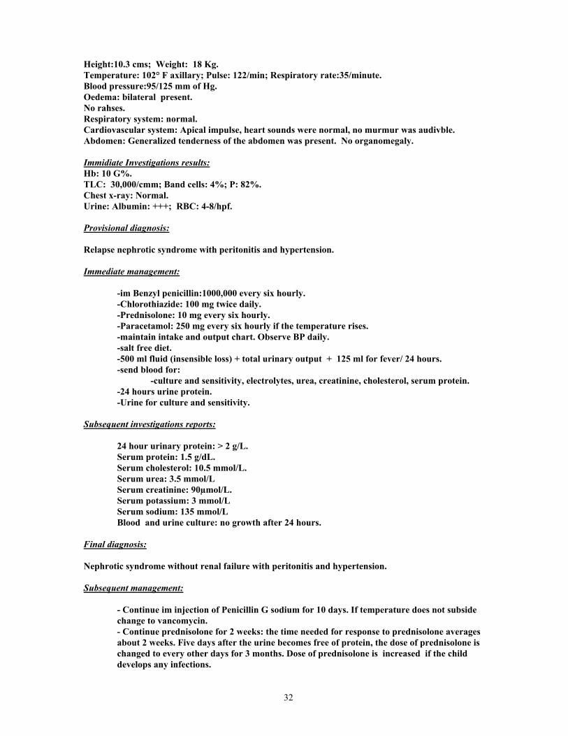
Height:10.3 cms; Weight: 18 Kg. Temperature: 102° F axillary; Pulse: 122/min; Respiratory rate:35/minute. Blood pressure:95/125 mm of Hg. Oedema: bilateral present. No rahses. Respiratory system: normal. Cardiovascular system: Apical impulse, heart sounds were normal, no murmur was audivble. Abdomen: Generalized tenderness of the abdomen was present. No organomegaly. Immidiate Investigations results: Hb: 10 G%. TLC: 30,000/cmm; Band cells: 4%; P: 82%. Chest x-ray: Normal. Urine: Albumin: +++; RBC: 4-8/hpf. Provisional diagnosis: Relapse nephrotic syndrome with peritonitis and hypertension. Immediate management: -im Benzyl penicillin:1000,000 every six hourly. -Chlorothiazide: 100 mg twice daily. -Prednisolone: 10 mg every six hourly. -Paracetamol: 250 mg every six hourly if the temperature rises. -maintain intake and output chart. Observe BP daily. -salt free diet. -500 ml fluid (insensible loss) + total urinary output + 125 ml for fever/ 24 hours. -send blood for: -culture and sensitivity, electrolytes, urea, creatinine, cholesterol, serum protein. -24 hours urine protein. -Urine for culture and sensitivity. Subsequent investigations reports: 24 hour urinary protein: > 2 g/L. Serum protein: 1.5 g/dL. Serum cholesterol: 10.5 mmol/L. Serum urea: 3.5 mmol/L Serum creatinine: 90µmol/L. Serum potassium: 3 mmol/L Serum sodium: 135 mmol/L Blood and urine culture: no growth after 24 hours. Final diagnosis: Nephrotic syndrome without renal failure with peritonitis and hypertension. Subsequent management: - Continue im injection of Penicillin G sodium for 10 days. If temperature does not subside change to vancomycin. - Continue prednisolone for 2 weeks: the time needed for response to prednisolone averages about 2 weeks. Five days after the urine becomes free of protein, the dose of prednisolone is changed to every other days for 3 months. Dose of prednisolone is increased if the child develops any infections.

33
If the child continues to have proteinuria (2+ or greater) after one month of continuous prednisolone the nephrotic syndrome is lebelled as steroid resistance and renal biopsy is idicate. -If hypokalaemia develops an oral potassium (36 meq/day) is supplemented and chlorthiazide is replaced with spironolactone. Once the blood pressure is controlled the antihypertensive is stopped. -Continue salt free diet till the oedema subsides and diuresis starts. - Observe the urine for proteinuria and asses for oedema. Recurrence of oedema or proteinuria is treated as a new case. If proteinuria or oedema reappears after stopping the steroid (steroid dependent), optimum dose of steroid is used to keep the urine free of protein. In case of repeated relapse and if the child suffers severe corticosteroid toxicity cyclophosphamide therapy is considered along with alternate day therapy of prednisolone. 5. A ten year old boy is admitted with a 5-day history of sore throat, cough, frontal headache and posterior neck pain. He had been seen by a health worker earlier in the week when a diagnosis of URTI was made and co-trimoxazole was prescribed. On the day before admisson he was seen in emergency in view of his lack of improvement, and changed onto erythromycin. Over the next 24 hours he had become progressively distressed with difficulty in breathing and neck pain. In the past he had had tonsillitis at the age of 5 years but no other major problem. His father, mother and 5-year old sister had been well and there was no known infectious contact. Differential diagnosis: -pneumonia, penumothorax, pleural effusion. -peritonsillar abscess, epiglottitis, retropharyngeal abscess. .CCF, pericardial effusion, myocarditis.- Examination: Pyrexial: 38.5° C (oral). Respiratory rate: 40/min. Pulse: 100/min, regular, with marked diminution in volume during inspiration. Blood pressure: 110/95 mm of Hg. Height: 138 cms. Weight: 28 kg. Right submandibular lymphnode: palpable, nontender, mobile and smooth. Breath sounds: no wheeze or stridor but bronchial brathing over a small area beneath the angle of left scapula. Apical impulse can not be lcocated. Heart sounds muffled, no murmur. Provisional diagnosis: -pericardial effusion. -pneumonia. Investigations: - Hb: 10.8g%; WBC: 11,800/cmm; P: 84%; send blood for culture and sensitivity. - Chest x-ray : enlarged csrdiac shadow with water bottle condiguration and clear lung fields.. ( provide the student x-ray film showing pericardial effusion). - Electrodardiography: Low voltages of QRS complexes; generalized mild elevation of ST segments and generalized T wave inversion. Working diagnosis:
34
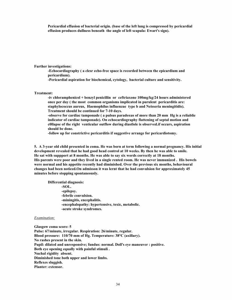
Pericardial effusion of bacterial origin. (base of the left lung is compressed by pericardial effusion produces dullness beneath the angle of left scapula: Ewart's sign). Further investigations: -Echocardiagraphy ( a clesr echo-free space is recorded between the epicardium and pericardium). -Pericardial aspiration for biochemical, cytology, bacterial culture and sensitivity. Treatment: -iv chloramphenicol + benzyl penicillin or ceftriaxone 100mg/kg/24 hours administered once per day ( the most common organisms implicated in purulent pericarditis are: staphylococcus aureus, Haemophilus influenzae type b and Neisseria meningitidis). Treatment should be continued for 7-10 days. -observe for cardiac tamponade ( a pulsus paradoxus of more than 20 mm Hg is a reliablie indicator of cardiac tamponade). On echocardiography flattening of septal motion and c0llapse of the right venticular outflow during diastlole is observed.if occurs, aspiration should be done. -follow up for constrictive pericarditis if suggestive arrange for pericardiotomy. 5. A 3-year old child presented in coma. He was born at term following a normal pregnancy. His initial development revealed that he had good head control at 10 weeks. By then he was able to smile. He sat with suppport at 8 months. He was able to say six words correctly at 18 months. His parents were poor and they lived in a single rented room. He was never immunized . His bowels were normal and his appetite recently had diminished. Over the previous six months, behavioural changes had been noticed.On admisson it was lernt that he had convulsion for approximately 45 minutes before stopping spontaneously. Differential diagnosis: -SOL. -epilepsy. -febrile convulsion. -miningitis, encephalitis. -encephalopathy: hypertensive, toxic, metabolic. -acute stroke syndromes. Examination: Glasgow coma score: 5 Pulse: 67/minute, irregular. Respiration: 26/minute, regular. Blood pressure: 110/70 mm of Hg. Temperature: 38°C (axillary). No rashes present in the skin. Pupil: dilated and unresponsive; fundus: normal. Doll's eye maneuver : positive. Both eye opening equally with painful stimuli . Nuchal rigidity absent. Diminished tone both upper and lower limbs. Reflexes sluggish. Planter: extensor.
35
Immediate management: -iv line is established with 5% dextrose. -blood is obtained for: complete blood count, electrolytes, glucose, creatinine and liver function test. Store 5ml of heparinized blood for later investigations. Investigations: -Dextrostix: blood glucose 4mmol/l. -TLC: 6700/cmm; P: 64%; L:36%, Hb: 11g%. - Na+: 137 mmol/l; K+: 3.2mmol/l; -creatinine:110µmol/l. Working diagnosis: -Postictal coma: -SOL. -Acute stroke syndromes. Further investigations: -MRI scan of brain. -EEG. -Cerebral angiography. 6. A 12 months old male child presented in the OPD with the presenting complaint of increased sweating particularly in the head since last few months. Mother has observed that the child is also floppy. The child was delivered at term by cesarean section because of contracted pelvis of the 30 year old mother. His birth weight was 2200 gms. During his Perinaatal period he had an episode of convulsion on the 48 hours of life and was treated in the nursery of the hospital. He was kept there for 48 hours. Mother had also noticed yellowish discoloration of skin on the fifth day but he was discharged from the hospital on sixth day of his life. Mother consulted a health worker for the jaundice and he prescribed an Ayurvedic medicine in drops. He also advised the mother not to do oil massage to the baby and not to take oily foods by herself. He was breast fed for the first six months of life. He is fully immunized. He had two episodes of pneumonia and two episodes of diarrhoea in the past. Differential diagnosis: -undernutrition, rickets. -Down's syndrome, mental defficiency. -benign congenital hypotonia. -neuromuscular disorders. Examination: Temperature: 36°C axillary. Pusle rate: 86/minute. Length: 70 cms. Weight: 8 Kg. OFC: 44 cms. Chest circumference: 43 cms. Anterior fontanalle: measuring 20mm in transverse diameter. Face: appeared normal. Chest: pigeon shaped. Both wrist swollen mildly but nontender, mobility normal. At present he sits with support and says 2 words with meaning. All reflexes are normal. All muscles are hypotonic. Working diagnosis: -rickets.

36
Investigations: X-ray wrist: cupping and fraying of the distal ends of radius and ulna. Serum phosphorus: 3.2 mg/dL. Serum calcium: 7mg/dL. Final diagnosis: Rickets. Management: - daily administration of 50-150µg of vitamin D³ or 0.5-2µg of of 1,25- dihydroxycholecalceferol for 4 week. -roentgenograms will show healing within 2-4 weeks. -15,000 µg of vitamin D in a single dose without further therapy for several months may be advantageous. -after healing is complete , the dose of vitamin D should be lowered to 10µg/day. -if no healing occurs, the rickets is probably resistant to vitamin D. 7. Thirteen year old female child presented in the OPD with the chief complaints of fever and upper abdominal pain of 5 days duration. Five years back she was operated for diagnostic lymphnode biopsy of the left axilla and subsequently treated with antitubercular drugs for nine months. Her father also had tuberculosis. Differential diagnosis: -hepatitis. -liver abscess (pyaemic, amoebic). -acute cholecystitis. -pneumonia, pleural effusion. -pericardial effusion, myocarditis. Examination:

37
Temperature: 39°C (axillary). Pulse rate: 116/min. Respiratory rate: 32/minute (thoraco-abdominal). Blood pressure: 115/80 mm of Hg. Weight: 40kg. Height: 148 cms. Pallor; jaundice; cyanosis and clubbing: absent. Abdominal examination: Tender right upper quadrant of abdomen. Liver: 4 cms bellow the R mid clavicular line. Soft, smooth and normal margin. Spleen not palpable. Working diagnosis: Liver abscess( pyogenic). Investigations: TLC: 15600.cmm; P: 78%. Hb: 9.6G%. ESR: 46 mm in first hour. Serum bilirubin: 18 micro mol/l. Serum alkaline phosphatase: 90 U/l. Blood culture for salmonella and non-salmonella organism: negetive. Stool for OPC: negetive. Ultrasound abdomen: mild hepatomegaly with an abscess in the left lobe mesuring 6 x4 cms. Final diagnosis: Pyaemic liver abscess. Treatment: 1. Ultrasound guided aspiration of the abscess. Send the aspirated fluid for culture and sensitivity. 2. iv. ceftriaxone 60mg/kg/day for one week. 3. Oral ferrous sulphate. (6mg/kg of elemental iron in three divided doses; ferrous sulphate is 20% elemental iron by weight) The aspirated fluid culture was positive for salmonella. Second batch: 2053. mangsir. 1. Five year old male child was brought to the emergency of Kanti Children's Hospital by his mother with the presenting complaints of : drowsiness since this morning. On further narration mother mentioned that the child has mild cough since one week. There was a shaking chill last evening which was followed by fever. This period was accompanied by restlessness, and occasionally delirium. Differential diagnosis: Pneumonia. Meningitis, encephalitis. Malaria Pyelonephritis. Septicaemia. Examination: He was lying on the right lateral side away from the window with the knees drawn up on the chest. Height: 100 cms. Weight: 11.4kg. Pulse rate: 128 /min. Respiratory rate: 51/min. Temp: 39° C. Blood pressure: 115/80 mm of Hg. Circumoral cyanosis was noticed. The right infra axillary region of chest revealed: increased fremitus, dullness on percussion, and tubular breath sound. Working diagnosis: Consolidation right lung probably bacterial (pneumococcal) origin .

38
Investigation: TLC: 4,000/cmm. P: 80%; Blood culture report awaited (30% of patients have bacteraemia) Chest x-ray supplied. Diagnosis: Consolidation R lung, severe bacterial (pneumococcal ) infection. Treatment plan: Admit. iv penicillin (100,000 units/kg/24 hours) Oxygen if the child develops respiratory distress through nasal canula at the rate of 2l/min Paracetamol 125 mg/dose every six hourly if the temperature is more than 39°C. Blood culture report: Gram positive, lancet-shaped, encapsulated diplococci isolated. 2. Mandira, aged 14 years female attended the OPD of Kanti Children's Hospital with the presenting complaints of prominent cheeks and headache of 4 and 1 month duration respectively and are not increasing significantly since then.The proominent cheeks are non tender. There is no history of fever. Her mother has observed deterioration in school work and deepening of voice since last six month. Differential diagnosis: -nephrotic syndrome on steroid. -cushing syndrome -SOL Examination: Pulse rate: 90/min; Respiratory ratae: 25/min; Temperature: 36.4 degree C (orally); Blood pressure: 135/100 mm of Hg. Height: 135 cms. Weight: 44 Kg. Pallor: absent; Jaundice: absent; Cyanosis: absent; Oedema: absent; Clubbing: absent. Hypertrichosis of face: present. Purplish striae were noted in the abdomen. Sexual maturity rating: stage 2. Other systemic examination: normal. Working diagnosis: Cushing syndrome. Investigations: TLC: 12000/cmm; P:82%; L:12% Urine routine examination: Normal. Stool: normal. ENT: openion for deepening of voice: Lyryngoscopic examination revealed normal anatomy. Ultrasound abdomen: Bilateral enlarged adrenal glands. Final diagnosis: Benign cortical adenoma. Further investigations: Blood cortisol level. Urinary cortisol. Dexamethasone supression test. CT scanning.

39
3. Anususan, a two year old girl was brought in the Emergency of Kanti Children's Hospital with the presenting complaints of fainting episode lasting for 10 minutes, this morning. There was no jerky movements or fever with this fainttng episode. Differential fiagnosis: -Cyanotic spell -Pallid spell -Cough syncope -The prolonged QT syndrome. The fainting episodes is not associated with excessive coughing, crying , vomiting or bed weting. There were no evidences of any precipitating factors. Since last one week she is breathing fast, coughing on and off, and not feeding well. Mother has noticed mild swelling of both feet since last few days and she has stopped playing since last one month. Anususan is not growing well in comparison with her other siblings. There was history of four such episode in the past each episode lasting for 5-10 minutes. There was no predilection of time for these faintings. There was no such history in the family. She was delivered at Prasuti Griha at term by normal vaginal delivary. Her birth weight was 2370 gms. Perinatal period was uneventful. She was fully breast fed for first 4months of her life and subsequently mother itroduced bottle milk and lito. After she was supplemented with bottle milk, mother had noticed she could suck the bottle continuously only for nearly 5 minutes. She is fully immunized and her developmental milestones were normal. During her first year of life she has one episode of diarrhoea which was preceeded by vomiting of one day duration. During this episode she was advised to take ORS at home. Eight months back she had cough and fever for which she was treated from the OPD of Kanti Children's Hospital for 5 days. Working diagnosis: - Syncope from congestive cardiac failure; secondary to congenital acyanotic heart disease. Exanination: Conscious Pulse: 123/ minute; low volume, regular. BP: not measured. Resp: 43/minute. T: 36.2 ºC (axillary). Extremities cold, pallor present; Cyanosis: absent. Oedema of feet present. Weight: 10 Kg; Height: 80 cms. All other systeic examination were normal except: Apical impulse on the left 5th intercostal space outsiede the midclavivular line. Systolic murmur present over the mitral and tricuspid area. Crepitations over scapular regions. Hepatomegaly: 3 cms below the right costal margin on the MCL. Clinical diagnosis: -congestive cardiac failure secondary to congenital acyanotic heart diseases VSD; Mitral /tricuspid regurgitation; cardiomyopathy. Investigation: CXR: Cardiomegaly and pulmonary congestions at base of both lungs. Echo: dilatation of left atrium and ventricle, and poor contractility. ECG: combination of atrial enlargement with left ventricular hypertophy.

40
Final Diagnosis: Primary dilated cardiomyopathy with clot in R ventiricle. 4. A 3 year old female child presented with a history of recurrent pallor , weakness and decreased appetite of almost two years duration. There was no history of associated fever, nausea, vomiting, diarrnoea , constipation, malaena, haematemesis, jaundice, skin lesions, dysuria, headache, incoordiation, or trauma. There was no associated loss of consciousness nor history of recurrent purulent infection. The family history included a mother with a history of recurrent abdominal pain and loose motion. Past history, developmental history, social history, and review of system were unremarkable. Differential diagnosis; Anaemia due to: Nutritonal Intrinsic factor Congenital RBC defficiency Drug causing deficiency Examination: Vitals: normal Height: 95 cms; weight:: 14 kg. Pallor present; jaundice, cyanosis, oedema : absent. Regional examination were normal except : smooth tongue. Systemic examination of the systems were normal except soft systolic murmur in the left second intercostal space. Working diagnosis: Anaemia of deficiencies: vitamin b 12; folic acid, iron. Investigations: Haemoglobin: 9.2% Haematocrit: 27% WBC: 10,000/cmm; PMN: 38% , large and hypersegmented; L: 50%; M:1%; E:1%. ESR:13mm/hr. Peripheral smear: macrocytic with prominent macro-ovalocytosis. Urine, stool: normal. LFT:normal; Diagnosis: Megaloblastic (pernicious) anaemia probably due to deficiency of intrinsic factor. Further investigations: (Vellore) Serum vitamin B 12 levels: < 100pg/ml. (low) Serum iron and folic acid is normal. Schilling test: abnormal Final diagnosis: juvenile pernicious anaemia. Treatment: Monthly im injection of 1 mg of vitamin B 12. 5. An 12 year old girl presented with a 4-week history of recurrent abdominal pain, epigastric location, gnawing in type, with radiation around to both sides and the adjacent back area, occurying almost daily for the past 1-2 weeks. There had been associated nausea and vomiting for almost one week. There was no history of fever, haematemesis, diarrhoea, malaena, skin lesions, anorexia, or trauma. The activity had remained essentially unchanged altough she seeme to tire more easily. Her bowel habit pattern had generally been once daily, soft and brown in nature. Medication had included antacid tablets. The family history

41
included a sister and maternal grandmother with diabetes mellitus. The remainder of the past history, family history, social history, developmental history, and review of systems were unremarkable. Differential diagnosis: Reflux oesophagitis: viral or peptic Functional abdominal pain Cholelithiasis Abdominal migraine/epilepsy Henoch-Schonleing purpura Examination: Vitals normal. Height: 145 cms; Weight 37 kg. All other system examination normal Working diagnosis: Reflux oesphagitis Investigation: Hb: 12.4 gm%; SGPT: 20 Units; Alkaline phosphatase: 195 Units; Amylase: 106 Units; Ultrosound abdomen: normal. Endoscopy: mild erythema with increasesed friability of the distal oesophagus. Final diagnosis: reflux oesophagitis: peptic. 6. SKC, 11 years old male child was brought in the OPD of KCH with the presenting complaints of cough lasting for 2 months and pain over the left side of chest of one and half month. His mother has noticed lethargy in SKC within this period. She has also noticed a swelling in the left side of the neck which is gradually increasing since then. There was no history of associated chills, rigor, vomiting, diarrhoea, constipation, haemoptysis, noisy breathing, headache, personality changes, joint pain, and contact history of tuberculosis.

42
Differential diagnosis: Tuberculosis of lung Hodgkins disease Non-Hodgkin Lymphoma Examnation: Pulse rate: 95/min; Respration rate: 60/min; Temperature: 36ºC axillary; Blood pressure: 110/75 mm of Hg. Pallor: ++; Cyanosis, jaundice, oedema and clubbing : absent. Lymphnode: single, over the posterior triangle of neck on the left side, nontender, firm, smooth and mobile, measuring 2.5 X 2 cms. Another over the left axilla: nontender, firm, smooth, mobile mesuring 4X4 cms. Respiratory system: subcostal indrawing present; right side of chest bulged; diminshed expansion ; trachea central; diminished breath sound and dull on percussion. Other system examination: normal. Working diagnosis: right sided plerual effusion with cervical and axillary lymphadenopathy, probably of tuberculous origin. Investigations: Pleural fluid aspiration: Sp.gravity: 1.034. Sugar: 30mg/dl. Protein: >100mg/dl. Cells: 20,00/cmm; N: 65%, L:45%; RBC:plenty. Blood: Hb: 9G%; WBC:5,100/cmm; N:45%, L:54%. FNAC of left axillary lymphnode: aspiration smear shows highly cellular and shows medium sized atypycal lymphocyte with scanty to thin basophilic cytoplasm and non cleaved nuclei compatible with Non Hodgkins Lymphoma. Final diagnosis: Non Hodgkins Lymphoma with metastasis to left cervical, right axillary lymphnodes and right pleural cavity. 7. Puspa., aged 11 years presented in the ER of KCH with the history of severe difficulty in breathing since last one day. His father has also noticed puffy face since then. On further questioning it was revealed that Puspa used to complain of pain mainly localized in the front of chest. There was no history of cough, vomiting, diarrhoea, constipation, noisy breathing, and dysuria.

43
Differential diagnosis: Myocarditis Pneumothorax Bornholm disease Examination: Pulse: 89/minute, regular and of low volume; Respiration: 35/minute and regular. Temperature: 36.2ºC. Blood pressure: 90/70 mmof Hg during inspiration and 110/75 mm of Hg during expiration. Patient in propped up position in respiratory distress. ema: present; Pallor, yanosis, jaundice and clubbin: absent. Cardiovascular system examination: Slight precordial bulge, apex beat is not visible, neck veins are prominent. Apical impulse is not palpable. Heart sounds are muffled and murmurs are not audible. Liver is 3 cm below the left costal margin on the mid clavicular line, tender, smooth, rounded margin and hepatojugular reflex is present. Respiratory system examination was normal except few bilateral basal crepitations. Working diagnosis: Pericardial effusion with cardiac tamponade probably of tubercualr origin. Investigation: CXR ECG: low voltage QRS complex, mild elevation of ST segment, some T wave inversion suggestive of pericardial effusion with pressure on the myocardium by fluid and associated myocardial inflammation. Echocardiogram: a clear echo free space is recorded between the epicardium and pericardium, there is also flattening of septal motion and contractlility is diminished. Pericardial aspiration: Colour: Straw coloured Protein: >3 gm%. Spicific gravity: 1.030. LDH: >200 iu. Cell count: L:78. Blood: TLC: 8700/cmm; P:40%, L:60%; ESR: 64 mm/hr. Mantaux test: 18 mm +; BCG scar +. Diggnosis: Pericardial effusion, with cardiac tamponade of tubercular aetiology.

44
8. A ninteen months old female child was seen in the OPD with the hsitory of high fever since last one week. Historical summary: Her mother did not have any antinatal check-up. She was born by normal vaginal delivery at home that was attended by her grand mother. Her birth weight was similar to the sibling. The umbilicus was cut with a new blade.She passed urine immediately after birth and meconium on the same evening. She was breast fed after nearly six hours of birth. On the fourth day of her life she developed yellow tinge on the skin. Her stool was dark yellow. The first 10 days of her life was uneventful. Her mother introduced lito in her second month. She did not receive any vaccination. She began to sit at six months and can identify her nose, mouth and eyes. She had three episodes of diarrhoea in her first year, each episode lasted for nearly one week and subsided without any treatment. Six months back she developed cough and fever for which she was treated by a local traditional medical practitioner and lasted for nearly two weeks and since then she is having cough on and off. She has not lost weight but she is not growing well in comparison with her brother who is now three years old. Six month back 56 year old relative came to stay in her house who was having cough. The detailed history of this visitor was not available. Her father, mother and brother are all well. Brother goes to local school and is studying in class one. Parent works in their own land. The earning from the land is sufficient to maintain their daily life. They also have one milking cow. They consume dairy products that is made by themselves. She feels warmer since last one month. At present she is lethargic and eats very little. She does not sleep well in the night and becomes irritable most of the time. She has 2-3 loose motions daily which is often blood stained and mixed with mucous. She is passing urine normally. Her abdomen seems to be distended within this period. She is being treated by a local practitioner who has prescribed tablets that are to be given mixed with water three times daily and a three different bottles of liquid. Examination: Temperature: 101ºF(axillary); Pulse: 124/minute, regular and normal volume. Respiration: 54/minute. Blood pressure: 90/60 mm of Hg.
45
Pallor and pedal oedema present. cyanosis, jaundice, and clubbing were absent. Weight: 9Kg; Height: 75 cms. MUAC: 12.5cms. OFC: 46cms. Chest circumference: 44 cms. Anterior fontanalle: open. Hair: brownish, lustureless, and easily pluckeble. Teeth: 8 in number. Tonsills were enlarged but not congested. Erythematous, sigle punctate lesion was noticed in the right eye over the lateral part of sclera. Tender erythematous nodules were present over the front of both legs. Three cervical lymphnodes were palpable on the right posterior triangle: each masuring 1cm x.5cm, non tender, firm, mobile and smooth. Bilateral basal crepetations were audible on both the lung fields. The breath sounds were vesicular except ronchi was noticed over the right infra axillary region. Heart sounds were normal. Liver and spleen was palpable 2 cms bellow the subcostal margin, and were nontender, smooth, firm with normal border. Shifting dullness was present. Investigations: Blood: TLC: P: 26%, L: 64%, E: 8%, M: 2%. ESR: 42mm in first hour. Hb: 9g%. CXR: milliary shadow. Ultrasound abdo: Hepatosplenomegaly with genralized hyperechogenicity, ascitis and paraaortic lymphadenopathy. Mantaux test: 18mm. FNAC of cervical lymphadenopathy: Central area of caseation with Langhans giant cells and plenty of lymphocyts in the periphery. Stool: mucous and red blood cells present. OPC: absent. Culture within 48 hours: sterile. Urine: normal. Diagnosis: milliary tuberculosis, involving lung, cervical lymphnodes, liver, spleen, intestine with PEM., phlyctenular conjunctivits and erythema nodosum. 9. Anil Kumar aged 45 days was brought in the ER by his mother with the presenting complaint of difficulty in sucking since last one week. Anil was deliverd in the TUTH at term by elective caeserian section for CPD. The APGAR was 6 at one minute and 10 at five minutes. His birth weight was 3400 gms. He is having breast milk only and mother beleives that she is

46
producing enough milk for Anil. She had regular ante-natal check-up in TUTH. Anil was vaccinated with BCG on the second day of his life. Anil has one sister aged 3 years and she is healthy. His father owns a carpet factory and earning is satisfactory. Mother is a high school teacher science teacher. She is planning to join school after two months. Differential diagnosis: -cngenital defects: cardiac defects: cyanotic/ CCF. respiratory defects: TOE/ partial agenesis. -infections: pneumonias, septicaemia. -metabolic: hypoglycaemia Further informations: Anil is sucking frequently but can not suck for long. He stops after sucking for 2-3 minutes. Mother feels the oral temperature of Anil normal while suckling. He is not having cough excessively but she has noticed blueness in the perioral regions off and on. Examination findings: Resp.rate: 64/minute. Temperature, rectal: 97.4°F. Pulse: 135/min. Cyanosis: central, present. Pedal oedema: not detected. Pallor: absent. Alert but tachypnoeic and having subcostal and severe intercostal indrawing. OFC: 36 cms. Weight: 3.4 kg. Length:52 cms. Scalp, fontanalle, face, oral cavity are normal. Chest: Systolic murmur in the precordium more prominent over the xiphisternum. Lung fields: crepitations not heard. Abdomen: massive swelling on the left hypochondrium moving with respiration, soft and smooth. No mass felt on the right hypochondrium Umbilicus and hernial orifices normal. Diagnosis: congenital cyanotic heart disease, dextrocardia with failure. Suggested investigations: Chest x-ray. Echo-cardiography. Diagnosis: Dextrocardia with tricuspid regurgitation, ASD and pulmonary arterial hypertension.

47
Your presentation should be based on: - The differential diagnosis based on given information and important positive and negative points in history and examination that will help to establish the diagnosis*. -Investigations that are needed to establish the diagnosis with priority*. - Suggested management plan. * you must give reasons . 10. Mrs. Gauri attended the OPD of KCH with her son Gyanu, who is now 5 weeks old. She brought Gyanu to this OPD for general check-up as she was advised to do so during her discharge from TUTH. Gyanu was delivered normally with the birtht weight of 2500 gms at term. The APGAR score was 8 at one minute and 10 at 5 minutes. Mrs. Gauri was discharged from the hospital on the second day. She is breast feeding her baby whenever he cries. This is her first baby. Her husband works in military and she is a carpet weaver. Both of them are healthy. On asking for further informations it was revealed that Gyanu is not growing well and vomits off and on. He is not having fever and sucks well. Mrs. Gauri beleives her son is breathing normally and is not coughing but sometimes sneezes. He is passing urine and stool normally. Differential diagnosis: - faulty technique of breast feeding. - gastrooesophageal reflux. - milk curd syndrome - hiatal hernia, hypertrophic pyloric stenosis. - UTI/ Renal tubular lesions. - Adrenal insufficency. - inborn errors of metabolism. Further information: Gyanu sucks milk every 2-3 hourly. Sucking is very powerful. She also burps after each feeding. The vomitus is nonbilious in nature. The vomiting would occur occasionally within 15-30 minutes after some of feed. The vomiting was forceful but Mrs. Gauri did not feel it was projectile. There was no association of haemetesis, jaundice, convulsion or skin lesions. Examination: He was a relatively alert, thin infant in no apparent distress and with normal virtal signs. . Head circumference was 33 cm, weight 3 kg, length 50 cm.
48
Palpation of the abdomen did not reveal any abnormal mass. Active bowel sounds were present with no bruits. No peristaltic waves were noted. The remainder of the examination including genitalia were normal. Investigarions: Hb: 13.2 gm%; Haematocrit: 35%; WBC: 9,200/cmm (P:45%, L:48%, M:5%, E:2%); Platelets: 225,000/cmm. Serum: Na 132 meq/L; K 3.8 meq/L; Cl 92 meq/L; albumin 4.6g%; SGOT 24 units; alkaline phosphatase 90 Units;. Urine: normal. CXR: normal. Diagnosis: Congenital hypertrophic pyloric stenosis. Investigarions: Ultrasound abdomen: normal. Upper GI series: normal stomach and pylorus (X-ray 6a and b). Final diagnosis: Milk curd syndrome. - Jestha 2054: 1. Seven months old Mekhmaya was brought in the OPD by her mother with the complaints of loose motions off and on since 2 months and lethargy since this morning. Further historical informations: Home delivary. Cried immediatly after birth. Started breast feeding within two hour of delivary and the baby sucked well. Absolute breast feeding for first six months of life. Weanling started with rice eating ceremony and since then diarrhoea started. She is not putting on weight as before once she started eating lito and bottle at six months. She had two injections given in the upper right thigh six weeks apart when she was 2 months old. She stared sitting mommentarily without support at six months but she can not sit at present. Her mother is 19 year old and a carpet weaver, father also works as a daily wage earner. They live in a rented small room in Boudha. Mekhmaya is their only child. Examination findings: Length: 63 cms. Weight: 3.6 Kg. Head circumference: 43 cms,
49
chest circumference:42 cms. Pulse: 96/min, equally palpable in all the limbs. Resp.rate: 36/minute. Temperature: axillary 35°C. General appearences: slides provided. Investigations: Blood glucose: 56 mg/dl. Questions: 1. Diagnosis: Hypoglycaemia, hypothermia, severe undernutrition, chronic diarrhoea 2. Immediate mangement: a. 1-2ml/kg of 50% dextrose iv immediatly. continue with 1/5 normal saline in 10% dextrose. b. cover with blanket, keep in warm room in mothers lap. c. send blood for electrolytes. d. order for portable chest x-ray. e. send stool sample for OPC/pus cells and culture, senstivity. Further management: Calorie calculation and feeding. Nutrition eduacation. Case 2: Ramila is a 13 month old child. Her mother brought to the emergency at mid night because she has had severe difficulty breathing since last four hours. Previous day she was seen in the OPD for cough and fever. Historical summary: Ramila was well till 4 days back when she developed mild cough and cold after comming from a fair riding a motor-bike. Mother provided her paracetamol and a cough mixture for two days. Fever persisted and she bgan to breathe faster but was able to drink, for which she attended the OPD. Ramila was prescribed ampicillin and went home. She continued to develop high fever and fever persisted. Suddenly in the night she became

50
more irritable and her mother noticed she was grunting and was having severe difficulty in breathing. The OPD notes were as follows: Weight: 9.5 Kg. Immunization: complete. Temperature: axillary 39°C. Respiratory rate: 56/minute. Stridor and wheeze were absent. Subcostal indrawing: absent. Basal crepitations present over subscapular region bilaterally. Emergency examination: Grunting and restless. Not able to drink. Marked subcostal indrawing. Respiratory rate: 96/min. Not dehydrated. Temperature: 38.5°C Difficult to examine for the other clinical signs. Urgent Chest X-ray: provided. A case of pneumothorax. Further management: a. Oxygen Nasal canula at the rate of 1L/min. b. iv 1/5th Normal saline in 10% dextrose: 1000ml/24 hours. c. iv ceftriaxone 500 mg stat. d. blood for TLC/ differential and film. Further investigative informations: Blood: TLC: 19,200/cmm. P: 86%;L: 10%; Band cells 4%; shift to the left. Case 3 A five year old male child presented in the ER with the history of sudden loss of consciousness lasting for one hour. He was complaining of headache for the last few days.

51
Further relevant informations: Three weeks prior he had mild cough associated with fever. Mother had also noticed lethargy, fast breathing and occasional vomiting for the last one week. Examination findings: Unconscious. Pulse: 82/minute, bounding. Respiration: rate- 40/minute; abdomino-thoracic type. Temperature: 37.5°C. Blood pressure: 160/110 mm of Hg. Oedema: pitting, bilateral over foot. Liver: 3cms palpable below the costal margin on the mid clavicular line: soft, smooth with rounded margin. Bilateral basal crepitations. Glasgow coma scale (total score=7): best motor response-withdraws (flexion); verbal response- nil; eye opening- to pain. The right leg was extended and externally rotated. Deviation of the angle of the mouth towards the left side on deep pressure over the supraorbital ridge. Ankle jerk exaggerated on the right side. Babinski's sign: up-going great toe on the right side. Investigations: Blood: Hb- 9 g%; TLC- 9500/cmm; P- 58%; L-42%. glusose- 82mg/dl; urea- 10.2 mmol/l; creatinine- 200 micro mol/l; sodium- 136 mEq/l. Potassium- 4 mEq/l. X-ray chest: provided. Catheter urine specimen: RBC- plenty; Protein-absent. Next days reports: ASO titre: 552 units; CRP- positive. C3-reduced. Case 4 A 3 year old healthy looking female child was brought in the OPD with the presenting complaints swelling of both feet since last three days.

52
Further relevant informations: This child had mild cough and cold few days back. Mother had noticed swelling arround both eyes specially in morning for the last 5 days. Examination findings: Pulse: 96/minute. Respiratory rate: 28/min. Temperature: 37°C. BP: 100/65 mm of Hg. Height: 95 cms. Weight: 16 Kg. Bilateral pitting pedal oedema. Periorbital oedema. Shifting dullness present. Investigations: Urine: Protein- ++++; RBC- 4-8/hpf. Blood: Hb- 10 g%; TLC- 8900/cmm; P-48%, L-52% Serum albumin: 20 g/l; cholesterol: 10.5 mmol/l. Urea: 6.5 mmol/l; creatinine: 55 micro mol/l Na: 135 mEq/l; Potassium: 4 mEq/l. 24 hour urinary protein: 2.8 g/24 hrs. Serum calcium: 1.8 mmol/l. C3 normal. Case 5 A nine year old male child from Trisuli was seen in the OPD with the presenting comlaints of difficulty in breathing since last two weeks. There is no history of cough or fever within two weeks but mild coryzal symptoms were noted one month back. Further relevant informations: He was born at home without any perinatal problems. His neonatal and infancy periods were uneventful. He is fully immunized with primary vaccinations. There is no previous history of joint pain or similar episode. He is studying in class three in the local school which is half hour walk from his home and the performace is well. He was seen in the Trishuli hospital last week and was prescribed medicine but without any improvement. Examination findings:
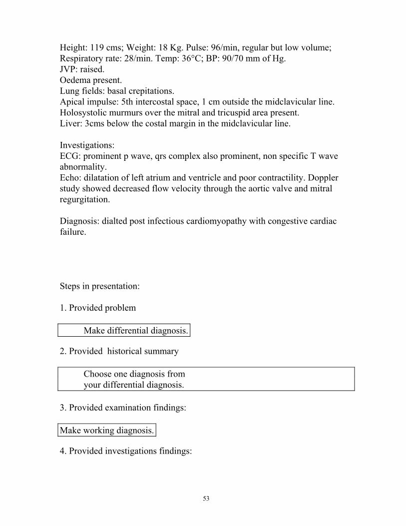
53
Height: 119 cms; Weight: 18 Kg. Pulse: 96/min, regular but low volume; Respiratory rate: 28/min. Temp: 36°C; BP: 90/70 mm of Hg. JVP: raised. Oedema present. Lung fields: basal crepitations. Apical impulse: 5th intercostal space, 1 cm outside the midclavicular line. Holosystolic murmurs over the mitral and tricuspid area present. Liver: 3cms below the costal margin in the midclavicular line. Investigations: ECG: prominent p wave, qrs complex also prominent, non specific T wave abnormality. Echo: dilatation of left atrium and ventricle and poor contractility. Doppler study showed decreased flow velocity through the aortic valve and mitral regurgitation. Diagnosis: dialted post infectious cardiomyopathy with congestive cardiac failure. Steps in presentation: 1. Provided problem
2. Provided historical summary
3. Provided examination findings:
4. Provided investigations findings:
Make differential diagnosis.
Choose one diagnosis from your differential diagnosis.
Make working diagnosis.

54
Make a clinical diagnosis. Outline the manaement plan.