Fossil remain of fungis alga, aned other organisms from ...
41
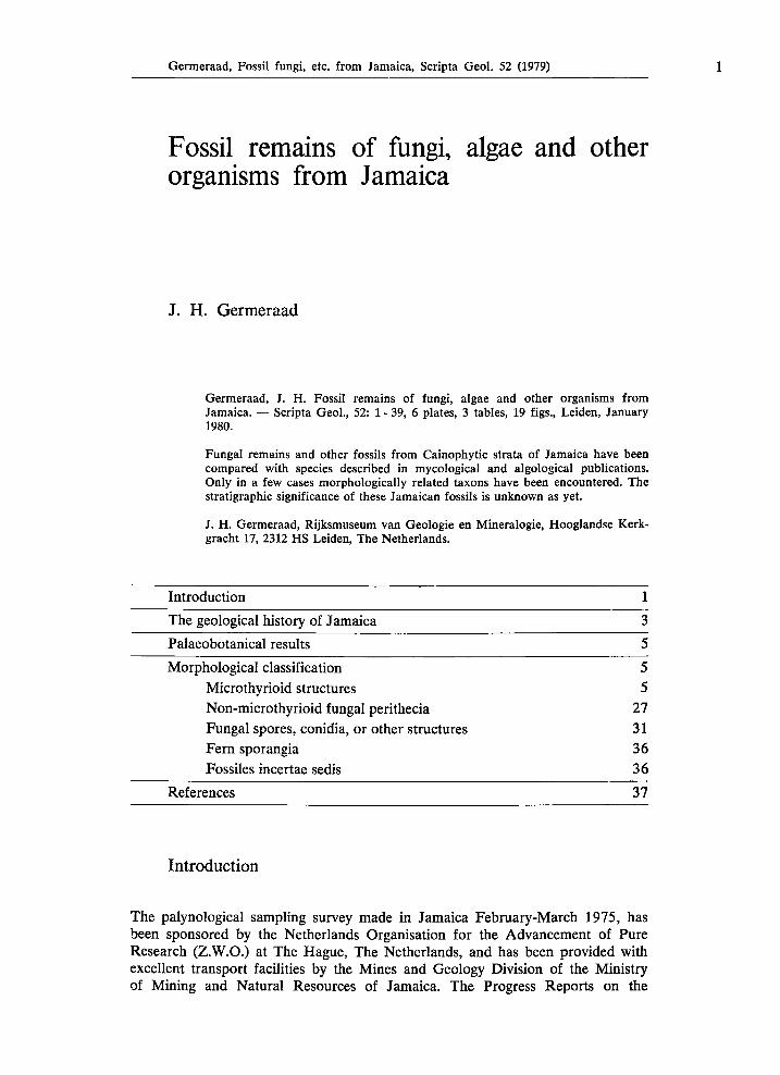
Germeraad, Fossil fungi, etc. from Jamaica, Scripta Geol. 52 (1979) 1 Fossil remains of fungi, algae and other organisms from Jamaica J. H. Germeraad Germeraad, J. H . Fossil remains of fungi, algae and other organisms from Jamaica. — Scripta Geol., 52: 1-39, 6 plates, 3 tables, 19 figs., Leiden, January 1980. Fungal remains and other fossils from Cainophytic strata of Jamaica have been compared with species described in mycological and algological publications. Only in a few cases morphologically related taxons have been encountered. The stratigraphie significance of these Jamaican fossils is unknown as yet. J. H . Germeraad, Rijksmuseum van Geologie en Mineralogie, Hooglandse Kerk- gracht 17, 2312 HS Leiden, The Netherlands. Introduction 1 The geological history of Jamaica 3 Palaeobotanical results 5 Morphological classification 5 Microthyrioid structures 5 Non-microthyrioid fungal perithecia 27 Fungal spores, conidia, or other structures 31 Fern sporangia 36 Fossiles incertae sedis 36 References 37 Introduction The palynological sampling survey made in Jamaica February-March 1975, has been sponsored by the Netherlands Organisation for the Advancement of Pure Research (Z.W.O.) at The Hague, The Netherlands, and has been provided with excellent transport facilities by the Mines and Geology Division of the Ministry of Mining and Natural Resources of Jamaica. The Progress Reports on the
Transcript of Fossil remain of fungis alga, aned other organisms from ...
Germeraad, Fossil fungi, etc. from Jamaica, Scripta Geol. 52 (1979) 1
Fossil remains of fungi, algae and other organisms from Jamaica
J. H. Germeraad
Germeraad, J. H. Fossil remains of fungi, algae and other organisms from Jamaica. — Scripta Geol., 52: 1-39, 6 plates, 3 tables, 19 figs., Leiden, January 1980.
Fungal remains and other fossils from Cainophytic strata of Jamaica have been compared with species described in mycological and algological publications. Only in a few cases morphologically related taxons have been encountered. The stratigraphie significance of these Jamaican fossils is unknown as yet.
J. H. Germeraad, Rijksmuseum van Geologie en Mineralogie, Hooglandse Kerk-gracht 17, 2312 HS Leiden, The Netherlands.
Introduction 1
The geological history of Jamaica 3 Palaeobotanical results 5
Morphological classification 5 Microthyrioid structures 5 Non-microthyrioid fungal perithecia 27 Fungal spores, conidia, or other structures 31 Fern sporangia 36 Fossiles incertae sedis 36
References 37
Introduction
The palynological sampling survey made in Jamaica February-March 1975, has been sponsored by the Netherlands Organisation for the Advancement of Pure Research (Z.W.O.) at The Hague, The Netherlands, and has been provided with excellent transport facilities by the Mines and Geology Division of the Ministry of Mining and Natural Resources of Jamaica. The Progress Reports on the
2 Germeraad, Fossil fungi, etc. from Jamaica, Scripta Geol. 52 (1979)
palynological investigation of the samples collected in Jamaica are mentioned in the list of references (Germeraad, 1978, 1979).
In the sampleresidues from the Jamaican Cainophytic strata fungal remains and other dark brown fossils were frequently observed. As far as possible single grain slides were made, the grains were arranged in morphotypes, each with a unique JAMnumber; the fossils were photographed and their morphological features were described and measured. Subsequently the types were compared with species described in the botanical and palaeobotanical literature to which ample reference is made in this article. A rather complete bibliography (about 700 titles) of all related literature can be obtained from the author upon request.
As so many palynologists are unfamiliar with the subject, and in order to stimulate the investigation of fungal remains, illustrations from publications of various authors have been copied and added to elucidate the text. As regards the postwar articles permission to do so was generously granted by Dr Ε. Müller of the Eidgenössische Technische Hochschule in Zürich, by Dr J. A . von Arx of the Centraal Bureau Schimmelcultures in Baarn and by Dr Ε. S. Luttrell of the Georgia Experiment Station of the University of Georgia, and the editor in chief of Mycologia, New York Botanical Garden, Bronx, New York.
From the very beginning of this study the expert guidance and constructive criticism of Dr R. A . Maas Geesteranus (Rijksherbarium, Leiden) were highly valued by the author, especially so, as the difficulties of identification were encountered at several levels: 1) The literature to be consulted is widely scattered. Apart from a series of hand
books on the classification of fungi in which in general only the genera and some
times the typespecies are defined, the majority of the species are described not only in the vast literature on mycology and paleobotany, but also in a wide range of periodicals that cover subjects from geology to phytopathology and allergology. Quite helpful proved the Bibliography of Systematic Mycology. 2) A part of the literature found in reference lists is rather old, not easy to consult, and often deals almost exclusively with the fungushost relationship sensu lato, while in old palaeobotanical publications microscopical details are often scarce or of little use. 3) In the description of extant fungus species the characteristics of the organs which are (partly) fossilizable, are not or very rarely specifically mentioned as such; much more emphasis is placed on biological aspects like growth, reproduc
tion and host relationship. 4) The number of living fungus species with fossilizable parts or spores is only a fraction of the number of totally unfossilizable species; it is not known whether, in the Cainophytic, the relatives of this majority never had fossilizable parts or spores. 5) The majority of the fungal spores have a simple structure and are hard to distinguish from each other; they may be spherical or spindleshaped, without or with septa, sometimes in irregular arrangement. Therefore it is questionable whether good stratigraphie markers will be found among the simple fossil spores. 6) An ornamentation is lacking in most spores; if present, rather simple Verrucae, echinae, or reticula prevail. These ornamental features have often been described as though not having much detail, and therefore are not very promising for strat
igraphie studies either, nor useful for determining the taxon of the fossil speci
men; more conspicuous features are needed.
Germeraad, Fossil fungi, etc. from Jamaica, Scripta Geol. 52 (1979) 3
7) Of some fossil structures the fungal origin seems doubtful. In a few cases an algal origin may be assumed. Supporting evidence may come from physico-chemical analysis (Good & Chapman, 1978; van Gijzel, 1966, 1967) but such investigations are scarce as yet. Those fossils that are assumably neither fungal nor algal will be even more difficult to identify.
The geological history of Jamaica
The geology of Jamaica is complex and, of course, can be explained here only in a highly condensed, simplified way (see geological map, Fig. 1). For more detailed information the reader is referred to the literature on the geology of Jamaica, among which the Synopsis by Zans et al. (1962) is recommended. From the viewpoint of a palynologist the floral changes which are related to the vertical movements of the island and the changing possibilities of plant-migration from nearby regions are the most fascinating aspects.
The oldest palynomorphs observed are of Carboniferous-Permian age, found in reworked position in Upper Cretaceous tuffs and shales (Germeraad, 1979a). The Upper Cretaceous flora itself has its own, probably insular character, differing essentially from the known Venezuelan, Colombian and Brazilian floras (Germeraad, Hopping & Muller, 1968; Herngreen, 1975a,b, 1976; Germeraad, 1978b). From the restricted information available at present, the floral changes within the Jamaican Upper Cretaceous and up into the Palaeocene appear gradual, with a distinct impoverishment in the Palaeocene, suggesting a gradual downward movement of the island with a reduction in the number of different environments. The Lower Eocene part of the Richmond Formation in the eastern section of the island contains beautifully preserved pollen grains and spores, in contrast with the sporomorphs of the nearby Palaeocene Providence Shales, thus supporting the assumption that this part of the Lower Eocene did not participate in the preceding tectonic cycle of the Upper Cretaceous and Palaeocene. The entirely different composition of the Eocene flora also suggests that a more or less complete submergence took place between the Palaeocene and this part of the Eocene, followed by the immigration of a new vegetation. In younger Eocene strata the flora shows an enrichment, obviously owing to the rising of this or a neighbouring island with a greater number of different types of environment. However, somewhere in the Middle Eocene a new downward movement started, not only noticeable in the increasingly marine facies of the spreading Yellow Limestone Formation, but also obvious from the decreasing number of pollen types. The almost complete disappearance of pollen in the above-lying White Limestone Formation suggests the (near-)total submergence of the island. The assumption of the field-geologists that the boundaries between the formations mentioned (Richmond Formation, Yellow Limestone Formation, White Limestone Formation) should not be regarded as time-levels, is confirmed by the palynological investigation (Germeraad, 1978a).
The next emergence of the island by young tectonic movements, starting in Miocene time and continuing, be it at a slower pace, afterwards, resulted in the geomorphology that, in broad outlines, exists today. In the southeast of the island the rising post-Eocene sediments slumped downwards, possibly already before

Fig
. 1.
Geo
logi
cal
map
of
Jam
aica
, a
slig
htly
mod
ified
cop
y of
the
map
of
Wri
ght (
1974
, p.
11).
Germeraad, Fossil fungi, etc. from Jamaica, Scripta Geol. 52 (1979) 5
emerging above sealevel, and these aggregated masses now compose the Bowden Formation. At many places along the coast today Pliocene - Quaternary sediments are found raised above sealevel; they generally contain very few pollen. In the peat deposits of the Great Morasses at the east and west end of the island and in the marshy deposits at Bowden a Holocene flora has been observed, very much resembling the Recent one.
From the geological map it can be seen that the eastern part of the island, with the Blue Mountains as its centre, is different from the rest of Jamaica. In this respect especially the Upper Cretaceous is intriguing, because a difference in the floras of east and west Jamaica may explain part of the pre-Tertiary tectonic history of the island (e.g. a possible post-Cretaceous unision of the two parts). As yet no pollen have been found in the available samples, but new material has lately been collected by Dr J. P. Krijnen.
Palaeobotanical results
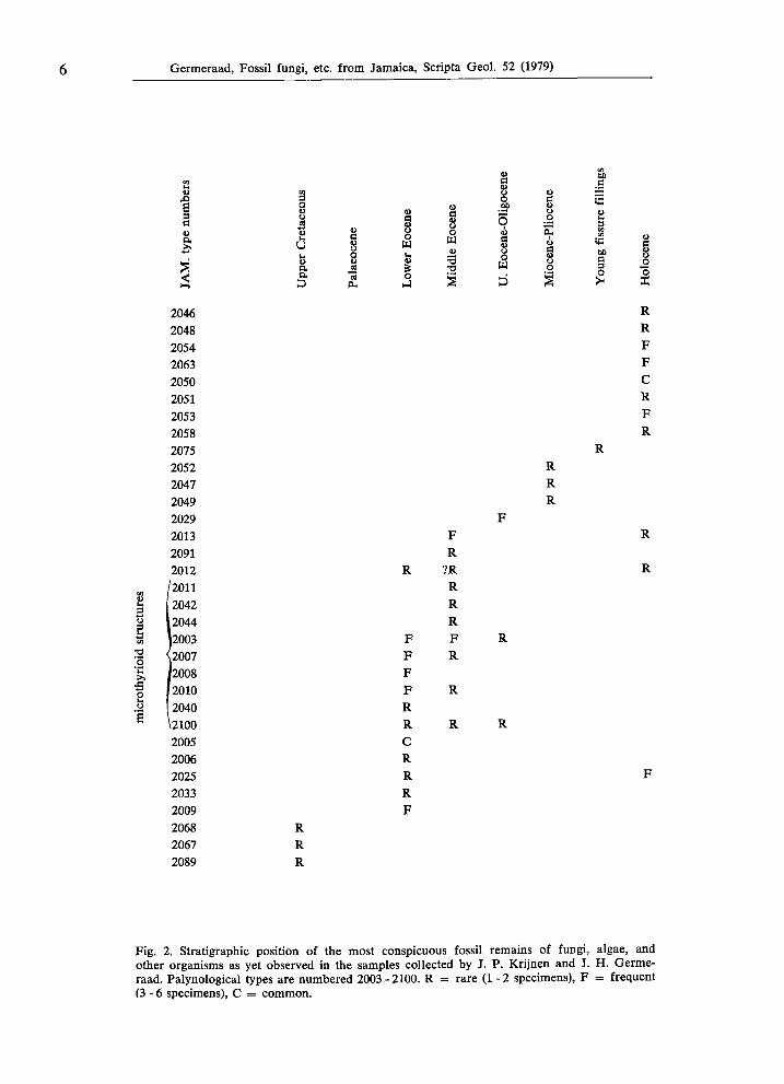
The stratigraphie results of studies like the present one are very limited owing to the restricted amount of material examined as yet (Sheffy & Dilcher, 1971, p. 49). As stated above the simple-structured fossils appear less promising; the emphasis should be laid on the study of more conspicuous forms. In time a compilation of the pan-tropical, or even worldwide data may yield, for some types, valuable stratigraphie ranges which may be interpreted either as a time-range or as a range resulting from geographically imposed environmental or sedimentary influence (Fig. 2).
Morphological classification
It would have been preferable to present the descriptions of the observed fossils in an arrangement based on assumed taxonomie position. Unfortunately that is not easily done. The following morphological interpretational classification seems feasible: I. Microthyrioid structures, comprising fungi and perhaps algae and lichenes:
types J A M . 2003, 2007, 2008, 2010, 2011, 2040, 2042, 2044. II. Non-microthyrioid fungal perithecia: types J A M . 2052, 2091. III. Fungal spores, conidia, or other structures: types J A M . 2005, 2006, 2012,
2013, 2025, 2029, 2033, 2046, 2047, 2048, 2049, 2054, 2063, 2068, 2075. IV. Fern sporangia: type J A M . 2009. V . Fossiles incertae sedis: types J A M . 2050, 2051, 2053, 2058, 2067, 2089.
MICROTHYRIOID STRUCTURES
Isolated, more or less circular disks composed of radially arranged cells, resembling at first glance the fruitbodies of Microthyriales (Fungi) detached from their
6 Germeraad, Fossil fungi, etc. from Jamaica, Scripta Geol. 52 (1979)
Fig. 2. Stratigraphie position of the most conspicuous fossil remains of fungi, algae, and other organisms as yet observed in the samples collected by J. P. Krijnen and J. H. Germe
raad. Palynological types are numbered 2003 2100. R = rare (1 2 specimens), F = frequent (36 specimens), C = common.
co ö
g
§
co Ο Ο α •4-» δ U
s
s υ δ
S 8 ο W u
Ο H4
I ο m τ3
S
! ο .SP δ ώ
I α
«υ d υ Ο
Ε ώ
8 ο §
co 60
«in
2 S3 co co
§ O ><
§ ο *o
2046 R
2048 R 2054 F 2063 F 2050 C 2051 R 2053 F 2058 R 2075 R 2052 R 2047 R 2049 R 2029 F 2013 F R 2091 R 2012 R ?R R
CO /2011 R 2042 R
υ 12044 R to 12003 F F R
yrio
id
\2007 J2008
F F
R
ρ 12010 F R
mie
i
2040 \2100 2005 2006 2025 2033 2009 2068 2067 2089
R R R
R R C R R R F
R R
F
Germeraad, Fossil fungi, etc. from Jamaica, Scripta Geol. 52 (1979) 7
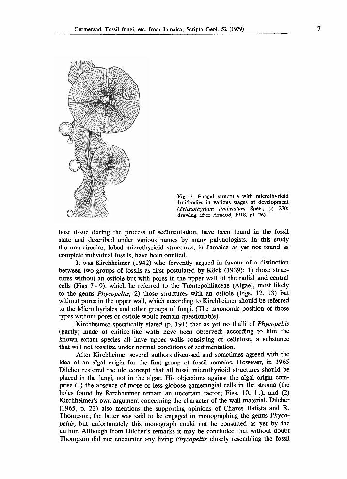
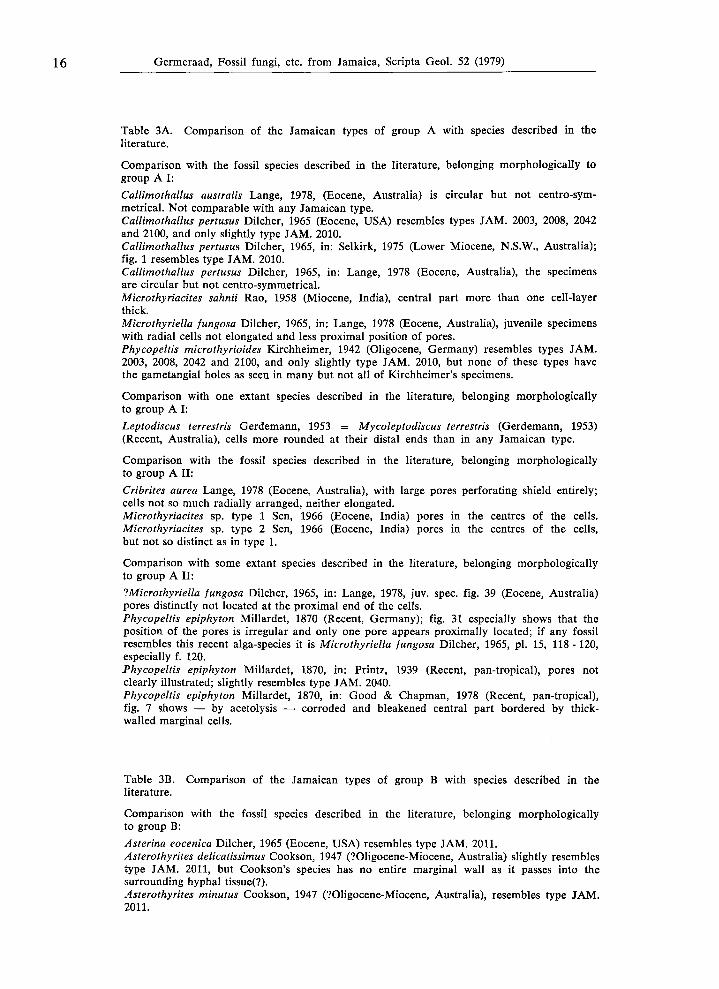
Fig. 3. Fungal structure with microthyrioid fruitbodies in various stages of development (Trichothyrium fimbriatum Speg., χ 270; drawing after Arnaud, 1918, pl. 26).
host tissue during the process of sedimentation, have been found in the fossil state and described under various names by many palynologists. In this study the noncircular, lobed microthyrioid structures, in Jamaica as yet not found as complete individual fossils, have been omitted.
It was Kirchheimer (1942) who fervently argued in favour of a distinction between two groups of fossils as first postulated by Köck (1939): 1) those struc
tures without an ostiole but with pores in the upper wall of the radial and central cells (Figs 7 9), which he referred to the Trentepohliaceae (Algae), most likely to the genus Phycopeltis; 2) those structures with an ostiole (Figs. 12, 13) but without pores in the upper wall, which according to Kirchheimer should be referred to the Microthyriales and other groups of fungi. (The taxonomie position of those types without pores or ostiole would remain questionable).
Kirchheimer specifically stated (p. 191) that as yet no thalli of Phycopeltis (partly) made of chitinelike walls have been observed: according to him the known extant species all have upper walls consisting of cellulose, a substance that will not fossilize under normal conditions of sedimentation.
After Kirchheimer several authors discussed and sometimes agreed with the idea of an algal origin for the first group of fossil remains. However, in 1965 Dilcher restored the old concept that all fossil microthyrioid structures should be placed in the fungi, not in the algae. His objections against the algal origin com
prise (1) the absence of more or less globose gametangial cells in the stroma (the holes found by Kirchheimer remain an uncertain factor; Figs. 10, 11), and (2) Kirchheimer's own argument concerning the character of the wall material. Dilcher (1965, p. 23) also mentions the supporting opinions of Chaves Batista and R. Thompson; the latter was said to be engaged in monographing the genus Phyco
peltis, but unfortunately this monograph could not be consulted as yet by the author. Although from Dilcher's remarks it may be concluded that without doubt Thompson did not encounter any living Phycopeltis closely resembling the fossil
8 Germeraad, Fossil fungi, etc. from Jamaica, Scripta Geol. 52 (1979)
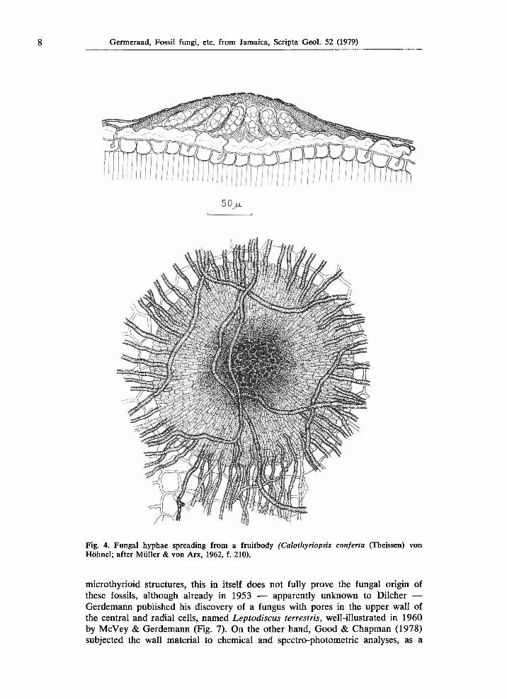
Fig. 4. Fungal hyphae spreading from a fruitbody (Calothyriopsis conferia (Theissen) von Höhnel; after Müller & von Arx, 1962, f. 210).
microthyrioid structures, this in itself does not fully prove the fungal origin of these fossils, although already in 1953 — apparently unknown to Dilcher — Gerdemann published his discovery of a fungus with pores in the upper wall of the central and radial cells, named Leptodiscus terrestris, well-illustrated in 1960 by McVey & Gerdemann (Fig. 7). On the other hand, Good & Chapman (1978) subjected the wall material to chemical and spectro-photometric analyses, as a
Germeraad, Fossil fungi, etc. from Jamaica, Scripta Geol. 52 (1979) 9
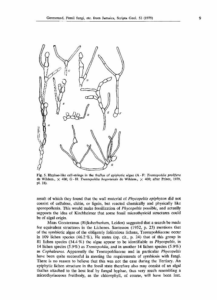
Fig. 5. Hyphae-like cell-strings in the thallus of epiphytic algae (A - F: Trentepohlia prolifera de Wildem., χ 400; G - Η: Trentepohlia bogoriensis de Wildem., χ 400; after Printz, 1939, pl. 18).
result of which they found that the wall material of Phycopeltis epiphyton did not consist of cellulose, chitin, or lignin, but reacted chemically and physically like sporopollenin. This would make fossilization of Phycopeltis possible, and actually supports the idea of Kirchheimer that some fossil microthyrioid structures could be of algal origin.
Maas Geesteranus (Rijksherbarium, Leiden) suggested that a search be made for equivalent structures in the Lichenes. Santesson (1952, p. 23) mentions that of the symbiotic algae of the obligately foliicolous lichens, Trentepohliaceae occur in 109 lichen species (46.2%). He states (op. cit., p. 24) that of this group in 81 lichen species (34.4%) the algae appear to be identifiable as Phycopeltis, in 14 lichen species (5.9%) as Trentepohlia, and in another 14 lichen species (5.9%) as Cephaleuros. Apparently the Trentepohliaceae and in particular Phycopeltis have been quite successful in meeting the requirements of symbiosis with fungi. There is no reason to believe that this was not the case during the Tertiary. An epiphytic lichen structure in the fossil state therefore also may consist of an algal thallus attached to the host leaf by fungal hyphae, thus very much resembling a microthyriaceous fruitbody, as the chlorophyll, of course, will have been lost.
10 Germeraad, Fossil fungi, etc. from Jamaica, Scripta Geol. 52 (1979)
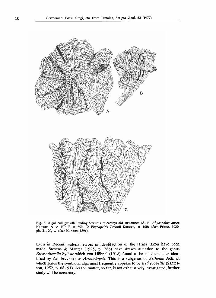
Fig. 6. Algal cell growth tending towards microthyrioid structures (A, B: Phycopeltis aurea Karsten, A χ 150, Β χ 250; C: Phycopeltis Treubii Karsten, χ 100; after Printz, 1939, pis. 21, 20, = after Karsten, 1891).
Even in Recent material errors in identifaction of the larger taxon have been made. Stevens & Manter (1925, p. 286) have drawn attention to the genus Eremothecella Sydow which von Höhnel (1918) found to be a lichen, later iden
tified by Zahlbruckner as Arthoniopsis. This is a subgenus of Arthonia Ach. in which genus the symbiotic alga most frequently appears to be a Phycopeltis (Santes
son, 1952, p. 6891). As the matter, so far, is not exhaustively investigated, further study will be necessary.

Germeraad, Fossil fungi, etc. from Jamaica, Scripta Geol. 52 (1979) 11
Fig. 7. The pores in the fruitbody of a fungus serve as an exit for the spores (Leptodiscus terrestris Gerdemann, 1953, X 450; drawing after McVey & Gerdemann, 1960, f. 14).
With the data available at present it appears possible to provide a list of relevant genera of fungi and algae, both extant and fossil (Table 1). In this list the less conspicuous, questionable group of microthyrioid structures without pores and without ostiole is incorporated. No doubt many of its species should be considered juvenile specimens, as it is known that the ostiole in fungal fruitbodies is formed at maturity. No such a mature condition is known to exist in Phycopeltis as regards the appearance of pores in the upper wall of the radial cells. As these pores serve as an exit for the gametes, a mature condition may be assumed to be present.
Apart from the major problem whether a fossil is an alga or a fungus, there is the question of the validity of the relationship of the various genera and species with microthyrioid structures. For the distinction of the extant taxa it appears that use is made principally of biological criteria such as growth, reproductive processes, and host relationship, phenomena that generally cannot be observed in or reconstructed from (isolated) fossils (Cookson, 1947, p. 208). It
Table 1. List of relevant genera of extant and fossil fungi and algae.
Group A
Group Β
Group C
Recent Phycopeltis Leptodiscus Mycoleptodiscus Asterina Microthyriella Microthyriolum Plochmopeltidella Actinopelte Asterina Ferrarisia Haplopeltis Leptothyrium Manginula Microthyrium Parasterina Stomiopeltis Trichopeltina Vizella
Fossil Callimothallus Cribrites
Asterothyrites Microthyriacites Microthyrites Phragmothy rites Asterothyrites Brefeldiellites Dictyotopileos Entopeltacites Mariusia Microthallites Notothyrites Plochmopeltinites Shortensis Stomiopeltites Trichothyrites
12 Germeraad, Fossil fungi, etc. from Jamaica, Scripta Geol. 52 (1979)
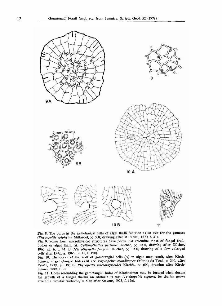
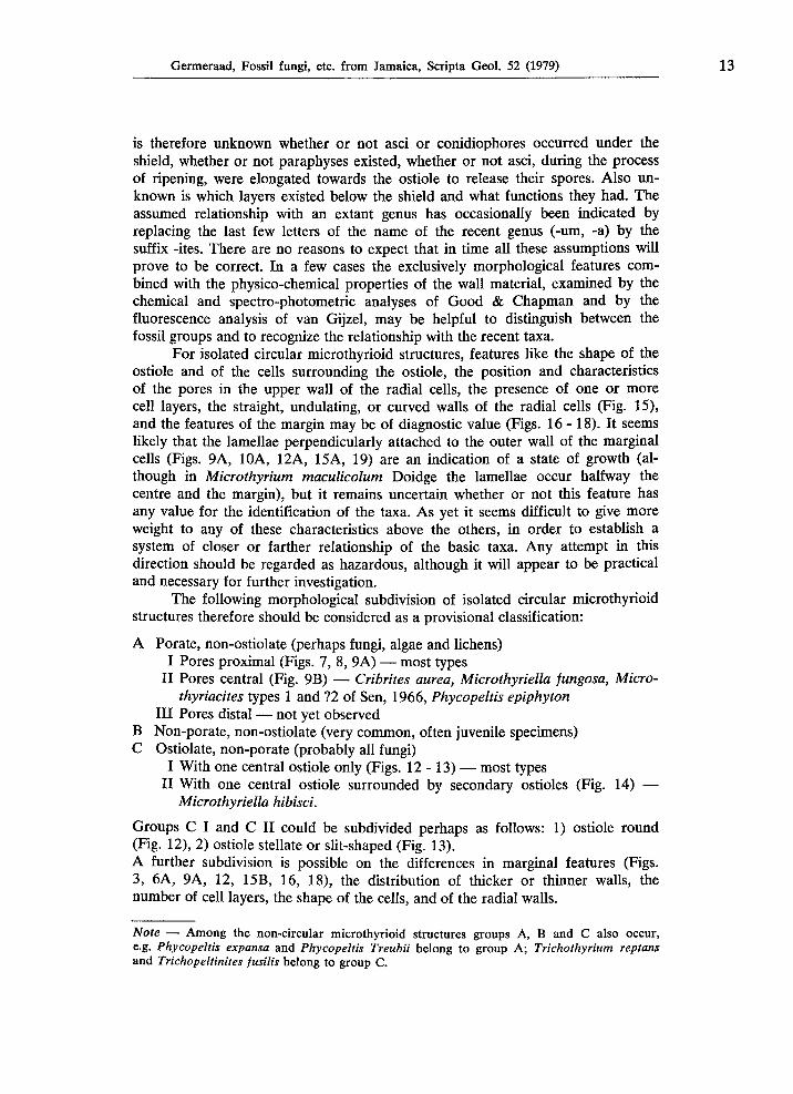
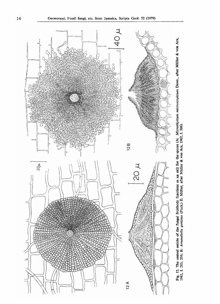
Fig. 8. The pores in the gametangial cells of algal thalli function as an exit for the gametes (Phycopeltis epiphyton Millardet, X 500; drawing after Millardet, 1870, f. 31). Fig. 9. Some fossil microthyrioid structures have pores that resemble those of fungal fruit-bodies or algal thalli (A: Callimothallus pertusus Dilcher, χ 1000, drawing after Dilcher, 1965, pi. 6, f. 44; B: Microthyriella fungosa Dilcher, χ 1000, drawing of a few enlarged cells after Dilcher, 1965, pi. 15, f. 120). Fig. 10. The decay of the wall of gametangial cells (A) in algae may result, after Kirchheimer, in gametangial holes (B). (A: Phycopeltis arundinacea (Mont.) de Toni, χ 300, after Printz, 1939, pl. 19; Β: Phycopeltis microthyrioides Kirchh., X 600, drawing after Kirchheimer, 1942, f. 8). Fig. 11. Holes resembling the gametangial holes of Kirchheimer may be formed when during the growth of a fungal thallus an obstacle is met (Trichopeltis reptans, its thallus grows around a circular trichome, χ 500; after Stevens, 1925, f. 17e).
Germeraad, Fossil fungi, etc. from Jamaica, Scripta Geol. 52 (1979) 13
is therefore unknown whether or not asci or conidiophores occurred under the shield, whether or not paraphyses existed, whether or not asci, during the process of ripening, were elongated towards the ostiole to release their spores. Also unknown is which layers existed below the shield and what functions they had. The assumed relationship with an extant genus has occasionally been indicated by replacing the last few letters of the name of the recent genus (-urn, -a) by the suffix -ites. There are no reasons to expect that in time all these assumptions will prove to be correct. In a few cases the exclusively morphological features combined with the physico-chemical properties of the wall material, examined by the chemical and spectro-photometric analyses of Good & Chapman and by the fluorescence analysis of van Gijzel, may be helpful to distinguish between the fossil groups and to recognize the relationship with the recent taxa.
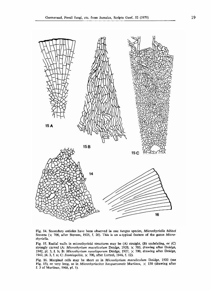
For isolated circular microthyrioid structures, features like the shape of the ostiole and of the cells surrounding the ostiole, the position and characteristics of the pores in the upper wall of the radial cells, the presence of one or more cell layers, the straight, undulating, or curved walls of the radial cells (Fig. 15), and the features of the margin may be of diagnostic value (Figs. 16 - 18). It seems likely that the lamellae perpendicularly attached to the outer wall of the marginal cells (Figs. 9A, 10A, 12A, 15A, 19) are an indication of a state of growth (although in Microthyrium maculicolum Doidge the lamellae occur halfway the centre and the margin), but it remains uncertain whether or not this feature has any value for the identification of the taxa. As yet it seems difficult to give more weight to any of these characteristics above the others, in order to establish a system of closer or farther relationship of the basic taxa. Any attempt in this direction should be regarded as hazardous, although it will appear to be practical and necessary for further investigation.
The following morphological subdivision of isolated circular microthyrioid structures therefore should be considered as a provisional classification:
A Porate, non-ostiolate (perhaps fungi, algae and lichens) I Pores proximal (Figs. 7, 8, 9A) — most types
II Pores central (Fig. 9B) — Cribrites aurea, Microthyriella fungosa, Micro-thyriacites types 1 and ?2 of Sen, 1966, Phycopeltis epiphyton
III Pores distal — not yet observed Β Non-porate, non-ostiolate (very common, often juvenile specimens) C Ostiolate, non-porate (probably all fungi)
I With one central ostiole only (Figs. 12-13) — most types II With one central ostiole surrounded by secondary ostioles (Fig. 14) —
Microthyriella hibisci.
Groups C I and C II could be subdivided perhaps as follows: 1) ostiole round (Fig. 12), 2) ostiole stellate or slit-shaped (Fig. 13). A further subdivision is possible on the differences in marginal features (Figs. 3, 6A, 9A, 12, 15B, 16, 18), the distribution of thicker or thinner walls, the number of cell layers, the shape of the cells, and of the radial walls.
Note — Among the non-circular microthyrioid structures groups A, Β and C also occur, e.g. Phycopeltis expansa and Phycopeltis Treubii belong to group A; Trichothyrium reptans and Trichopeltinites fusilis belong to group C.
14 Germeraad, Fossil fungi, etc. from Jamaica, Scripta Geol. 52 (1979)
$ d 0 >
ö
1 i Ρ s ο ! I
£^ S ο
<< ο\
&« "> α
II GO £ j ce ου
CO η Ο J2
£ ω
l 2 1 ä 1-1 eö «
•2 I • 60
ο -g •s § •S £ co ^
ι* α "fr S ο υ es Η CM
. CM bo vo
ES
Tab
le 2
A.
Porate
mic
roth
yrio
id st
ru
ctu
res f
ou
nd
in
the
Terti
ary o
f Jam
aic
a.
cen
tral
part t
hic
kw
all
ed
, th
inw
alled
arou
nd
ou
ter m
argin
w
ith
lam
ellae
cells n
ear m
argin
rath
er lo
ng
d
isti
nct
an
nu
li
arou
nd
pores
typ
e J
AM
. 2003
cen
tral
part
thin
walled
, th
ick
walled
arou
nd
ou
ter m
argin
n
ot
prese
rved
cells n
ear m
argin
sh
ort
no
an
nu
li
arou
nd
pores
typ
e J
AM
. 2100
cen
tral
part
thin
walled
, th
ick
(er)
walled
arou
nd
ou
ter m
argin
w
ith
ou
t la
mellae
cells n
ear m
argin
sh
ort
except a
few
an
nu
li
arou
nd
pores
typ
e J
AM
. 2008
thic
kw
alled
th
rou
gh
ou
t ou
ter m
argin
w
ith
ou
t la
mellae
cells n
ear m
argin
sh
ort
dis
tin
ct
an
nu
li
arou
nd
pores
typ
e J
AM
. 2042
thin
walled
th
rou
gh
ou
t ou
ter m
argin
w
ith
lon
g l
am
ellae
cells n
ear m
argin
lo
ng
d
isti
nct
an
nu
li
arou
nd
pores
typ
e J
AM
. 2010
thin
walled
th
rou
gh
ou
t ou
ter m
argin
w
ith
ou
t la
mellae
cells n
ear m
argin
lo
ng
in
dis
tin
ct
an
nu
li
arou
nd
pores
typ
e J
AM
. 2040
Tab
le
2B
. N
on
porate
, n
on
ost
iola
te m
icroth
yrio
id s
tru
ctu
res
fou
nd
in t
he
Terti
ary
of
Jam
aic
a.
cen
tral
part t
hic
kw
alled
, th
inw
alled
arou
nd
ou
ter m
argin
w
ith
lam
ellae
cells n
ear m
argin
lo
ng
ty
pe
JA
M. 2
011
thin
walled
th
rou
gh
ou
t ou
ter m
argin
w
ith
lon
g l
am
ellae
cells n
ear m
argin
rath
er lo
ng
ty
pe
JA
M. 2
007
thin
walled
th
rou
gh
ou
t ou
ter m
argin
w
ith
lon
g l
am
ell
ae
cells n
ear m
argin
very
lon
g
typ
e J
AM
. 2044
ο
Ρ
Ρ
ρ ο ?
3
Ο
ι»»
•ι
Ο
3
<»
Ρ
Õ*
Ρ
CO
ο ο
Ν>
/
Ν
Η^
V
O
t—k
U
i
16 Germeraad, Fossil fungi, etc. from Jamaica, Scripta Geol. 52 (1979)
Table 3A. Comparison of the Jamaican types of group A with species described in the literature.
Comparison with the fossil species described in the literature, belonging morphologically to group A I: Callimothallus australis Lange, 1978, (Eocene, Australia) is circular but not centro-sym-metrical. Not comparable with any Jamaican type. Callimothallus pertusus Dilcher, 1965 (Eocene, USA) resembles types JAM. 2003, 2008, 2042 and 2100, and only slightly type JAM. 2010. Callimothallus pertusus Dilcher, 1965, in: Selkirk, 1975 (Lower Miocene, N.S.W., Australia); fig. 1 resembles type JAM. 2010. Callimothallus pertusus Dilcher, 1965, in: Lange, 1978 (Eocene, Australia), the specimens are circular but not centro-symmetrical. Microthyriacites sahnii Rao, 1958 (Miocene, India), central part more than one cell-layer thick. Microthyriella fungosa Dilcher, 1965, in: Lange, 1978 (Eocene, Australia), juvenile specimens with radial cells not elongated and less proximal position of pores. Phycopeltis microthyrioides Kirchheimer, 1942 (Oligocène, Germany) resembles types JAM. 2003, 2008, 2042 and 2100, and only slightly type JAM. 2010, but none of these types have the gametangial holes as seen in many but not all of Kirchheimer's specimens.
Comparison with one extant species described in the literature, belonging morphologically to group A I: Leptodiscus terrestris Gerdemann, 1953 = Mycoleptodiscus terrestris (Gerdemann, 1953) (Recent, Australia), cells more rounded at their distal ends than in any Jamaican type.
Comparison with the fossil species described in the literature, belonging morphologically to group A II: Cribrites aurea Lange, 1978 (Eocene, Australia), with large pores perforating shield entirely; cells not so much radially arranged, neither elongated. Microthyriacites sp. type 1 Sen, 1966 (Eocene, India) pores in the centres of the cells. Microthyriacites sp. type 2 Sen, 1966 (Eocene, India) pores in the centres of the cells, but not so distinct as in type 1.
Comparison with some extant species described in the literature, belonging morphologically to group A II: Ί Microthyriella fungosa Dilcher, 1965, in: Lange, 1978, juv. spec. fig. 39 (Eocene, Australia) pores distinctly not located at the proximal end of the cells. Phycopeltis epiphyton Millardet, 1870 (Recent, Germany); fig. 31 especially shows that the position of the pores is irregular and only one pore appears proximally located; if any fossil resembles this recent alga-species it is Microthyriella fungosa Dilcher, 1965, pi. 15, 118 -120, especially f. 120. Phycopeltis epiphyton Millardet, 1870, in: Printz, 1939 (Recent, pan-tropical), pores not clearly illustrated; slightly resembles type JAM. 2040. Phycopeltis epiphyton Millardet, 1870, in: Good & Chapman, 1978 (Recent, pan-tropical), fig. 7 shows — by acetolysis — corroded and bleakened central part bordered by thick-walled marginal cells.
Table 3B. Comparison of the Jamaican types of group Β with species described in the literature.
Comparison with the fossil species described in the literature, belonging morphologically to group B: Asterina eocenica Dilcher, 1965 (Eocene, USA) resembles type JAM. 2011. Asterothyrites delicatissimus Cookson, 1947 (?01igocene-Miocene, Australia) slightly resembles type JAM. 2011, but Cookson's species has no entire marginal wall as it passes into the surrounding hyphal tissue(?). Asterothyrites minutus Cookson, 1947 (?01igocene-Miocene, Australia), resembles type JAM. 2011.
Germeraad, Fossil fungi, etc. from Jamaica, Scripta Geol. 52 (1979) 17
Asterothyrites sinuatus Cookson, 1947 (?01igocene-Miocene, Australia), slightly resembles type JAM. 2011, but Cookson's species has an irregular outer margin. Entopeltacites irregularis Selkirk, 1972 (Lower Miocene, N.S.W., Australia) resembles type JAM. 2008, but Selkirk's species has no pores. Microthyriacites baqueroensis Martinez, 1967 (Lower Cretaceous, Argentine) has very long radial cells. Microthyriacites cooksoni Rao, 1958 (Eocene and Miocene, India) resembles type JAM. 2011, but Rao's species has a more conspicuous central portion of compact cells (Rao, p. 45). Microthyriacites edwardsi Rao, 1958 (Miocene, India) resembles type JAM. 2044. Microthyriacites fimbriatus Cookson, 1947 (Oligocene-Miocene, Australia) resembles type JAM. 2011. Microthyriacites grandis Cookson, 1947 (?01igocene, Australia) has a rather thick-walled appearance. Microthyriacites sp.(?grandis) Cookson, 1947, pi. 13, f. 18, 19 (?Oligocene-Miocene, Australia) is rather thick-walled. Microthyrites dysodilis Pampaloni, 1902 (Miocene, Italy) is rather poorly illustrated. Pediastrum sp. in: Davis, 1916 (Eocene, USA), a ?juvenile specimen. Phragmothyrites delicatus Selkirk, 1975 (Lower Miocene, N.S.W., Australia) slightly resembles type JAM. 2011, but Selkirk's species is thin-walled with less slender marginal cells. Phragmothyrites eocenica Edwards, 1923 (Eocene, Scotland, UK) slightly resembles types JAM. 2008 and 2009, but Edwards' species has less elongated radial cells. Phragmothyrites cf. fimbriatus Cookson, 1947, in: Selkirk, 1975 (Lower Miocene, N.S.W., Australia). Phragmothyrites hibernicus Johnson, 1941 (Lower Tertiary, Ireland) with air-hyphae and intercellular mycelium. Phragmothyrites kiandrensis Selkirk, 1975 (Lower Miocene, N.S.W., Australia) resembles type JAM. 2010, but Selkirk's species has no pores. Plochmopeltidella antiqua Dilcher, 1965 (Eocene, USA) is endoparasitic and has strongly undulating radial walls and an irregular outer margin.
Comparison with some extant species described in the literature, belonging morphologically to group B: Calothyriopsis conferia (Theissen) von Höhnel, 1919, in: Müller & von Arx, 1962 (Recent, tropical America) with long hyphae extending from the margin. Cirsosiella globulifera (Pat.) Arnaud, 1918 (Recent, SE Asia) outer margin with extending radial cells. Lembosia bromeliacearum Rehm, 1900, in: Arnaud, 1918 (Recent, South America) has a margin with acuminate extensions. Lembosia rubiacearum Arnaud, 1918 (Recent, South America) has a margin with acuminate extensions. Manginulopsis lunariae Chaves Batista & Peres, 1963 (Recent, Brasil) with hardly elongated radial cells. Microthyrium maculicolum Doidge, 1942 (Recent, South Africa) with straight radial walls and lamellae not at outer margin; slightly resembling types JAM. 2008 and 2010. Microthyrium ranulisporum Doidge, 1942 (Recent, South Africa) outer margin with extending radial cells, radial walls undulating. Palawaniella orbiculata = Seynesia orbiculata Doidge, 1942 (Recent, South Africa) with undulating radial walls, and lamellae at outer margin. Phycopeltis aurea Karsten, 1891, in: Printz, 1939 (Recent, SE Asia) has a circular shield composed of fan-shaped segments. Phycopeltis expansa Jennings, 1896, in: Schmidle, 1897 (Recent, New Zealand) is circular, but not centro-symmetrical. Prillieuxina winteriana (Pazschke) Arnaud, 1918 (Recent, Brasil) has extending radial cells and straight radial walls. Seynesiella juniperi (Desm.) Arnaud, 1918 (Recent, Europe) is very dark.
The microthyrioid types found in Jamaica may be related to, but are not necessarily identical with, the following fossil and/or extant species:
18 Germeraad, Fossil fungi, etc. from Jamaica, Scripta Geol. 52 (1979)
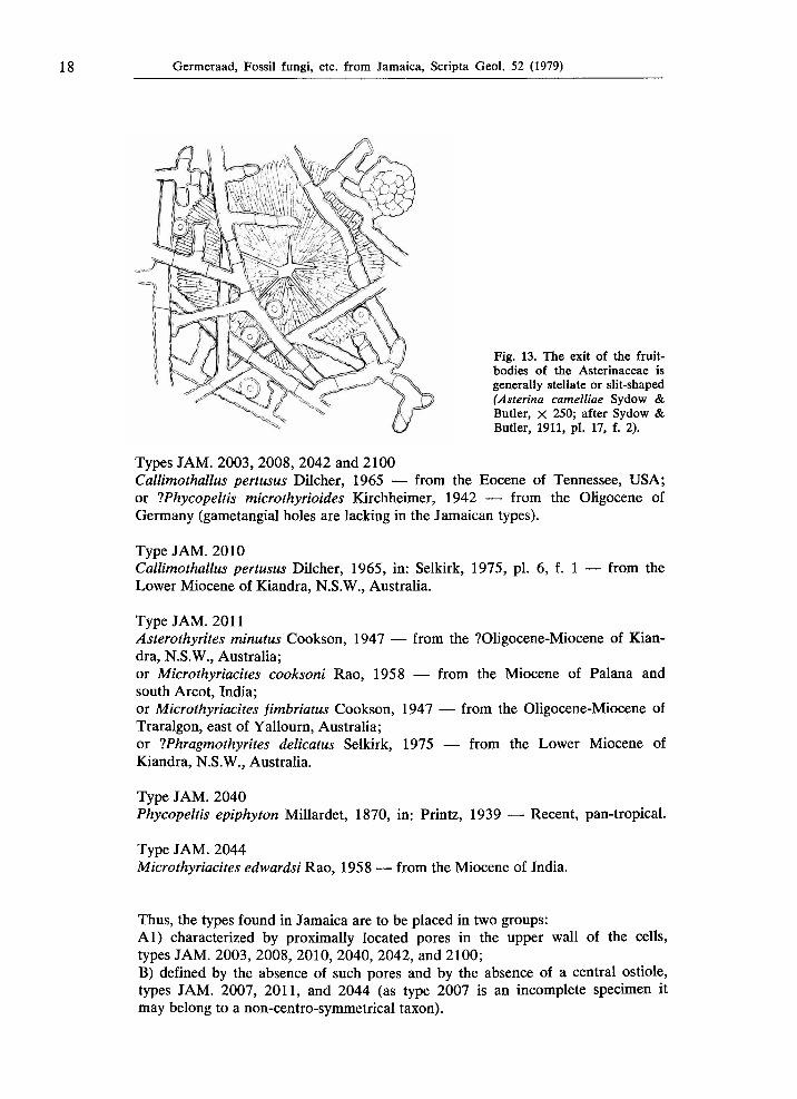
Fig. 13. The exit of the fruit-
bodies of the Asterinaceae is generally stellate or slit-shaped (Asterina camelliae Sydow & Butler, χ 250; after Sydow & Butler, 1911, pi. 17, f. 2).
Types J A M . 2003, 2008, 2042 and 2100 Callimothallus pertusus Dilcher, 1965 — from the Eocene of Tennessee, USA; or IPhycopeltis microthyrioides Kirchheimer, 1942 — from the Oligocène of Germany (gametangial holes are lacking in the Jamaican types).
Type J A M . 2010 Callimothallus pertusus Dilcher, 1965, in: Selkirk, 1975, pi. 6, f. 1 — from the Lower Miocene of Kiandra, N.S.W., Australia.
Type J A M . 2011 Asterothyrites minutus Cookson, 1947 — from the ?01igoceneMiocene of Kian
dra, N.S.W., Australia; or Microthyriacites cooksoni Rao, 1958 — from the Miocene of Palana and south Arcot, India; or Microthyriacites fimbriatus Cookson, 1947 — from the OligoceneMiocene of Traralgon, east of Yallourn, Australia; or Whragmothyrites delicatus Selkirk, 1975 — from the Lower Miocene of Kiandra, N.S.W., Australia.
Type J A M . 2040
Phycopeltis epiphyton Millardet, 1870, in: Printz, 1939 — Recent, pantropical.
Type J A M . 2044 Microthyriacites edwardsi Rao, 1958 — from the Miocene of India.
Thus, the types found in Jamaica are to be placed in two groups: AÍ) characterized by proximally located pores in the upper wall of the cells, types J A M . 2003, 2008, 2010, 2040, 2042, and 2100; B) defined by the absence of such pores and by the absence of a central ostiole, types J A M . 2007, 2011, and 2044 (as type 2007 is an incomplete specimen it may belong to a noncentrosymmetrical taxon).
Germeraad, Fossil fungi, etc. from Jamaica, Scripta Geol. 52 (1979) 19
Fig. 14. Secondary ostioles have been observed in one fungus species, Microthyriella hibisci Stevens ( χ 700, after Stevens, 1925, f. 20). This is an a-typical feature of the genus Microthyriella, Fig. 15. Radial walls in microthyrioid structures may be (A) straight, (B) undulating, or (C) strongly curved (A: Microthyrium maculicolum Doidge, 1920, X 700, drawing after Doidge, 1942, pi. 3, f. b; B: Microthyrium ranulisporum Doidge, 1927, X 700, drawing after Doidge, 1942, pi. 3, f. a; C: Stomiopeltis, X 700, after Luttrel, 1946, f. 12). Fig. 16. Marginal cells may be short as in Microthyrium maculicolum Doidge, 1920 (see Fig. 15), or very long, as in Microthyriacites bacqueroensis Martinez, χ 150 (drawing after f. 3 of Martinez, 1968, pi. 1).
20 Germeraad, Fossil fungi, etc. from Jamaica, Scripta Geol. 52 (1979)
Morphological descriptions
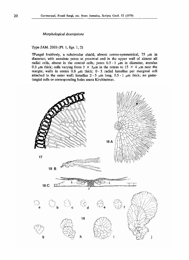
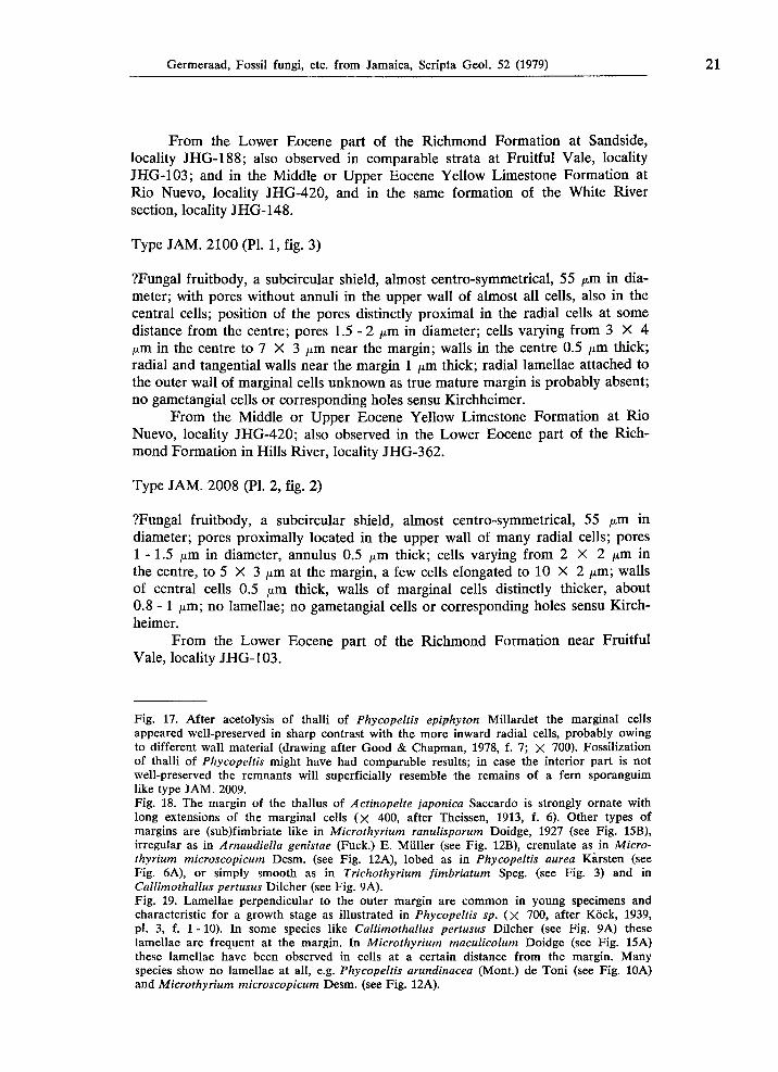
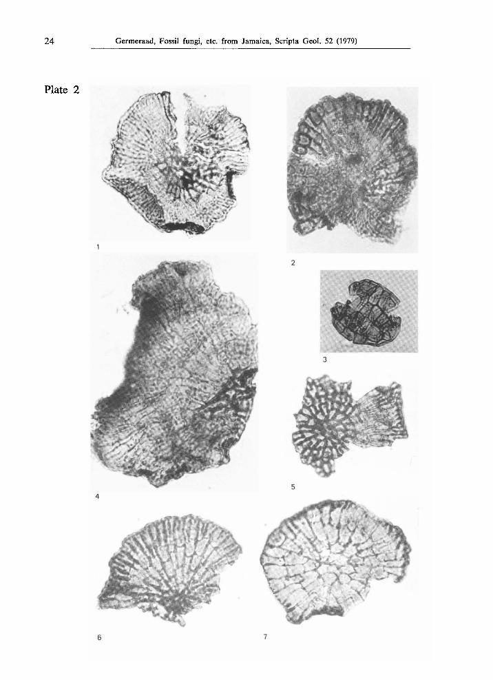
Type J A M . 2003 (PL 1, figs. 1, 2)
?Fungal fruitbody, a subcircular shield, almost centro-symmetrical, 75 μτη in diameter; with annulate pores at proximal end in the upper wall of almost all radial cells, absent in the central cells; pores 0.5 - 1 ^m in diameter, annulus 0.3 μτη thick; cells varying from 3 X 3//,m in the centre to 15 X 4 μτη near the margin; walls in centre 0.8 μτη thick; 0 -3 radial lamellae per marginal cell attached to the outer wall; lamellae 2 - 5 μτα long, 0.5 - 1 μτη thick; no gametangial cells or corresponding holes sensu Kirchheimer.
Germeraad, Fossil fungi, etc. from Jamaica, Scripta Geol. 52 (1979) 21
From the Lower Eocene part of the Richmond Formation at Sandside, locality JHG-188; also observed in comparable strata at Fruitful Vale, locality JHG-103; and in the Middle or Upper Eocene Yellow Limestone Formation at Rio Nuevo, locality JHG-420, and in the same formation of the White River section, locality JHG-148.
Type J A M . 2100 (Pl. 1, fig. 3)
?Fungal fruitbody, a subcircular shield, almost centro-symmetrical, 55 /im in diameter; with pores without annuli in the upper wall of almost all cells, also in the central cells; position of the pores distinctly proximal in the radial cells at some distance from the centre; pores 1.5-2 μτη. in diameter; cells varying from 3 X 4 μπι in the centre to 7 X 3 μΐη near the margin; walls in the centre 0.5 μτη. thick; radial and tangential walls near the margin 1 μτη thick; radial lamellae attached to the outer wall of marginal cells unknown as true mature margin is probably absent; no gametangial cells or corresponding holes sensu Kirchheimer.
From the Middle or Upper Eocene Yellow Limestone Formation at Rio Nuevo, locality JHG-420; also observed in the Lower Eocene part of the Richmond Formation in Hills River, locality JHG-362.
Type J A M . 2008 (PL 2, fig. 2)
?Fungal fruitbody, a subcircular shield, almost centro-symmetrical, 55 μτα in diameter; pores proximally located in the upper wall of many radial cells; pores 1-1.5 μτη. in diameter, annulus 0.5 thick; cells varying from 2 X 2 μτη in the centre, to 5 X 3 μτη at the margin, a few cells elongated to 10 X 2 μτη; walls of central cells 0.5 μτη thick, walls of marginal cells distinctly thicker, about 0.8-1 μπι; no lamellae; no gametangial cells or corresponding holes sensu Kirchheimer.
From the Lower Eocene part of the Richmond Formation near Fruitful Vale, locality JHG-103.
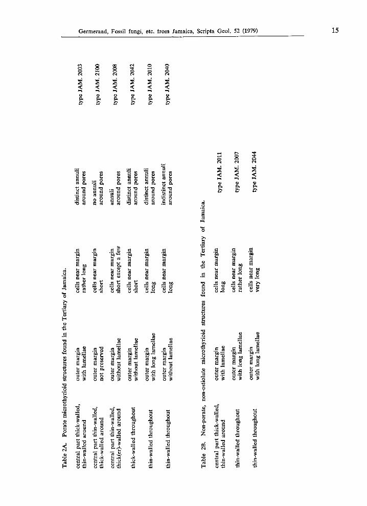
Fig. 17. After acetolysis of thalli of Phycopeltis epiphyton Millardet the marginal cells appeared well-preserved in sharp contrast with the more inward radial cells, probably owing to different wall material (drawing after Good & Chapman, 1978, f. 7; X 700). Fossilization of thalli of Phycopeltis might have had comparable results; in case the interior part is not well-preserved the remnants will superficially resemble the remains of a fern sporanguim like type JAM. 2009. Fig. 18. The margin of the thallus of Actinopelte japonica Saccardo is strongly ornate with long extensions of the marginal cells ( χ 400, after Theissen, 1913, f. 6). Other types of margins are (sub)fimbriate like in Microthyrium ranulisporum Doidge, 1927 (see Fig. 15B), irregular as in Arnaudiella genistae (Fuck.) E. Müller (see Fig. 12B), crenulate as in Microthyrium microscopicum Desm. (see Fig. 12A), lobed as in Phycopeltis aurea Karsten (see Fig. 6A), or simply smooth as in Trichothyrium fimbriatum Speg. (see Fig. 3) and in Callimothallus pertusus Dilcher (see Fig. 9A). Fig. 19. Lamellae perpendicular to the outer margin are common in young specimens and characteristic for a growth stage as illustrated in Phycopeltis sp. (X 700, after Köck, 1939, pl. 3, f. 1 -10). In some species like Callimothallus pertusus Dilcher (see Fig. 9A) these lamellae are frequent at the margin. In Microthyrium maculicolum Doidge (see Fig. 15A) these lamellae have been observed in cells at a certain distance from the margin. Many species show no lamellae at all, e.g. Phycopeltis arundinacea (Mont.) de Toni (see Fig. 10A) and Microthyrium microscopicum Desm. (see Fig. 12A).
22 Germeraad, Fossil fungi, etc. from Jamaica, Scripta Geol. 52 (1979)
Plate 1
Germeraad, Fossil fungi, etc. from Jamaica, Scripta Geol. 52 (1979) 23
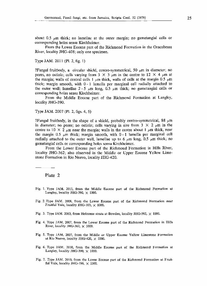
Type J A M . 2042 (PI. 3, figs. 1, 2)
?Fungal fruitbody, a subcircular shield, almost centro-symmetrical, 87 μΐη in diameter; with annulate pores in the upper wall of almost all cells, also in the central cells; position of the pores distinctly proximal in the radial cells; pores 1-1.5 μπι in diameter; cells varying from 2 X 2 μπι in the centre, to 5 X 4 μπι near the margin; radial and tangential walls aways about 1.5 μπι thick; no radial lamellae observed where the outer margin is well preserved; no gametangial cells or corresponding holes sensu Kirchheimer.
From the Middle or Upper Eocene Yellow Limestone Formation at Rio Nuevo, locality JHG-419.
Type J A M . 2010 (PI. 2, figs. 6,7)
?Fungal fruitbody, a subcircular shield, almost centro-symmetrical, 60 μπι in diameter; with annulate pores at proximal end in upper wall of almost all cells except the marginal cells; pores 1-1.5 μπι in diameter, annuli 0.5 μπι thick; cells varying from 3 X 3 μπι in the centre to 12 X 4 ^m at the margin; walls everywhere 0.5 μπι thick; 0 -3 lamellae per marginal cell attached to the outer wall, lamellae 1 - 5 μπι long, 0.5 μπι thick; no gametangial cells or corresponding holes sensu Kirchheimer.
From the Middle Eocene part of the Richmond Formation at Langley, locality JHG-390; also observed in the Lower Eocene part of the same formation at Sandside, localities JHG-195, 196 and 197, and in the same strata at Fruitful Vale, localities JHG-102 and 108.
Type J A M . 2040 (PI. 3, fig. 5)
?Fungal fruitbody, a subcircular shield, rather juvenile?, centro-symmetrical, 35 μπι in diameter; with ?annulate pores in a few cells only, proximally located, 1 - 1.5 jum in diameter; cells in the small centre about 4 μπι large, all cells around approximately radially arranged, 10 X 6 μπι; radial and tangential walls curved,
Plate 1
Figs.l, 2. Type JAM. 2003, from the Lower Eocene part of the Richmond Formation at Sandside, locality JHG-188, χ 1000.
Fig. 3. Type JAM. 2100, from the Middle or Upper Eocene Yellow Limestone Formation at Rio Nuevo, locality JHG-420, χ 1000.
Fig. 4. Type JAM. 2044, from the Middle or Upper Eocene Yellow Limestone Formation in Rio Nuevo, locality JHG-420, χ 1000.
Fig. 5. Type JAM. 2068, from Upper Cretaceous strata in Rio Minho, East of Frankfield, Central Inlier, locality JHG-471, χ 1000.
Fig. 6. Type JAM. 2013, from the Middle Eocene part of the Richmond Formation at Langley, locality JHG-390, χ 1000.
24 Germeraad, Fossil fungi, etc. from Jamaica, Scripta Geol. 52 (1979)
Plate 2
Germeraad, Fossil fungi, etc. from Jamaica, Scripta Geol. 52 (1979) 25
about 0.5 μτη thick; no lamellae at the outer margin; no gametangial cells or corresponding holes sensu Kirchheimer.
From the Lower Eocene part of the Richmond Formation in the Oracabessa River, locality JHG-408; only one specimen.
Type J A M . 2011 (PI. 2, fig. 1)
?Fungal fruitbody, a circular shield, centro-symmetrical, 50 μτη. in diameter; no pores, no ostiole; cells varying from 3 X 3 μία in the centre to 12 X 4 μτα at the margin; walls of central cells 1 μτη thick, walls of cells at the margin 0.5 μΐχι thick; margin smooth, with 0-1 lamella per marginal cell radially attached to the outer wall; lamellae 2 - 5 ^m long, 0.5 μτη thick; no gametangial cells or corresponding holes sensu Kirchheimer.
From the Middle Eocene part of the Richmond Formation at Langley, locality JHG-390.
Type J A M . 2007 (PL 2, figs. 4, 5)
?Fungal fruitbody, in the shape of a shield, probably centro-symmetrical, 88 ^m in diameter; no pores; no ostiole; cells varying in size from 3 X 2 μτη in the centre to 10 X 2 μτη near the margin; walls in the centre about 1 μτα thick, near the margin 0.5 μτη thick; margin smooth, with 0-1 lamella per marginal cell radially attached to the outer wall, lamellae up to 6 μτη long, 0.5 μΤΆ thick; no gametangial cells or corresponding holes sensu Kirchheimer.
From the Lower Eocene part of the Richmond Formation in Hills River, locality JHG-362; also observed in the Middle or Upper Eocene Yellow Limestone Formation in Rio Nuevo, locality JHG-420.
Plate 2
Fig. 1. Type JAM. 2011, from the Middle Eocene part of the Richmond Formation at Langley, locality JHG-390, X 1000.
Fig. 2.Type JAM. 2008, from the Lower Eocene part of the Richmond Formation near Fruitful Vale, locality JHG-103, χ 1000.
Fig. 3. Type JAM. 2063, from Holocene strata at Bowden, locality JHG-592, χ 1000.
Fig. 4. Type JAM. 2007, from the Lower Eocene part of the Richmond Formation in Hills River, locality JHG-362, χ 1000.
Fig. 5. Type JAM. 2007, from the Middle or Upper Eocene Yellow Limestone Formation at Rio Nuevo, locality JHG-420, χ 1000.
Fig. 6. Type JAM. 2010, from the Middle Eocene part of the Richmond Formation at Langley, locality JHG-390, χ 1000.
Fig. 7. Type JAM. 2010, from the Lower Eocene part of the Richmond Formation at Fruitful Vale, locality JHG-196, χ 1000.
26 Germeraad, Fossil fungi, etc. from Jamaica, Scripta Geol. 52 (1979)
Plate 3
Germeraad, Fossil fungi, etc. from Jamaica, Scripta Geol. 52 (1979) 27
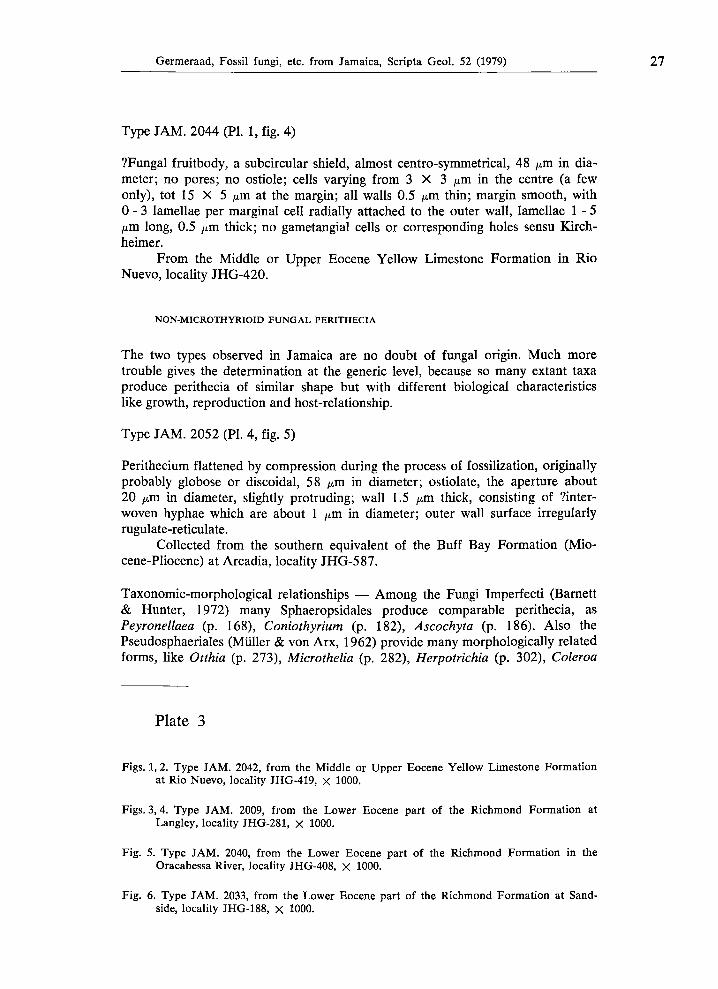
Type J A M . 2044 (PI. 1, fig. 4)
?Fungal fruitbody, a subcircular shield, almost centro-symmetrical, 48 μτη in diameter; no pores; no ostiole; cells varying from 3 X 3 μτα in the centre (a few only), tot 15 X 5 /Am at the margin; all walls 0.5 μπι thin; margin smooth, with 0 -3 lamellae per marginal cell radially attached to the outer wall, lamellae 1-5 μπι long, 0.5 μπι thick; no gametangial cells or corresponding holes sensu Kirchheimer.
From the Middle or Upper Eocene Yellow Limestone Formation in Rio Nuevo, locality JHG-420.
NON-MICROTHYRIOID FUNGAL PERITHECIA
The two types observed in Jamaica are no doubt of fungal origin. Much more trouble gives the determination at the generic level, because so many extant taxa produce perithecia of similar shape but with different biological characteristics like growth, reproduction and host-relationship.
Type J A M . 2052 (PI. 4, fig. 5)
Perithecium flattened by compression during the process of fossilization, originally probably globose or discoidal, 58 ^m in diameter; ostiolate, the aperture about 20 μΐη in diameter, slightly protruding; wall 1.5 μτα thick, consisting of ?inter-woven hyphae which are about 1 μία in diameter; outer wall surface irregularly rugulate-reticulate.
Collected from the southern equivalent of the Buff Bay Formation (Miocene-Pliocene) at Arcadia, locality JHG-587.
Taxonomic-morphological relationships — Among the Fungi Imperfecta (Barnett & Hunter, 1972) many Sphaeropsidales produce comparable perithecia, as Peyronellaea (p. 168), Coniothyrium (p. 182), Ascochyta (p. 186). Also the Pseudosphaeriales (Müller & von Arx, 1962) provide many morphologically related forms, like Otthia (p. 273), Microthelia (p. 282), Herpotrichia (p. 302), Coleroa
Plate 3
Figs. 1, 2. Type JAM. 2042, from the Middle or Upper Eocene Yellow Limestone Formation at Rio Nuevo, locality JHG419, X 1000.
Figs. 3, 4. Type JAM. 2009, from the Lower Eocene part of the Richmond Formation at Langley, locality JHG281, χ 1000.
Fig. 5. Type JAM. 2040, from the Lower Eocene part of the Richmond Formation in the Oracabessa River, locality JHG408, χ 1000.
Fig. 6. Type JAM. 2033, from the Lower Eocene part of the Richmond Formation at Sand
side, locality JHG188, χ 1000.
28 Germeraad, Fossil fungi, etc. from Jamaica, Scripta Geol. 52 (1979)
Plate 4
Germeraad, Fossil fungi, etc. from Jamaica, Scripta Geol. 52 (1979) 29
(p. 413), Botryostroma (p. 462), Lizonia (p. 500). Among the Amphisphaeriaceae (Müller & von Arx, 1962) Amphisphaeria (p. 691). Lilliputia gaillardi Boud. & Pat., 1900, according to Hughes (1951, p. 21, pi. 1, f. 9) a synonym of L. rufula (Berk. & Broome, 1873), original name Chaetomium rufulum, shows a super
ficial resemblance to type J A M . 2052. Hughes states that the apex is composed of about three rows of coarse cells and that the ostiolum, if present, is very in
conspicuous; moreover the size of Lilliputia (800 μτη) is of a different order.
Type J A M . 2091 (PI. 4, fig. 1)
Perithecium pearshaped, largest diameter 176 μτη; ostiolate, aperture about 15 μτη in diameter (slightly damaged); wall consisting of interwoven hyphae, giving an irregular rugulatereticulate impression; wall thickness 56 μτη, hyphae less than 2 μτη. in diameter.
Collected from the Middle Eocene part of the Richmond Formation at Langley, locality JHG377.
Taxonomicmorphological relationships — Some Sphaeropsidales of the Fungi Imperfecti (Barnett & Hunter, 1972) produce comparable structures as in Phoma (p. 166), Phomopsis (p. 168) and Diplodia (p. 186). Comparable perithecia are also produced by many Sphaeriaceae like Chaetosphaeria, Ohleria and Melanom-
ma (Berlese, 1984, vol. I, generaplate 9), and Fenestella (Berlese, 1984, vol. II, pi. 106112). Chaetosphaeria (Müller & von Arx, 1962, p. 583, f. 231) has no hyphal structure but is composed of cells. Some resemblance shows Gelasinospora calospora (Mouton) C. & M . Moreau (Ellis, 1960; Maniotis, 1965, f. 18) but it has no ostiole and the perithecium is about 800 μτη large.
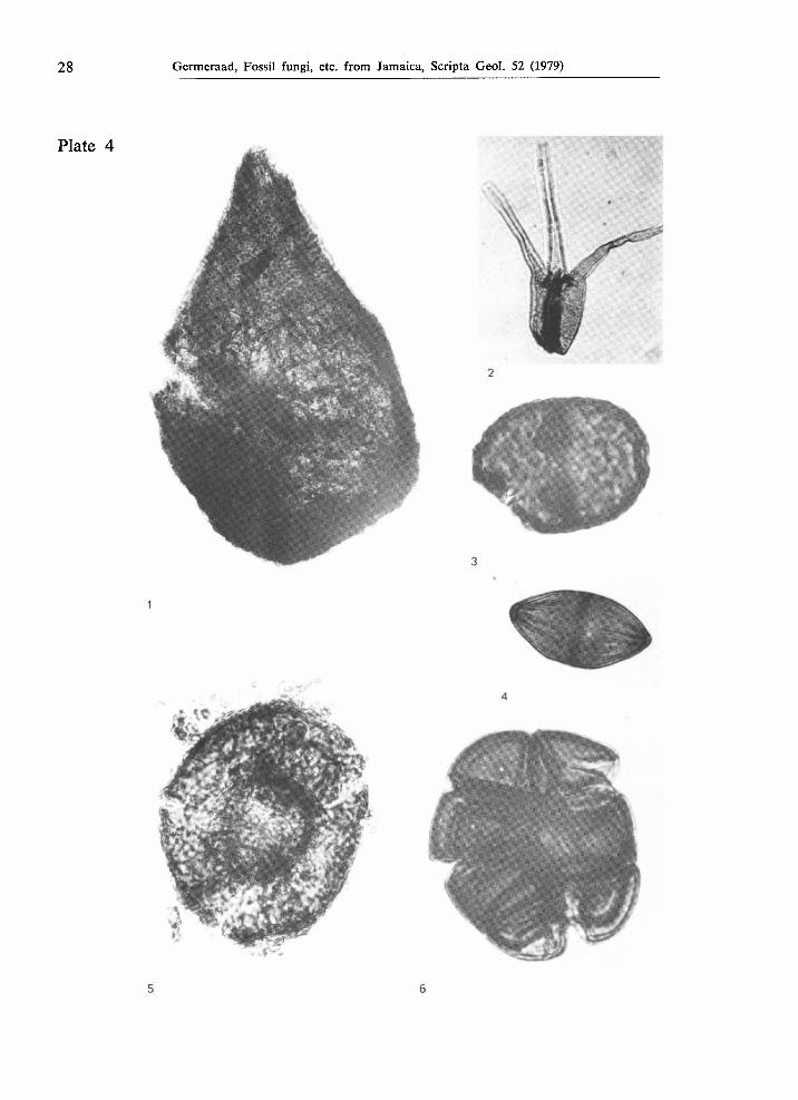
Plate 4
Fig. 1. Type JAM. 2091, from the Middle Eocene part of the Richmond Formation, locality JHG377, χ 650.
Fig. 2. Type JAM. 2049, from the southern equivalent of the Buff Bay Formation (Mio
cenePliocene) at Arcadia, locality JHG590, X 1000.
Fig. 3. Type JAM. 2048, from Holocene strata near Bowden, locality JHG591, χ 1000.
Fig. 4. Type JAM. 2025, from the Lower Eocene part of the Richmond Formation at Richmond, locality JHG426, X 1000.
Fig. 5. Type JAM. 2052, from the southern equivalent of the Buff Bay Formation (Mio
cenePliocene) at Arcadia, locality JHG587, X 1000.
Fig. 6. Type JAM. 2012, from Holocene strata near Bowden, locality JHG591, X 1000.
30 Germeraad, Fossil fungi, etc. from Jamaica, Scripta Geol. 52 (1979)
Plate 5
Germeraad, Fossil fungi, etc. from Jamaica, Scripta Geol. 52 (1979) 31
FUNGAL SPORES, CONIDIA, OR OTHER STRUCTURES
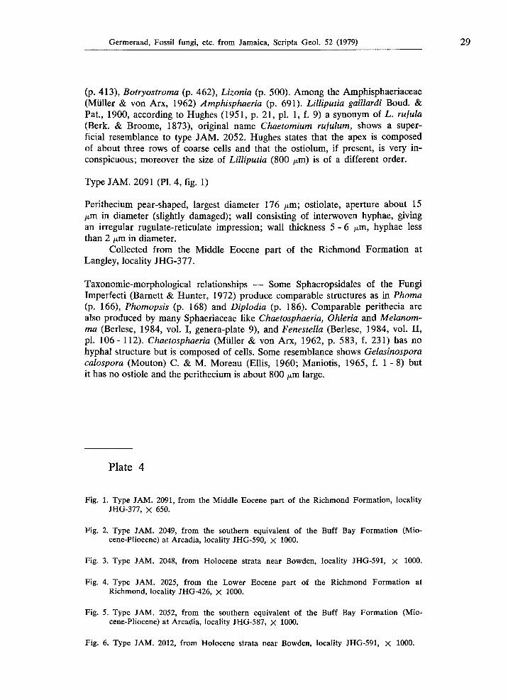
Type J A M . 2005 (PL 5, fig. 7)
Ascospore or conidium, oval with bluntly pointed apex and slightly rounded-truncate antapex, largest diameter 28 μτη; diporate, sometimes one pore indistinct, seemingly monoporate; largest, antapical pore slightly intruding, 6 μτα wide; apical pore less distinct owing to the protruding apex; wall consisting of a 1 /am thin continuous layer (except at the apices) covered by a coarse reticulum (which at low magnification gives a striate impression); muri with rounded crests, 2 -3 μπι wide and high, 1 μτη higher where muri anastomose; lumina elliptical, largest diameter 5 - 9 μπι in approximately polar direction, 3 - 5 ^m in perpendicular direction.
Collected from the Lower Eocene part of the Richmond Formation in the Oracabessa River, locality JHG-407. Also observed in similar strata at Sandside, localities JHG-196, 197; at Langley, locality JHG-216; and near Richmond, locality JHG-426.
Morphological relationships — Closely related to the Eocene Striadiporites sanctaebarbarae Elsik & Jansonius, 1974, in all respects except that the apical pore in type J A M . 2005 is less clear to indistinct. Striadiporites reticulatus Varma & Rawat, 1963 (p. 137, pi. 1, f. 21) from the Lower Miocene of India has smaller lumina. Striadiporites sp. (Jansonius, 1976, pi. 1, f. 3) from the Palaeogene of arctic Canada has much thinner muri and is more pointing at both polar ends. Type J A M . 2005 differs basically from Gelasinospora retispora Cain, 1950 and from G. reticulispora (Greis-Dengler) C. & M . Moreau (van Geel, 1972, 1976) by the presence of pore(s) in type J A M . 2005 and by the smaller lumina (2 - 4 μΐη wide) in the Gelasinospora species.
Plate 5
Figs. 1-3. Type JAM. 2051, from Holocene strata near Bowden, locality JHG-591; fig. 1: X 500; figs. 2 - 3: χ 1000.
Fig. 4. Type JAM. 2047, from the southern equivalent of the Buff Bay Formation (Miocene-Pliocene) at Arcadia, locality JHG-587, χ 1000.
Fig. 5. Type JAM. 2029, from the Upper Eocene Swanswick Limestone in Widcomb bore hole no. 1 at 110' depth, χ 1000.
Fig. 6. Type JAM. 2006, from the Lower Eocene part of the Richmond Formation in the Oracabessa River, locality JHG-407, χ 1000.
Fig. 7. Type JAM. 2005, from the Lower Eocene part of the Richmond Formation in the Oracabessa River, locality JHG-407, χ 1000.
32 Germeraad, Fossil fungi, etc. from Jamaica, Scripta Geol. 52 (1979)
Plate 6
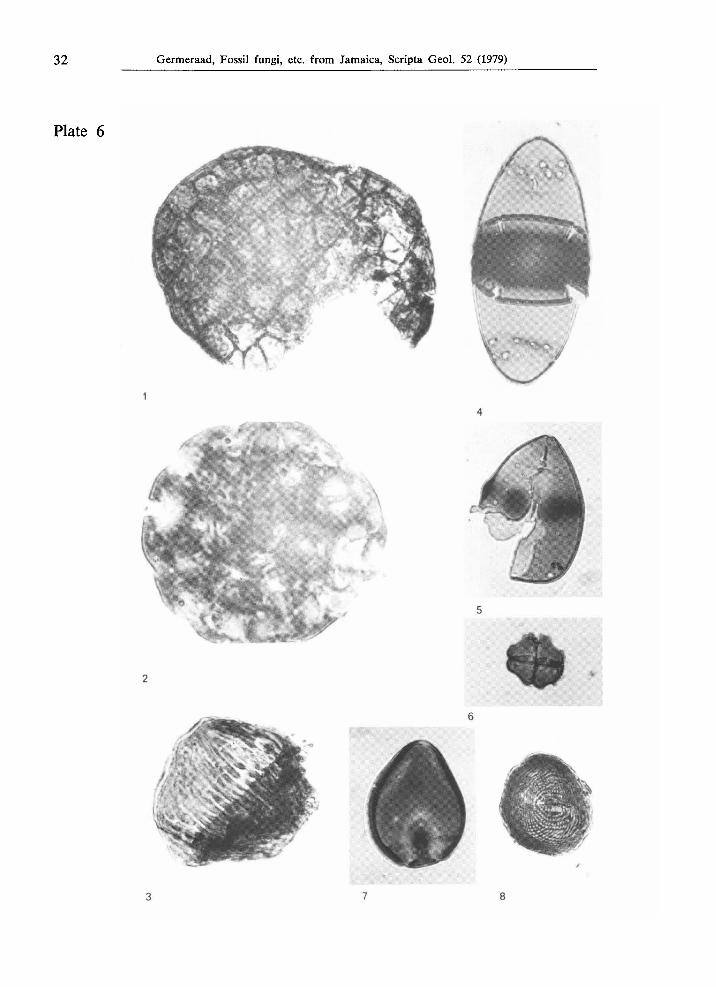
Germeraad, Fossil fungi, etc. from Jamaica, Scripta Geol. 52 (1979) 33
Type J A M . 2006 (PI. 5, fig. 6)
Morphologically very close to type J A M . 2005 but less close to Striadiporites sanctaebarbarae by the much wider lumina (up to 8 X 15 μΐη), the wider antapical pore (14 μ,πι), and the larger overall diameter (35 μπι).
From the Lower Eocene part of the Richmond Formation in the Oracabessa River, locality JHG-407.
Morphological relationship — Perhaps within the variability of type J A M . 2005 (see Corner, 1947).
Type J A M . 2012 (PI. 4, fig. 6)
Fungal ?conidium, ovoid in shape, largest diameter 52 μπι; consisting of 12-25 cells; each cell approximately 15 μπι large; wall psilate, 1.5 μπι thick at the outer side; septa 1 μπι thick; each cell has a distinct pore in the outer wall; pores 1 - 1.5 μπι in diameter, surrounded by an 1 μπι thick annulus.
From the Holocene strata near Bowden, locality JHG-591. Also found in the Lower Eocene part of the Richmond Formation in Dry River, locality JHG-334, and in the Middle Eocene part of the Richmond Formation at Langley, locality JHG-390.
Type J A M . 2013 (PI. 1, fig. 6)
Fungal spore or conidium, a string of cells separated by centrally perforated septa; length of string 72 μΐη and more (often broken into pieces), width 8-10 μπι;
Plate 6
Fig. 1. Type JAM. 2046, from Holocene strata at Bowden, locality JHG-585, X 1000.
Fig. 2. Type JAM. 2054, from Holocene strata near Bowden, locality JHG-591, X 1000.
Fig. 3. Type JAM. 2089, from Upper Cretaceous strata at Lottery, Sunderland Inlier, locality JHG-177, X 1000.
Fig. 4. Type JAM. 2058, from Holocene strata near Bowden, locality JHG-592, χ 1000.
Fig. 5. Type JAM. 2053, from Holocene strata near Bowden, locality JHG-591, X 1000.
Fig. 6. Type JAM. 2075, from younger Tertiary and Quaternary fissure fillings in the Chepstow Limestone from Bantimore River, west of Bloomfield, locality Krijnen K74-4, χ 1000.
Fig. 7. Type JAM. 2050, from Holocene strata near Bowden, locality JHG-592, χ 1000.
Fig. 8. Type JAM. 2067, from Upper Cretaceous strata at Lottery, Sunderland Inlier, locality JHG-178, χ 1000.
34 Germeraad, Fossil fungi, etc. from Jamaica, Scripta Geol. 52 (1979)
septa 2-3 μτη thick, 10- 12 μτη apart; outer wall less than 1 μτη. thick, psilate; pores in septa all protruding into one direction, giving the impression of a thicker central part of the septa.
From the Middle Eocene part of the Richmond Formation at Langley, locality JHG-390. Also observed in the same strata at Langley, locality JHG-389; moreover from Holocene strata at Bowden, locality JHG-591.
Morphological relationships — Van der Hammen (1954) established the genus Pluricellaesporites in which he distinguished five species, all from the Upper Cretaceous Guaduas Formation in Colombia. The species P. filiformis (pl. 21) has the same type of cell-arrangement as type J A M . 2013, but the pore-structure in the septa is different. Of all the species described by Elsik (1968, pi. 3) from the Palaeocene of Texas only the type named 'Hyphae type 2' comes very close to type J A M . 2013. Jansonius (1976, p. 131, pi. 1, f. 9, 11) illustrates two specimens from the Palaeogene of arctic Canada which are morphologically related to type J A M . 2013. Also rather similar seems P. aff. psilatus Clarke, 1965 described by Srivastava (1968, p. 1115, pi. 1, f. 1 -3) from the Maestrichtian Edmonton Formation in Alberta, Canada. Reduviasporites described by Wilson (1962) from the Permian of Oklahoma, only superficially resembles type J A M . 2013.
Type J A M . 2025 (PI. 4, fig. 4)
Fungal spore or conidium, spindle-shaped, bilaterally and axially symmetrical, iso-hemispherical, 33 X 17 μπι; one septum, 3 ^m thick, ?perforate in the centre; pore 4 μπι wide; wall 0.8 μτη thick but at the apices 1.5 μτη thick; striate, striae 1.5 μτη. thick, 4 μτα apart, gradually fading out towards the septum.
From the Lower Eocene part of the Richmond Formation at Richmond, locality JHG-426. Also observed in Holocene strata at Holland Bay, locality JHG-447, and at Bowden, locality JHG-591.
Morphological relationships — Cookeina tricholoma Wolf (1966, p. 150, pi. 1, f. 28; 1967, p. 402, f. 4, nr 44) both from the Holocene of Tanzania, Africa. Also resembling Fusiformisporites pseudocrabbii Elsik (1968, p. 270, pi. 2, f. 13, 14) from the Palaeocene of Texas, USA, and Fusiformisporites rugosus Sheffy & Dilcher (1971, pis. 14 + 16, f. 73) from the Eocene of Tennessee and Kentucky, USA.
Type J A M . 2029 (PI. 5, fig. 5)
Fungal structure, largest diameter 28 /xm, consisting of 30-40 cells; apparently in coiled arrangement, non-planispiral; cells 6 -7 /xm in diameter; wall thickness 1 μπι; each cell with a pore of 1-1.5 μΐη diameter with slightly thickened rim or annulus.
From the Upper Eocene Swanswick Limestone in Widcomb bore hole no. 1 at 110' depth.
Morphological relationships — The type sligthly resembles Dictyosporium Corda (Barron, 1968, p. 152; Barnett & Hunter, 1972, p. 142) which is a conidium that branches to a multicellular stage, arising from a single basal cell, usually
Germeraad, Fossil fungi, etc. from Jamaica, Scripta Geol. 52 (1979) 35
U-shaped, but in D. toruloides of irregular shape. Dictyosporium is a soil fungus that belongs to the Moniliales of the Fungi Imperfecti.
Type J A M . 2033 (PL 3, fig. 6)
Fungal spore or conidium, ?asymmetrical, constricted ellipsoidal, 20 X 10 μπι; one septum 1.5 μπι thick; wall 0.5 μπι thick; densely covered with gemmae, each 1.5 μπι thick and high.
From the Lower Eocene part of the Richmond Formation at Sandside, locality JHG-188.
Morphological relationships — Englerulaster asperuliporus (Gaillard) Theissen, 1912 (Arnaud, 1918, p. 184, pi. 39), a finely gemmate, Recent species. Englerulaster macowanianus (Thum.) Arnaud, 1918 (p. 183, pl. 39) is psilate.
Type J A M . 2046 (PI. 6, fig. 1)
Fungal structure subglobose, 77 X 50 μπν large, consisting of compactly aggregated, irregularly distributed cells; each cell 6-8 ^m in diameter; cell walls less than 0.5 μπι thick, no perforations in the walls.
From Holocene strata near Bowden, locality JHG-585.
Morphological relationship — only slightly resembling ?Coniosporium from the Holocene of Tanzania, Africa (Wolf, 1966a, p. 60, pi. 3, f. 29).
Type J A M . 2047 (PI. 5, fig. 4)
Fungal structure of irregular shape, largest diameter 30 μΐη; consisting of 5 - 6 subglose cells, 10-12 μτπ in diameter, with 1.5 μπι wide perforations in the outer walls and 1 μπι wide pores in some of the septa; wall 1-2 μπι thick, covered with minute ?gemmae, 0.5 μπι in diameter.
From the southern equivalent of the Buff Bay Formation (Miocene-Pliocene) at Arcadia, locality JHG-587.
Morphological relationship — Pluricellaesporites clarkei Srivastava (1968, p. 1116, pi. 1, f. 7) from the Maestrichtian Edmonton Formation of Alberta, Canada.
Type J A M . 2048 (PI. 4, fig. 3)
Fungal spore or conidium, ovoid, 40 X 30 μπι; one septum, 3 μπι thick, folded at the central pore which is 3 μπι wide; septum 3 μπι thick near the outer wall, with an 8 μπι thickening around the septal pore; septal pore with an 1 μπι thick membrane; outer wall 2 μπι thick, consisting of an 1 μπι thick continuous layer covered with Verrucae, each 2 - 3 μ,πι wide, less than 1 μπι high; space between Verrucae 3 -4 μπι; two apical pores, each 6 μπι wide, with distinct, 2 μπι thick annuli.
From Holocene strata near Bowden, locality JHG-591.
36 Germeraad, Fossil fungi, etc. from Jamaica, Scripta Geol. 52 (1979)
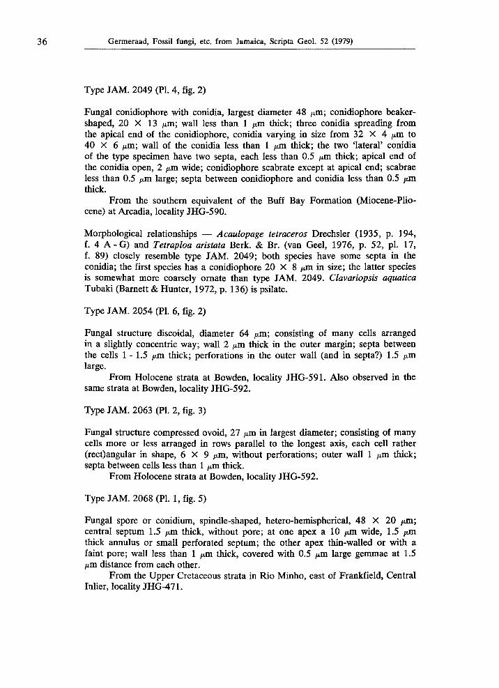
Type J A M . 2049 (PI. 4, fig. 2)
Fungal conidiophore with conidia, largest diameter 48 μπι; conidiophore beaker-shaped, 20 X 13 μπι; wall less than 1 μπι thick; three conidia spreading from the apical end of the conidiophore, conidia varying in size from 32 X 4 μπι to 40 X 6 μπι; wall of the conidia less than 1 μπι thick; the two 'lateral' conidia of the type specimen have two septa, each less than 0.5 μπι thick; apical end of the conidia open, 2 μπι wide; conidiophore scabrate except at apical end; scabrae less than 0.5 μία large; septa between conidiophore and conidia less than 0.5 μπι thick.
From the southern equivalent of the Buff Bay Formation (Miocene-Pliocene) at Arcadia, locality JHG-590.
Morphological relationships — Acaulopage tetraceros Drechsler (1935, p. 194, f. 4 A - G ) and Tetraploa aristata Berk. & Br. (van Geel, 1976, p. 52, pi. 17, f. 89) closely resemble type J A M . 2049; both species have some septa in the conidia; the first species has a conidiophore 20 X 8 μπι in size; the latter species is somewhat more coarsely ornate than type J A M . 2049. Clavariopsis aquatica Tubaki (Barnett & Hunter, 1972, p. 136) is psilate.
Type J A M . 2054 (PI. 6, fig. 2)
Fungal structure discoidal, diameter 64 μπι; consisting of many cells arranged in a slightly concentric way; wall 2 μπι thick in the outer margin; septa between the cells 1-1.5 μπι thick; perforations in the outer wall (and in septa?) 1.5 μπι large.
From Holocene strata at Bowden, locality JHG-591. Also observed in the same strata at Bowden, locality JHG-592.
Type J A M . 2063 (PI. 2, fig. 3)
Fungal structure compressed ovoid, 27 μπι in largest diameter; consisting of many cells more or less arranged in rows parallel to the longest axis, each cell rather (rect)angular in shape, 6 X 9 μπι, without perforations; outer wall 1 μπι thick; septa between cells less than 1 μπι thick.
From Holocene strata at Bowden, locality JHG-592.
Type J A M . 2068 (PI. 1, fig. 5)
Fungal spore or conidium, spindle-shaped, hetero-hemispherical, 48 X 20 μπι; central septum 1.5 μπι thick, without pore; at one apex a 10 μπι wide, 1.5 μπι thick annulus or small perforated septum; the other apex thin-walled or with a faint pore; wall less than 1 μπι thick, covered with 0.5 μπι large gemmae at 1.5 μπι distance from each other.
From the Upper Cretaceous strata in Rio Minho, east of Frankfield, Central Inlier, locality JHG-471.
Germeraad, Fossil fungi, etc. from Jamaica, Scripta Geol. 52 (1979) 37
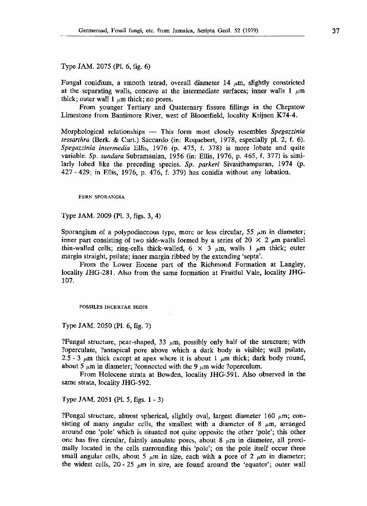
Type J A M . 2075 (PL 6, fig. 6)
Fungal conidium, a smooth tetrad, overall diameter 14 /xm, slightly constricted at the separating walls, concave at the intermediate surfaces; inner walls 1 /xm thick; outer wall 1 /xm thick; no pores.
From younger Tertiary and Quaternary fissure fillings in the Chepstow Limestone from Bantimore River, west of Bloomfield, locality Krijnen K74-4.
Morphological relationships — This form most closely resembles Spegazzinia tessarthra (Berk. & Curt.) Saccardo (in: Roquebert, 1978, especially pi. 2, f. 6). Spegazzinia intermedia Ellis, 1976 (p. 475, f. 378) is more lobate and quite variable. Sp. sundara Subramanian, 1956 (in: Ellis, 1976, p. 465, f. 377) is similarly lobed like the preceding species. Sp. parkeri Sivasithamparan, 1974 (p. 427-429; in Ellis, 1976, p. 476, f. 379) has conidia without any lobation.
FERN SPORANGIA
Type J A M . 2009 (PL 3, figs. 3, 4)
Sporangium of a polypodiaceous type, more or less circular, 55 /xm in diameter; inner part consisting of two side-walls formed by a series of 20 X 2 μΐη parallel thin-walled cells; ring-cells thick-walled, 6 X 3 /xm, walls 1 /xm thick; outer margin straight, psilate; inner margin ribbed by the extending 'septa'.
From the Lower Eocene part of the Richmond Formation at Langley, locality JHG-281. Also from the same formation at Fruitful Vale, locality JHG-107.
FOSSILES INCERTAE SEDIS
Type J A M . 2050 (PL 6, fig. 7)
?Fungal structure, pear-shaped, 33 /an, possibly only half of the structure; with ?operculate, ?antapical pore above which a dark body is visible; wall psilate, 2.5 - 3 /xm thick except at apex where it is about 1 /xm thick; dark body round, about 5 /xm in diameter; ?connected with the 9 /xm wide ?operculum.
From Holocene strata at Bowden, locality JHG-591. Also observed in the same strata, locality JHG-592.
Type J A M . 2051 (PL 5, figs. 1 - 3)
?Fungal structure, almost spherical, slightly oval, largest diameter 160 /xm; consisting of many angular cells, the smallest with a diameter of 8 /xm, arranged around one 'pole' which is situated not quite opposite the other 'pole'; this other one has five circular, faintly annulate pores, about 8 /xm in diameter, all proxi-mally located in the cells surrounding this 'pole'; on the pole itself occur three small angular cells, about 5 /xm in size, each with a pore of 2 /xm in diameter; the widest cells, 20-25 /xm in size, are found around the 'equator'; outer wall
38 Germeraad, Fossil fungi, etc. from Jamaica, Scripta Geol. 52 (1979)
psilate, 1.5 μπι thick; septa between cells much thinner, 0.5 μπι; the inner part of the structure seems to have been destroyed during the process of fossilization.
From Holocene strata at Bowden, locality JHG-591.
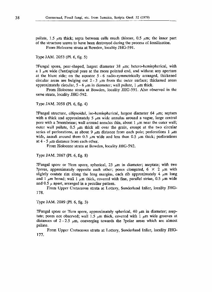
Type J A M . 2053 (PI. 6, fig. 5)
?Fungal spore, pear-shaped, largest diameter 38 μπι; hetero-hemispherical, with a 1 μπι wide (?ant)apical pore at the more pointed end, and without any aperture at the blunt side; on the equator 5 -6 radio-symmetrically arranged, thickened circular areas are bulging out 2 -3 μπι from the outer surface; thickened areas approximately circular, 5 - 6 μπι in diameter; wall psilate, 1 μπι thick.
From Holocene strata at Bowden, locality JHG-591. Also observed in the same strata, locality JHG-592.
Type J A M . 2058 (PI. 6, fig. 4)
?Fungal structure, ellipsoidal, iso-hemispherical, largest diameter 64 μπι; septum with a thick and approximately 5 μπι wide annulus around a vague, large central pore with a ?membrane; wall around annulus thin, about 1 μπι near the outer wall; outer wall psilate, 0.5 μπι thick all over the grain, except at the two circular series of perforations, at about 9 μπι distance from each pole; perforations 1 μπι wide, annuli around them 0.5 μπι wide and less than 0.5 μπι thick; perforations at 4 - 5 μπι distance from each other.
From Holocene strata at Bowden, locality JHG-592.
Type J A M . 2067 (PL 6, fig. 8)
?Fungal spore or ?fern spore, spherical, 23 μπι in diameter; aseptate; with two ?pores, approximately opposite each other; pores elongated, 6 X 2 jum with slightly costate rim along the long margins, each rib approximately 4 μπι long and 1 μπι broad; wall 1 μπι thick, covered with fine, parallel striae, 0.5 μπι wide and 0.5 μ apart, arranged in a peculiar pattern.
From Upper Cretaceous strata at Lottery, Sunderland Inlier, locality JHG-178.
Type J A M . 2089 (PL 6, fig. 3)
?Fungal spore or ?fern spore, approximately spherical, 40 μπι in diameter; aseptate; pores not observed; wall 1.5 μπι thick, covered with 1 μπι wide grooves at distances of 2 - 2.5 μπι, converging towards the ?polar areas which are almost psilate.
From Upper Cretaceous strata at Lottery, Sunderland Inlier, locality JHG-177.
Germeraad, Fossil fungi, etc. from Jamaica, Scripta Geol. 52 (1979) 39
References
Arnaud, G., 1918. Les Astérinées. — Coulet, Montpellier, Thèses Fac. Sei. Paris, A, 805, no. d'ordre 1598: 288 pp., 53 pis.
Barnett, H. L. & B. B. Hunter, 1972. Illustrated genera of imperfect Fungi. — Burgess, Minneapolis, 3rd ed.: 241 pp.
Barron, G. L., 1968. The genera of Hyphomycetes from soil. — Williams & Wilkins, Balti
more: 364 pp., 226 figs. Berlese, A. N., 1894. ícones fungorum. Vols. 14. — Typis E. Pergola, Abellini. Bibliography of Systematic Mycology. Vols. 16. — Commonw. Mycol. Inst., Kew, Surrey,
England, 1 (1939 1949); 2 (1950 1960); 3 (1961 1965); 4 (1966 1970); 5 (1971 1975); (1976 ).
Cain, R. F., 1950. Studies of coprophilous Ascomycetes. I. Gelasinospora. — Canad. Jour. Research, C, 28: 566576, 4 pis., 24 figs.
Chaves Batista, Α., 1963a. Novos fungos Peltasteraceae. — Inst. Micol. Univ. Recife., 222: 114, 5 figs.
Chaves Batista, Α., 1963b. Dois novos fungos da família Manginulaceae. — Inst. Micol. Univ. Recife, 223: 19, 2 figs.
Chaves Batista, Α., 1963c. Drummondia. — novo gênero de Rhizothyriaceae. — Inst. Micol. Univ. Recife, 224: 18, 2 figs.
Cookson, I. C , 1947. Fossil fungi from the Tertiary deposits of the southern hemisphere, Part I. — Proc. Linnean Soc. N.S.W., 72: 207214.
Corner, E. J. H., 1947. Variation in size and shape of spores, basidia and cystidia in Basidio
mycetes. — New Phytol., 46: 195228. Davis, C. Α., 1916. On the fossil algae of the petroleumyielding shales of the Green River
Formation of Colorado and Utah. — Proc. Nat. Acad. Sei., 2: 114119. Dilcher, D. L., 1965. Epiphyllous fungi from Eocene deposits in western Tennessee, U.S.A. —
Palaeontographica, B, 116, 14: 154, pis. 126. Doidge, Ε. M., 1942. A revision of South African Microthyriaceae. — Bothalia, 4: 273420,
pis. 1 76. Drechsler, C , 1935. Some noncatenulate conidial Phycomycetes preying on terricolous
amoebae. — Mycologia, 27: 176205. Edwards, W. N., 1923. An Eocene mycrothyriaceous fungus from Mull, Scotland. — Trans.
Br. mycol. Soc, 8: 6672, pi. 8. Ellis, Μ. B., 1960. Plasmogamy and ascocarp development in Gelasinospora calospora. —
Mycologia 52: 557572. Ellis, Μ. B., 1976. More dematiaceous Hyphomycetes. — Commonw. Mycol. Inst., Kew,
Surrey, England: 793 pp., 40 pis. Elsik, W. C , 1968. Palynology of a Paleocene Rockdale lignite, Milam Co., Texas. I. Mor
phology and Taxonomy. — Pollen Spores, 10,2: 263314; 10,3: 599664. Elsik, W. C. & J. Jansonius, 1974. New genera of Paleogene fungal spores. — Canad. Jour.
Bot., 52, 5: 953958, 20 figs. Geel, Β. van, 1972. Palynology of a section from the raised peat bog Wietmarscher Moor',
with special reference to fungal remains. — Acta Bot. Neerl., 21: 261284. Geel, Β. van, 1976. A palaeoecological study of Holocene peat bog sections, based on the
analysis of pollen, spores and macro and microscopic remains of fungi, algae, cormo
phytes and animals. — Thesis, Hugo de Vries Lab. Univ. Amsterdam: 175, 18 pis., 10 figs.
Gerdemann, J. W., 1953. An undescribed fungus causing a root rot of red clover and other Leguminosae. — Mycologia, 45: 548554, figs. 17.
Germeraad, J. H., 1978a. Contribution to the palynology of Jamaica (B.W.I.). A progress report. — Rijksmus. Geol. Miner., Leiden: 18 (mimeographed).
Germeraad, J. H., 1978b. Contribution to the palynology of the Cretaceous of Jamaica (B.W.L). A progress report. — Rijksmus. Geol. Miner., Leiden: 16, pi. 1 (mimeo
graphed). Germeraad, J. H., 1979a. Displaced sporomorphs and dinoflagellates in Jamaican Cainophytic
strata. A progress report. — Rijksmus. Geol. Miner., Leiden: 14, pis. 2, 3 (mimeo
graphed).
40 Germeraad, Fossil fungi, etc. from Jamaica, Scripta Geol. 52 (1979)
Germeraad, J. H., 1979b. Fossil fungi, algae and other organisms from Jamaica. A progress report. — Rijksmus. Geol. Miner., Leiden: 112, pis. 4, 5 (mimeographed).
Germeraad, J. H., C. A. Hopping & J. Muller, 1968. Palynology of Tertiary sediments from tropical areas. — Rev. Palaeobot. Palynol., 6: 189348, 18 pis., 17 figs.
Gijzel, P. van, 1966. Die FluoreszenzPhotometrie von Mikrofossilien mit dem Zweistrahl
Mikroskopphotometer nach Berek. — Leitz Mitt. Wiss. Technik, 7, 3: 206213. Gijzel, P. van, 1967. Autofluorescence of fossil pollen and spores with special reference to
age determination and coalification. — Thesis Leiden Univ., Leidse Geol. Meded., 40: 263317, 1 pi.
Good, B. H. & R. L. Chapman, 1978. The ultrastructure of Phycopeltis (Chroolepidaceae: Chlorophyta). I. Sporopollenin in the cell walls. — Amer. Jour. Bot., 65, 1: 2733, 9 figs.
Hammen, T. van der, 1954. El desarollo de la flora Colombiana en los períodos geológicos, I. Maestrichtiano hasta Terciário mas inferior. — Bol. Geol. Bogota, 2, 1: 49106.
Herngreen, G. F. W., 1975a. An Upper Senonian pollen asemblage of borehole 3PIAlOAL, State of Alagoas, Brazil. — Pollen Spores: 17, 1: 93140, 14 pis., 1 fig., 1 table.
Herngreen, G. F. W., 1975b. Palynology of Middle and Upper Cretaceous strata in Brazil. — Meded. Geol. Dienst, Ν. S., 26, 3: 3990, 5 pis., 13 figs., 6 tables.
Herngreen, G. F. W., 1976. Microfloral relationships between Africa and South America during the Middle and Upper Cretaceous. — Abstr. 4th. Intern. Palynol. Conf. (Byrbal Sahni Inst. Lucknow): 289304.
Höhnel, F. von, 1918. Fragmente zur Mykologie No. 1152. Ueber Eremothecella calamicola Sydow. — Sber. Kais. Akad. Wiss. Wien, 127: 630.
Hughes, S. J., 1951. Studies on Microfungi VIII. Orbicula and Lilliputia. — Commonw. Mycol. Inst. Kew, Surrey, Mycol. Paper, 42: 127, 1 pl., 12 figs.
Jansonius, J., 1976. Palaeogene fungal spores and fruiting bodies of the Canadian arctic. — Geoscience and Man 15: 129132, 1 pi.
Jennings, Α. V., 1896. On two new species of Phycopeltis from New Zealand. — Proc. R. Irish. Acad. Dublin, 3, 3: 753766.
Johnson, T., 1941. List of fossil plants from Co. Tyrone. — Natl. Mus. Dublin, Dublin. Johnson, T., 1949. Fossil plants from Washing Bay, Co. Tyrone. Fungi. Part I. — Irish. Nat.
Jour., 9: 253256, 1 pi. Journal of the Geological Society of Jamaica. — Univ. West Indies, Mona, Kingston, Jamaica. Karsten, G., 1891. Untersuchungen über die Familie der Chroolepideen. — Ann. Jard. Bot.
Buitenzorg, 10: 166, pis. 16. Kirchheimer, F., 1942. Phycopeltis microthyrioides n. sp., eine blattbewohnende Alge aus
dem Tertiär. — Bot. Archiv, 44: 172204. Köck, C , 1939. Fossile Kryptogamen aus der Eozänen Braunkohle des Geiseltales. — Nova
Acta Acad. Leopol.Carol., N.S., 6: 333359, 9 pis. Lange, R. T., 1978. Southern Australian Tertiary epiphyllous fungi, modern equivalents in
the Australian region, and habitat indicator value. — Canad. Jour. Bot., 56, 5: 532541. Luttrell, E. S., 1946. The genus Stomiopeltis (Hemisphaeriaceae). — Mycology, 38, 5: 565586. Maniotis, J., 1965. A cleistothecial mutant of the perithecial fungus Gelasinospora calospora.
— Jour. Mycol., 57, 1: 2325, 8 figs. Martinez, Α., 1968. Microthyriales (Fungi, Ascomycetes) fosiles del Cretáceo Inferior de la
Província de Santa Cruz, Argentina. — Ameghiniana, 5: 257263, 1 pi. McVey, D. V. & J. W. Gerdemann, 1960. The morphology of Leptodiscus terrestris and the
function of setae in spore dispersal. — Mycologia, 52, 2: 193200, figs. 116. Millardet, Μ. Α., 1870. De la germination des zygospores dans les genres Closterium et
Staurastrum, et sur un genre nouveau d'algues chlorosporées. — Mem. Soc. Sei. nat. Strasbourg, 6: 3750, 35 figs.
Müller, Ε. & J. Α. von Arx, 1962. Die Gattungen der didymosporen Pyrenomyceten. — Büchler, Wabern, Bern, 922 pp., 323 figs.
Pampaloni, L., 1902. I resti organici nel disolile di Melilli in Sicilia. — Paleontographia Itálica, 8: 121130, pis. 1011.
Printz, H., 1939/40. Vorarbeiten zu einer Monographie der Trentepohliaceen. — N. Mag. Naturfid., 80: 137210.
Rao, A. R., 1958. Fungal remains from some Tertiary deposits of India. — Palaeobotanist, 7, 1: 4346.
Roquebert, M. F., 1978. Conidiogenese et conidies chez Spegazzinia tessarthra (Berk. & Curt.) Sacc. — Rev. Mycol., 42, 4: 309320.
Germeraad, Fossil fungi, etc. from Jamaica, Scripta Geol. 52 (1979) 41
Saccardo, P. Α., 18771882. Fungorum Italicorum Autographice Delineatorum, Vol. I: figs. 11280.
Saccardo, P. Α., 18831886. Fungi Italici Autographice Delineati, Vol. II: figs. 12811500. Santesson, R., 1952. Filiicolous Lichens. I. A revision of the taxonomy of the obligately
filiicolous lichenized fungi. — Inaug. Diss., Almqvist & Wicksells, Uppsala. Schmidle, W., 1897. Einige Baumalgen aus Samoa. — Hedwigia, 36: 277287, pis. 69. Selkirk, D. R., 1972. Fossil Manginula-lïke fungi and their classification. — Proc. Linnean
Soc. N.S.W., 97, 2: 141149, pis. 37. Selkirk, D. R., 1975. Tertiary fossil fungi from Kiandra, New South Wales. — Proc. Linnean
Soc. N.S.W., 100, 1: 7094, 12 pis. Sen, J., 1966. Occurrence of Microthyriaceae in the Eocene of west India. — Sei. Cult., 32:
140142. Sheffy, Μ. V. & D. L. Dilcher, 1971. Morphology and taxonomy of fossil fungal spores. —
Palaeontographica, 133, 13: 3451, pis. 1316. Sivasithamparan, K., 1974. Spegazzinia parkeri sp. nov. — Trans. Br. Mycol. Soc, 62: 427
429, 1 fig. Srivastava, S. K., 1968. Fungal elements from the Edmonton Formation (Maestrichtian),
Alberta, Canada. — Canad. Jour. Bot. 46: 11151118. Stevens, F. L., 1925. Hawaiian Fungi. — Bull. Bernice P. Bishop Mus., 19: 1189. Stevens, F. L. & H. W. Manter, 1925. The Hemisphaeriaceae of British Guiana and Trinidad.
— Bot. Gaz., 79: 265294. Subramanian, C. V., 1971. Hyphomycetes, an account of Indian species, except Cercosporae.
— Indian Council Agric. Res. New Dehli: 930 pp., 475 figs. Sydow, H., P. Sydow & E. J. Butler, 1906. Fungi indiae orientalis. Pars 3. — Ann. Mycol.,
9, 4: 372421, pi. 17, 9 figs. Theissen, F., 1913. Ueber einige Microthyriazeen. — Ann. Mycol. 11, 6: 493511, 7 figs. Varma, C. P. & M. S. Rawat, 1963. A note on some diporate grains recovered from Tertiary
horizons of India and their potential marker value. — Grana Palyn. 4: 130139, 1 pi. Wilson, L. R., 1962. A Permian fungus spore type from the Flowerpot Formation of Okla
homa. — Oklahoma Geol. Notes, 22, 4: 9196, 1 pi. Wolf, F. Α., 1966a. Fungus spores in East African lake sediments. — Bull. Torrey Bot. Club,
93: 104113, 6 figs. Wolf, F. Α., 1966b. Fungus spores in East African lake sediments. 2. — Jour. Elisha Mitchell
Sei. Soc, 82: 5761, 3 pis. Wolf, F. Α., 1967a. Fungus spores in East African lake sediments. 4. — Bull Torrey Bot.
Club, 94: 3134. Wolf, F. Α., 1967b. Fungus spores in East African lake sediments. 5. — Mycologia, 59:
397404. Wright, R. M., 1974. Field Guide to selected Jamaican geological localities. — Mines Geol.
Div. Ministry Mining Nat. Resources, Special Publ. No 1: 157. Zans, V. Α., L. J. Chubb, H. R. Versey, J. B. Williams, E. Robinson & D. L. Cooke, 1962.
Synopsis of the Geology of Jamaica. An explanation of the 1958 provisional geological map of Jamaica. — Geol. Survey Dept. Jamaica, Bull. 4: 172.
Manuscript received 25 October 1979.