Forskolin-induciblecAMPPathwayNegativelyRegulates T ... ·...
11
Forskolin-inducible cAMP Pathway Negatively Regulates T-cell Proliferation by Uncoupling the Interleukin-2 Receptor Complex * Received for publication, August 7, 2012, and in revised form, December 21, 2012 Published, JBC Papers in Press, January 22, 2013, DOI 10.1074/jbc.M112.408765 Georgialina Rodriguez, Jeremy A. Ross, Zsuzsanna S. Nagy, and Robert A. Kirken 1 From the Department of Biological Sciences, The University of Texas at El Paso, El Paso, Texas 79902 Background: Within activated T-cells, the binding of IL-2 to its receptor initiates the Jak3/Stat5 cascade culminating in proliferation. Results: Elevated levels of cAMPi within activated T-cells suppresses proliferation by targeting multiple levels of the IL-2R cascade. Conclusion: Cross-talk occurs between cAMP/PKA and the IL-2R/Jak3/Stat5 cascade in human T-cells. Significance: Therapeutic potential exists in manipulating the described cross-talk for the treatment of various immune diseases. Cytokine-mediated regulation of T-cell activity involves a complex interplay between key signal transduction pathways. Determining how these signaling pathways cross-talk is essen- tial to understanding T-cell function and dysfunction. In this work, we provide evidence that cross-talk exists between at least two signaling pathways: the Jak3/Stat5 and cAMP-mediated cascades. The adenylate cyclase activator forskolin (Fsk) signif- icantly increased intracellular cAMP levels and reduced prolif- eration of the human T-cells via inhibition of cell cycle regula- tory genes but did not induce apoptosis. To determine this inhibitory mechanism, effects of Fsk on IL-2 signaling was inves- tigated. Fsk treatment of MT-2 and Kit 225 T-cells inhibited IL-2-induced Stat5a/b tyrosine and serine phosphorylation, nuclear translocation, and DNA binding activity. Fsk treatment also uncoupled IL-2 induced association of the IL-2R and c chain, consequently blocking Jak3 activation. Interestingly, phosphoamino acid analysis revealed that Fsk-treated cells resulted in elevated serine phosphorylation of Jak3 but not Stat5, suggesting that Fsk can negatively regulate Jak3 activity possibly mediated through PKA. Indeed, in vitro kinase assays and small molecule inhibition studies indicated that PKA can directly serine phosphorylate and functionally inactivate Jak3. Taken together, these findings suggest that Fsk activation of adenylate cyclase and PKA can negatively regulate IL-2 signal- ing at multiple levels that include IL-2R complex formation and Jak3/Stat5 activation. Interleukin-2 receptor (IL-2R) 2 signaling in T-cells provides a necessary third signal for driving cellular proliferation during an immune response. The transmission of a proliferative signal by IL-2 requires a heterotrimeric receptor complex comprised of , , and common ( c ) chains. Subsequently, Janus tyrosine kinase 1 (Jak1) and Jak3 are recruited to the and c chains, respectively, allowing for autoactivation of both proteins. Acti- vated Jaks then phosphorylate tyrosine residues on the IL-2R chain, creating docking sites for downstream effectors, includ- ing signal transducer and activator of transcription 5a/b (Stat5a/b), which associate via their Src homology 2 domains (1). Stat5a/b become phosphorylated on Tyr 694 /Ser 726 /Ser 780 and Ser 193 /Tyr 699 /Tyr 725 /Ser 731 /Tyr 740 /Tyr 743 , respectively, dissociate from the receptor, form hetero- or homodimers, and translocate to the nucleus where they mediate transcription of genes important for cell growth and differentiation (2– 8). Con- versely, this signaling pathway is negatively regulated by three families of proteins, suppressor of cytokine signaling, protein tyrosine phosphatases, and protein inhibitors of activated Stats (PIAS), which target different levels within the Jak/Stat signal- ing cascade (reviewed in Ref. 9). T-cell activity is also tightly controlled by other effectors, including Mapk and PI3K pathways, as well as non-protein sig- naling molecules such as cAMP (10). cAMP is produced by adenylate cyclase (AC), a 12-transmembrane-spanning enzyme that catalyzes the conversion of ATP to 3,5-cAMP and pyro- phosphate (11). Cellular phosphodiesterases then degrade newly synthesized cAMP by hydrolyzing the phosphodiester bonds to produce 5AMP (12). While cAMP has multiple func- tions including activation of cyclic nucleotide-gated (CNG) ion channels (13) and exchange protein activated by cAMP (EPAC) (14), in T-cells it predominately exerts its effects through Pro- tein kinase A (PKA) (15). PKA is a ubiquitous serine/threonine kinase comprised of two catalytic and two regulatory subunits. Interestingly, cAMPi levels can be elevated via addition of for- skolin (Fsk), phosphodiesterase inhibitors, prostaglandin E2, or adenosine, which results in suppression of T-cell proliferation * This work was supported by grants from the Lizanell and Colbert Coldwell Foundation, the Edward N. and Margaret G. Marsh Foundation, and Grant G12MD007592 from the National Institutes on Minority Health and Health Disparities, National Institutes of Health. 1 To whom correspondence may be addressed: The University of Texas at El Paso, Dept. of Biological Sciences, 500 W. University Ave., El Paso, TX 79968. Tel.: 915-747-5844; Fax: 915-747-5808; E-mail: [email protected]. 2 The abbreviations used are: IL-2, interleukin-2; Fsk, forskolin; c , common chain; Jak, Janus tyrosine kinase; Stat, signal transducer and activator of transcription; AC, adenylate cyclase; PKA, protein kinase A; IBMX, 3-isobu- tyl-1-methyl-xanthine; DSMO, dimethyl sulfoxide; PARP, poly (ADP-Ribose) polymerase; PKAc, protein kinase A catalytic subunit. THE JOURNAL OF BIOLOGICAL CHEMISTRY VOL. 288, NO. 10, pp. 7137–7146, March 8, 2013 © 2013 by The American Society for Biochemistry and Molecular Biology, Inc. Published in the U.S.A. MARCH 8, 2013 • VOLUME 288 • NUMBER 10 JOURNAL OF BIOLOGICAL CHEMISTRY 7137 by guest on March 21, 2019 http://www.jbc.org/ Downloaded from
Transcript of Forskolin-induciblecAMPPathwayNegativelyRegulates T ... ·...
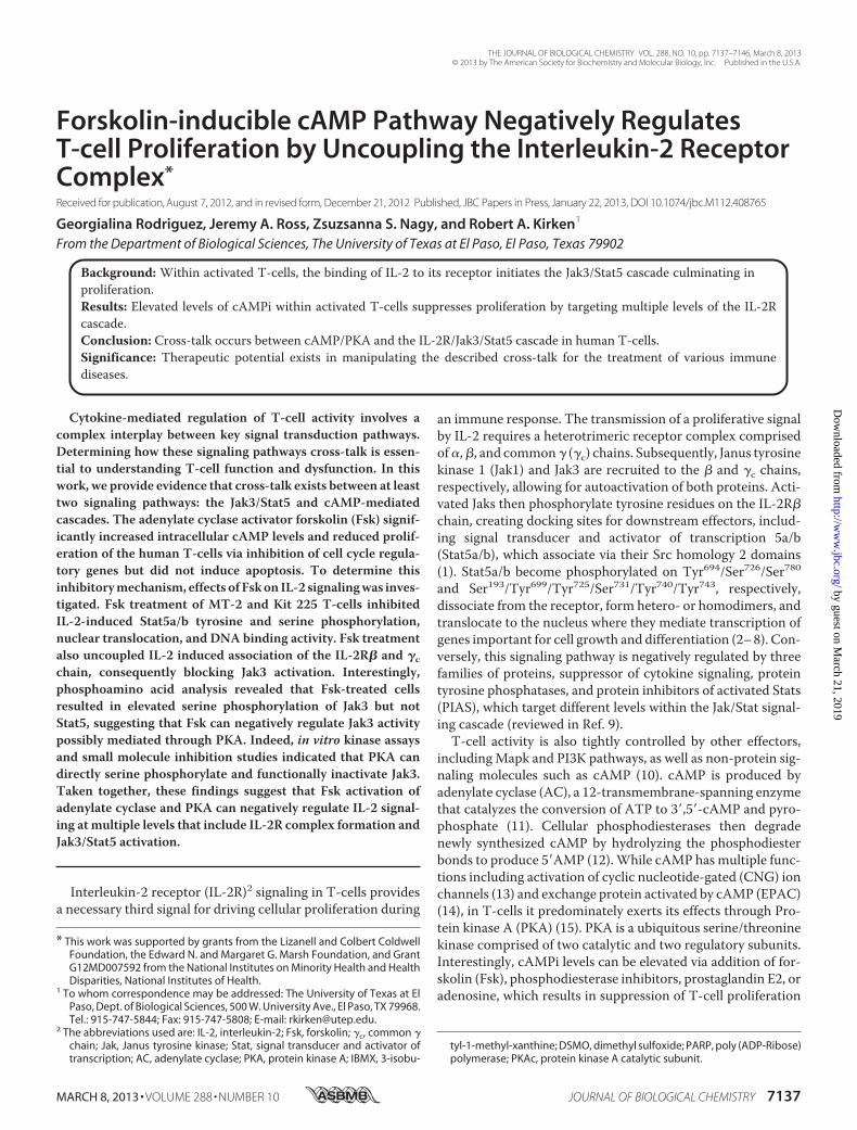
Forskolin-inducible cAMP Pathway Negatively RegulatesT-cell Proliferation by Uncoupling the Interleukin-2 ReceptorComplex*
Received for publication, August 7, 2012, and in revised form, December 21, 2012 Published, JBC Papers in Press, January 22, 2013, DOI 10.1074/jbc.M112.408765
Georgialina Rodriguez, Jeremy A. Ross, Zsuzsanna S. Nagy, and Robert A. Kirken1
From the Department of Biological Sciences, The University of Texas at El Paso, El Paso, Texas 79902
Background: Within activated T-cells, the binding of IL-2 to its receptor initiates the Jak3/Stat5 cascade culminating inproliferation.Results: Elevated levels of cAMPi within activated T-cells suppresses proliferation by targeting multiple levels of the IL-2Rcascade.Conclusion: Cross-talk occurs between cAMP/PKA and the IL-2R/Jak3/Stat5 cascade in human T-cells.Significance: Therapeutic potential exists in manipulating the described cross-talk for the treatment of various immunediseases.
Cytokine-mediated regulation of T-cell activity involves acomplex interplay between key signal transduction pathways.Determining how these signaling pathways cross-talk is essen-tial to understanding T-cell function and dysfunction. In thiswork, we provide evidence that cross-talk exists between at leasttwo signaling pathways: the Jak3/Stat5 and cAMP-mediatedcascades. The adenylate cyclase activator forskolin (Fsk) signif-icantly increased intracellular cAMP levels and reduced prolif-eration of the human T-cells via inhibition of cell cycle regula-tory genes but did not induce apoptosis. To determine thisinhibitorymechanism, effects of Fskon IL-2 signalingwas inves-tigated. Fsk treatment of MT-2 and Kit 225 T-cells inhibitedIL-2-induced Stat5a/b tyrosine and serine phosphorylation,nuclear translocation, and DNA binding activity. Fsk treatmentalso uncoupled IL-2 induced association of the IL-2R� and �cchain, consequently blocking Jak3 activation. Interestingly,phosphoamino acid analysis revealed that Fsk-treated cellsresulted in elevated serine phosphorylation of Jak3 but notStat5, suggesting that Fsk can negatively regulate Jak3 activitypossibly mediated through PKA. Indeed, in vitro kinase assaysand small molecule inhibition studies indicated that PKA candirectly serine phosphorylate and functionally inactivate Jak3.Taken together, these findings suggest that Fsk activation ofadenylate cyclase and PKA can negatively regulate IL-2 signal-ing at multiple levels that include IL-2R complex formation andJak3/Stat5 activation.
Interleukin-2 receptor (IL-2R)2 signaling in T-cells providesa necessary third signal for driving cellular proliferation during
an immune response. The transmission of a proliferative signalby IL-2 requires a heterotrimeric receptor complex comprisedof�,�, and common� (�c) chains. Subsequently, Janus tyrosinekinase 1 (Jak1) and Jak3 are recruited to the � and �c chains,respectively, allowing for autoactivation of both proteins. Acti-vated Jaks then phosphorylate tyrosine residues on the IL-2R�chain, creating docking sites for downstream effectors, includ-ing signal transducer and activator of transcription 5a/b(Stat5a/b), which associate via their Src homology 2 domains(1). Stat5a/b become phosphorylated on Tyr694/Ser726/Ser780and Ser193/Tyr699/Tyr725/Ser731/Tyr740/Tyr743, respectively,dissociate from the receptor, form hetero- or homodimers, andtranslocate to the nucleus where they mediate transcription ofgenes important for cell growth and differentiation (2–8). Con-versely, this signaling pathway is negatively regulated by threefamilies of proteins, suppressor of cytokine signaling, proteintyrosine phosphatases, and protein inhibitors of activated Stats(PIAS), which target different levels within the Jak/Stat signal-ing cascade (reviewed in Ref. 9).T-cell activity is also tightly controlled by other effectors,
includingMapk and PI3K pathways, as well as non-protein sig-naling molecules such as cAMP (10). cAMP is produced byadenylate cyclase (AC), a 12-transmembrane-spanning enzymethat catalyzes the conversion of ATP to 3�,5�-cAMP and pyro-phosphate (11). Cellular phosphodiesterases then degradenewly synthesized cAMP by hydrolyzing the phosphodiesterbonds to produce 5�AMP (12).While cAMP has multiple func-tions including activation of cyclic nucleotide-gated (CNG) ionchannels (13) and exchange protein activated by cAMP (EPAC)(14), in T-cells it predominately exerts its effects through Pro-tein kinase A (PKA) (15). PKA is a ubiquitous serine/threoninekinase comprised of two catalytic and two regulatory subunits.Interestingly, cAMPi levels can be elevated via addition of for-skolin (Fsk), phosphodiesterase inhibitors, prostaglandin E2, oradenosine, which results in suppression of T-cell proliferation
* This work was supported by grants from the Lizanell and Colbert ColdwellFoundation, the Edward N. and Margaret G. Marsh Foundation, and GrantG12MD007592 from the National Institutes on Minority Health and HealthDisparities, National Institutes of Health.
1 To whom correspondence may be addressed: The University of Texas at ElPaso, Dept. of Biological Sciences, 500 W. University Ave., El Paso, TX 79968.Tel.: 915-747-5844; Fax: 915-747-5808; E-mail: [email protected].
2 The abbreviations used are: IL-2, interleukin-2; Fsk, forskolin; �c, common �chain; Jak, Janus tyrosine kinase; Stat, signal transducer and activator oftranscription; AC, adenylate cyclase; PKA, protein kinase A; IBMX, 3-isobu-
tyl-1-methyl-xanthine; DSMO, dimethyl sulfoxide; PARP, poly (ADP-Ribose)polymerase; PKAc, protein kinase A catalytic subunit.
THE JOURNAL OF BIOLOGICAL CHEMISTRY VOL. 288, NO. 10, pp. 7137–7146, March 8, 2013© 2013 by The American Society for Biochemistry and Molecular Biology, Inc. Published in the U.S.A.
MARCH 8, 2013 • VOLUME 288 • NUMBER 10 JOURNAL OF BIOLOGICAL CHEMISTRY 7137
by guest on March 21, 2019
http://ww
w.jbc.org/
Dow
nloaded from
(16–18). Although the antiproliferative effects of cAMPwithincells of the immune system are well documented (19–23), themechanismof action is less clear. It is tempting to speculate thatcAMP-mediated effectors can suppress a T-cell proliferativesignaling pathway such as Jaks and Stats. Nearly a decade ear-lier, Zhang et al. (16) demonstrated that adenosine, a cAMPactivator, can suppress T-cell proliferation that may involveStat5 but not Jak3.Similarly, previous studies have reported that high intensity
activation of the TCR can inhibit IL-2, IL-4, IL-6, and IFN-�signaling in murine T-cells by uncoupling Erk and Mek (24).Ivashkiv and co-workers (25) have also shown that inhibition ofcytokine signaling was independent of new protein synthesisnot dependent on suppressor of cytokine signaling, but impli-cated Stat5, Jak3, and Akt as possible targets. His work suggeststhat PKC/Mek/Erk signaling may uncouple this pathway (25).However, a role for PKA was not investigated or linked to neg-ative regulation of these cytokine-dependent pathways.The objective of this study was to determine the role cAMP-
PKA plays in regulating IL-2R signaling, how it affects T-cellactivity, and consequently immune function. Evidence is pro-vided that Fsk activation of AC/cAMP/PKA disrupts IL-2Rcomplex formation, Jak3 catalytic activity, Stat5 signaling, andexpression of cell cycle-dependent genes. This work suggeststhat a novel cross-talk pathway dependent on cAMPi can neg-atively affect T-cell proliferation and Jak/Stat signaling.
EXPERIMENTAL PROCEDURES
Cell Culture and Treatment—The human leukemic cell lineMT-2 (26) and human embryonic kidney cells 293 (Hek293)were maintained as described previously (27). Human IL-2dependent leukemic Kit 225 cells (28) were similarly main-tained but supplemented with 100 IU/ml human recombinantIL-2 (NCI Preclinical Repository). Prior to cell treatment withFsk or 3-isobutyl-1-methylxanthine (IBMX), MT-2 cells weremade quiescent by growing them to exhaustion or culturingthem in 1% FBS overnight at 37 °C. Kit 225 cells were madequiescent by CO2 washes and subsequently culturing in 10%FBS overnight at 37 °C. Quiescent cells were subsequentlytreated with the indicated concentrations of Fsk (Calbiochem),IBMX (Sigma-Aldrich), or anti-CD3 monoclonal antibody(Santa Cruz Biotechnology) and cultured at 37 °C for differentamounts of time as indicated. Control cells were treated with1% dimethyl sulfoxide (DMSO, vehicle) and cultured as treatedcells. Following pre-treatment, cells were stimulated with 1 �104 IU IL-2 at 37 °C for the times shown.MT-2 cells exposed toultraviolet (UV) light were subjected to 120,000 mJ/cm2/minfor 1 min prior to the final 5 h of incubation.Proliferation Assays—Quiescent Kit 225 or MT-2 cells were
seeded into 96-well plates at 5 � 104 cells per well. Cells werethen pretreated for 1 h with 1% DMSO (vehicle) or Fsk at 1, 5,10, 25, 50, and 100 �M concentrations. The cells were stimu-lated with IL-2 as above and cultured for an additional 20 h at37 °C. Control cells were treated with 1% DMSO for 20 h. Dur-ing the final 4 h of incubation, the cells were pulsed with[3H]thymidine (PerkinElmer Life Sciences) at a concentrationof 0.5 �Ci/200 �l. Cells were harvested onto fiberglass filtersand analyzed using liquid scintillation counting.
cAMP Production Assay—Kit 225 orMT-2 cells were treatedwith 1, 5, 10, 20, 50, or 100�MFsk for 20min at 37 °C.Cellswerelysed and clarified by centrifugation, and concentration ofcAMP was detected by direct cAMP ELISA (Assay Designs).Optical density wasmeasured at 405 nM, and the concentrationof intracellular cAMP was calculated using a weighted fourparameter logistic curve according to the manufacturesinstructions.Cell Solubilization, Protein Immunoprecipitation, and West-
ern Blotting—Cell pelletswere lysed, clarified by centrifugation,and subjected to immunoprecipitation and Western blot anal-ysis as reported previously (27). Briefly, immunoprecipitationreactionswere performed by addition of 2�g of antisera againstJak3 (29), Stat5b (30), IL-2R� (Santa Cruz Biotechnology,C-20), or IL-2R� (Santa Cruz Biotechnology, M-20). Immuno-blotting was performed using antibodies to Jak3, Stat5b,anti-pY (4G10, Millipore), pY939 Jak3 (31), glutathioneS-transferase (GST) (Amersham Biosciences), IL-2R�, IL-2R�,poly(ADP-ribose) polymerase (PARP) (Cell Signaling), glycer-aldehyde 3-phosphate dehydrogenase (GAPDH) (ResearchDiagnostics, Inc.), or �-actin (Santa Cruz Biotechnology,AC-40). Secondary antibodies and Western blot conditionswere performed as described previously (32).In Vitro Kinase Assays—For Jak3 kinase assays, Fsk-treated
MT-2 cells were lysed, clarified, and immunoprecipitated usingJak3 antibody as described above. Kinase reactionswere carriedout as described previously (31) at 30 °C for 20 min. For PKAkinase assays, untreated MT-2 cells were lysed, and Jak3 wasimmunoprecipitated and bound to PAS beads as described pre-viously (31). Immunoprecipitated Jak3 was washed with kinasebuffer (50 mM Hepes-NaOH (pH 7.4), 10 mM MgCl2, 0.5 mM
EGTA, 0.5mMDTT, 20�g/ml aprotinin, 10�g/ml leupeptin, 1�g/ml pepstatin A) and incubated with 200 �M ATP and puri-fied protein kinase A catalytic subunit (PKAc) (Invitrogen) asindicated in the figure legends. Kinase reactions were carriedout at 32 °C for 30 min followed by vigorous washing of thebeads with cold kinase wash buffer as described previously (31).For [�-32P]ATP radiolabeled kinase assays using recombinantJak3, Hek293 cells were transfectedwithwild type (WT) Jak3 orkinase-dead Jak3K855A (31) using Lipofectamine 2000 accord-ing to the manufacturer’s instructions (Invitrogen). Cells werelysed and immunoprecipitated with Jak3 antibody (describedabove). Jak3-bound PAS beads were washed three times in coldlysis buffer (described above) followed by kinase buffer. Kinasereactions were initiated by adding 10 �Ci [�-32P]ATP, 10 �M
unlabeled ATP, and 1 �g of purified PKAc (Invitrogen) to Jak3-bound PAS bead reaction mixtures. Kinase reactions were per-formed at 32 °C for 30min. Jak3-boundPASbeadswerewashedthree times in radioimmunoassay buffer (10 mM Tris-HCl, pH7.4, 75mMNaCl, 20mMEDTA, 10mMEGTA, 20mMNa4P2O7,50 mM NaF, 20 mM 2-glycerolphosphate, 1 mM p-nitrophenyl-phosphate, 0.1% Triton X-100) and one time in kinase washbuffer (described above). The reactions were stopped by adding2� SDS-PAGE sample buffer followed by SDS-PAGE. Coo-massie stainable Jak3 bands were excised from the PVDFmem-brane and subjected to phosphoamino acid analysis.
Forskolin/cAMP Uncouples Interleukin-2 Receptor Signaling
7138 JOURNAL OF BIOLOGICAL CHEMISTRY VOLUME 288 • NUMBER 10 • MARCH 8, 2013
by guest on March 21, 2019
http://ww
w.jbc.org/
Dow
nloaded from
[32P]Orthophosphate Labeling and Phosphoamino AcidAnalysis—Jak3 and Stat5b obtained from MT-2, Kit 225, orHek293 (transfectedwith Jak3) weremetabolically labeled, sep-arated by SDS-PAGE, transferred to PVDF membrane, andvisualized using autoradiography. Coomassie blue staining orWestern blotting was used to determine total protein; bandscorresponding to Jak3 and Stat5b were excised from the mem-branes and subjected to two-dimensional phosphoamino acidanalysis as described previously (33).Nuclear Extract and Electrophoretic Mobility Shift Assays—
Nuclear extracts were prepared from MT-2 cell pellets asreported previously (34). Oligonucleotide sequences corre-sponding to the �-casein promoter (5�-AGATTTCTAG-GAATTCAATCC-3�) were purchased from Santa CruzBiotechnology.RT2 Profiler PCR Array—Kit 225 cells were treated with 1%
DMSO (vehicle) or 100 �M Fsk for 1 h prior to stimulation withIL-2 as above for 30 h at 37 °C. Total RNA was isolated from�4 � 106 cells using the RNeasy kit (Qiagen) and then wereDNase-treated (Qiagen RNase-free DNase set). Complemen-tary DNAwas synthesized using RT2 first strand kit (SA Biosci-ences) as recommended by themanufacturer from 2 �g of totalRNA/each sample/plate. SA Biosciences’ human cell cycle PCRarray (PAHS-020) was used according to the manufacturer’sinstructions. Quantification based on real-time monitoring ofamplification was determined using a Bio-Rad iQ5 machineand 2� SYBR Green Mastermix (SA Biosciences). Sampleswere run in 25-�l reaction volumes. Treatments were per-formed in duplicate, and data were analyzed using the ��Ctmethod normalized to the average of four housekeeping genes(B2M, RPL13A, GAPDH, and ACTB) using SA Biosciences’data analysis web tool. Genes with Ct � 32 were ignored in theanalysis.
RESULTS
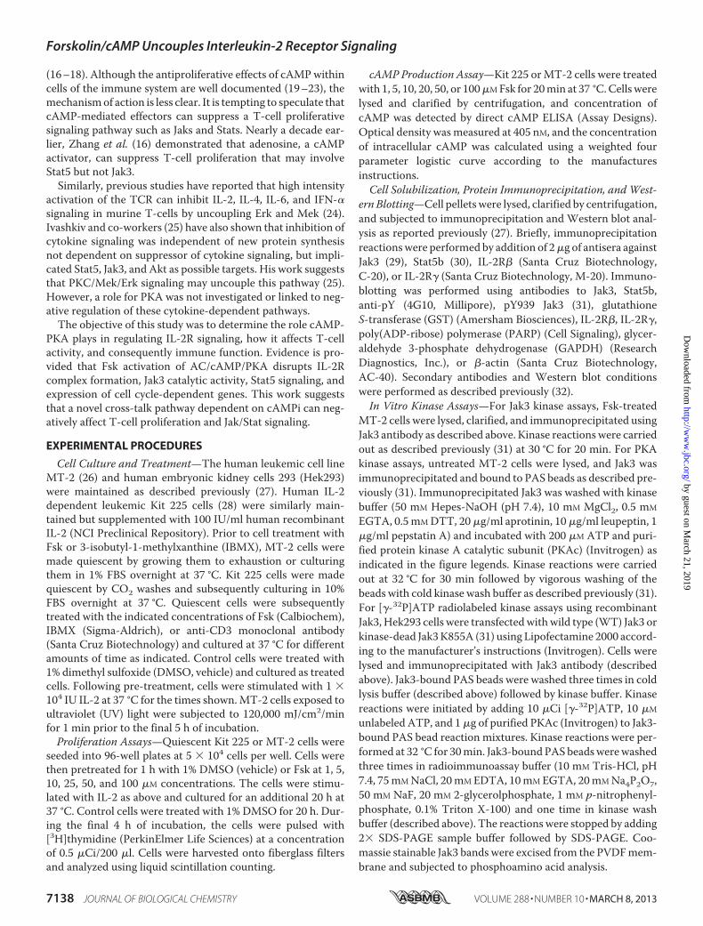
Forskolin-induced cAMPi Inhibits T-cell Proliferation andDisrupts Expression of Cell Cycle Progression Genes—Publishedreports have extensively documented the inhibitory effects ofadenylate cyclase linked G protein-coupled receptor agonistson lymphocyte proliferation (16, 19, 21, 35). To identify themolecular mechanism responsible for this effect, we firstsought to determine whether Fsk, a potent adenylate cyclaseactivator, affects the proliferation of the human T-cell linessuch as Kit 225 (Fig. 1A) andMT-2 (Fig. 1B). For this investiga-tion, cells were pretreated with increasing concentrations ofFsk (0–100 �M) or vehicle prior to stimulation with IL-2. Cellswere subsequently pulsed with [3H]thymidine to detect DNAsynthesis in dividing cells. As shown in Fig. 1, Fsk treatmentinhibited the proliferation of both Kit 225 (Fig. 1A) and MT-2(Fig. 1B) cells in a dose-dependentmannerwith an IC50 equal to�5 �M Fsk. To verify that Fsk concentrations were not causinga nonspecific decrease in cell viability, cleavage of the apoptoticmarker PARP was examined (36). PARP cleavage (Fig. 1C) wasnot detected in negative control or cells treated with increasingconcentrations of Fsk compared with the positive apoptoticcontrol (UV light, lane a). Similar results were observed for Kit225 cells (data not shown). To further support the notion thatFsk-mediated inhibition of MT-2 and Kit 225 cell proliferation
was due to an increase in cAMPi, cAMPi generation was meas-ured using an ELISA. Indeed, Fsk treatment (10–100 �M)increased cAMPi levels�5- to 20-fold above basal levels, whichreached maximum levels between 50–100 �M Fsk (Fig. 1D).Several reports have indicated that cAMPi can block expres-
sion of cell cycle genes (37, 38). To determine whether thereduction in T-cell proliferation was due to Fsk similarly affect-ing cell cycle regulators, these genes were analyzed (Fig. 1E) byquantitative RT-PCR. Indeed, cell cycle-dependent genes,CCNB1 and CCNB2, as well as the cell cycle checkpoint genesCDKN2A, CDKN2B, and CDKN3, were found to be down-reg-ulated following Fsk treatment of Kit 225 cells. Expression ofthe G2/M transition cell cycle regulatory genes GTSE1 andBIRC5 were similarly down-regulated. Interestingly, the p53tumor suppressor gene, TP53, was shown to be up-regulated.Similar results were obtained for MT-2 cells (data not shown).These data suggest that cAMP can inhibit cell cycle geneexpression, which may serve as a possible inhibitory mecha-nism of cell proliferation.Forskolin-cAMPi Inhibits IL-2-induced Stat5 Activation,
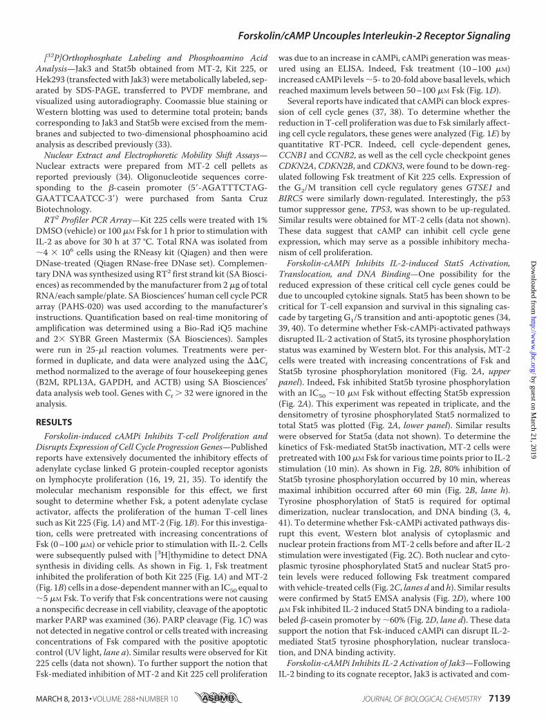
Translocation, and DNA Binding—One possibility for thereduced expression of these critical cell cycle genes could bedue to uncoupled cytokine signals. Stat5 has been shown to becritical for T-cell expansion and survival in this signaling cas-cade by targeting G1/S transition and anti-apoptotic genes (34,39, 40). To determine whether Fsk-cAMPi-activated pathwaysdisrupted IL-2 activation of Stat5, its tyrosine phosphorylationstatus was examined by Western blot. For this analysis, MT-2cells were treated with increasing concentrations of Fsk andStat5b tyrosine phosphorylation monitored (Fig. 2A, upperpanel). Indeed, Fsk inhibited Stat5b tyrosine phosphorylationwith an IC50 �10 �M Fsk without effecting Stat5b expression(Fig. 2A). This experiment was repeated in triplicate, and thedensitometry of tyrosine phosphorylated Stat5 normalized tototal Stat5 was plotted (Fig. 2A, lower panel). Similar resultswere observed for Stat5a (data not shown). To determine thekinetics of Fsk-mediated Stat5b inactivation, MT-2 cells werepretreatedwith 100�MFsk for various time points prior to IL-2stimulation (10 min). As shown in Fig. 2B, 80% inhibition ofStat5b tyrosine phosphorylation occurred by 10 min, whereasmaximal inhibition occurred after 60 min (Fig. 2B, lane h).Tyrosine phosphorylation of Stat5 is required for optimaldimerization, nuclear translocation, and DNA binding (3, 4,41). To determine whether Fsk-cAMPi activated pathways dis-rupt this event, Western blot analysis of cytoplasmic andnuclear protein fractions fromMT-2 cells before and after IL-2stimulation were investigated (Fig. 2C). Both nuclear and cyto-plasmic tyrosine phosphorylated Stat5 and nuclear Stat5 pro-tein levels were reduced following Fsk treatment comparedwith vehicle-treated cells (Fig. 2C, lanes d and h). Similar resultswere confirmed by Stat5 EMSA analysis (Fig. 2D), where 100�M Fsk inhibited IL-2 induced Stat5 DNA binding to a radiola-beled �-casein promoter by �60% (Fig. 2D, lane d). These datasupport the notion that Fsk-induced cAMPi can disrupt IL-2-mediated Stat5 tyrosine phosphorylation, nuclear transloca-tion, and DNA binding activity.Forskolin-cAMPi Inhibits IL-2 Activation of Jak3—Following
IL-2 binding to its cognate receptor, Jak3 is activated and com-
Forskolin/cAMP Uncouples Interleukin-2 Receptor Signaling
MARCH 8, 2013 • VOLUME 288 • NUMBER 10 JOURNAL OF BIOLOGICAL CHEMISTRY 7139
by guest on March 21, 2019
http://ww
w.jbc.org/
Dow
nloaded from
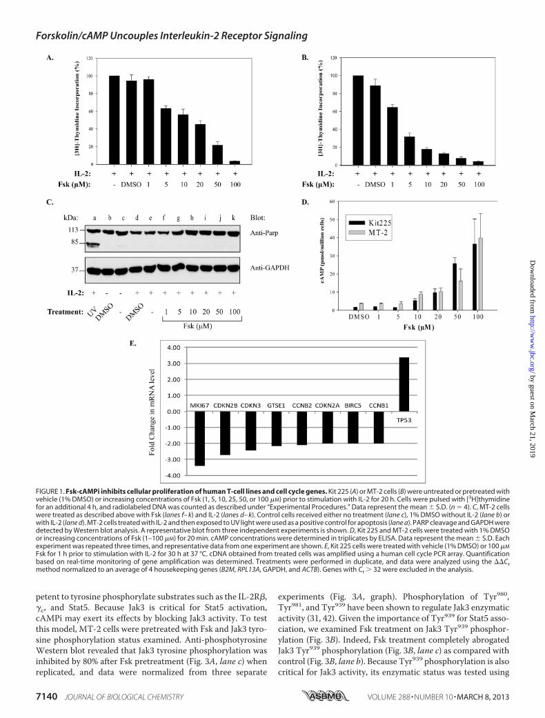
petent to tyrosine phosphorylate substrates such as the IL-2R�,�c, and Stat5. Because Jak3 is critical for Stat5 activation,cAMPi may exert its effects by blocking Jak3 activity. To testthis model, MT-2 cells were pretreated with Fsk and Jak3 tyro-sine phosphorylation status examined. Anti-phosphotyrosineWestern blot revealed that Jak3 tyrosine phosphorylation wasinhibited by 80% after Fsk pretreatment (Fig. 3A, lane c) whenreplicated, and data were normalized from three separate
experiments (Fig. 3A, graph). Phosphorylation of Tyr980,Tyr981, and Tyr939 have been shown to regulate Jak3 enzymaticactivity (31, 42). Given the importance of Tyr939 for Stat5 asso-ciation, we examined Fsk treatment on Jak3 Tyr939 phosphor-ylation (Fig. 3B). Indeed, Fsk treatment completely abrogatedJak3 Tyr939 phosphorylation (Fig. 3B, lane c) as compared withcontrol (Fig. 3B, lane b). Because Tyr939 phosphorylation is alsocritical for Jak3 activity, its enzymatic status was tested using
FIGURE 1. Fsk-cAMPi inhibits cellular proliferation of human T-cell lines and cell cycle genes. Kit 225 (A) or MT-2 cells (B) were untreated or pretreated withvehicle (1% DMSO) or increasing concentrations of Fsk (1, 5, 10, 25, 50, or 100 �M) prior to stimulation with IL-2 for 20 h. Cells were pulsed with [3H]thymidinefor an additional 4 h, and radiolabeled DNA was counted as described under “Experimental Procedures.” Data represent the mean � S.D. (n � 4). C, MT-2 cellswere treated as described above with Fsk (lanes f– k) and IL-2 (lanes d– k). Control cells received either no treatment (lane c), 1% DMSO without IL-2 (lane b) orwith IL-2 (lane d). MT-2 cells treated with IL-2 and then exposed to UV light were used as a positive control for apoptosis (lane a). PARP cleavage and GAPDH weredetected by Western blot analysis. A representative blot from three independent experiments is shown. D, Kit 225 and MT-2 cells were treated with 1% DMSOor increasing concentrations of Fsk (1–100 �M) for 20 min. cAMP concentrations were determined in triplicates by ELISA. Data represent the mean � S.D. Eachexperiment was repeated three times, and representative data from one experiment are shown. E, Kit 225 cells were treated with vehicle (1% DMSO) or 100 �M
Fsk for 1 h prior to stimulation with IL-2 for 30 h at 37 °C. cDNA obtained from treated cells was amplified using a human cell cycle PCR array. Quantificationbased on real-time monitoring of gene amplification was determined. Treatments were performed in duplicate, and data were analyzed using the ��Ctmethod normalized to an average of 4 housekeeping genes (B2M, RPL13A, GAPDH, and ACTB). Genes with Ct � 32 were excluded in the analysis.
Forskolin/cAMP Uncouples Interleukin-2 Receptor Signaling
7140 JOURNAL OF BIOLOGICAL CHEMISTRY VOLUME 288 • NUMBER 10 • MARCH 8, 2013
by guest on March 21, 2019
http://ww
w.jbc.org/
Dow
nloaded from
theGST-tagged cytoplasmic portion of�c as a substrate (31). Asshown in Fig. 3C, Fsk treatment inhibited Jak3 tyrosine phos-phorylation of �c in a dose-dependent manner with an IC50between 1 and 10 �M. These results closely parallel those Fskconcentrations required to reduceT-cell proliferation (Fig. 1B).
cAMPi Elevators Induce Jak3 Serine Phosphorylation but Dis-rupt IL-2-inducible Stat5 Serine Phosphorylation—The posi-tive regulatory role of tyrosine phosphorylation of IL-2R signal-ing proteins has been well documented (3, 42, 43), and manyalso undergo serine phosphorylation to positively or negativelyregulate their activity (8, 44–46). The role of serine phosphor-ylation within IL-2R signaling, however, is not fully established.Because cAMPi most notably activates the serine-threonineprotein kinase PKA (10), we sought to determine the effects of
FIGURE 2. Fsk-cAMPi inhibits Stat5b activation, nuclear translocation,and DNA binding activity. A, MT-2 cells were untreated (lane a) or pretreatedwith the indicated concentrations of Fsk for 40 min prior to stimulation withIL-2 for 10 min at 37 °C (lanes b–i). Immunoprecipitated Stat5b was Westernblotted with anti-phosphotyrosine (pY; upper panel) or anti-Stat5b (lowerpanel). Representative data from three independent experiments are shown.Densitometric analysis of phosphotyrosine Stat5b was normalized to totalStat5b and graphed (percentage of phosphotyrosine Stat5b) (lane b). Datarepresent the mean � S.D. from three independent experiments. B, MT-2 cellswere treated as above with vehicle (lanes a and b) or pretreated with 100 �M
Fsk for the times indicated before IL-2 stimulation for 10 min at 37 °C (lanesc– h). Immunoprecipitated Stat5b was Western blotted with anti-phosphoty-rosine (upper panel) or anti-Stat5b (lower panel). C, MT-2 cells were eitheruntreated or pretreated with 100 �M Fsk for 40 min prior to IL-2 stimulation.Immunoprecipitated nuclear (lanes a– d) and cytoplasmic (lanes e– h) local-ized Stat5b proteins were separated by SDS-PAGE and Western blotted usingantibodies for phosphotyrosine, Stat5, or �-actin (internal standard). D, EMSAanalysis was performed on MT-2 cells treated as in C. Nuclear proteinswere isolated and incubated with [�-32P]ATP-labeled �-casein promoteralone (lanes a– d), with anti-Stat5 antibody (lane e) or normal rabbit serum(NRS; lane f) or free probe without cell lysate (lane g). Stat5 migration is indi-cated by the arrow.
FIGURE 3. Fsk-cAMPi inhibits IL-2-induced Jak3 activation and catalyticactivity. A, MT-2 cells were left untreated (lane a) or treated with vehicle (1%DMSO) (lane b) or 100 �M Fsk for 40 min (lane c) prior to stimulation with 100nM IL-2 for 10 min (lanes b and c) at 37 °C. Immunoprecipitated Jak3 proteinwas Western blotted using anti-phosphotyrosine (pY) antibody (upper panel)or total Jak3 (lower panel). Densitometric analyses were carried out (n � 3),and tyrosine phosphorylation levels of Jak3 were normalized to total Jak3protein and plotted (lower panel). B, cells were treated as described in A andWestern blotted with anti-phosphotyrosine 939 (pY939). C, MT-2 cells weretreated with increasing concentrations of Fsk for 40 min prior to stimulationwith 100 nM IL-2 for 10 min. Immunocaptured Jak3 was used for in vitro kinaseassays with GST-�c as a substrate and Western blotted with anti-phosphoty-rosine or anti-GST antibodies. Densitometric analysis was performed, andtyrosine phosphorylation of GST-�c was normalized to total GST expression.The results are presented as percentage of IL-2 activated Jak3 activity in theabsence or presence of Fsk.
Forskolin/cAMP Uncouples Interleukin-2 Receptor Signaling
MARCH 8, 2013 • VOLUME 288 • NUMBER 10 JOURNAL OF BIOLOGICAL CHEMISTRY 7141
by guest on March 21, 2019
http://ww
w.jbc.org/
Dow
nloaded from
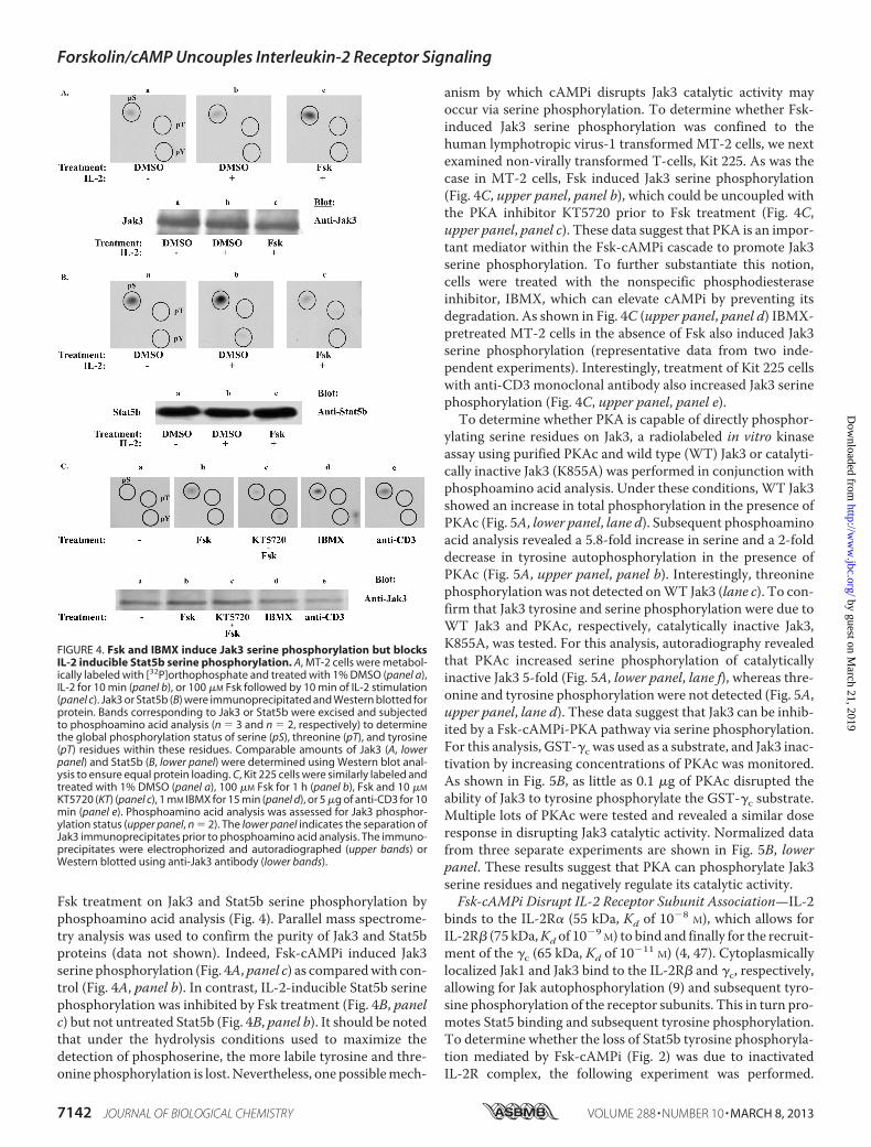
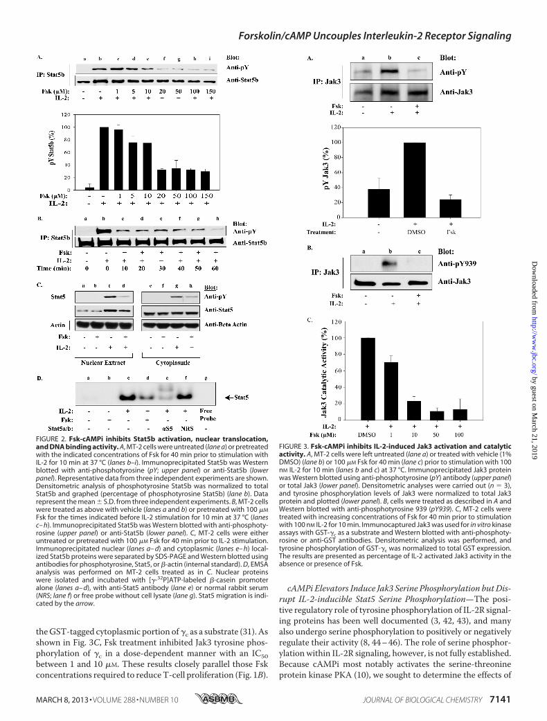
Fsk treatment on Jak3 and Stat5b serine phosphorylation byphosphoamino acid analysis (Fig. 4). Parallel mass spectrome-try analysis was used to confirm the purity of Jak3 and Stat5bproteins (data not shown). Indeed, Fsk-cAMPi induced Jak3serine phosphorylation (Fig. 4A,panel c) as comparedwith con-trol (Fig. 4A, panel b). In contrast, IL-2-inducible Stat5b serinephosphorylation was inhibited by Fsk treatment (Fig. 4B, panelc) but not untreated Stat5b (Fig. 4B, panel b). It should be notedthat under the hydrolysis conditions used to maximize thedetection of phosphoserine, the more labile tyrosine and thre-onine phosphorylation is lost. Nevertheless, one possiblemech-
anism by which cAMPi disrupts Jak3 catalytic activity mayoccur via serine phosphorylation. To determine whether Fsk-induced Jak3 serine phosphorylation was confined to thehuman lymphotropic virus-1 transformed MT-2 cells, we nextexamined non-virally transformed T-cells, Kit 225. As was thecase in MT-2 cells, Fsk induced Jak3 serine phosphorylation(Fig. 4C, upper panel, panel b), which could be uncoupled withthe PKA inhibitor KT5720 prior to Fsk treatment (Fig. 4C,upper panel, panel c). These data suggest that PKA is an impor-tant mediator within the Fsk-cAMPi cascade to promote Jak3serine phosphorylation. To further substantiate this notion,cells were treated with the nonspecific phosphodiesteraseinhibitor, IBMX, which can elevate cAMPi by preventing itsdegradation. As shown in Fig. 4C (upper panel, panel d) IBMX-pretreated MT-2 cells in the absence of Fsk also induced Jak3serine phosphorylation (representative data from two inde-pendent experiments). Interestingly, treatment of Kit 225 cellswith anti-CD3 monoclonal antibody also increased Jak3 serinephosphorylation (Fig. 4C, upper panel, panel e).To determine whether PKA is capable of directly phosphor-
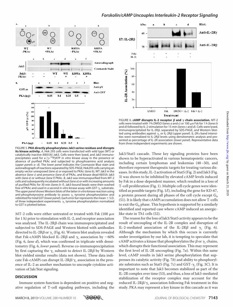
ylating serine residues on Jak3, a radiolabeled in vitro kinaseassay using purified PKAc and wild type (WT) Jak3 or catalyti-cally inactive Jak3 (K855A) was performed in conjunction withphosphoamino acid analysis. Under these conditions, WT Jak3showed an increase in total phosphorylation in the presence ofPKAc (Fig. 5A, lower panel, lane d). Subsequent phosphoaminoacid analysis revealed a 5.8-fold increase in serine and a 2-folddecrease in tyrosine autophosphorylation in the presence ofPKAc (Fig. 5A, upper panel, panel b). Interestingly, threoninephosphorylationwas not detected onWT Jak3 (lane c). To con-firm that Jak3 tyrosine and serine phosphorylation were due toWT Jak3 and PKAc, respectively, catalytically inactive Jak3,K855A, was tested. For this analysis, autoradiography revealedthat PKAc increased serine phosphorylation of catalyticallyinactive Jak3 5-fold (Fig. 5A, lower panel, lane f), whereas thre-onine and tyrosine phosphorylation were not detected (Fig. 5A,upper panel, lane d). These data suggest that Jak3 can be inhib-ited by a Fsk-cAMPi-PKA pathway via serine phosphorylation.For this analysis, GST-�c was used as a substrate, and Jak3 inac-tivation by increasing concentrations of PKAc was monitored.As shown in Fig. 5B, as little as 0.1 �g of PKAc disrupted theability of Jak3 to tyrosine phosphorylate the GST-�c substrate.Multiple lots of PKAc were tested and revealed a similar doseresponse in disrupting Jak3 catalytic activity. Normalized datafrom three separate experiments are shown in Fig. 5B, lowerpanel. These results suggest that PKA can phosphorylate Jak3serine residues and negatively regulate its catalytic activity.Fsk-cAMPi Disrupt IL-2 Receptor Subunit Association—IL-2
binds to the IL-2R� (55 kDa, Kd of 10�8 M), which allows forIL-2R� (75 kDa,Kd of 10�9 M) to bind and finally for the recruit-ment of the �c (65 kDa, Kd of 10�11 M) (4, 47). Cytoplasmicallylocalized Jak1 and Jak3 bind to the IL-2R� and �c, respectively,allowing for Jak autophosphorylation (9) and subsequent tyro-sine phosphorylation of the receptor subunits. This in turn pro-motes Stat5 binding and subsequent tyrosine phosphorylation.To determine whether the loss of Stat5b tyrosine phosphoryla-tion mediated by Fsk-cAMPi (Fig. 2) was due to inactivatedIL-2R complex, the following experiment was performed.
FIGURE 4. Fsk and IBMX induce Jak3 serine phosphorylation but blocksIL-2 inducible Stat5b serine phosphorylation. A, MT-2 cells were metabol-ically labeled with [32P]orthophosphate and treated with 1% DMSO (panel a),IL-2 for 10 min (panel b), or 100 �M Fsk followed by 10 min of IL-2 stimulation(panel c). Jak3 or Stat5b (B) were immunoprecipitated and Western blotted forprotein. Bands corresponding to Jak3 or Stat5b were excised and subjectedto phosphoamino acid analysis (n � 3 and n � 2, respectively) to determinethe global phosphorylation status of serine (pS), threonine (pT), and tyrosine(pT) residues within these residues. Comparable amounts of Jak3 (A, lowerpanel) and Stat5b (B, lower panel) were determined using Western blot anal-ysis to ensure equal protein loading. C, Kit 225 cells were similarly labeled andtreated with 1% DMSO (panel a), 100 �M Fsk for 1 h (panel b), Fsk and 10 �M
KT5720 (KT) (panel c), 1 mM IBMX for 15 min (panel d), or 5 �g of anti-CD3 for 10min (panel e). Phosphoamino acid analysis was assessed for Jak3 phosphor-ylation status (upper panel, n � 2). The lower panel indicates the separation ofJak3 immunoprecipitates prior to phosphoamino acid analysis. The immuno-precipitates were electrophorized and autoradiographed (upper bands) orWestern blotted using anti-Jak3 antibody (lower bands).
Forskolin/cAMP Uncouples Interleukin-2 Receptor Signaling
7142 JOURNAL OF BIOLOGICAL CHEMISTRY VOLUME 288 • NUMBER 10 • MARCH 8, 2013
by guest on March 21, 2019
http://ww
w.jbc.org/
Dow
nloaded from
MT-2 cells were either untreated or treated with Fsk (100 �M
for 1 h) prior to stimulation with IL-2, and receptor associationwas analyzed. The IL-2R� chain was immunoprecipitated andsubjected to SDS-PAGE and Western blotted with antibodiesdirected to IL-2R� or �c (Fig. 6).Western blot analysis revealedthat Fsk-cAMPi blocked IL-2R� and �c association by �60%(Fig. 6, lane d), which was confirmed in triplicate with densi-tometry (Fig. 6, lower panel). Reverse co-immunoprecipitationby first capturing the �c subunit to detect IL-2R� by Westernblot yielded similar results (data not shown). These data indi-cate Fsk-cAMPi can disrupt IL-2R�/�c association in the pres-ence of IL-2 as another mechanism to uncouple cytokine acti-vation of Jak/Stat signaling.
DISCUSSION
Immune system function is dependent on positive and neg-ative regulation of T-cell signaling pathways, including the
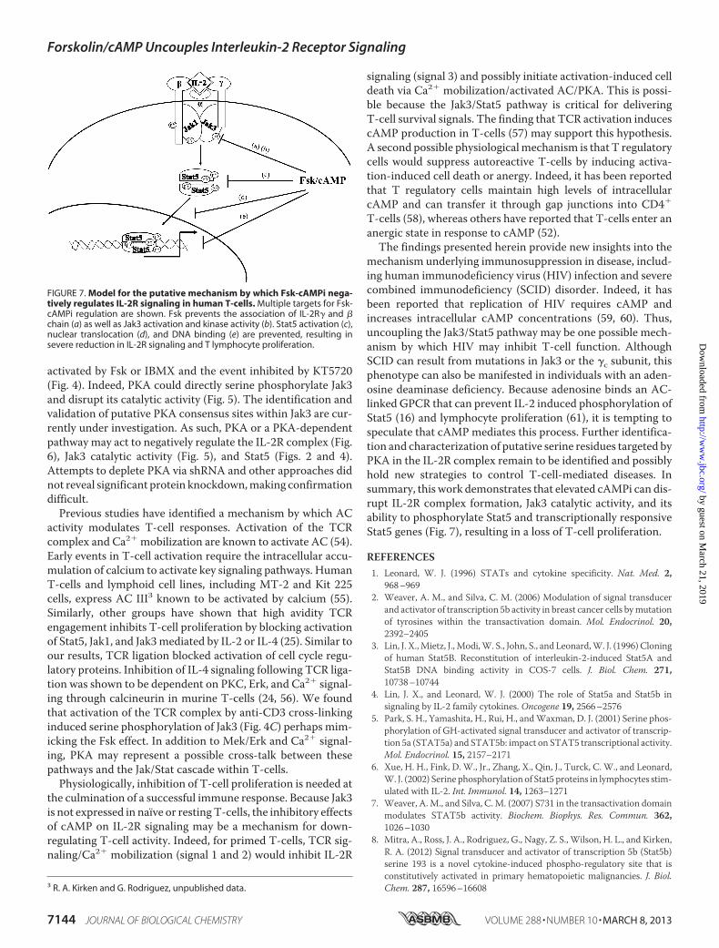
Jak3/Stat5 cascade. These key signaling proteins have beenshown to be hyperactivated in various hematopoietic cancers,including certain lymphomas and leukemias (48–50), andtherefore represent therapeutic targets for treating various dis-eases. In this study, IL-2 activation of Stat5 (Fig. 2) and Jak3 (Fig.3) was shown to be inhibited by elevated cAMP levels inducedby Fsk in a dose-dependent manner, which resulted in a loss ofT-cell proliferation (Fig. 1).Multiple cell cycle genes were iden-tified as possible targets (Fig. 1E), including the gene for KD-67,a protein present during all phases of the cell cycle except G0(51). It is likely that cAMPi accumulation does not allowT-cellsto exit the G0 phase. This hypothesis is supported by a similarlyidentified and reported case where cAMP induced an anergic-like state in Th1 cells (52).The reason for the loss of Jak3/Stat5 activity appears to be the
result of uncoupling of the IL-2R complex and disruption ofIL-2-mediated association of the IL-2R� and �c (Fig. 6).Although the mechanism by which this occurs is currentlyunder investigation by our lab, it is tempting to speculate thatcAMP activates a kinase that phosphorylates the � or �c chains,which disrupts their functional association. Thismay representthe first level of IL-2R uncoupling (Fig. 7a). Within this samelevel, cAMP results in Jak3 serine phosphorylation that sup-presses its catalytic activity (Fig. 7b) and ability to phosphoryl-ate substrates such as Stat5 (Fig. 7c) and GST-�c (Fig. 3C). It isimportant to note that Jak3 becomes stabilized as part of theIL-2R complex over time (53), and thus, a loss of Jak3-mediatedstabilization of the receptor complex may account for thereduced IL-2R�/�c association following Fsk treatment in thisstudy. PKA may represent a key kinase in this cascade as it was
FIGURE 5. PKA directly phosphorylates Jak3 serine residues and disruptsits kinase activity. A, Hek 293 cells were transfected with wild type (WT) orcatalytically inactive (K855A) Jak3. Cells were then lysed, and Jak3 immuno-precipitates used for a [�-32P]ATP in vitro kinase assay in the presence orabsence of purified PKAc and subjected to phosphoamino acid analysis(upper panels a– d). The lower panel indicates the Coomassie Blue stain andautoradiograph of reactions separated by SDS-PAGE; Hek293 cells carrying anempty vector unexposed (lane a) or exposed to PKAc (lane b), WT Jak3 in theabsence (lane c) and presence (lane d) of PKAc, and kinase dead K855A Jak3with (lane e) or without (lane f) PKAc. B, Jak3 was immunopurified from MT-2cells and subsequently incubated without (lane a) or with increasing amountsof purified PKAc for 30 min (lanes b–f). Jak3-bound beads were then washedfree of PKAc and used in a second in vitro kinase assay with GST-�c substrate.The upper panel shows Western blots of the latter in vitro kinase reaction usinganti-phosphotyrosine antibody to assess �c tyrosine phosphorylation andreblotted for total GST (lower panel). Each error bar represents the mean � S.D.of three independent experiments. �c tyrosine phosphorylation normalizedto GST is plotted below.
FIGURE 6. cAMP disrupts IL-2 receptor � and � chain association. MT-2cells were treated with 1% DMSO (lanes a and c) or 100 �M Fsk for 1 h (lanes band d) followed by IL-2 stimulation for 15 min (lanes c and d). Cells were lysed,immunoprecipitated for IL-2R�, separated by SDS-PAGE, and Western blot-ted using antibodies against �c or IL-2R� (upper panel). IL-2R� band intensi-ties were normalized to IL-2R� levels using densitometric analysis and pre-sented as percentage of IL-2R association (lower panel). Representative datafrom three independent experiments are shown.
Forskolin/cAMP Uncouples Interleukin-2 Receptor Signaling
MARCH 8, 2013 • VOLUME 288 • NUMBER 10 JOURNAL OF BIOLOGICAL CHEMISTRY 7143
by guest on March 21, 2019
http://ww
w.jbc.org/
Dow
nloaded from
activated by Fsk or IBMX and the event inhibited by KT5720(Fig. 4). Indeed, PKA could directly serine phosphorylate Jak3and disrupt its catalytic activity (Fig. 5). The identification andvalidation of putative PKA consensus sites within Jak3 are cur-rently under investigation. As such, PKA or a PKA-dependentpathway may act to negatively regulate the IL-2R complex (Fig.6), Jak3 catalytic activity (Fig. 5), and Stat5 (Figs. 2 and 4).Attempts to deplete PKA via shRNA and other approaches didnot reveal significant protein knockdown,making confirmationdifficult.Previous studies have identified a mechanism by which AC
activity modulates T-cell responses. Activation of the TCRcomplex and Ca2 mobilization are known to activate AC (54).Early events in T-cell activation require the intracellular accu-mulation of calcium to activate key signaling pathways. HumanT-cells and lymphoid cell lines, including MT-2 and Kit 225cells, express AC III3 known to be activated by calcium (55).Similarly, other groups have shown that high avidity TCRengagement inhibits T-cell proliferation by blocking activationof Stat5, Jak1, and Jak3mediated by IL-2 or IL-4 (25). Similar toour results, TCR ligation blocked activation of cell cycle regu-latory proteins. Inhibition of IL-4 signaling following TCR liga-tion was shown to be dependent on PKC, Erk, and Ca2 signal-ing through calcineurin in murine T-cells (24, 56). We foundthat activation of the TCR complex by anti-CD3 cross-linkinginduced serine phosphorylation of Jak3 (Fig. 4C) perhaps mim-icking the Fsk effect. In addition to Mek/Erk and Ca2 signal-ing, PKA may represent a possible cross-talk between thesepathways and the Jak/Stat cascade within T-cells.Physiologically, inhibition of T-cell proliferation is needed at
the culmination of a successful immune response. Because Jak3is not expressed in naïve or restingT-cells, the inhibitory effectsof cAMP on IL-2R signaling may be a mechanism for down-regulating T-cell activity. Indeed, for primed T-cells, TCR sig-naling/Ca2 mobilization (signal 1 and 2) would inhibit IL-2R
signaling (signal 3) and possibly initiate activation-induced celldeath via Ca2 mobilization/activated AC/PKA. This is possi-ble because the Jak3/Stat5 pathway is critical for deliveringT-cell survival signals. The finding that TCR activation inducescAMP production in T-cells (57) may support this hypothesis.A second possible physiologicalmechanism is that T regulatorycells would suppress autoreactive T-cells by inducing activa-tion-induced cell death or anergy. Indeed, it has been reportedthat T regulatory cells maintain high levels of intracellularcAMP and can transfer it through gap junctions into CD4
T-cells (58), whereas others have reported that T-cells enter ananergic state in response to cAMP (52).The findings presented herein provide new insights into the
mechanism underlying immunosuppression in disease, includ-ing human immunodeficiency virus (HIV) infection and severecombined immunodeficiency (SCID) disorder. Indeed, it hasbeen reported that replication of HIV requires cAMP andincreases intracellular cAMP concentrations (59, 60). Thus,uncoupling the Jak3/Stat5 pathway may be one possible mech-anism by which HIV may inhibit T-cell function. AlthoughSCID can result from mutations in Jak3 or the �c subunit, thisphenotype can also be manifested in individuals with an aden-osine deaminase deficiency. Because adenosine binds an AC-linked GPCR that can prevent IL-2 induced phosphorylation ofStat5 (16) and lymphocyte proliferation (61), it is tempting tospeculate that cAMP mediates this process. Further identifica-tion and characterization of putative serine residues targeted byPKA in the IL-2R complex remain to be identified and possiblyhold new strategies to control T-cell-mediated diseases. Insummary, this work demonstrates that elevated cAMPi can dis-rupt IL-2R complex formation, Jak3 catalytic activity, and itsability to phosphorylate Stat5 and transcriptionally responsiveStat5 genes (Fig. 7), resulting in a loss of T-cell proliferation.
REFERENCES1. Leonard, W. J. (1996) STATs and cytokine specificity. Nat. Med. 2,
968–9692. Weaver, A. M., and Silva, C. M. (2006) Modulation of signal transducer
and activator of transcription 5b activity in breast cancer cells bymutationof tyrosines within the transactivation domain. Mol. Endocrinol. 20,2392–2405
3. Lin, J. X.,Mietz, J.,Modi,W. S., John, S., and Leonard,W. J. (1996) Cloningof human Stat5B. Reconstitution of interleukin-2-induced Stat5A andStat5B DNA binding activity in COS-7 cells. J. Biol. Chem. 271,10738–10744
4. Lin, J. X., and Leonard, W. J. (2000) The role of Stat5a and Stat5b insignaling by IL-2 family cytokines. Oncogene 19, 2566–2576
5. Park, S. H., Yamashita, H., Rui, H., andWaxman, D. J. (2001) Serine phos-phorylation of GH-activated signal transducer and activator of transcrip-tion 5a (STAT5a) and STAT5b: impact on STAT5 transcriptional activity.Mol. Endocrinol. 15, 2157–2171
6. Xue, H. H., Fink, D. W., Jr., Zhang, X., Qin, J., Turck, C. W., and Leonard,W. J. (2002) Serine phosphorylation of Stat5 proteins in lymphocytes stim-ulated with IL-2. Int. Immunol. 14, 1263–1271
7. Weaver, A.M., and Silva, C.M. (2007) S731 in the transactivation domainmodulates STAT5b activity. Biochem. Biophys. Res. Commun. 362,1026–1030
8. Mitra, A., Ross, J. A., Rodriguez, G., Nagy, Z. S., Wilson, H. L., and Kirken,R. A. (2012) Signal transducer and activator of transcription 5b (Stat5b)serine 193 is a novel cytokine-induced phospho-regulatory site that isconstitutively activated in primary hematopoietic malignancies. J. Biol.Chem. 287, 16596–166083 R. A. Kirken and G. Rodriguez, unpublished data.
FIGURE 7. Model for the putative mechanism by which Fsk-cAMPi nega-tively regulates IL-2R signaling in human T-cells. Multiple targets for Fsk-cAMPi regulation are shown. Fsk prevents the association of IL-2R� and �chain (a) as well as Jak3 activation and kinase activity (b). Stat5 activation (c),nuclear translocation (d), and DNA binding (e) are prevented, resulting insevere reduction in IL-2R signaling and T lymphocyte proliferation.
Forskolin/cAMP Uncouples Interleukin-2 Receptor Signaling
7144 JOURNAL OF BIOLOGICAL CHEMISTRY VOLUME 288 • NUMBER 10 • MARCH 8, 2013
by guest on March 21, 2019
http://ww
w.jbc.org/
Dow
nloaded from
9. Ross, J. A., Nagy, Z. S., Cheng, H., Stepkowski, S. M., and Kirken, R. A.(2007) Regulation of T cell homeostasis by JAKs and STATs.Arch. Immu-nol. Ther. Exp. 55, 231–245
10. Taskén, K., andAandahl, E.M. (2004) Localized effects of cAMPmediatedby distinct routes of protein kinase A. Physiol. Rev. 84, 137–167
11. Vandamme, J., Castermans, D., and Thevelein, J. M. (2012) Molecularmechanisms of feedback inhibition of protein kinase A on intracellularcAMP accumulation. Cell Signal. 24, 1610–1618
12. Houslay,M.D. (2010)Underpinning compartmentalised cAMP signallingthrough targeted cAMP breakdown. Trends Biochem. Sci. 35, 91–100
13. Biel, M., and Michalakis, S. (2009) Cyclic nucleotide-gated channels.Handb. Exp. Pharmacol. 191, 111–136
14. Grandoch, M., Roscioni, S. S., and Schmidt, M. (2010) The role of Epacproteins, novel cAMP mediators, in the regulation of immune, lung andneuronal function. Br. J. Pharmacol. 159, 265–284
15. Mosenden, R., and Taskén, K. (2011) Cyclic AMP-mediated immuneregulation–overview of mechanisms of action in T cells. Cell Signal. 23,1009–1016
16. Zhang, H., Conrad, D.M., Butler, J. J., Zhao, C., Blay, J., and Hoskin, D.W.(2004) Adenosine acts through A2 receptors to inhibit IL-2-induced tyro-sine phosphorylation of STAT5 in T lymphocytes: role of cyclic adenosine3�,5�-monophosphate and phosphatases. J. Immunol. 173, 932–944
17. Elliott, L., Brooks, W., and Roszman, T. (1992) Inhibition of anti-CD3monoclonal antibody-induced T-cell proliferation by dexamethasone,isoproterenol, or prostaglandin E2 either alone or in combination. CellMol. Neurobiol. 12, 411–427
18. Ekholm, D., Mulloy, J. C., Gao, G., Degerman, E., Franchini, G., andMan-ganiello, V. C. (1999) Cyclic nucleotide phosphodiesterases (PDE) 3 and 4in normal, malignant, and HTLV-I transformed human lymphocytes.Biochem. Pharmacol 58, 935–950
19. Bury, T. B., Corhay, J. L., and Radermecker, M. F. (1992) Histamine-in-duced inhibition of neutrophil chemotaxis and T-lymphocyte prolifera-tion in man. Allergy 47, 624–629
20. Essayan, D. M., Huang, S. K., Undem, B. J., Kagey-Sobotka, A., and Lich-tenstein, L. M. (1994) Modulation of antigen- and mitogen-induced pro-liferative responses of peripheral bloodmononuclear cells by nonselectiveand isozyme selective cyclic nucleotide phosphodiesterase inhibitors.J. Immunol. 153, 3408–3416
21. Huang, S., Apasov, S., Koshiba, M., and Sitkovsky, M. (1997) Role of A2aextracellular adenosine receptor-mediated signaling in adenosine-medi-ated inhibition of T-cell activation and expansion. Blood 90, 1600–1610
22. Lingk, D. S., Chan, M. A., and Gelfand, E. W. (1990) Increased cyclicadenosine monophosphate levels block progression but not initiation ofhuman T cell proliferation. J. Immunol. 145, 449–455
23. Maca, R. D. (1984) The effects of cyclic nucleotides on the proliferation ofcultured human T-lymphocytes. Immunopharmacology 8, 53–60
24. Zhu, J., Huang, H., Guo, L., Stonehouse, T., Watson, C. J., Hu-Li, J., andPaul, W. E. (2000) Transient inhibition of interleukin 4 signaling by T cellreceptor ligation. J. Exp. Med. 192, 1125–1134
25. Lee, I. H., Li, W. P., Hisert, K. B., and Ivashkiv, L. B. (1999) Inhibition ofinterleukin 2 signaling and signal transducer and activator of transcription(STAT)5 activation during T cell receptor-mediated feedback inhibitionof T cell expansion. J. Exp. Med. 190, 1263–1274
26. Miyoshi, I., Kubonishi, I., Yoshimoto, S., and Shiraishi, Y. (1981) A T-cellline derived from normal human cord leukocytes by co-culturing withhuman leukemic T-cells. Gann 72, 978–981
27. Kirken, R. A., Erwin, R. A., Taub, D., Murphy, W. J., Behbod, F., Wang, L.,Pericle, F., and Farrar, W. L. (1999) Tyrphostin AG-490 inhibits cytokine-mediated JAK3/STAT5a/b signal transduction and cellular proliferationof antigen-activated human T cells. J. Leukoc Biol. 65, 891–899
28. Hori, T., Uchiyama, T., Tsudo,M., Umadome, H., Ohno, H., Fukuhara, S.,Kita, K., and Uchino, H. (1987) Establishment of an interleukin 2-depen-dent human T cell line from a patient with T cell chronic lymphocyticleukemia who is not infected with human T cell leukemia/lymphoma vi-rus. Blood 70, 1069–1072
29. Malabarba,M. G., Rui, H., Deutsch, H. H., Chung, J., Kalthoff, F. S., Farrar,W. L., and Kirken, R. A. (1996) Biochem. J. 319 (Pt 3), 865–872
30. Behbod, F., Nagy, Z. S., Stepkowski, S. M., Karras, J., Johnson, C. R., Jarvis,
W. D., and Kirken, R. A. (2003) Specific inhibition of Stat5a/b promotesapoptosis of IL-2-responsive primary and tumor-derived lymphoid cells.J. Immunol. 171, 3919–3927
31. Cheng,H., Ross, J. A., Frost, J. A., andKirken, R. A. (2008) Phosphorylationof human Jak3 at tyrosines 904 and 939 positively regulates its activity.Mol. Cell Biol. 28, 2271–2282
32. Ross, J. A., Cheng, H., Nagy, Z. S., Frost, J. A., and Kirken, R. A. (2010)Protein phosphatase 2A regulates interleukin-2 receptor complex forma-tion and JAK3/STAT5 activation. J. Biol. Chem. 285, 3582–3591
33. Ross, J. A., Nagy, Z. S., and Kirken, R. A. (2008) The PHB1/2 phosphoc-omplex is required for mitochondrial homeostasis and survival of humanT cells. J. Biol. Chem. 283, 4699–4713
34. Nagy, Z. S., Rui, H., Stepkowski, S. M., Karras, J., and Kirken, R. A. (2006)A preferential role for STAT5, not constitutively active STAT3, in pro-moting survival of a human lymphoid tumor. J. Immunol. 177, 5032–5040
35. Bartik, M. M., Brooks, W. H., and Roszman, T. L. (1993) Modulation of Tcell proliferation by stimulation of the �-adrenergic receptor: lack of cor-relation between inhibition of T cell proliferation and cAMP accumula-tion. Cell Immunol. 148, 408–421
36. Mullen, P. (2004) PARP cleavage as a means of assessing apoptosis.Meth-ods Mol. Med. 88, 171–181
37. Zambon, A. C., Zhang, L., Minovitsky, S., Kanter, J. R., Prabhakar, S.,Salomonis, N., Vranizan, K., Dubchak, I., Conklin, B. R., and Insel, P. A.(2005) Gene expression patterns define key transcriptional events in cell-cycle regulation by cAMP and protein kinase A. Proc. Natl. Acad. Sci.U.S.A. 102, 8561–8566
38. Stork, P. J., and Schmitt, J. M. (2002) Crosstalk between cAMP and MAPkinase signaling in the regulation of cell proliferation.Trends Cell Biol. 12,258–266
39. Moriggl, R., Topham, D. J., Teglund, S., Sexl, V., McKay, C., Wang, D.,Hoffmeyer, A., van Deursen, J., Sangster, M. Y., Bunting, K. D., Grosveld,G. C., and Ihle, J. N. (1999) Stat5 is required for IL-2-induced cell cycleprogression of peripheral T cells. Immunity 10, 249–259
40. Nagy, Z. S., LeBaron, M. J., Ross, J. A., Mitra, A., Rui, H., and Kirken, R. A.(2009) STAT5 regulation of BCL10 parallels constitutive NF�B activationin lymphoid tumor cells.Mol. Cancer 8, 67
41. Gouilleux, F., Wakao, H., Mundt, M., and Groner, B. (1994) Prolactininduces phosphorylation of Tyr694 of Stat5 (MGF), a prerequisite forDNA binding and induction of transcription. EMBO J. 13, 4361–4369
42. Zhou, Y. J., Hanson, E. P., Chen, Y. Q., Magnuson, K., Chen, M., Swann,P. G., Wange, R. L., Changelian, P. S., and O’Shea, J. J. (1997) Distincttyrosine phosphorylation sites in JAK3 kinase domain positively and neg-atively regulate its enzymatic activity. Proc. Natl. Acad. Sci. U.S.A. 94,13850–13855
43. Ellery, J. M., and Nicholls, P. J. (2002) Alternate signalling pathways fromthe interleukin-2 receptor. Cytokine Growth Factor Rev. 13, 27–40
44. Kirken, R. A., Malabarba, M. G., Xu, J., DaSilva, L., Erwin, R. A., Liu, X.,Hennighausen, L., Rui,H., and Farrar,W. L. (1997)Twodiscrete regions ofinterleukin-2 (IL2) receptor � independently mediate IL2 activation of aPD98059/rapamycin/wortmannin-insensitive Stat5a/b serine kinase.J. Biol. Chem. 272, 15459–15465
45. Nagy, Z. S., Wang, Y., Erwin-Cohen, R. A., Aradi, J., Monia, B., Wang,L. H., Stepkowski, S. M., Rui, H., and Kirken, R. A. (2002) Interleukin-2family cytokines stimulate phosphorylation of the Pro-Ser-Pro motif ofStat5 transcription factors in human T cells: resistance to suppression ofmultiple serine kinase pathways. J. Leukoc. Biol. 72, 819–828
46. Decker, T., and Kovarik, P. (2000) Serine phosphorylation of STATs. On-cogene 19, 2628–2637
47. Boyman, O., and Sprent, J. (2012) The role of interleukin-2 during home-ostasis and activation of the immune system. Nat. Rev. Immunol. 12,180–190
48. Chai, S. K., Nichols, G. L., and Rothman, P. (1997) Constitutive activationof JAKs and STATs in BCR-Abl-expressing cell lines and peripheral bloodcells derived from leukemic patients. J. Immunol. 159, 4720–4728
49. Weber-Nordt, R. M., Egen, C., Wehinger, J., Ludwig, W., Gouilleux-Gru-art, V., Mertelsmann, R., and Finke, J. (1996) Constitutive activation ofSTAT proteins in primary lymphoid and myeloid leukemia cells and inEpstein-Barr virus (EBV)-related lymphoma cell lines. Blood 88, 809–816
Forskolin/cAMP Uncouples Interleukin-2 Receptor Signaling
MARCH 8, 2013 • VOLUME 288 • NUMBER 10 JOURNAL OF BIOLOGICAL CHEMISTRY 7145
by guest on March 21, 2019
http://ww
w.jbc.org/
Dow
nloaded from
50. Takemoto, S., Mulloy, J. C., Cereseto, A., Migone, T. S., Patel, B. K., Mat-suoka, M., Yamaguchi, K., Takatsuki, K., Kamihira, S., White, J. D., Leon-ard,W. J.,Waldmann, T., and Franchini, G. (1997) Proliferation of adult Tcell leukemia/lymphoma cells is associated with the constitutive activa-tion of JAK/STAT proteins. Proc. Natl. Acad. Sci. U.S.A. 94, 13897–13902
51. Gerdes, J., Schwab, U., Lemke, H., and Stein, H. (1983) Production of amouse monoclonal antibody reactive with a human nuclear antigen asso-ciated with cell proliferation. Int. J. Cancer 31, 13–20
52. Cone, R. E., Cochrane, R., Lingenheld, E. G., and Clark, R. B. (1996) Eleva-tion of intracellular cyclic AMP induces an anergic-like state in Th1clones. Cell Immunol. 173, 246–251
53. Kirken, R. A., Rui, H., Malabarba, M. G., Howard, O. M., Kawamura, M.,O’Shea, J. J., and Farrar, W. L. (1995) Activation of JAK3, but not JAK1, iscritical for IL-2-induced proliferation and STAT5 recruitment by aCOOH-terminal region of the IL-2 receptor�-chain.Cytokine7, 689–700
54. Taskén, K., and Stokka, A. J. (2006) The molecular machinery for cAMP-dependent immunomodulation in T-cells. Biochem. Soc. Trans. 34,476–479
55. Sadana, R., and Dessauer, C. W. (2009) Physiological roles for G protein-regulated adenylyl cyclase isoforms: insights from knockout and overex-pression studies. Neurosignals 17, 5–22
56. Ivashkiv, L. B. (2008) A signal-switch hypothesis for cross-regulation ofcytokine and TLR signalling pathways. Nat. Rev. Immunol. 8, 816–822
57. Kammer, G. M., Boehm, C. A., Rudolph, S. A., and Schultz, L. A. (1988)Mobility of the human T lymphocyte surface molecules CD3, CD4, andCD8: regulation by a cAMP-dependent pathway. Proc. Natl. Acad. Sci.U.S.A. 85, 792–796
58. Bopp, T., Becker, C., Klein, M., Klein-Hessling, S., Palmetshofer, A., Ser-fling, E., Heib, V., Becker, M., Kubach, J., Schmitt, S., Stoll, S., Schild, H.,Staege,M. S., Stassen,M., Jonuleit, H., and Schmitt, E. (2007) Cyclic aden-osine monophosphate is a key component of regulatory T cell-mediatedsuppression. J. Exp. Med. 204, 1303–1310
59. Nokta, M. A., and Pollard, R. B. (1992) Human immunodeficiency virusreplication: modulation by cellular levels of cAMP. AIDS Res. Hum. Ret-roviruses 8, 1255–1261
60. Nokta, M., and Pollard, R. (1991) Human immunodeficiency virus infec-tion: association with altered intracellular levels of cAMP and cGMP inMT-4 cells. Virology 181, 211–217
61. Antonysamy, M. A., Moticka, E. J., and Ramkumar, V. (1995) Adenosineacts as an endogenous modulator of IL-2-dependent proliferation of cy-totoxic T lymphocytes. J. Immunol. 155, 2813–2821
Forskolin/cAMP Uncouples Interleukin-2 Receptor Signaling
7146 JOURNAL OF BIOLOGICAL CHEMISTRY VOLUME 288 • NUMBER 10 • MARCH 8, 2013
by guest on March 21, 2019
http://ww
w.jbc.org/
Dow
nloaded from
Georgialina Rodriguez, Jeremy A. Ross, Zsuzsanna S. Nagy and Robert A. KirkenUncoupling the Interleukin-2 Receptor Complex
Forskolin-inducible cAMP Pathway Negatively Regulates T-cell Proliferation by
doi: 10.1074/jbc.M112.408765 originally published online January 22, 20132013, 288:7137-7146.J. Biol. Chem.
10.1074/jbc.M112.408765Access the most updated version of this article at doi:
Alerts:
When a correction for this article is posted•
When this article is cited•
to choose from all of JBC's e-mail alertsClick here
http://www.jbc.org/content/288/10/7137.full.html#ref-list-1
This article cites 61 references, 24 of which can be accessed free at
by guest on March 21, 2019
http://ww
w.jbc.org/
Dow
nloaded from