Forest tree seedhealth - Gene bank · 2013-09-19 · Forest tree seedhealth for germplasm...
89
Forest tree seed health for germplasm conservation J.R. Sutherland, M. Diekmann and P. Berjak, editors IPGRI TECHNICAL BULLETIN NO. 6
Transcript of Forest tree seedhealth - Gene bank · 2013-09-19 · Forest tree seedhealth for germplasm...

Forest tree seedhealthfor germplasm conservation
J.R. Sutherland, M. Diekmann and P. Berjak, editors
IPGRI is a Future Harvest Centresupported by theConsultative Group onInternational AgriculturalResearch (CGIAR)
ISBN 92-9043-515-1
Fore
st tree se
ed h
ealth
Te
chnic
al B
ulle
tin N
o. 6
IPG
RI
IPGRI TECHNICAL BULLETIN NO. 6

IPGRI Technical Bulletins are published by the InternationalPlant Genetic Resources Institute with the intention of puttingforward definitive recommendations for techniques in geneticresources. They are specifically aimed at National Programme andgenebank personnel.
Previous titles in this series:
A protocol to determine seed storage behaviourT.D. Hong and R.H. EllisIPGRI Technical Bulletin No. 1, 1996.
Molecular tools in plant genetic resources conservation:a guide to the technologiesA. Karp, S. Kresovich, K.V. Bhat, W.G. Ayad and T. HodgkinIPGRI Technical Bulletin No. 2, 1997.
Core collections of plant genetic resourcesTh.J.L. van Hintum, A.H.D. Brown, C. Spillane and T. HodgkinIPGRI Technical Bulletin No. 3, 2000.
Design and analysis of evaluation trials of genetic resourcescollectionsStatistical Services Centre and University of ReadingIPGRI Technical Bulletin No. 4, 2001.
Accession management: combining or splitting accessionsas a tool to improve germplasm management efficiencyN.R. Sackville Hamilton, J.M.M. Engels, Th.J.L. van Hintum, B. Kooand M. SmaleIPGRI Technical Bulletin No. 5, 2002.
Copies can be obtained in PDF format from IPGRI’s Web site(www.ipgri.cgiar.org) or in printed format by sending a requestto [email protected].

Forest tree seedhealthfor germplasm conservation
J.R. Sutherland, M. Diekmann and P. Berjak, editors

Introduction to the Series
The Technical Bulletin series is targeted at scientists and techniciansmanaging genetic resources collections. Each title will aim toprovide guidance on choices while implementing conservationtechniques and procedures and in the experimentation required toadapt these to local operating conditions and target species.Techniques are discussed and, where relevant, options presentedand suggestions made for experiments. The Technical Bulletins areauthored by scientists working in the genetic resources area. IPGRIwelcomes suggestions of topics for future volumes. In addition,IPGRI would encourage, and is prepared to support, the exchangeof research findings obtained at the various genebanks andlaboratories.

IPGRI TECHNICAL BULLETIN NO. 6
The International Plant Genetic Resources Institute (IPGRI) is anautonomous international scientific organization, supported by theConsultative Group on International Agricultural Research (CGIAR).IPGRI’s mandate is to advance the conservation and use of geneticdiversity for the well-being of present and future generations. IPGRI’sheadquarters is based in Maccarese, near Rome, Italy, with offices inanother 19 countries worldwide. The Institute operates through threeprogrammes: (1) the Plant Genetic Resources Programme, (2) the CGIARGenetic Resources Support Programme and (3) the International Networkfor the Improvement of Banana and Plantain (INIBAP).
The international status of IPGRI is conferred under an EstablishmentAgreement which, by January 2001, had been signed and ratified by theGovernments of Algeria, Australia, Belgium, Benin, Bolivia, Brazil, BurkinaFaso, Cameroon, Chile, China, Congo, Costa Rica, Côte d’Ivoire, Cyprus,Czech Republic, Denmark, Ecuador, Egypt, Greece, Guinea, Hungary,India, Indonesia, Iran, Israel, Italy, Jordan, Kenya, Malaysia, Mauritania,Morocco, Norway, Pakistan, Panama, Peru, Poland, Portugal, Romania,Russia, Senegal, Slovakia, Sudan, Switzerland, Syria, Tunisia, Turkey,Uganda and Ukraine.
In 2000 financial support for the Research Agenda of IPGRI wasprovided by the Governments of Armenia, Australia, Austria, Belgium,Brazil, Bulgaria, Canada, China, Croatia, Cyprus, Czech Republic,Denmark, Estonia, F.R. Yugoslavia (Serbia and Montenegro), Finland,France, Germany, Greece, Hungary, Iceland, India, Ireland, Israel, Italy,Japan, Republic of Korea, Latvia, Lithuania, Luxembourg, Macedonia(F.Y.R.), Malta, Mexico, the Netherlands, Norway, Peru, the Philippines,Poland, Portugal, Romania, Slovakia, Slovenia, South Africa, Spain,Sweden, Switzerland, Thailand, Turkey, Uganda, the UK and the USA andby the African Development Bank (AfDB), Asian Development Bank(ADB), Center for Development Research (ZEF), Center for ForestryResearch (CIFOR), Centre de Coopération Internationale en RechercheAgronomique pour le Développement (CIRAD), Centro AgronómicoTropical de Investigación y Enseñanza, Costa Rica (CATIE), Common Fundfor Commodities (CFC), Technical Centre for Agricultural and RuralCooperation (CTA), European Environmental Agency, European Union,Food and Agriculture Organization of the United Nations (FAO), Food andFertilizer Technology Center for the Asia and Pacific Region (FFTC), FutureHarvest, Global Forum on Agricultural Research (GFAR), InstitutoColombiano para el Desarollo de la Cienca y la Technología(COLCIENCIAS), Inter-American Drug Abuse Control Commission(CICAD), International Association for the Promotion of Cooperation withScientists from the New Independent States of the former Soviet Union(INTAS), International Development Research Centre (IDRC), InternationalFoundation for Science (IFS), International Fund for AgriculturalDevelopment (IFAD), International Service for National AgriculturalResearch (ISNAR), Japan International Research Centre for AgriculturalSciences (JIRCAS), National Geographic Society, Natural Resources

Forest tree seed health
Institute (NRI), Programme on Participatory Research and GenderAnalysis for Technology Development and Institutional Innovation(PGRA), Regional Fund for Agricultural Technology (FONTAGRO),Rockefeller Foundation, Taiwan Banana Research Institute (TBRI),Technova, United Nations Development Programme (UNDP), UNDPGlobal Environment Facility (UNDP-GEF), United Nations EnvironmentProgramme (UNEP), UNEP Global Environment Facility (UNEP-GEF),United States Department of Agriculture (USDA), Vlaamse Vereinigingvoor Ontwikkelingssasamenwerking en Technische Bijstand (VVOB) andthe World Bank.
The geographical designations employed and the presentation ofmaterial in this publication do not imply the expression of any opinionwhatsoever on the part of IPGRI or the CGIAR concerning the legal statusof any country, territory, city or area or its authorities, or concerning thedelimitation of its frontiers or boundaries. Similarly, the views expressedare those of the authors and do not necessarily reflect the views of theseorganizations.
Mention of a proprietary name does not constitute endorsement of theproduct and is given only for information.
Citation: Sutherland, J.R., M. Diekmann and P. Berjak. Forest Tree SeedHealth. 2002. IPGRI Technical Bulletin No. 6. International Plant GeneticResources Institute, Rome, Italy.
Cover: Pod of Pterocarpus macrocarpus (Vietnam). Jarkko Koskela, IPGRI.
ISBN 92-9043-515-1
IPGRIVia dei Tre Denari 472/a00057 Maccarese Rome, Italy
© International Plant Genetic Resources Institute, 2002
6 IPGRI TECHNICAL BULLETIN NO. 6
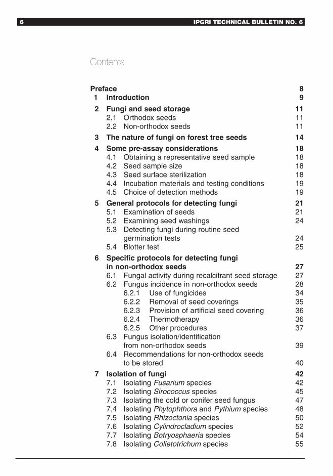
Contents
Preface 81 Introduction 9
2 Fungi and seed storage 112.1 Orthodox seeds 112.2 Non-orthodox seeds 11
3 The nature of fungi on forest tree seeds 14
4 Some pre-assay considerations 184.1 Obtaining a representative seed sample 184.2 Seed sample size 184.3 Seed surface sterilization 184.4 Incubation materials and testing conditions 194.5 Choice of detection methods 19
5 General protocols for detecting fungi 215.1 Examination of seeds 215.2 Examining seed washings 245.3 Detecting fungi during routine seed
germination tests 245.4 Blotter test 25
6 Specific protocols for detecting fungiin non-orthodox seeds 276.1 Fungal activity during recalcitrant seed storage 276.2 Fungus incidence in non-orthodox seeds 28
6.2.1 Use of fungicides 346.2.2 Removal of seed coverings 356.2.3 Provision of artificial seed covering 366.2.4 Thermotherapy 366.2.5 Other procedures 37
6.3 Fungus isolation/identification from non-orthodox seeds 39
6.4 Recommendations for non-orthodox seeds to be stored 40
7 Isolation of fungi 427.1 Isolating Fusarium species 427.2 Isolating Sirococcus species 457.3 Isolating the cold or conifer seed fungus 477.4 Isolating Phytophthora and Pythium species 487.5 Isolating Rhizoctonia species 507.6 Isolating Cylindrocladium species 527.7 Isolating Botryosphaeria species 547.8 Isolating Colletotrichum species 55

Forest tree seed health 7
7.9 Isolating Trichoderma species 567.10 Isolating Botrytis species 587.11 Isolating saprophytic or weakly pathogenic fungi 59
8 Molecular approaches to fungus detection 618.1 Detection of pathogens by PCR 62
9 The nature of viruses on forest tree seeds 6410 Protocols for detecting viruses 66
10.1 Double antibody sandwich, enzyme-linked immunosorbent assay (DAS-ELISA) 6610.1.1 Antibody preparation 6610.1.2 Coating to adsorb antibodies to the plate 6610.1.3 Adding the virus 6610.1.4 Blocking to improve the results (optional) 6610.1.5 Adding enzyme-labelled virus antibody
(conjugation) 6610.1.6 Adding the substrate 67
10.2 Immunocapture-reverse transcriptase-polymerasechain reaction (IC-RT-PCR) 6710.2.1 Sample preparation 6710.2.2 Coating and reverse transcription 6710.2.3 Polymerase chain reaction 6810.2.4 Gel electrophoresis of the amplification
products 6810.3 Dot blot hybridization 68
10.3.1 Extracting RNA from plant tissue 6810.3.2 Sample preparation 6910.3.3 Blotting procedure 6910.3.4 Hybridization 6910.3.5 Detection 69
11 Growing on tests 71Glossary 72References 74

8 IPGRI TECHNICAL BULLETIN NO. 6
Preface
The conservation and use of forest genetic resources worldwideposes several challenges to scientists, policy-makers and, inparticular, to local stakeholders interested in long-term strategiesto manage these biological resources in a sustainable manner. Thevast diversity of tree species, many of which are still unknown, thehigh level of threats and the increased demand for forest productsrequire prioritization of actions, clear indications for research anddevelopment, and strategies to mitigate the current trends in thedepletion of forest resources.
The strategy of conservation ‘through-use’ of forest geneticresources is a very important alternative to an in situ approachand, as such, is to be promoted and developed. However, basicknowledge and understanding of species’ reproductive biology,seed production, seed quality and health aspects, limit the use ofa larger number of species in important activities such as restoration,rehabilitation, agroforestry and on-farm conservation practices.Increasingly, the use of forest genetic diversity in research andbreeding requires a greater movement of germplasm.
This Technical Bulletin, prepared by Drs J. R. Sutherland, M.Diekmann and P. Berjak, all well-known scientists in their respectiveareas of specialization, aims to breach some of the knowledge gapsin forest seed biology and technology and, more importantly, tocontribute to future research on priority forest seed health aspects.This is an area of extreme importance for an effective and safeuse of existing diversity of tree species in either agroforestryprojects or in conservation of genetic resources in managedlandscapes programmes, and also to widen the scope of thisdiversity in the activities listed above. Protocols for fungi and virusdetection using different techniques are also presented and discussedextensively.
In addition, this Technical Bulletin aims to increase awarenessamongst technical staff involved in conservation and use activities. Tothis end it presents state-of-the-art tools for the identification of themost important tree seed pathogens and provides clear and ready-to-use molecular-based tools for the screening of fungi and virus in seeds.
We hope that with this publication some of the existing gaps inthe knowledge of forest seed health management are addressed insuch a way that seed scientists, pathologists and foresters mayfully benefit from existing forest biodiversity, and contribute totheir effective conservation and efficient and safe use.
Weber A. N. Amaral, PhDSenior Scientist
Forest Genetic Resources Coordinator

Forest tree seed health 9
1 Introduction
Everyone who works with orthodox and/or recalcitrant tree seedsshould be concerned with seed health issues. Seed health analystswould like to know if germination failure is the result of seed-bornepathogens, whereas foresters and seed dealers often discuss theimportance of moulds on the quality of the tree seeds they collector sell. Reduced germination is of particular importance forgenebanks storing forest seeds. Forest nursery managers areinterested in seed-borne pathogens affecting seed germination orcausing disease in their crop. In tree improvement programmes,pathogens may be transported long distances along with the plantgermplasm used. Plant quarantine officials should know if seedsbeing moved domestically or internationally harbour pathogens ofimportance to local forests.
A number of publications list micro-organisms of tree seeds(e.g. Ivory and Tompsett 1994; Mittal et al. 1990; Mohanan andSharma 1991; Prochazkova and Jancarek 1991; Sutherland et al.1987), but there is not a publication dealing solely with forest treeseed health testing. Thus, the purpose of this publication is toassemble several protocols for detecting pathogens of conifer andhardwood seeds.
Most of the reports on seed-borne pathogens of forest trees, dealwith fungi. However, data on seed-borne viruses of trees areaccumulating and, consequently, we have included a section onviruses of hardwood seeds. Although numerous species of bacteriamay be detected on tree seeds (Mittal et al. 1990), fruits and cones,little is known about their role in seed health. Some, like thebacterial wilt pathogen, Ralstonia (Burkholderia/ Pseudomonas)solanacearum, of Eucalyptus spp. (Ciesla et al. 1996), may eventuallyprove to be seed-transmitted, whereas other seed-borne bacteriamight be beneficial, i.e. in stimulating seedling growth, as occurswith many soil-borne bacteria (Chanway 1997).
Considering the limited knowledge of the role of bacteria ontree seeds, the general protocols described here, complementthose procedures recommended for individual pathogens onspecific seed species, published by the International SeedTesting Association (ISTA). Also included in this technicalbulletin, are assays for several pathogens and species of treeseeds, which have not been previously covered. We have alsoincluded a section on non-orthodox or recalcitrant tree seeds,contributed by an expert in the field, Dr Patricia Berjak,University of Natal, South Africa, to cover this importantgroup of forest tree species. She discusses different disease

10 IPGRI TECHNICAL BULLETIN NO. 6
detection methods and other procedures for pathogen detectionand control.
We expect that this information will be useful to seed analysts andgeneral diagnosticians working in genebanks, plant quarantine, andother research facilities, especially in countries where pertinentreferences may not be available. No attempt has been made to list allof the pathogens found on tree seeds. Refer to Mittal et al. (1990) andRichardson (1990) for such information.
Whenever possible, taxonomic references are given foridentifying fungi. We also recommend consulting ‘A LiteratureGuide for the Identification of Plant Pathogenic Fungi’ (Rossmanet al. 1987) which gives taxonomic references for identifying plantpathogenic fungi. Many seed-borne fungi produce only ananamorph (asexual) stage. Barnett and Hunter (1998) give anexcellent taxonomic key to the genera of these fungi. Generallaboratory procedures or recipes for commonly used culturemedia such as PDA are not covered in this publication. Detailscan be found for example in Johnston and Booth (1983) orHawksworth et al. (1995). Although we describe severaltechniques for isolating specific fungi or groups of fungi,particularly the use of selective media, many seed-borne fungiare easily isolated from surface-sterilized seeds plated ontowater agar, potato–dextrose agar (PDA), malt agar (Uniyal andUniyal 1996), or other standard culture media (Diekmann andSutherland 1998). Try these less-complicated procedures beforemoving on to more specific protocols.

Forest tree seed health 11
2 Fungi and seed storage
2.1 Orthodox seedsMost temperate tree species produce seeds with an orthodox seedstorage behaviour. Orthodox seed storage behaviour is defined byHong et al. (1996) as “mature whole seeds (which) not only surviveconsiderable desiccation (to at least 5% moisture content) but theirlongevity in air-dry storage increases in a predictable way byreduction in seed storage moisture content and temperature”.Recalcitrant seeds in contrast, are “unable to tolerate more than alimited amount of desiccation, for example to moisture contents inequilibrium at 20°C, with about 96–98% relative humidity”. Inbetween these two categories, are some species with an intermediatestorage behaviour, which “are able to tolerate desiccation to seedmoisture contents in equilibrium at 20°C with about 40–50% relativehumidity but where further desiccation often reduces viability andalways results in more rapid deterioration in subsequent hermeticstorage the more the seeds are dried below this value”. Examples fortrees with orthodox storage behaviour are Fagus, Fraxinus, Pinus andPrunus. Recalcitrant species are cacao (Theobroma cacao), rubber(Hevea brasiliensis), durian (Durio zibethinus) and jackfruit (Artocarpusheterophyllus); temperate recalcitrant species are oak (Quercus robur),maple (Acer saccharinum) and horsechestnut (Aesculus hippocastanum).Coffee (Coffea spp.), African oil palm (Elaeis guineensis), papaya(Carica papaya) and several Citrus species belong to the intermediategroup.
2.2 Non-orthodox seedsSeeds that may be collectively categorized as non-orthodox,especially those commonly described as being recalcitrant,differ from orthodox types in terms of the final stages of pre-shedding maturation and, notably, in their post-harvestresponses. Most orthodox seeds undergo maturation drying asthe final phase of their pre-shedding development and willcome to water content equilibrium with the relative humidity(RH) of the atmosphere. Even where this is not the case, afterharvest orthodox seeds will tolerate a substantial degree offurther dehydration (to ca. 5% moisture content [dry massbasis]), and are storable for predictable periods under definedconditions of RH and temperature (Ellis and Roberts 1980).Seeds of any species that do not behave in this way areconsidered to be non-orthodox (Berjak et al. 1989). While mostof the species considered elsewhere in this publication arestrictly orthodox, this section deals with those that are not.

12 IPGRI TECHNICAL BULLETIN NO. 6
The original, formal definition of seeds as being orthodox orrecalcitrant and, indeed, the introduction of these terms was basedon their storage behaviour (Roberts 1973), which is a manifestationof post-harvest seed physiology. When they are shed, not onlyare recalcitrant seeds characterized by relatively to very high watercontents, but they are also actively metabolic (Berjak et al. 1989). Inthis condition, such seeds will withstand only very restricteddehydration before severely damaging or lethal effects occur, andconsequently are described as being desiccation-sensitive (e.g. Chinand Roberts 1980). This is, in fact, the major criterion by whichseeds of particular species are categorized as being recalcitrant.Although there is an enormous gap in dehydration responsebetween recalcitrant and orthodox seeds, thus far only one furthercategory has been formally defined––that comprised by seedswhich will withstand a substantial degree of dehydration, but notto as low water contents as will orthodox types: these have beendescribed as showing intermediate storage behaviour (e.g. Ellis etal. 1980), and those of tropical origin may, especially in thedehydrated condition, be adversely affected by chilling (Hong andEllis 1996).
Despite the non-orthodox behaviour of recalcitrant andintermediate seeds–– and seeds of the many species that may fallsomewhere between these two categories (Berjak and Pammenter1994)–– it is essential that storage strategies be developed, albeitthat only short-term conservation for planting programmes andgermplasm exchange be facilitated. The immediate problem is thatrecalcitrant seeds cannot be dehydrated to any water contentwhich would allow low temperature–low RH storage, and it islikely that intermediate seeds of many species too could notwithstand the conditions developed to optimize storage longevityfor orthodox types.
In fact, overcoming the problems associated with conservationof non-orthodox seeds has been a much-debated topic for at leasttwo decades (e.g. Chin and Roberts 1980; Ouédraogo et al. 1996),but very little progress has been made in terms of improved short-term storage of the intact propagules.
As regards recalcitrant seeds, the only conservation strategy forthe intact propagules involves their storage at high water contents(essentially undiminished from those characteristic of the newlyshed/harvested condition), at the minimum temperature tolerated.In the case of many tropical species, however, these minima must berelatively high, as the seeds are chilling-sensitive. Even for seeds oftemperate recalcitrant species, as the tissues are highlyhydrated–– and continue to be metabolic–– storage at sub-zero

Forest tree seed health 13
temperatures is precluded. Low-temperature storage is also not anoption for chilling-sensitive seeds showing intermediate post-harvestbehaviour.
The very conditions necessary for viability retention ofrecalcitrant, and probably most other non-orthodox seeds, arealso those that facilitate fungal proliferation (Berjak 1996).Furthermore, any manipulations of such seeds, for example,attempting to lower the water content of recalcitrant types toprevent ongoing events of germinative metabolism, may wellprove sufficiently stressful (Drew et al. 2000) to exacerbate thedeleterious effects of seed-associated micro-organisms,particularly fungi. The same is likely to be the case fordehydrated intermediate seeds (Berjak 1996).
During short-term storage of whole non-orthodox seeds, theproliferation of micro-organisms, particularly of fungi, must becurtailed. It is, of course, desirable that microbial propagules beeliminated completely from the seeds prior to storage, but this isextremely difficult to achieve. Long-term conservation of thegenetic resources of species producing non-orthodox seeds islikely to be achieved only by cryostorage of suitably smallexplants, such as excised zygotic axes (Engelmann 1999). Thesemust harbour no microbial propagules ––particularly as allrequire a period in vitro following retrieval from cryostorage,during which any associated fungal or bacterial propagules willflourish. However, the advantage when working with isolatedaxes, is that these structures which are usually tightly enclosedby the surrounding tissues in the intact seed, are able to besurface-sterilized after excision.

14 IPGRI TECHNICAL BULLETIN NO. 6
3 The nature of fungi on forest tree seeds
One of the main features of forest tree seeds is their greatdiversity in size, shape and texture. The size and texture of treeseeds range from small and hard, as the seeds of Eucalyptus spp.,to the relatively large and fleshy acorns of some Quercus spp. orhard walnuts of some Juglans spp. The longevity of tree seedsvaries from a few days to many years.
The main effects of seed-transmitted fungi are the diseasesthey cause, and to some extent also the reduced seed viability.However, they rarely destroy the seeds completely. Examplesare Sphaeropsis sapinea, causing Diplodia shoot blight of pines andother conifers, Sirococcus conigenus, causing Sirococcus blight ofpines and other conifers, Botryodiplodia theobromae, causing rotsin a wide host range and many Fusarium spp. causing damping-off of seedlings.
Seed-borne micro-organisms may reduce germination andseed longevity in storage of all types of seeds. When seeds aremoved internationally, pathogens may become a quarantineconcern and jeopardize seed trade and germplasm exchange.The micro-organisms that are mostly associated with tree seedsare fungi, bacteria and, to a lesser extent, viruses. Acomprehensive list was published by Mittal et al. (1990).Phytoplasmas, which are known to cause a number of so-calledlittle leaf or witches’ broom diseases, are not seed-borne due tothe nature of their transmission (phloem-limited, and noconnection from phloem to seeds). It is important to distinguishbetween seed-borne micro-organisms and seed-transmittedmicro-organisms. The term seed-borne describes the state ofany micro-organism being carried with, on or in the seed. Theterm seed-transmitted includes the act of infection of theseedlings from seed-borne inoculum. Thus seed-borne micro-organisms include the pathogens causing plant diseases, the so-called ‘field fungi’ as well as the so-called ‘storage fungi’.
Examples for typical ‘storage fungi’ include Penicillium spp.,Aspergillus spp. and Caloscypha fulgens, the seed or cold fungus thatkills pine seeds under cool conditions. Their main effect is toreduce seed viability, under certain conditions to even kill theseeds. Poor germination of seedlots can often be attributed tocontamination with micro-organisms (e.g. Mwanza and Kellas1987; Sutherland et al. 1987; Huang and Kuhlman 1990). Micro-organisms in general thrive under conditions of high moisture anda temperature range between 20 and 25°C. These are also theconditions that ensure survival of recalcitrant seeds. Although few
Forest tree seed health 15
systematic surveys of the contamination of recalcitrant seeds withmicro-organisms have been conducted (e.g. Mittal and Sharma1983; Mycock and Berjak 1990; Pongpanich 1990), it appears thatthey suffer more from effects of micro-organisms on seed qualitythan orthodox seeds (Berjak 1996). In addition, recalcitrant seedsare more sensitive to common seed treatment with heat orfungicides. Recalcitrant seeds are often conserved in vitro, whereparticularly the storage fungi may cause problems bycontaminating the media.
There is a need to conduct more survey work on pathogens ofrecalcitrant tree seeds, both with regard to quality aspects and withregard to phytosanitary issues. It is very important to keep thesetwo aspects separate in the evaluation of research results. Pest RiskAnalysis (FAO 1996) can help in identifying pests of quarantineconcern and in suggesting management options. Treatmenttechniques (chemical, biological or physical) that do not affectseed viability need to be identified.
The majority of pathogens associated with forest tree seeds arefungi, producing only or predominantly the asexual (anamorph)stage (Deuteromycetes). There are exceptions though, as theoomycetes (Oomycota) or water moulds, which can be seed-borne on hardwood seeds, e.g. Phytophthora cactorum onbeechnuts (Prochazkova and Jancarek 1991). Except for Rhizoctoniaspp. that are sometimes seed-borne and may have a basidium-producing (Basidiomycotina) sexual stage, the reproductive sporesor structures of other Basidiomycotina, are rarely found on treeseeds, and even when present, they are of no consequence. Forexample, Heterobasidion annosum, which causes root rots of foresttrees, has been isolated from Abies sp. seeds (Mittal et al. 1990).However, there is no evidence of this or similar fungi affectingseeds or serving as inoculum to cause root rots. Although rustfungi, which are also Basidiomycotina, frequently attack conifercones, seeds from diseased cones do not carry the pathogen.
There are many mechanisms by which tree seeds acquirepathogens. Indeed, both the occurrence and severity of manyseed-borne fungi, is often traced to the mishandling of fruits,cones or seeds. A major factor in the acquisition of seed-bornefungi is the contamination of fruits or cones with soil, as oftenoccurs when they are collected from the forest floor, the groundbeneath seed orchard trees, or when forest trees are felled tofacilitate fruit or cone picking. Beechnuts (Fagus spp.), forexample, collected from the forest floor may be infested withPhytophthora cactorum (Prochazkova and Jancarik 1991). Plasticnetting can help reduce infection (Fig. 1). Improper handling of

16 IPGRI TECHNICAL BULLETIN NO. 6
cones or fruits may also contribute to the occurrence of seed-bornefungi, when moulds build up on wet conifer cones stored forprolonged periods following collection. Storing cones wet can alsoincrease temperature within the collection bags, further enhancingmoulding. The inclusion of old, infested cones or fruits in currentyear’s collections, can be a source of seed-borne pathogens.Sirococcus blight of conifers, caused by S. conigenus, is a classicexample. The pathogen occurs on old, pathogen-infested conifercones, especially of spruces, Picea spp. Seedlots become affected whensuch cones, containing infected seeds, are included in collections ofcurrent year, disease-free cones. Another source of contaminationresults from improperly cleaned seedlots containing bits ofpathogen-infested needles, leaves, cones or other debris andinfected seeds.
Seeds often carry fungi that are considered as saprophytes orweak pathogens, e.g. species of Penicillium or Aspergillus. Such socalled ‘storage fungi’ are common on stored, but not fresh, seeds(Prochazkova, pers. comm.). This observation invariably leads tothe question of what role these fungi play. Are they harmful or dothey simply colonize weakened seeds? For example, seeds may beweakened by numerous factors, including long-term or improperstorage, or high processing temperatures that are sometimesrequired for drying or opening cones. Seeds harbouring such fungiinvariably germinate poorly or slowly, but it is hard to define the
Fig. 1. Plastic netting suspended above the floor in a beech forest to preventinfection of beechnuts by duff-inhabiting, pathogenic fungi. (Dr Eva Palatova,Mendel University of Agriculture and Forestry, Brno, Czech Republic)

Forest tree seed health 17
cause and effect. Not all seed-borne fungi are detrimental andsometimes they may be beneficial. For instance, Trichodermaharzianum is sold commercially as seed dressing to protect seedsand seedlings from damping-off and root rots. However, underoptimal conditions even Trichoderma species may be pathogenic(Vaartaja 1957).
Seed analysts are interested in detecting and defining the role ofseed-borne pathogens but, except for seeds destined for export orimport, it may be impractical to assay all seed lots. The followingproblems should be avoided when seeds are selected for storage:• Moulds on the fruits or cones from which the seeds originated.• Moulds on the seeds during germination tests or following
moist treatments such as stratification.• Presence of pathogenic fungus structures such as sclerotia on or
within seeds.• A high percentage of the seeds fail to germinate and often their
contents are rotted.• Fruits or cones originate from forest stands, seed orchards or
mother trees with potential seed-borne diseases, such asSirococcus blight on pines and spruces or pitch canker, causedby Fusarium subglutinans f. sp. pini on pines.
• Low percentage seed germination, slow germination or bothfollowing stratification or other treatments that stress theseeds. Slow germination is an excellent indicator of poorquality seeds.
• Radiographs reveal many seeds that are empty, with crackedseedcoats, abnormal contents or insect damage.
• Fruits or cones harshly treated before or during processing,usually after being subjected to higher that normaltemperatures to dry or open them.
• Seeds originated from fruits or cones that had contacted seedorchard soil or the forest floor, e.g. cones collected from squirrelcaches.
• Seeds originated from collections containing old or insect-damaged fruits or cones.
• Seed lots contain many immature seeds and much debris.• Seed lots in which seed viability in long-term storage declines
faster than expected.• Specific fungi that regularly appear on seeds during routine
germination tests.• Disease that consistently appears on specific seedlots of young
nursery seedlings.Specific recommendations for storage of non-orthodox seeds aregiven in Section 6.4.

18 IPGRI TECHNICAL BULLETIN NO. 6
4 Some general pre-assay considerations
4.1 Obtaining a representative seed sampleBefore assaying for seed-borne pathogens, it is first necessary toobtain a representative seed sample. The most recent edition of theISTA International Rules for Seed Testing (1999) and Edwards andWang (1995) cite methods and equipment needed for seedsampling. We recommend that these procedures andrecommendations for selection of reliable equipment be followedto guarantee the accuracy of the assays.
4.2 Seed sample sizeThe number of seeds required for testing is determined accordingto the expected incidence of the test pathogen in specific seed lots.In general, sample size depends upon the sensitivity required (themore seeds tested the greater the accuracy) and the availability ofseed and resources available to process the samples. Determiningproper sample size requires that preliminary assays be done toobtain data about the incidence of seed-borne pathogens presentin seed lots. Once this information is obtained, sample size canbe determined using a binomial distribution (Zar 1984). Whenusing such procedures it is common for sample size to vary bypathogen, tree seed species and accuracy level desired. See Zar(1984) or other statistical texts for help in this area or consult abiometrician whenever there is doubt about the number of seedsneeded for an assay. A rule of thumb: if 300 seeds are tested with areliable method and found healthy, one can be 95% sure that theinfection percentage is less than 1%, or: to be 95% sure that theinfection in a seed lot is below 0.3%, one has to test 1000 seeds(Diekmann 1993).
4.3 Seed surface sterilizationAnother consideration is how to prepare the seeds for assay.Before testing forest seed it can be washed with water ordisinfesting chemicals. Exposure to physical agents, such asheat, cold, scarification, or removal of the seed coat, may alsobe considered as pre-treatments of forest seed. Removing theseedcoat from small seeds is too tedious and time consuming tobe justified. However, it may be worthwhile for large seeds suchas acorns, particularly if the suspected pathogen is within seedsthat have many surface fungi and bacteria. Seedcoats ofhardwood seeds, or seedcoats adhering to hardwood germinants,must be removed before assaying for viruses. If the isolationmedium is selective for a particular fungus or group of fungi it is

Forest tree seed health 19
often not necessary to surface-sterilize the seeds as the mediuminhibits growth of most contaminants or stimulates the growth of thedesired fungus. The most common surface disinfectant isordinary bleach (NaOCl). While effective, bleach is also residualand so the treated materials must be washed two or three timeswith sterile water to remove the bleach, which would otherwiseinhibit growth of the fungus. We have used 3–30% hydrogenperoxide for surface-sterilizing tree seeds (Sutherland et al. 1987).It works well and does not have to be washed off the seeds, butit is very corrosive and must be handled with extreme caution.Handling samples of small seeds is facilitated by using small,cylindrical-shaped (e.g. 5 cm long × 2 cm diameter) ‘baskets’made from plastic window screen. The open end of the basket isplugged with a rubber stopper to keep the seeds in the basketthat is moved in and out of surface disinfectants or washingwater with laboratory tweezers.
4.4 Incubation materials and testing conditionsThe protocol dictates the type of apparatus to be used for incubatingseeds or seedlings. Most assays in which small to medium-sizedseeds are incubated on blotter paper or agar media are done in90–100 mm diameter Petri plates. Large seeds require biggercontainers. Ideally the incubation temperature should be optimumfor growth of the target pathogen, but not for other fungi andbacteria. As fruiting bodies and spores are a prerequisite for fungusidentification, sometimes methods to promote fungus sporulationmay be required. These could be incubation of cultures in ambientdaylight or under near ultraviolet (NUV) light (‘black light’).Details are given for the respective species.
4.5 Choice of detection methodsThe method to be used depends on the target pathogen(s), thepurpose of testing, the accuracy level desired and cost. For thedetection of ilarviruses and cucumoviruses, the immuno-enzymaticELISA test is recommended, whereas the immuno-capture-reversetranscriptase polymerase chain reaction (IC-RT-PCR) is used fordetecting a nepovirus. The dot blot nucleic acid hybridizationtechnique can be used for detecting tobamoviruses in oaks andmaples.
Reeves (1995) reviewed various immunological and nucleic acidmethods for detecting seed-borne fungi, bacteria and viruses. AsMaude (1996) points out, these techniques are used for detectingfungi in soil and diseased plants, but seldom used in assays forseed-borne fungi. This is mainly because existing tests for detecting

20 IPGRI TECHNICAL BULLETIN NO. 6
fungi on seeds, including blotter tests and use of selective media,are fairly effective and cheaper to develop and use. However, thisdoes not preclude the need for developing molecular assays forfungi and other pathogens on tree seeds. Simple and reliableprocedures are needed, especially for testing seed health of treespecies which are widely used in large scale re- and afforestationprogrammes (Mohanan and Sharma 1991). Already someprogress has been made in this direction with the developmentof monoclonal antibody protocols for detecting Sirococcusconigenus on spruce seeds (Mitchell and Sutherland 1986;Mitchell 1988). One of the main advantages of moleculartechniques is that they are much more sensitive than existingprocedures. Consequently, extremely low levels of seed-bornepathogens can be detected with these advanced techniques,which is particularly important to certify seed for export, or evenfor long-distance movement within countries, to minimize the riskof disseminating seed-borne pathogens.

Forest tree seed health 21
5 General protocols for detecting fungi
5.1 Examination of seedsBefore proceeding to more elaborate procedures, first examinedry seeds using a magnifying lens or stereomicroscope. Seedsthat are mouldy (Fig. 2), cracked or broken (Fig. 3), or which
Fig. 2. Mouldy seeds of Douglas-fir. (Dr J. Sutherland, Victoria, BC,Canada)
Fig. 3. Acorns with cracked seedcoats, showing black mycelium ofCiboria batachiana infection. (Dr Zdenka Prochazkova, Forestry andGame Management Institute, Research Station Uherske Hradiste, Czech Republic)

22 IPGRI TECHNICAL BULLETIN NO. 6
have insect damage (Fig. 4) are easily detected. Poor quality seedlots may contain small or abnormal seed. Resin drops (Fig. 5) onconifer seeds indicate the cones or seeds were subjected toexcessively high temperatures during processing. If so, the seedsmay be weakened and thus susceptible to saprophytes andopportunistic pathogens and not suitable for long-term storagein genebanks. Bits of pathogen-infested needles and leaves andother debris (Figs 6 and 7) can also be seen, as can sclerotia(Fig. 8) and other fungal structures. Poor quality seedlots maycontain many small or abnormally shaped seeds. Radiographsreveal many of the same problems and, in addition, help todetect minute cracks in seedcoats, internal insects or abnormalcontents of pathogen-infected seeds which would otherwise goundetected. Poor quality seed should be discarded.
Fig. 5. Resin on Douglas-fir seeds indicates high temperatures were usedduring cone or seed processing. (Dr J. Sutherland, Victoria, BC, Canada)
Fig. 4. Insect-damaged (hole) Douglas-fir seeds. (Dr J. Sutherland,Victoria, BC, Canada)

Forest tree seed health 23
Fig. 7. Western redcedar, Thuja plicata, seeds with needle debris (green), apossible source of pathogenic fungi. (Dr J. Sutherland, Victoria, BC, Canada)
Fig. 8. Round, black sclerotia of the fungus Botrytis cinerea.(Dr J. Sutherland, Victoria, BC, Canada)
Fig. 6. Douglas-fir seeds with debris, a possible source of pathogenicfungi. (Dr J. Sutherland, Victoria, BC, Canada)

24 IPGRI TECHNICAL BULLETIN NO. 6
5.2 Examining seed washingsRecommended for detecting surface-contaminating fungus spores onseedsThe seeds are placed in a flask or other container with waterand shaken. For a quantitative test, seed weight and watervolume should be known, e.g. 50 g seeds and 50 ml water.Spore counts with the help of a haemocytometer can becalculated as number of spores per millilitre of water, whichthen equals the number of spores per gram of seed. Adding adrop or two of detergent helps dislodge spores from seeds, butit may create foaming problems. If a low spore load is expected,the seed washings should be centrifuged and the sedimentexamined by compound microscope for spores.
Yuan et al. (1990) used 50 seeds of Acacia spp., 200 mg ofCasuarina spp. and 100 mg of Eucalyptus spp. seeds each in 10 mlof water shaken on a ‘wrist action shaker’ for 10 min. Suspensionsof particulate matter were decanted from the seed washings andcentrifuged 5 min at 5000 rpm. A compound microscope andhaemocytometer were used to examine the sediment in the tubesfor spores. Using this procedure they detected Pestalotiopsis sp. andUlocladium sp. in seed washings of all three tree genera and Phomasp. was found in washings from Casuarina cunninghamiana andseveral Eucalyptus species. Other fungi detected in washings (treeseed species not given) were Curvularia lunata, Drechslera spiciferaand Penicillium sp.
5.3 Detecting fungi during routine seed germinationtestsWith valuable germplasm, this method helps reduce the numberof test seeds. Germinate the seeds according to recognizedprotocols, usually the ISTA rules (1999). During or at the end ofthe test, remove and identify the fungi that grow on the seedsor germinants. Transfer those that do not sporulate to PDA, maltagar or other culture media to induce sporulation. However, theserecommendations vary according to the test species. For instance,seed may be germinated on top of, or between, paper, in sand, orseed may be soaked in concentrated phosphoric acid or water.Some tree seed requires 14–70 days of incubation before germinationcan be evaluated.
A major advantage of this procedure is that it is carried outconcurrently with routine germination tests. Thus, except formicroscopes and references needed to identify the fungi, otherequipment and resource requirements are minimal. Otheradvantages are that many of the fungi sporulate on the seeds or

Forest tree seed health 25
germinants, and so it is usually not necessary to subculture them onagar or other culture media to induce sporulation. It is also easy torelate and quantify fungus occurrence and abundance to diseasessuch as seed decay and radicle rot. Some disadvantages are that fast-growing, saprophytic fungi may obscure pathogen growth and thetest is not specific for detecting one or a group of pathogens.
Prochazkova and Jancarik (1991) used this technique to identify141 fungi in almost 6000 conifer seedlots and 170 fungi in over2200 broadleaf seedlots. The conifer seeds were Abies alba, A.concolor and A. grandis, Larix decidua, Pseudotsuga menziesii, Piceaabies, P. glauca, P. omorika, P. pungens and P. sitchensis and Pinussylvestris, P. nigra, P. strobus, P. mugo var. mughus, P. mugo var.uncinata, P. cembra, and P. contorta. Some of the pathogenic fungiisolated were Botrytis species from species of Picea, Pinus and Larix,Fusarium spp. from species of Pseudotsuga, Pinus and Larix andVerticillium spp. from Pinus and Larix spp. The hardwood seedsassayed were Alnus glutinosa and A. viridis, Betula verrucosa,Carpinus betulus, Fagus sylvatica, Fraxinus excelsior, F. americana andF. angustifolia, Sorbus aucuparia, Tilia cordata and T. platyphyllos andUlmus glabra. Among the pathogenic fungi obtained were Ciboriaalni, C. batschiana and C. betulae from the seeds of species of Alnusand Betula (<1% of the seedlots tested), respectively. Fusariumspp. were detected in all the hardwood seedlots, especiallyFraxinus sp. seeds where >50% of the lots were infested.Rhizoctonia solani was obtained from <1% of the beechnutseedlots. The grey mould pathogen, Botrytis cinerea, was found inover two-thirds of the Carpinus betulis seedlots. Trichoderma viride,which is often associated with poor quality seeds, occurred on over40% of the Pseudotsuga, Larix and Carpinus seedlots, 30% of the Tilialots, and 15–30% of the lots of Acer, Quercus and Sorbus.
Sometimes pathogens may be detected in tests that are madein conjunction with routine germination tests. For example,cross or longitudinal sections of seeds that germinate poorly areoften cut with a razor or scalpel blade to determine embryodevelopment and size. Internal seed decay and presence ofmoulds can often be seen in such sections
5.4 Blotter testRecommended for detecting a wide variety of fungi in or on hardwoodand conifer seeds.Place seeds on water-soaked blotter paper in sterilized Petri dishesor other such containers. Tap water can be used as long as it is boiledfor 10 min. Sterile water safeguards against contamination by water-borne fungi. Incubate the seeds at 20–25°C, or temperatures favouring

26 IPGRI TECHNICAL BULLETIN NO. 6
the suspected pathogen, for one to two weeks, or until fungi develop.They can then be identified using the fruiting bodies or spores on thepaper, seeds or germinants (Fig. 9), or removed and plated onto aculture medium to induce sporulation. The principle here is thatseeds are kept in a humid environment favouring fungusdevelopment. The technique is thus similar to that of detectingpathogens during routine seed germination tests and so it is notsurprising that the two assays detect many of the same fungi. Themajor advantage of the blotter test is that it more flexible, e.g. it canbe used to assay one to several seedlots using incubationtemperatures and lighting regimes that favour fungus detection overseed germination. Another advantage is that it is possible to relatepathogen occurrence and abundance to seed decay and otherdamage. Sometimes agricultural seeds are killed or weakened byfreezing or herbicides, to enhance pathogen development, beforebeing placed on the blotter papers (Maude 1996). Working with bothnon-and surface-sterilized seeds Sharma and Mohamed Ali (1997)used the blotter technique to isolate a variety of non-pathogenic,potentially pathogenic and pathogenic fungi, e.g. Fusarium solani, F.moniliforme and Botryodiplodia theobromae, from seeds of the tropicalhardwoods Lagerstroemia microcarpa and Pterocarpus marsupium. As inthis case seeds may be surface-sterilized before being assayed or theseedcoat can be removed from large seeds with profuse surfacecontamination. This technique is easy to use and requires minimalequipment; however, rapidly growing saprophytes or weakpathogens such as species of Penicillium, Trichoderma or Aspergillusmay overgrow certain pathogens. Also, pathogens such asPhytophthora that may lack conspicuous spores and vegetativegrowth can go undetected.
Fig. 9. Seed-borne Sirococcus conigenus sporulating on a killed,spruce germinant. (Dr J. Sutherland, Victoria, BC, Canada)
Forest tree seed health 27
6 Specific protocols for detecting fungi in non-orthodox seeds
6.1 Fungal activity during recalcitrant seed storageAn in-depth study on the effects of fungi during hydrated storageof Avicennia marina has proved very revealing of the significantrole of mycoflora in promoting deterioration of recalcitrant seeds(Calistru et al. 2000). In that study, after pericarp removal, hand-harvested seeds were treated initially and periodically duringhydrated storage by aerosol spraying with a relatively effectivefungicide, Previcur N (see below for details). The peculiarmorphology of A. marina seeds (see Farrant et al. 1993 fordiagrammatic details) allows fungicide, applied as a fine spray,access to the inner cotyledonary surfaces as well as to the exteriorof much of the embryonic axis. The results showed unequivocallythat this treatment, which continued to curtail fungal activity,extended the storage lifespan of these ‘clean’ seeds by almost 50%compared with previous studies in which no equivalent measureswere taken (e.g. Berjak et al. 1989). In the investigations of Calistruet al. (2000), seeds that had been initially treated with the fungicide,but then inoculated with Fusarium moniliforme, were all deadwithin a third of the time for which the ‘clean’ seeds remainedvigorous and showed high viability. In the experimentally infectedseeds, rapid fungal proliferation occurred, and was accompaniedby profound deterioration of both cotyledonary and embryonicaxis cells. What was a highly significant finding though, was thatif the seeds had been stored ‘clean’ for a few days prior to beingexperimentally infected with F. moniliforme, then they wereconsiderably more resilient to fungal depredation, arguing for thedevelopment of active defence mechanisms as germinativemetabolism progressed under hydrated conditions. This response,however, postponed rather than prevented the seeminglyinevitable fungal degradation and viability loss of the stored seeds.
The change in susceptibility of the seeds to fungal attackunderscores the importance of developmental stage in these activelymetabolic plant propagatory units. Even in the ‘clean’ seeds though,inherent infection by F. moniliforme was not completely eliminated,becoming apparent in localized association with only cotyledonarysurfaces, about half way through the experimental storage periodalthough there was no vigorous fungal proliferation. This was thesituation in 30% of the ‘clean’ seed population by the end of thestorage period when viability (perhaps, or perhaps not,coincidentally) was 70%. Electron microscopical studies showedthat cotyledonary cells contiguous with the fungal mycelium were

28 IPGRI TECHNICAL BULLETIN NO. 6
extensively degraded but, significantly, cells of the embryonic axiswith which no mycelium was associated also showed markeddeteriorative changes.
This study confirms the premise (Berjak 1996) that active fungalmetabolism during hydrated storage of recalcitrant seeds wouldimpose significant limitations––not only on the total (storage)lifespan––but also on seed vigour, and thus quality. The findingsunderscore the necessity of finding whatever solutions may bepossible, to curtail the incidence––or ideally, to eliminate––theassociated fungi from recalcitrant, and all other non-orthodoxseeds, prior to storage.
6.2 Fungus incidence in non-orthodox seedsFungal propagules may gain access to the seed tissues at any timefrom flowering to the post-shedding phase. Recalcitrant seeds maybe internally infected by fungi ab initio by systemic transmissionvia the parent plant, as has been shown for developing maizecaryopses, which are orthodox (Mycock and Berjak 1992; Kabeereet al. 1997), or through the stigma-style continuum duringflowering (Marsh and Payne 1984). The problem with infectionthat has originated in these ways during seed development, is thatthe mycelium has the opportunity to become established deepwithin the tissyitself––and is consequently very difficult (if notimpossible) to eradicate. As is the case for orthodox seeds, insectsmay both cause damage and act directly as vectors for fungalpropagules during seed development. Recalcitrant (and probablyall non-orthodox) seeds offer a further advantage to opportunisticinvading fungi, in that being shed at high water content, they arevery prone to contamination once on the ground (see below) or instorage containers. If the seeds are collected soon after shedding,however, such fungal propagules may be only peripherally locatedand easy to eradicate if prompt action is taken. Additionally, ofcourse, the storage containers and all devices used to elevate theseeds within them, must be rigorously sterilized.
In Durban, South Africa, scientists have worked with non-orthodox seeds of a wide variety of species and, although theproportion harbouring fungi and/or bacteria varied from onebatch to another, some contamination was invariable. In general,fungi were the major contaminants of recalcitrant seeds of tropical,sub-tropical and temperate southern African origin but frequentlybacteria occurred as co-contaminants (Mycock and Berjak 1990).These findings have been consistent for tropical and temperatespecies since then, and we have become particularly aware of theproblem since embarking on cryopreservation of embryonic axes

Forest tree seed health 29
excised from a variety of recalcitrant seed species (Berjak et al.1999a,b). Our experience with intermediate seeds has been limited,but in the case of extensive trials with coffee seeds where fungicould be controlled, bacteria posed an almost intractable problem(unpublished data). It should be noted though that seed-associatedmicro-organisms are not confined to recalcitrant and other non-orthodox types originating only in the tropical to warm temperatezones (see below). It is possible, however, that the spectrum of fungalspecies may differ with seed provenance where the geographicallocalities are widely separated (Table 1). As an example (althoughspecific relationships among seed and fungal species cannot be ruledout as the present state of knowledge is scanty), it seems thatrecalcitrant seeds harvested in 1990 in southern Africa harboured abasically common spectrum of fungi (Mycock and Berjak 1990),which was essentially different from that associated with seedscollected in the Asia-Pacific region (Pongpanich 1990). In a fewinstances, particular fungal genera have been isolated from seeds ofboth cool temperate regions and widely separated tropical/sub-tropical zones (Table 1). This is the case for Phomopsis spp., whichwere associated with a low percentage of acorns (Quercus spp., Kehrand Schroeder 1996); some species of the Dipterocarpaceae(Pongpanich 1990); Trichilia dregeana (Meliaceae, our unpublisheddata); Hevea brasiliensis (Singh and Singh 1990); as well as with neem(Azadirachta indica, Sateesh and Shankara Bhat 1999). However,although identifications to the fungus species level were notpresented for all the seeds from which Phomopsis was isolated, it islikely that these were different: for example, the species isolatedfrom seeds of commercial rubber and of neem, were P. heveae and P.azadirachtae, respectively. In contrast, while Phytophthora spp. havenot been generally recorded as being associated with recalcitrantseeds, this genus has been shown to be seed-transmitted in Ghana,where it poses a serious threat to Theobroma cacao causing blackpod, which is a widespread and destructive disease of cocoa(Kumi et al. 1996).
In the southern African context, Mycock and Berjak (1990)investigated the fungal status of newly harvested recalcitrant seedsof seven unrelated species, ranging in provenance from sub-tropicalsalt-water estuaries to warm-temperate montane areas. These were:Avicennia marina, Castanospermum australe, Litchi chinensis, Podocarpushenkelii (a gymnosperm), Landolpia kirkii, Scadoxus membranaceus andCamellia sinensis. The findings were that Fusarium spp. were presenton, or in, the tissues in all but one (L. kirkii, a latex-producing speciespossessed also of apocyanaceous alkaloids) and that species ofAlternaria, Cladosporium, Aspergillus and Penicillium with Fusarium

30 IPGRI TECHNICAL BULLETIN NO. 6
spp. dominated the spectrum of fungi associated with the freshseeds. Although, unfortunately, in that study fungi were generallynot identified to species level, it was unequivocally established thatnone was of the xerotolerant group collectively described as theseed storage fungi (e.g. Christensen and Kaufmann 1974). Referenceto Table 1 shows that the only storage fungi that have been recordedamong the many major isolates, are Aspergillus flavus group speciesin association with seeds of the Dipterocarpaceae (Mittal andSharma 1982; Pongpanich 1990) and those of Hevea brasiliensis(Singh and Singh 1990).
The relative absence of demonstrable storage fungi in associationwith recalcitrant seeds is not surprising. This ecological groupingcomprises xerotolerant species (mainly of Aspergillus andPenicillium) which generally become apparent on, and in, the tissuesof air-dry orthodox seeds in storage (Christensen and Kaufmann1974), when the competition imposed by the so-called field fungi iscurtailed by the low water activity and osmotic challenges of seedsstored at low RH. It is probable though that in intermediate seedsdehydrated to relatively low water contents xerotolerant storagefungal species may manifest themselves, provided that theirpropagules are present intra-seminally, or in the storage containers.The field fungi, in contrast, require relatively high seed-watercontents in order to proliferate–– which are precisely thosenecessary to maintain viability of metabolically active, recalcitrantseeds in storage (Berjak 1996). Whereas species of Fusarium,Cladosporium and Alternaria are considered to be major componentsof the field fungi on, and in, developing and newly harvestedorthodox seeds, this does not preclude others. The essentialdifferences between orthodox seeds–– which become increasinglyinert–– and recalcitrant types, which continue to be activelymetabolic, are basic to the spectrum of seed-associated fungi thatwill be manifested.
In fact, unless they are dormant, recalcitrant seeds might bedescribed as seeds in name only, and are actually far more likeseedlings once they have been shed. In many cases, the metabolismassociated with their development graduates without any obviousmarker event into that of germination (Berjak et al. 1989; Farrant et al.1989; Pammenter et al. 1994). As a consequence, it may be reasonableto suppose that these propagatory units would be open to invasionby a broad spectrum of fungal species, and not only those classicallyconsidered to be field fungi. This is borne out by a consideration ofthe range comprising the mycoflora found to be associated withsome of the few species of recalcitrant seeds that have beenexamined: the spectrum of fungi is, in fact, considerably moreextensive than those listed as major isolates in Table 1.

Forest tree seed health 31
A further complication is that whatever associated fungi areisolated at any one stage of recalcitrant seed development, thecomposition of the mycoflora is likely to be different at otherstages, both before and after the seeds are shed from the parentplant. This is well illustrated by the information from Kehr andShroeder (1996) that the most virulent of the primary pathogens,Ciboria batschiana, associated with seeds of temperate Quercus spp.,infects the acorns only after they have been shed onto the soil. Ourstudies on the stored seeds of four species of tropical or warmtemperate origin showed that whereas a mixed mycofloraoccurred when the seeds were newly shed, the spectrum narrowedto become dominated by Fusarium spp. during storage (Mycockand Berjak 1990). A recent, major survey on fungi associated withdeveloping and mature fruits and seeds and shed seeds of Trichiliadregeana has been very revealing of the changing nature of themycoflora (Table 1). Lack of overt disease symptoms is also noguarantee that the seeds are not infected: the embryos of themajority of seeds of Theobroma cacao––97%––were found to beinfected by Phytophthora, although taken from fruits which weresymptomless for black pod (Kumi et al. 1996).
In the case of Trichilia dregeana, many of the fungi associatedwith immature or mature fruits were not transmitted to the seeds,although in most cases these were isolated from the interior of thepericarp. These included Alternaria spp. and notably A. alternata,Colletotrichum gloeosporioides group species, Penicilliumaurantiogriseum, Pestalotiopsis maculans and Rhizopus nigricans.However, in other cases, fungal species isolated from fruit tissuesduring seed development, were isolated from the arils of the seedsafter they had been shed: the fungi concerned were aColletotrichum species, Fusarium semitectum, F. solani, F. subglutinansand two Penicillium species. Seeds of T. dregeana are enclosed by asubstantial waxy aril which, in the shortest-term, might prove tobe a barrier to the ingress of fungal structures to the seed tissuesthemselves. In the trials with these seeds only one species each ofPhoma and Phomopsis, isolated from the fruit tissues duringdevelopment, became associated with seed tissues, sensu stricto,after shedding. However, what seems to be the case from thesestudies on fruits and seeds of T. dregeana is that the fungi thatultimately became seed-associated were the more serious in termsof their potential pathogenicity.
This would have serious consequences in terms of even short-term storage of these seeds as, if they are kept enclosed for even afew hours after collection, the mycofloral propagules in the arilproliferate vigorously, enveloping the seeds in a mass of mycelium.However, it is vital in terms of T. dregeana seed survival, that water

32 IPGRI TECHNICAL BULLETIN NO. 6Ta
ble
1.
Maj
or is
olat
es f
rom
fru
its a
nd s
eeds
of
som
e re
calc
itran
t sp
ecie
s di
fferin
g w
idel
y in
pro
vena
nce
A, a
ril;C
, cot
yled
on;E
A, e
mbr
yoni
c ax
is;P
, per
icar
p;T,
test
a;ex
t/int
, ext
erna
l/int
erna
l sur
face
;im
m/m
at, i
mm
atur
e/m
atur
e;pr
e-/p
ost-s
s, b
efor
e/af
ter s
eed
surf
ace-
ster
iliza
tion;
pre-
/pos
t-tt,
bef
ore/
afte
r th
erm
othe
rapy
;tis
s., w
ithin
tiss
ues
of s
truc
ture
indi
cate
d;%
, ind
icat
ed p
erce
ntag
e of
see
ds/s
truc
ture
sfr
om w
hich
fun
gus
(bac
teria
) is
olat
ed.
Qu
ercu
ssp
p.1
Dip
tero
-S
ho
rea
Tric
hili
aT.
dre
gea
na
Avi
cen
nia
A.m
arin
aH
evea
carp
acea
e2ro
bust
a3d
reg
ean
a4(s
hed
)4m
arin
a5,6
sto
red
5,6
bra
silie
nsi
s7
Alte
rnar
iasp
p.im
m:P
, ex
tP,
ext
A.a
ltern
ata
P 4
8% C
20%
2%im
m:P
, ex
t/int
A.t
enui
s6/
12 s
pp.
Asp
ergi
llus
spp.
P,C
,EA
A.f
lavu
sgp
.5/
12 s
pp.
12%
pres
ent
A.n
iger
gp.
12/1
2 sp
p.10
0%E
A6
pres
ent
Aur
eoba
sidi
um p
ullu
lans
P 5
% C
1%
A.a
pocr
yptu
mP
2%
C 3
%
Bot
ryod
iplo
dia
sp.
6/12
spp
.
B.t
heob
rom
ae>
20%
–58%
Bot
rytis
cin
erea
P 3
% C
3%
Cha
etom
ium
spp.
3/12
spp
.2%
Cib
oria
bat
schi
ana
P <
2%‡ ,
†
Cla
dosp
oriu
msp
p.P
4%
C 2
%7/
12 s
pp.
mat
:P,
int;
TC
(po
st-s
s)
C.c
lado
spor
ioid
esP
13%
C 1
2%6%
Col
leto
tric
hum
sp.
3/12
spp
.im
m/m
at:P
, A
ext/i
nt;m
at:A
/T
C.g
loeo
spor
ioid
esgp
.im
m:P
, in
tup
to
24%
Cur
vula
riasp
p.8/
12 s
pp.
C6
Dis
cula
um
brin
ella
(10
–13
%8 )
Epi
cocc
um n
igru
mP
31%
C 6
%
Fus
ariu
msp
.1P
10%
C 6
%
Fus
ariu
msp
./spp
.6/
12 s
pp.
14%
P,C
,EA
ext
;P,
C,E
A e
xt,
C,E
A t
iss5
int,
tiss5
F.m
onili
form
e1/
12 s
pp.
EA
ext
C 3
0% @
21
dpo
st-s
s6st
or.+
fun
gici
de6
F.ox
yspo
rum
(rec
orde
d9 )up
to
15%
F.se
mite
ctum
1/12
spp
.im
m/m
at:P
, A
C 6
5%
C 1
5% @
14
dex
t;mat
:P,e
xt/in
t;A
/Tpo
st-s
s6st
or.+
fung
icid
e,0%
@ 2
1 d
6
F.so
lani
imm
/mat
:P,
Aup
to
10%
ext/i
nt

Forest tree seed health 33
Qu
ercu
ssp
p.1
Dip
tero
-S
ho
rea
Tric
hili
aT.
dre
gea
na
Avi
cen
nia
A.m
arin
aH
evea
carp
acea
e2ro
bust
a3d
reg
ean
a4(s
hed
)4m
arin
a5,6
sto
red
5,6
bra
silie
nsi
s7
F.su
bglu
tinan
sim
m/m
at:
AP,
ext
Mac
roph
omin
asp
.8/
12 s
pp.
Pen
icill
ium
sp./s
pp.
(6 P.g
rand
icol
a &
P.cr
usto
sum
)P
6%
C 9
%12
/12
spp.
C,E
A e
xt5,
6pr
esen
t
P.al
bica
ns16
%
P.au
rant
iogr
iseu
mim
m:P
, ex
tm
at:P
, in
tP.
cana
dens
e80
% p
re-&
po
st-s
sP.
freq
uent
ans
12%
P.ol
soni
mat
:P,
int;
T;
AC
tis
s
P.ox
alic
um &
P.cf
.bre
vico
mpa
ctum
mat
:C,
int
AP
esta
lotio
psis
sp.
4/12
spp
.
Pes
talo
tiops
is m
acul
ans
imm
:P e
xt/
T;m
at:P
int
Pho
ma
spp.
P <
2% (
?8 )2/
12 s
pp.
imm
:P e
xt;
Tpr
esen
tm
at:A
Pho
mop
sis
sp./s
pp.
P 5
% C
6%
2/12
spp
.im
m/m
at:
C t
iss
pres
ent
in 9
/15
P.he
veae
post
-tt;
P e
xt/in
t;A
sam
ples
, up
to 4
4%(H
.bra
silie
nsis
onl
y)pr
esen
t8,9
Rhi
zopu
ssp
.1/
12 s
pp.
imm
/mat
:P e
xtm
at:P
int
Rhi
zopu
s ni
gric
ans
8%
R.o
ryza
e36
%U
locl
adiu
m c
hart
arum
P 7
% p
re-t
t, 10
%
post
-tt
bact
eria
P 7
% C
10%
ex
t/int
po
st-t
tP
/C/E
A5 ;
ext
C6
1 K
ehr
and
Sch
roed
er (
1996
);2 P
ongp
anic
h (1
990)
;3 Mitt
al a
nd S
harm
a (1
982)
;4 our
unp
ublis
hed
data
;5 Myc
ock
and
Ber
jak
(199
0);6 C
alis
tru
et a
l.(2
000)
;7 Sin
gh a
nd S
ingh
(19
90);
8 Del
atou
r et
al.
(198
0);9 M
urra
y (1
974)
.†
Cib
oria
bats
chia
na(s
ee t
ext)
, an
agg
resi
ve p
rimar
y pa
thog
en o
f Q
uerc
ussp
p.,
occu
rs in
onl
y is
olat
ed in
stan
ces
whe
n ac
orns
are
har
vest
edfr
om t
he t
ree,
but
sig
nific
antly
incr
ease
s af
ter
they
hav
e be
en s
hed
(Del
atou
r et
al.
1980
;Keh
r an
d S
chro
eder
199
6).
‡R
ecor
ded
on a
corn
s al
so b
y M
urra
y (1
974)
and
on
Cas
tane
asp
p.(D
elat
our
1978
).

34 IPGRI TECHNICAL BULLETIN NO. 6
content is not lowered (Drew et al. 2000) and the intact aril initiallyserves as a device limiting water loss. However, for storage, thesolution to the problem posed by the fungi is for aril removal beforethe seeds are enclosed at high RH (in fact, in a saturated atmosphere)for storage. This is achieved by soaking the seeds in water for a shorttime, which softens the aril and allows its removal by gentle rubbing.Thereafter, the seed surfaces have been successfully treated with aBenlate-type fungicide (Fundazol WP, Sanachem, South Africa[active ingredient, benomyl/benzimidazole]) which has ensuredtheir survival for several months under conditions permitting nowater loss. However, if the mycelium is internal, then neither thisapproach–– nor any other so far attempted–– will facilitate storageof T. dregeana seeds, and the same is true for those of all other species.However, it is notable that in no case were any fungi isolated fromthe axis tissues among the many hundreds of T. dregeana seeds usedfor the trials reported here. This is, however, not always the case: thesituation varies from season to season and from one seed-collectionlocality to another, and there have been instances where T. dregeanaseeds collected have harboured serious internal fungal infections,which (thus far) we have found to be irremedial and, in the currentseason, unusually the bacteria have posed a considerable problemafter anti-fungal treatments have been used.
6.2.1 Use of fungicidesIn cases of established internal fungal infections, the application ofsystemic types of fungicides may prove effective, considering thatrecalcitrant (and other non-orthodox) seeds are hydrated whenthey are shed. Such procedures would necessarily require that theseeds are dried back to the original water contents, as otherwisethe problem of their germination in storage would be exacerbated.A very important aspect of drying back, however, is that even ifoverall seed water content appears to be the same as the initialvalue, generally that of the embryonic axes is higher than it wasbefore treatment. One must be aware of the fact that for mostspecies, the embryonic axis is only an insignificant fraction of thetotal seed mass or volume; hence its water content per se will makea negligible contribution to that of the whole seed. Yet it is thedegree of hydration of the axis that primarily determines themetabolic events that will occur, so elevated axis water contentsmay mean that germinative metabolism is facilitated at anincreased rate, thus limiting even further the storage lifespan of theseeds. Additionally, elevated water content of the axis may wellencourage fungal activity, if any inoculum remains in its vicinity.Therefore, assessment of the extent of drying back needs to be

Forest tree seed health 35
carried out on the separated seed components, until the watercontent of the embryonic axes approximates to pre-treatment levels.With this proviso in mind, trials with a range of systemic fungicidesurgently need to be undertaken for a variety of seed species, toascertain whether or not this approach could usefully extend theirhydrated lifespan, essentially by curbing the proliferation ofinternally seed-borne fungi.
The essential approach must encompass application of anti-fungalmeasures when the seeds are newly harvested, and preferably, theyshould be harvested directly from the parent plant, so as to obviatecontact with the reservoir of fungal propagules in the soil. Thisprinciple is well-illustrated in the report of Kehr and Schroeder(1996) who showed that although the incidence of fungalcolonization by a variety of species (Table 1) increased steadilythrough the pericarp and cotyledons during maturation on theparent oak tree, it was only after these fruits were shed that theaggressive primary pathogen, Ciboria batschiana, infected the acorns.
6.2.2 Removal of seed coveringsThe fungicide(s) of choice must be arrived at by identifying thefungi concerned, as well as by testing seed tolerance to itsapplication. As much of the fungal inoculum may be located in theseed coverings, it is expedient to remove these structures, as alsosuggested by Bonner (1996) but only if this can be done non-injuriously and without curtailing the duration in hydrated storageor causing any damage to the seeds themselves. We have found thisto be a beneficial procedure for the recalcitrant seeds of severalspecies. However, the now-exposed seed surfaces must besterilized, which itself can be a damaging procedure if it is over-rigorous or otherwise too harsh (Berjak et al. 1999a, b). Where it hasbeen ascertained that there is no inoculum harboured internally,then surface application of fungicide will suffice, for example, inthe case of seeds of Trichilia dregeana (see above). In this case,application of a benomyl preparation, Fundazol WP (Sanachem,South Africa), proved effective. However, in experiments withseeds of Avicennia marina, dusting of the exposed seed surfaces withthis fungicide or Benlate (DuPont, USA) was very damaging. As analternative, aerosol application of the fungicide, Previcur N as asolution (2.5 g l–1), proved non-injurious and relatively effective(Calistru et al. 2000––see above). Previcur (active ingredientpropamocarb––HCl [AgrEvo, South Africa]) appears to have adefinite potential as a systemic fungicide for the treatment ofrecalcitrant (and other non-orthodox) seeds prior to storage, andtrials on its use alone, or in combination with other fungicides, are

36 IPGRI TECHNICAL BULLETIN NO. 6
presently being undertaken. As Previcur proved to be fungistaticrather than fungicidal in relation to Fusarium moniliforme (Calistruet al. 2000), these trials incorporate the use of ‘cocktails’: inparticular, combinations of Previcur and Fundazol or Benlate, andPrevicur with Early Impact (Zeneca Agrochemicals, South Africa),are being tested. Early Impact (active ingredients flutriafol[triazole] and carbendazim [benzamidazole]) is being used toreplace benomyl fungicides, where these appear to be damaging.
6.2.3 Provision of artificial seed coveringWhere the fungi are principally located in the seed coverings, theirremoval and surface-sterilization of the underlying tissues (as in thecase of Trichilia dregeana) may be relatively effective in curtailing theproblems caused by the mycoflora during hydrated storage.However, it may be advantageous to provide such seeds with anartificial covering, especially one in which a fungicide can beincorporated, or which has natural fungicidal properties. In thisregard, the use of alginate gel offers a convenient mode of seedencapsulation. However, it is essential that the degree of hydrationof the gel be controlled, so not to afford a source of additional waterto the seeds, which would accelerate germinative metabolism in thestorage containers.
In Durban, South Africa the research group has access to acrude alginate gel which is custom-made from a brown alga. Weare presently experimenting with this gel as an encapsulationmedium for de-coated recalcitrant seeds of a variety of species,and have found its use highly effective in significantly extendingthe storage lifespan of some species. Interestingly, this effect forAvicennia marina, could not be correlated with any metabolicparameters of the seeds, but no fungal proliferation occurredduring hydrated storage lifespan––which was extended fourfoldin comparison with unencapsulated seeds (Motete et al. 1997).
6.2.4 ThermotherapyThis is another approach that should be tested for non-orthodoxseeds of any species, prior to their hydrated storage. In fact, thisshould be the first possibility to be assessed, although its success isfar from assured in all cases (see below). Thermotherapy, however,has been extremely successful in the treatment of seeds of Quercusspp. prior to storage of the acorns. This approach, which wasdeveloped by Delatour (1978), involves submerging the acorns inwater held at temperatures of around 40°C for 2 h. As discussedabove, it is essential that the seeds are then dried back to acceptablestorage water contents. Kehr and Schroeder (1996) showed that

Forest tree seed health 37
many of the fungi associated with Q. robur acorns were eliminatedby thermotherapy, for example, Cladosporium cladosporioides, whichoriginally occurred as a pericarp and cotyledon infection in 13% and12%, respectively, of the seeds tested. Notably, those authorsreported that Ciboria batschiana, although its incidence was very lowinitially (Table 1), was also eliminated. Delatour et al. (1980), whostored treated acorns with the further precaution of inclusion of athiram-based fungicide (TMTD) in the water during thermotherapy(39°C, 8 h), showed zero recurrence of infection by C. batschiana afteran 8-month period. It is notable that in those studies, C. batschianawas isolated from 36% of the acorns prior to treatment. Theincidence of other fungi has been found to be significantlydiminished by thermotherapy of acorns: for example, Kehr andSchroeder (1996) reported that Alternaria alternata, which wasisolated at 48% from pericarps and 20% from cotyledons of controlQ. robur, occurred in 26% and 4% of the pericarp and cotyledonarysamples tested, following thermotherapy.
However, thermotherapy is not a ‘cure-all’ for all seed-associatedfungal species: Penicillium spp., for example, increased in incidenceto 21% and 7% in acorn pericarps and cotyledons afterthermotherapy, compared with their isolation from 6% and 9% ofthese structures, respectively, before the hot-water treatment (Kehrand Schroeder, 1996). It is probable that the Penicillium spp. present,which could obviously tolerate the 41°C thermotherapy, couldproliferate far more readily in the absence of certain of the fungalspecies that were eliminated or reduced by the treatment.
In other cases, thermotherapy just cannot be used. Work on seedsof Avicennia marina, showed them to be far more sensitive toelevated temperatures than were the fungi that they harboured. Theseeds were found to be lethally damaged at temperatures of 45°C,while the virulent strain of Fusarium moniliforme that was isolatedfrom them, could withstand temperatures in excess of 60°C (ourunpublished data). A vital aspect of thermotherapy trials for non-orthodox seeds therefore, is that at the outset tolerance of the seedsthemselves to various temperatures for different periods of time,must be assessed. Additionally, testing the effect of variouscombinations of temperature and time tolerated by the seeds, on thefungi isolated from those seeds, is ultimately a time-saving andrational preliminary approach.
6.2.5 Other proceduresAre there other treatments that could be used to eliminate or curtailthe fungi associated with recalcitrant seeds during hydrated storage?Again, it is impossible to generalize: Kehr and Schroeder (1996)

38 IPGRI TECHNICAL BULLETIN NO. 6
have discussed the possibility of cold-hardening acorns, so thatthey will tolerate lower storage temperatures than if untreated.Those authors cite the possibility that Q. robur acorns mightsurvive storage at –9°C, while those of Q. petraea could survive at–5 to –6°C. Such treatment would go far to minimize fungal effectsduring seed storage, but it must be borne in mind that Quercusspp. are temperate, and are not chilling-sensitive in the first place.It would be surprising if non-orthodox seeds of tropical/sub-tropical species could be so-acclimated.
Another approach suggested as promising for acorns by Kehrand Schroeder (1996) is the use of what has been termed ETS(electron treatment of seeds). While those authors report thatthis approach has been developed in Germany for control of seed-borne pathogens in cereal seeds, it is noteworthy that its use forimprovement of maize seeds was first reported by Pammenter etal. in 1974. Those authors, who termed the technique ‘cathodicprotection’ showed a dramatic improvement in vigour andviability retention of the caryopses during storage on a negativelycharged conductor, under age-accelerating conditions, butascribed the benefit to free-radical reducing effects in the seedsthemselves. This does not, of course, rule out the possibleeffectiveness of the treatment by reducing the incidence of seed-associated fungi and their metabolic effects, especially as free-radical generation is considered to be one of the deleteriousconsequences in the host tissue of fungal metabolism.
It is, however, presently considered unlikely that this type ofapproach will protect recalcitrant seeds. In the first place, even ifelectron treatment were to affect peripherally located fungi, inmany cases in recalcitrant seeds the fungi occur in the deeper seedtissues. Secondly–– and what may be a more importantconsideration–– is the fact that cereal seeds are orthodox and dry,but the associated mycoflora is hydrated and active, which maywell be the basis for its destruction by the applied charge, as hasbeen proposed for the success of thermotherapy of superficiallywetted maize caryopses (Berjak et al. 1992). Recalcitrant seeds, incontrast, are relatively highly hydrated and metabolically active,and it is essential that they remain so. It is thought likely, therefore,that such treatment might be as deleterious to the seeds as it couldbe to peripherally located mycoflora. However, in the case of non-orthodox seeds that will withstand dehydration–– e.g.intermediate seeds–– ETS might well be effective, but perhapsonly if any inoculum present is peripherally located.
A further approach, mentioned by Kehr and Schroeder (1996)as apparently having a beneficial effect on the elimination of fungi

Forest tree seed health 39
from acorns, is the use of combined microwave/steam treatment.However, as the basis of microwave activity resides in water (andlipid) heating, one must be critically aware of the possible deleteriouseffects on the seed tissues. The balance among microwaveintensity, the duration of its application and seed water content,is absolutely critical: for example, without the provision of anenergy-absorbing buffer, application of microwave therapy tosoybeans at the low water content around 0.08 g g–1 for less than 30 s,has been found to cause seed bursting (Reddy et al. 2000).Recalcitrant seeds are generally large and have water contentsanywhere in the range of approximately 0.3 g g–1–0.45 g g–1
depending on the species and the developmental status (personalobservations). As such, it is difficult to envisage the successfuluse of microwave energy to eliminate fungi that are locatedanywhere other than in the peripheral seed tissues. This is becausethe time taken for the interior of the seed to be sufficiently affected bythe microwave energy to eliminate deep-seated inoculum wouldpresumably devastate the peripheral tissues, but more significantly,unless the surface were perforated, the build-up of steam withinthe seed, would be likely to cause bursting.
Bonner (1996), in discussing possible treatment of non-orthodoxseeds against seed-borne pathogens and non-pathogenic micro-organisms, mentions the problem of their internal localization.That author makes the point that, because of the nature of theseeds which is so different from orthodox types, gaseousfumigants are also not an option for recalcitrant seeds.
6.3 Fungus isolation/identification from non-orthodox seedsIn terms of fungal isolations and diagnoses that have been reported,in some instances there is a commonality with the methods reportedelsewhere in this publication for fungi associated with orthodoxseeds. This is the case for species of e.g. Fusarium, Aspergillus,Penicillium, Colletotrichum and Phytophthora.
In other cases, specific or general isolation media are describedor mentioned. For the mycoflora of acorns, Kehr and Schroeder(1996) used 2% malt agar containing 50 mg l–1 streptomycin, withthe subculture of developing fungal colonies on malt agar: thesewere induced to sporulate after growth for two weeks (at roomtemperature) by maintaining the cultures for a week at 15°C underblack light (Philips UVA 40W/08). Delatour et al. (1980) mentionculturing Ciboria batschiana in tubes on malt agar.
Kumi et al. (1996) have detailed the medium they used for selectiveisolation of Phytophthora spp. Washed seeds removed from newly

40 IPGRI TECHNICAL BULLETIN NO. 6
opened cocoa fruits were surface sterilized with 93% alcohol(presumably ethanol) and incubated singly in Petri dishes onthe following medium: 23 g l–1 Lima Bean agar (Difco Bacto)containing 10 mg each of pimaricin and benomyl, 200 mgvancomycin, 50 mg hymexazol, 60 mg each chloramphenicol andpenicillin G. Incubation was for 3–5 days, in 12 h alternating cyclesof NUV light and dark. Certain of the isolations were made fromseparated seed components.
In work on the fungal flora of dipterocarp seeds in general(Pongpanich 1990), Shorea robusta, in particular (Mittal and Sharma1982) and Hevea brasiliensis [Euphorbiaceae] (Singh and Singh1990), aside from using the blotter test, all authors also used PDAplates as the agar method of choice. Isolation of Phomopsisazadirachtae and other fungi from neem seeds, Sateesh and Bhat(1999) used a standard agar method based on Czapek–Doxmedium.
For recalcitrant seeds of the various species studied in Durban,South Africa, the general approach is to isolate fungi from separatedcomponents (coverings, storage tissues [cotyledons/endosperm] andembryonic axes). For all isolations other than those from the outersurfaces of the pericarp, the seeds/seed components are surface-sterilized, generally using 1% sodium hypochlorite with a trace of awetting agent such as Tween 20 (15 drops l–1 sterilant). The durationof surface sterilization varies from 10 to 25 min, depending on thematerial and the effectiveness of this treatment. Generally PDA isused for the initial isolations, and when necessary for identification,sub-culturing onto other media is carried out.
For fungal isolation from fruit and seed material of Trichiliadregeana, initial isolation was in Petri dishes on Czapek–Dox(CDA) medium containing 50 µg ml–1 rifampicin. The pH of themedium was adjusted to 6.6 prior to autoclaving, and the filter-sterilized rifampicin added after the autoclaved medium hadcooled. Cultures were incubated for 5–10 days in the dark at25–27°C. The initial isolates were sub-cultured onto CDA to obtainaxenic cultures for expert identification. These, in turn, weresub-cultured onto malt extract agar slants, for storage at 4–5°C:stored cultures were sub-cultured every 2–3 months, and re-storedafter initial incubation at 25–27°C for 7 days in the dark.
6.4 Recommendations for non-orthodox seeds to bestoredAs has emerged from the limited investigations on the fungalproblem in non-orthodox seeds generally, and recalcitrant seeds inparticular, there is little pattern in the presence of seed-associated

Forest tree seed health 41
fungi, whether pathogens (sensu Plant Pathology) or not.However, irrespective of the pathogenic status of the fungi, thehigh RH conditions necessary for recalcitrant seed storage demandthat the primary approach must be to develop treatments that willideally eliminate, but at least curtail, the mycoflora. Certainly atpresent, such treatments will have to be determined empirically,on a seed-species basis––but also taking into account the prevalentfungal species.
Thus, unlike the situation in orthodox seed health testing, it isnot so much a matter of what seed-associated fungi are present––andhence of diagnosis––but the imperative of the development ofseed treatments, if any reasonable opportunity of short-term seedstorage and exchange is envisaged. Hence:1. the infection status of the seeds needs to be ascertained, on the
basis of the separated components (seed coverings, storagetissues, embryonic axes);
2. expert identification of axenic isolates should be made;3. suitable surface-sterilization procedures need to be established;4. the effects of removal of the seed coverings must be ascertained;5. experimentation with artificial seed coverings should be
undertaken;6. extensive experimentation with fungicide treatments is required,
particularly concentrated on the possibility of using systemicfungicides;
7. the possible use of thermotherapy requires to be assessed;8. other treatments should also be entertained;9. if effective fungicidal treatments can be developed, then it might
be necessary (e.g. if in vitro culture is to be used, as in the case ofcryostorage) to develop suitable anti-bacterial treatments.

42 IPGRI TECHNICAL BULLETIN NO. 6
7 Isolation of fungi
7.1 Isolating Fusarium speciesMany pathogenic and non-pathogenic species of Fusarium(Deuteromycotina, Hyphomycetes) occur on and in tree seeds. Forexample, Ciesla et al. (1996) reported seven Fusarium spp. fromeucalyptus (Eucalyptus spp.) seeds while Mittal et al. (1990) list 29Fusarium spp. or f. sp. on numerous species of tree seeds. In onestudy alone, Prochazkova and Jancarik (1991) isolated 28 Fusariumspp. from seeds of many temperate broadleaf and conifer species.While not all seed-borne Fusarium spp. are pathogenic, at leastunder the conditions tested to date (e.g. Axelrood et al. 1995), manycause seed rots, damping-off (Fig. 10), root rots (Fig. 11) and blightsin nurseries (e.g. James et al. 1991). Others, including the pine pitchcanker pathogen F. subglutinans f. sp. pini, affect older trees(Barrows-Broaddus 1987; Diekmann and Sutherland 1998). WhileFusarium spp. can be detected on seeds during routine germinationor by the blotter test, it is best to isolate the fungus directly fromseeds using selective media (Fig. 12). When surface contaminationis severe, seeds can be surface-sterilized before plating them ontothe medium or the seedcoat can be removed from large seeds. Thetwo most frequently used media for isolating Fusarium species arethose of Nash and Snyder (1962) and Komada (1975), ormodifications of these.
Fig. 10. Fusarium sp. growing from a conifer seedling killed by damping-off. (Dr J. Sutherland, Victoria, BC, Canada)

Forest tree seed health 43
Fig. 12. Colonies of Fusarium growing from beechnuts plated onto aFusarium-selective medium. (Dr Zdenka Prochazkova, Forestry andGame Management Institute, Research Station Uherske Hradiste,Czech Republic)
Fig. 11. Douglas-fir seedling killed by Fusarium root rot. (Dr J. Sutherland,Victoria, BC, Canada)

44 IPGRI TECHNICAL BULLETIN NO. 6
The following are the ingredients and procedures for making Nashand Snyder’s (1962) medium (modified from original):
Agar..............................................................................................16 gPeptone ........................................................................................12 gKHPO4 (monobasic) .............................................................800 mgMgSO4 ..................................................................................40 mgDistilled water ........................................................................800 ml
Autoclave for 15 min, then immediately add:PCNB (pentachloronitrobenzene) .........................................0.6 g†
Allow the medium to cool to 50°C, then add:Neomycin sulphate, 1% solution...........................................8 mlStreptomycin sulphate, 5% solution..............................................8 ml
Incorporate the ingredients into the medium, e.g. using a magneticstirrer to cool and just before the medium solidifies, dispense intoPetri plates.
The ingredients and procedures for Komada’s (1975) mediumare:
Agar..............................................................................................15 gKH2P4 (monobasic) ......................................................................1 gKCl ..........................................................................................500 mgMgSO4
. 7H2O........................................................................500 mgEDTA (C10H14N208Na . 2 H2O)............................................100 mgD-Galactose ..................................................................................20 gL-Asparagine .................................................................................2 gDistilled water ......................................................................1000 ml
Autoclave the above mixture for 15 min, allow it to cool to 50°C,before adding:
Na2B4O7..........................................................................................1 gPCNB ............................................................................................1 g†
Bovine ox gall powder .........................................................500 mgStreptomycin sulphate .........................................................300 mgChlortetracycline hydrochloride ...............................................100 mg
Then add sufficient 10% phosphoric acid to take the pH to 4.Seifert (http://res.agr.ca/brd/fusarium/) gives the recipes for
two other Fusarium isolation media, i.e. dichloran chloramphenicolpeptone agar and peptone PCNB agar. Both are from Burgess et al.(1988) and differ slightly from the above recipes. Singh et al. (1991)have also formulated a recipe for a Fusarium selective medium [seesection 5.5.10].
† Note: fungicides in the selective media are given as a.i. (active ingredient,i.e. the amount of active fungicide in the formulated product). For example,a fungicide listed as 80 WP contains 80% active ingredient in a wettablepowder formulation; thus 2 g of commercial product would have to be addedto obtain 1.6 g of active ingredient.

Forest tree seed health 45
A 100 mm diameter Petri dish accommodates about 20medium-sized seeds, e.g. of most conifers, and since theseselective media inhibit the growth of many contaminants theseeds can be added to the dishes without following strict asepticconditions. Incubate the plates at 24–26°C for 7–10 days (James etal. 1991) under cool, fluorescent light to induce Fusariumsporulation (Komada 1975) which is required for identification.
Based on cultural and spore characteristics, hosts, etc., there areseveral taxonomic keys for identifying Fusarium species, e.g. forthose with access to the Internet Seifert’s FUSKEY––FusariumInteractive Key (http://res.agr.ca/brd/fusarium/) is available inEnglish and French. Gerlach and Nirenberg (1982) give other keys.Singh et al. (1991) give cultural and spore characteristics, andcoloured illustrations of colonies, for many species of Fusariumfrom cereal and other seeds, several of which could occur on treeseeds.
7.2 Isolating Sirococcus speciesThe fungi of concern here are Sirococcus conigenus, the Sirococcusblight pathogen, and S. clavigignenti-juglandacearum, which causesSirococcus canker of butternut. S. conigenus (syn. S. strobilinus andS. piniperda) affects several species of conifer, notably Pinus spp.(Fig. 13), Picea spp. and Douglas-fir (Pseudotsuga menziesii).
Fig. 13. Sirococcus blight of container-grown, yellow pine, Pinusponderosa, nursery seedlings. Note the disease spreading outwardfrom where it originated on seed-borne inoculum. (Dr J. Sutherland,Victoria, BC, Canada)

46 IPGRI TECHNICAL BULLETIN NO. 6
S. clavigignenti-juglandacearum affects butternut (Juglans cinerea)trees in nature and, via artificial inoculation, other Juglans species(Sinclair et al. 1993). No sexual state is known for either fungus(Deuteromycotina, Coelomycetes) and both fungi are seed-borne, i.e.S. conigenus on conifer seeds (Sutherland et al. 1987) and S.clavigignenti-juglandacearum on butternut (Orchard, 1984; Prey et al.1997). Orchard (1984) demonstrated the seed-borne nature ofbutternut canker by isolating the pathogen from necrotic tissue nearthe point of seed attachment of up to 13% of seedlings of infested,butternut seedlots. S. conigenus often fruits on germinants (Fig. 9),thus it may also be detected during routine seed testing or by blotterplate testing. However, such tests are less reliable than the onedescribed here since S. conigenus, which is often within seeds, iseasily overgrown by contaminants.
The following procedures are recommended for detecting S.conigenus on conifer seeds and S. clavigignenti-juglandacearum onbutternut (Juglans cinerea) seeds. Surface-sterilize seeds with 30%H2O2 for 30 min at room temperature and then plate them, 20–25 perPetri dish, directly onto 2% water agar If H2O2 is unavailable,surface-sterilize the seeds with 0.5% NaOCl for 5 min, rinse themtwice with sterile, distilled water then plate them onto water agar.Incubate the seeds at 20 to 25°C and every 2–3 days, for up to 2weeks, use a stereomicroscope to check for Sirococcus growing fromthe seeds or sporulating (Fig. 14) on the germinants. Since incidenceof the fungus within infested seedlots is seldom above 3%, test500–1000 seeds/seedlot to detect the fungus. S. conigenus can also bedetected in seedlots using a monoclonal antibody protocol (see ourdiscussion on molecular techniques). Sutherland et al. (1987) give thecharacteristics of S. conigenus spores and cultures.
Fig. 14. Spindle-shaped pycnidiospores of Sirococcus conigenus. (Dr J.Sutherland, Victoria, BC, Canada)

Forest tree seed health 47
Isolations were made by surface-sterilizing diseased tissue in0.5% NaOCl (time not specified) followed by plating onto PDA.S. clavigignenti-juglandacearum was also isolated from aqueoussolutions of butternut meats of many of the same seedlots forwhich seed-borne inoculum of the fungus had been found. Nair etal. (1979) and Sinclair et al. (1993) give the spore and fruiting bodycharacteristics for identifying this pathogen.
7.3. Isolating the cold or conifer seed fungusCaloscypha fulgens (Ascomycotina, Pezizales; anamorphGeniculodendron pyriforme) is seed-borne and attacks seeds beforethey germinate in nurseries and forests. The fungus gets itscommon names because it is seed-borne and it can grow at fairlylow temperatures. Seeds are not susceptible to infection oncegermination begins. Affected seeds are mummified rather thanrotted, thus seed lots suspected of being infested should beassayed. Seed lots that germinate poorly following stratification(when the fungus spreads) are prime candidates for assay.
The following procedure is recommended for isolatingCaloscypha fulgens from conifer seeds, especially of species ofPicea and Pinus. Surface-sterilize seed samples for 30 min in 30%H2O2, then plate them 20–25 per 90 or 100 mm Petri dish onto1.5–2% water agar. Incubate the plates at 15–20°C in either thedark or under no more than 12 h of fluorescent lighting per day.After 10–14 days incubation use a stereomicroscope to look forthe distinctive indigo or orange fungus colonies growing fromdiseased seeds (Fig. 15). Individual hyphae are thick, verrucose
Fig. 15. Indigo-coloured hyphae of the seed or cold fungus growing froma surface-sterilized spruce seed on water agar. (Dr J. Sutherland, Victoria,BC, Canada)

48 IPGRI TECHNICAL BULLETIN NO. 6
and often branch at right-angles (Fig. 16), reminiscent ofRhizoctonia spp. The characteristics of the cultures, the asexualspores and the orange ascocarps of the fungus, which areproduced on the forest floor soon after spring snow melt, aregiven by Sutherland et al. (1987).
7.4 Isolating Phytophthora and Pythium speciesWhereas species of Phytophthora and Pythium (Peronosporales)are rather common on broadleaf tree seeds, they are rare onconifer seeds (Mittal et al.1990). Perhaps this is because conifercones are woody and dry at harvest and unsuitable for thesefungi, while hardwood fruits that are often succulent or fleshy, orboth, are better substrates for Phytophthora and Pythium species.Infection occurs when fruits contact infested soil, usually in theforest before collection for seed extraction. Phytophthora andPythium can be isolated directly from surface-sterilized fruits orseeds, however, since contaminants sometimes overgrow them orinhibit their growth it is best to isolate them using baits orselective media.
Fig. 16. Thick, verrucose, right-angle branching hyphae of the seed orcold fungus. (Dr J. Sutherland, Victoria, BC, Canada)

Forest tree seed health 49
A baiting method for isolating Phytophthora and Pythium fromsoil or plant material, should work well for seeds, is the appletechnique (Campbell 1949).
Use a cork borer to remove a core of tissue from a ripe apple andplace the test seeds or other plant tissues into the hole. If the seedsor other test samples are dry then moisten them with sterile,distilled water. Seal the opening of hole with masking tape andincubate the apple at room temperature. When the apple begins torot around the inoculation core, surface-sterilize it with ethylalcohol or weak bleach (ca. 5%), remove the skin over the rot, thenaseptically remove small segments of apple tissue just ahead of themargin between the rotted and firm tissue. Plate this tissue onto amedium (e.g. PDA) for isolation of Phytophthora or Pythium spp., orpreferably onto one of the selective media given below. Otherfruits, including pears, can also be used (Fig. 17).
Fig. 17. Pear baits can be used to isolate Phytophthora or Pythium fromwater, soil or seeds. Pear tissue, taken from the margin of the lesions, isplated onto the Phytophthora-selective medium in the Petri dish. (Dr J.Sutherland, Victoria, BC, Canada)
Hamm and Hansen (1991) give the recipes for several media forisolating and identifying species of Phytophthora and Pythium spp.The ingredients and procedures for making two of these follow:
Medium 1. Corn meal agar with pimiricin (CMP)Recommended for isolating and determining the growth rate ofPhytophthora spp. where bacteria are not a problem, and for long-termstorage of these fungi.

50 IPGRI TECHNICAL BULLETIN NO. 6
Cornmeal .....................................................................................17 gDistilled water ......................................................................1000 mlPimiricin (concentration of active ingredient)† ..................20 mgCombine, then autoclave these ingredients for 15 min.
Medium 2. CMP+ampicillin+rifamycin (CMP+A+R)An excellent medium for isolating Phytophthora and Pythium fromplant tissues, where bacteria is a problem.
Corn meal agar ...........................................................................17 gDistilled water .....................................................................1000 mlPimiricin ...................................................20 mg in distilled water
Combine, then autoclave the ingredients for 15 min. Allow themedium to cool to 45°C, then add 250 mg of ampicillin and 10 mgof rifamycin. Prepare new stock solutions for each batch ofmedium using sterile water for ampicillin and 50% ethanol forrifamycin (store both at 5°C).
Hamm and Hansen (1991) also give the recipes for severalmedia to induce sporulation in Pythium and Phytophthora, plus ataxonomic key for identifying Phytophthora spp. important asforest pathogens. Stamps et al. (1990) present a detailed tabular keyto Phytophthora species and Van Der Plaats-Niterink (1981) gives ataxonomic key for Pythium species.
7.5 Isolating Rhizoctonia speciesFungi in the genus Rhizoctonia are well known, ubiquitouspathogens, causing innumerable diseases on a wide variety of hostplants including damping-off and root rots in forest nurseries. R.solani is among the most widespread and prevalent species. It isthe mycelial, sclerotia-forming state of the fungus Thanatephoruscucumeris (Tulasnellales, Ceratobasidiaceae). R. solani typifies thesefungi that ordinarily produce no spores, have pale to dark brownmycelium, and large-diameter hyphae which branch frequently atright angles. R. solani produces black sclerotia, both in nature andin culture, varying in size from spheres less than 1 mm across tocrusts several millimetres in diameter (Sinclair et al. 1993).
Mittal et al. (1990) cite 10 incidences of Rhizoctonia on treeseeds, i.e. three for Rhizoctonia. sp. and seven for R. solani. Therecords for Rhizoctonia sp. are on Cedrus deodora in India,Cupressus sempervirens in Egypt and Pinus elliottii in Taiwan. R.solani is reported from a variety of conifer and broadleaf species
† Also sold as Devoced (Gist-Brocades, King of Persia, Pennsylvania), availableas a 50% wettable powder, store the powder in a refrigerated, amber-colouredbottle out of direct light. Always prepare fresh solutions before using.

Forest tree seed health 51
as for example Abies spp. in the USA, Pinus nigra in Italy, Fagussylvatica in France and Araucaria cunninghamii in Australia.
Usually Rhizoctonia species are easily isolated by plating surface-sterilized seeds or tissues onto culture media such as PDA or in theblotter test. However, it is also possible to use baits and selectivemedia for isolation. Chandelier (1994) gives a baiting technique forisolating R. solani from soil, which also seems appropriate forisolating the fungus from seeds, although it may be too laborious.The technique consists of soaking 10 mm diameter, filter paper disksin a liquid culture medium, allowing the fungus to colonize thedisks, then isolating it from the disks. The ingredients andprocedures for making this medium are:
Galactose......................................................................................20 gGlycine......................................................................................2.25 gKH2PO4 .........................................................................................1 gMgSO4....................................................................................100 mgFeSO4 ..........................................................................................5 mgZnSO4..........................................................................................5 mgMnCl2..........................................................................................2 mgCuSO4.........................................................................................5 mgStreptomycin sulphate.........................................................100 mgRose bengal............................................................................100 mgBenomyl.....................................................................................5 mg†
Propamocarb...........................................................................50 mg†
Distilled water.....................................................................1000 mlCombine the ingredients, autoclave the medium for 30 min,allow it to cool to ca. 50°C, then immerse the disks in themedium for 30 min, shaking it every 5 min. Next drain the disksand air-dry them for 2 h at 50°C. They can be stored up to 2weeks, preferably in a closed, refrigerated container. Add anappropriate number of disks to a sample of moistened seeds andincubate in the dark for 5 days at 25°C. Then plate out the discs(5–10 per 100 mm diameter Petri dish) onto the followingmedium:
K2HPO4..........................................................................................1 gMgSO4·72HO.........................................................................500 mgKCl..........................................................................................500 mgFeSO4
. 7H2O...........................................................................10 mgCuSO4.................................................................................... ..1.3 mgNaNO3....................................................................................200 mgAgar..............................................................................................15 gDistilled water......................................................................1000 ml
Combine these ingredients, autoclave for 30 min, allow to cool to45–50°C, then add the following:

52 IPGRI TECHNICAL BULLETIN NO. 6
Tannic acid ............................................................................120 mgPropamocarb .......................................................................800 mg†
Streptomycin sulphate ........................................................100 mgChlortetracycline.....................................................................50 mgEtridiazole..................................................................................5 mgBenomyl .................................................................................1.3 mg†
Dispense the medium to Petri dishes, add the disks, incubate at25°C and after 2–4 days examine the plates for Rhizoctonia usinga stereomicroscope.
Ko and Hora (1971) were among the first to develop a selectivemedia for isolating R. solani. Later Gangopadhyay and Grover(1985) improved the efficacy of this medium and although it wasdeveloped for isolating R. solani from soil it should suffice forisolating this Rhizoctonia, and others, from seeds and fruits.
The ingredients and procedures for making this medium are:K2PO4...........................................................................................1gMgSO4
. 7H2O ...................................................................500 mgKCl.......................................................................................500 mgFeSO4
. 7H2O ........................................................................10 mgNaNO2 ................................................................................200 mgAgar ..........................................................................................20 gDistilled water...................................................................1000 mlMix these ingredients, autoclave the medium, then add:Gallic acid ...........................................................................400 mgFosetyl-aluminium ............................................................250 mg
The latter two are prepared as stock solutions (w/v) using sterile,distilled water.
We recommend that surface disinfected seeds, or portions offruits, be plated directly onto this medium in Petri dishes.Incubate and examine the plates using a stereomicroscope. Snehet al. (1991) give several other isolation techniques and selectivemedia for isolating various Rhizoctonia species, mainly from soil.Chandelier (1994) outlines identification methods for Rhizoctoniaspecies and Sneh et al. (1991) provide techniques and taxonomickeys for identifying Rhizoctonia.
7.6 Isolating Cylindrocladium speciesSpecies of Cylindrocladium (Deuteromycotina, Hyphomycetes)cause damping-off, root rots and foliage blight of many speciesof broadleaf and conifer seedlings. Unidentified species ofCylindrocladium have been reported as being seed-borne forShorea assamica in Malaysia and Pinus lambertiana in the USA(Mittal et al. 1990) whereas C. clavatum has been reported onseeds of Eucalyptus tereticornis in India (Mohanan and Sharma

Forest tree seed health 53
1991). Ivory (1987) and Domsch et al. (1980) illustrated thedistinctive conidiophores and spores of Cylindrocladium. Thesespores suggest that it may be possible to identify infested seedlots using the seed washing technique described earlier.
The fungus is fairly easily isolated by plating surface-sterilizedseed or host tissue on to standard culture media such as PDA.(Phipps, pers. comm.) recommends home-made PDA amendedwith 100 ppm (100 mg l–1) each of chloramphenicol andchlortetracycline for isolating Cylindrocladium from plant tissues.The latter two components should be added only after the mediumhas been autoclaved and allowed to cool to 50°C. The PlantPathologist’s Pocketbook (Anonymous 1968) and Hawksworth etal. (1995) give procedures for making PDA. Griffin (1977) gives aselective medium for isolating C. crotalariae from soil which shouldalso work for other species of Cylindrocladium on tree seeds.
The ingredients and procedures for making this medium calledSucrose-QT medium, are:
Sucrose (to give an osmotic potential of –1000 kPa)...............70 gDL-Tyrosine................................................................................400 mgKH2PO4.........................................................................................1 gMgSO4
. 7H2O...........................................................................500 mgStreptomycin sulphate..............................................................50 mgChlortetracycline HCl..............................................................50 mgBovine ox gall powder..............................................................4 gPentachloronitrobenzene (PCNB)..........................................75 mg†
Thiabendazole...........................................................................2.3 mg†
Dimethyldicoco ammonium chloride(1 ml Adogen 46 [Ashland Chemical Co.])..........................750 mgMethyldodecylbenzyltrimethyl ammonium chloride.......400 mgMethydodecylxylenbis (trimethyl ammonium chloride), addedas 1 ml of Hyamine 2389, a mixture of two quaternaryammonium compounds (Rohm and Haas)........................ 100 mgAgar................................................................................................20 gDistilled water.......................................................................1000 ml
The medium is adjusted to pH 4 before autoclaving.Newhouse and Hunter (1983) tried two media for isolating
species of Cylindrocladium and Fusarium from roots and stems oftree seedlings. Both were equally effective for isolatingCylindrocladium spp. The recipe for the least complicated and moreeffective of the two for isolating Fusarium spp. follows:
Glucose–Yeast Extract Rose Bengal Agar (GYRA):Glucose.......................................................................................100 gYeast extract (Difco).......................................................................5 gAgar...............................................................................................15 g
54 IPGRI TECHNICAL BULLETIN NO. 6
Rose bengal ................................................................................0.5 gDistilled water ......................................................................1000 ml
Mix and autoclave these ingredients, allow the medium to cool to60–70°C then add:
Aureomycin .............................................................................50 mgChloramphenicol ....................................................................50 mgPlate surface-sterilized seeds onto either of the above media
and incubate the plates at 20–25°C for 5–10 days. Use astereomicroscope to detect Cylindrocladium spp. growing frominfected seeds. Confirm identification by examining cultures forthe characteristic spores and conidiophores of Cylindrocladium(Domsch et al. 1980; Ivory 1987). Peerally (1991), Schubert et al.(1989) and most recently Crous and Wingfield (1994) givetaxonomic keys for identifying species of Cylindrocladium. Crous etal. (1992) also describe how cultural conditions affect vesicle andconidiospore morphology of species of Cylindrocladium and theclosely related genus Cylindrocladiella.
7.7 Isolating Botryosphaeria spp.Species of Botryosphaeria (Pleosporales, Botryosphaeriaceae) haveconidial states in six form genera, including Lasiodiplodia andSphaeropsis (Sinclair et al. 1993). Lasiodiplodia theobromae (=Diplodiagossypina) and Sphaeropsis sapinea (=Diplodia pinea) are two closelyrelated pathogens that cause severe losses of pine seeds in a varietyof widely geographically separated areas. For example, Lasiodiplodiatheobromae, a widespread, unspecialized rot pathogen with a widehost range, causes seed rot in slash pine seeds (Pinus elliottii var.elliottii) in the southeastern USA (e.g. Fraedrich 1996) and rotting ofcones and seeds of this species in South Africa (Cilliers et al. 1995)and seed rot of P. caribaea and P. oocarpa seeds in Central America(Rees 1988; Rees and Webber 1988). Sphaeropsis sapinea, besidescausing seed rot in slash pine seeds (Fraedrich 1996), is a foliagepathogen of numerous pine species in over 25 countries in bothhemispheres (Swart and Wingfield 1991). Undoubtedly it attacksseeds of many of these pines. Cilliers et al. (1995) developed thefollowing selective medium to isolate L. theobromae from surface-sterilized (3.5% (m/v) NaOCl for 5 min) P. elliottii seeds:
Malt agar (malt extract agar) .................................................33.6 gDistilled wate........................................................................1000 ml
Autoclave this basal medium for 20 min, then add (per millilitre ofthe basal medium)
Tannic acid..........................................................................3 g [/ml]Benodanil ................................................................................50 mg†
Tridemorph ............................................................................0.5 mg†

Forest tree seed health 55
Etaconazole ............................................................................0.1 mg†
Chlorothalonil ..........................................................................5 mg†
Rose bengal ..............................................................................50 mgUse distilled water to make the stock solutions or suspensions.Incubate the Petri plates at 25°C for 4 days under near-
ultraviolet (black) light to induce sporulation of L. theobromae.Swart et al. (1987) give a selective medium for isolating S.
sapinea from Pinus radiata needle and stem tissue which is alsoeffective for isolating the fungus from seeds. We suggest that seedsbe surface-sterilized, e.g. as for isolating L. theobromae, before theyare plated onto the following medium:
Malt extract..................................................................................20 gAgar..............................................................................................20 gDistilled, deionized water...................................................1000 ml
Autoclave this basal medium for 15 min, allow it to cool to 50°C,then add the following fungicides (per millilitre of the basalmedium):
Rose bengal ..............................................................................50 mgBenodanil ................................................................................10 mg†
Chlorothalonil ..........................................................................1 mg†
O-phenylphenol ........................................................................1 mgAgitate the medium for 2 min after adding the componentsmentioned above, and incubate the inoculated plates at 25°C.
7.8 Isolating Colletotrichum speciesFungi in the genus Colletotrichum, such as C. gloeosporioides(conidial state of Glomerella cingulata, Phyllachorales,Melogrammataceae) often occur on seeds of trees (Fig. 18) andwoody shrubs. For example, a Colletotrichum sp. was isolatedfrom Eucalyptus citriodora seeds in India (Mohanan and Sharma1991) and C. gloeosporioides from seeds of the woody shrubLupinus arboreus in New Zealand (Dick 1994). C. dematium hasbeen found on seeds of Dipterocarpus alatus; C. gloeosporioides onseeds of Dalbergia cochinchinensis, D. cultrata and Pterocarpusmacrocarpus; and another Colletotrichium species on D. alatus andShorea siamensis seeds in Thailand (Pongpanich 1990). Mittal etal. (1990) report these and other species of Colletotrichum onseeds of seven tropical tree species in Malaysia, the Philippinesand India plus Acer palmatum in South Korea.
Species of Colletotrichum are ordinarily isolated from surface-sterilized tissues plated onto media such as PDA. However, ifseeds or fruits contain an inordinate amount of debris or soil-inhabiting microbes, other techniques may prove useful. Forexample, Russo et al. (1983) isolated C. gloeosporioides from Indian

56 IPGRI TECHNICAL BULLETIN NO. 6
coral tree (Erythrina variegata var. orientalis) leaf tissues washed inwarm, soapy water, rinsed in deionized, distilled water, platedonto PDA (Difco) and incubated at 22–24°C in the dark. Eastburnand Gubler (1992), studying C. acutatum survival in soil, isolatedthe fungus from strawberry (Fragaria spp.) petiole pieces bywashing them in running tap water for 10–15 min, followed by a30 s soak in 0.5% NaOCl and then a rinse in sterile, distilled water.The pieces were then plated onto acidified PDA or PDA containingbenomyl (5 mg a.i. l–1), streptomycin and tetracycline (each at 30mg l–1) followed by incubation at 24°C for 3–4 days.
Sutton (1992) gives the cultural and spore characteristics andmany hosts for 39 species of Colletotrichum, including C.gloeosporioides and C. acutatum and C. acutatum f. sp. pinea which areamong the most common species found on tree seeds and fruits.
7.9 Isolating Trichoderma speciesTrichoderma species (Deuteromycotina, Hypocreaceae) areamong the most common fungi occurring on seeds and fruits.It is rare to find seedlots in which some seeds do not harbourthese fungi. They are so common that sometimes they may notbe recorded, but disregarded as contaminants. Mittal et al.(1990) give 30 records of Trichoderma species on seeds ofvarious hardwoods and conifers around the world. The role ofTrichoderma spp. on seeds is not well understood, as thesespecies may be beneficial antagonists to plant-pathogenic
Fig. 18. Fruit bodies of Colletotrichum acutatum on western hemlockneedles. (Dr J. Sutherland, Victoria, BC, Canada)

Forest tree seed health 57
fungi, or they may indicate poor quality seeds. Conversely,some high-quality Douglas-fir seedlots may harbour largepopulations of Trichoderma spp. (Bloomberg 1996). Sometimes,as with Abies spp. seeds (Edwards and Sutherland 1979),removing Trichoderma with surface sterilants does notimprove seed germination.
The ingredients and procedures to make the Trichodermaselective medium of Papavizas and Lumsden (1982) are:
Glucose...........................................................................................1 gAgar..............................................................................................20 gSodium propionate ...............................................................500 mgV-8 juice ...................................................................................200 mlDistilled water ........................................................................800 ml
Combine these ingredients, autoclave, allow the medium to cool to50°C, then add the following:
Neomycin sulphate (1%)......................................................100 mgBacitracin................................................................................100 mgPenicillin G.............................................................................100 mgChloroneb hydrochloride ......................................................25 mgNystatin....................................................................................20 mgAlkylaryl polyether alcohol......................................................2 mlThoroughly mix these ingredients into the medium, e.g. using
a magnetic stirrer, then disperse the medium into Petri dishes.Plate seeds or fruits, or parts thereof, on to this medium andincubate at 20–25°C, preferably using ambient lighting tostimulate Trichoderma sporulation. Check the plates 2–4 days laterfor Trichoderma growth which at least for the common T. viride ischaracteristically green-blue (Fig. 19).
Fig. 19. Blue-green colonies of Trichoderma sp. following isolation fromDouglas-fir seeds. (Dr J. Sutherland, Victoria, BC, Canada)

58 IPGRI TECHNICAL BULLETIN NO. 6
There are several taxonomic keys to certain species ofTrichoderma, e.g. Domsch et al. (1980) give detailed descriptions anda key to six soil-borne species while Bissett (1984) and Rifai (1969)describe and give keys for six and nine species, respectively.
7.10 Isolating Botrytis speciesThe genus Botrytis (Deuteromycotina, Hyphomycetes), anamorphof the genus Botryotinia, contains some of the most ubiquitous fungiin the world. They are both pathogenic and saprophytic on a widevariety of plant material and tissues. Grey mould, caused by B.cinerea, is one of the most damaging forest nursery diseases. Mittalet al. (1990) recorded this fungus on the seeds of 15 tree species in atleast a dozen countries, e.g. seeds of Acer spp. in South Korea, A.saccharum and Betula papyrifera in the USA, Eucalyptus spp. andCassia fistula in India, Larix decidua in the former USSR, Pinus caribaeain Cuba and Quercus spp. in the former Czechoslovakia. Yuan et al.(1990) showed that B. cinerea, isolated from tree seeds, significantlyreduces emergence of Acacia auriculiformis and Casuarinacunninghamiana, but not Eucalyptus camaldulensis seeds.
Botrytis spp. are readily isolated from surface-sterilized tissueplated onto PDA and other culture media. Another diagnostic toolis to incubate non-sterilized seeds or tissues in a humiditychamber at room temperature where Botrytis quickly fruits onaffected tissues. Botrytis spp. are easily detected during routineseed germination testing (e.g. Prochazkova and Jancarek 1990) andthe blotter technique, both of which we describe earlier. Since allBotrytis species form sclerotia (Jarvis 1977) these could be detectedby examining seed washings (described earlier).
Fig. 20. Distinct conidiophores, and spores, of Botrytis cinerea (Ms R.Sturrock, Pacific Forestry Centre, Canadian Forest Service, Victoria, BC,Canada)

Forest tree seed health 59
Botrytis spp. are readily isolated from seeds and other plantmaterials, and usually sporulate readily (Fig. 20), so there hasnot been much need for selective media for their isolation. AsJarvis (1977) points out those selective media which have beendeveloped have been for distinguishing various species ofBotrytis (e.g. Netzer and Dishon 1967), rather than for isolatingBotrytis species. However, Kerssies (1990) gives a selectivemedium for isolating B. cinerea from spore traps. It could beuseful for isolating this fungus from tree seeds and fruits,particularly where contaminating fungi, especially air-bornespores of Penicillium, Aspergillus and other fungi and bacteriaas well, are of concern. The ingredients and procedures formaking this medium follow:
Glucose.........................................................................................20 gNaNO3............................................................................................1 gK2HPO4 .......................................................................................1.2 gMgSo4·7H2O..........................................................................200 mgKCl ..........................................................................................150 mgAgar..............................................................................................25 gDistilled water ......................................................................1000 ml
Autoclave for 20 min, allow to cool to 65°C, then add:CuSO4..........................................................................................2.2 gTannic acid.....................................................................................5 gPCNB .......................................................................................15 mg†
Maneb .....................................................................................10 mg†
Chloramphenicol ...................................................................50 mgFenarimol ................................................................................0.1 ml†
Adjust the pH of the medium to 4.5 with 5.0 N NaOH beforedispensing it to Petri dishes. Plate either surface-sterilized orunsterilized seeds, or fruits or pieces of cones, onto the medium andincubate at room temperature. Most Botrytis species grow fast, thusregardless of the isolation technique used, observe the cultures dailyfor up to 7 days. To induce sporulation Jarvis (1977) recommendsgrowing cultures of Botrytis spp. at 21°C in Pyrex glass containersand exposing them to near-ultraviolet light (wavelength 320–380nm). The characteristic conidiophores and spores are easilydistinguishable, and often dark sclerotia form in culture. Hennebert(1973) gives a taxonomic key to species of Botrytis and Botrytis-likegenera and Jarvis (1977) lists some 22 Botrytis species, plus anotherthree possibly valid species.
7.11 Isolating saprophytic or weakly pathogenic fungiFungi such as those in the genera Penicillium and Aspergillus areubiquitous on tree seeds, fruits and cones. For example, Mittal et

60 IPGRI TECHNICAL BULLETIN NO. 6
al. (1990) list almost 200 records of Aspergillus species on numerousspecies of tree seeds. While such fungi are sometimes weaklypathogenic (e.g. Mittal and Wang 1986; 1987) their presence oftenindicates that the seeds have been previously weakened by factorssuch as harsh handling or improper extraction procedures, or thatthe seeds are immature (Bloomberg 1966). Furthermore, thepresence of xerotolerant species of both Aspergillus and Penicilliumcan have devastating effects on the vigour and viability of seedsthat are stored under RH conditions that permit their proliferation.Such conditions are common in the tropics and sub-tropics.
Usually Penicillium and similar fungi are isolated withoutdifficulty by surface-sterilizing and plating seeds onto PDA ormalt extract agar media. These fungi may also be detected usingeither the routine seed germination assay or the blotter papertechnique described earlier. Since they seldom grow well on mediawhich are selective for other fungi they can sometimes be detectedby plating seeds onto such media where their growth is confinedto the seeds. Singh et al. (1991) give the recipe for a medium (DG-18) for isolating species of Aspergillus, Penicillium and Fusariumplus recipes for purifying isolates of these fungi. The ingredientsand procedures for making the DG-18 medium are:
Glucose.........................................................................................10 gPeptone ..........................................................................................5 gKH2PO4 ..........................................................................................1 gMgSO4
. 7H2O........................................................................500 mgDicloran......................................................................................2 mgChloramphenicol ..................................................................100 mgGlycerol......................................................................................220 gAgar..............................................................................................15 gDistilled water ......................................................................1000 ml
Dissolve the ingredients in the water, then add the glycerol justbefore autoclaving. Plate the surface-sterilized seeds onto themedium in Petri plates and incubate at 25°C for 7 days under 12-h,alternating cycles of near ultraviolet (NUV) light and darkness toinduce fungus sporulation. For NUV use black light tubes, e.g.Philips TLD 36 W/08, suspended about 40 cm above the seeds (20 cmbetween the tubes). If these tubes are not available, use cool, whiteflorescent light tubes, such as Philips TLD 36 W/84, which emitsome NUV light. Examine the fungi using either a stereo- orcompound microscope.
Singh et al. (1991) give the cultural and spore characteristics formany species of Penicillium, Aspergillus and Fusarium. Domsch etal. (1980) give taxonomic keys for numerous species of Penicilliumand Aspergillus.

Forest tree seed health 61
8 Molecular approaches to fungus detection
Molecular biological and biochemical approaches to the detection,identification and quantification of micro-organisms in situ carrycertain advantages over, and are complementary to, traditionalculture-based methods of analysis. In addition to helping avoidlaborious and time-consuming aspects of laboratory culture, suchmethods circumvent the inherent bias in favour of organisms thatare most amiable to culture on commonly used laboratory media.In fact, it has long been recognized that some important groups ofmicro-organisms may be completely resistant to laboratory culture(Skinner et al. 1952). The following is necessarily brief and isintended as a starting-point for the interested reader.
Culture-independent methods target microbial nucleic acid(DNA or RNA), protein or lipid molecules that can be distinguishedfrom that of the host (in this case, plant), and such informativemolecules are collectively referred to as biomarkers (White et al.1999). Methods for detection and quantification of these biomarkersgenerally arrive in fields such as plant pathology and microbialecology as a function of ‘trickle-down’ from clinical biology.Therefore, despite their growing popularity throughout thebiological sciences, it is the medical literature that must be surveyedin order to appreciate fully the potential impact that theseapproaches hold in monitoring microbial populations growing onnon-orthodox seeds or embryonic axes during, and prior to, storage.
The most recent non-clinical review addressing the moleculardetection of fungi was provided by Kowalchuk (1999). MicrobialDNA and RNA are usually detected by use methods based uponthe polymerase chain reaction (PCR), which is used to amplifynucleic acid fragments specific to the desired taxonomic level,which may range from microbial strain to one of the three domainsof life, or target groups of viruses. In studies related to microbialinfection of plant material, such methods must be able todiscriminate between the target populations and the host geneticmaterial. Furthermore, the genetic marker chosen must contain thenecessary information to identify the microbial population(s) ofinterest. Amplification products may be subjected to a number ofsecondary analyses, designed to glean information on the presenceand identity of target populations. Primary purposes are detectionof a specific microbial species or strains (presence/absence and
† Contributed by John R. Stephen and Bill Finch-Savage, Crop and WeedScience Dept, Horticultural Research International, UK and George A.Kowalchuk, Dept Plant–Micro-organism Interactions, Netherlands Institute forEcology, Netherlands.

62 IPGRI TECHNICAL BULLETIN NO. 6
quantification), taxonomic profiling of the target community todetermine diversity and associations, and comparison of samplesto detect effects of, and recovery from, perturbation. Often, a givenapproach provides information on more than one of the above.
8.1 Detection of pathogens by PCRThe simplest application of PCR-technology is in the detectionof the presence of a particular pathogen or taxonomicallyrelated group of pathogenic species. For these purposes, all thatis required is a method for extracting nucleic acids from seedtissue (numerous proprietary kits are marketed by Ambion,Biorad, Hybaid, Quaigen and others), suitable primers andreagents, and a thermocycling block. However, in this form theinformation recovered is not quantitative, the recovery of a PCRproduct warns of the presence of a pathogen, but does notindicate the severity of the infection (Henson and French 1993;Elliot et al. 1993; Schubert et al. 1999). Several methods exist togenerate quantitative information from the PCR, which includespiking the reaction with a known amount of an internalstandard (which may be a modified version of the targetsequence; e.g. Nicholson et al. 1998), extinction–dilution of thetemplate (Chandler 1998), and ‘real-time’ PCR, requiringexpensive and specialized equipment (Loeffler et al. 2000).
The above methods can provide results in less than a singleworking day, and have considerable application in determiningseed quality prior to storage. Further information on the identity ofcontaminant species may be derived by hybridising the PCRamplification product to taxonomically informative probes(Posteraro et al. 2000), or by generating a profile of the targetpopulation by denaturing gradient gel electrophoresis (DGGE,Muyzer et al. 1993; Kowalchuk et al. 1997) or ribosomal intergenicspacer analysis (RISA; García-Martínez et al. 1999). Both methodsmay be used to monitor microbial biocontrol agents (e.g. Stephen etal. 1999), and the former of these, DGGE (and its relatives, TTGEand TGGE) can be made to hold a quantitative aspect by use ofreal-time PCR or internal standards (Felske et al. 1998; Brüggemannet al. 2000). These methods target any user-definable taxonomicgroup, from kingdom to species, and are usually used to discovernovel organisms that are resistant to laboratory cultivation: suchorganisms are often of more environmental relevance than theircultivated relatives (Watanabe and Baker 2000).
For simplicity, the methods described above have assumed thatthe user is targeting microbial DNA: however, this may not alwaysbe useful. For example, if the investigator wished to use a

Forest tree seed health 63
molecular approach to test the efficacy of an antifungal treatment,PCR methods targeting DNA may be quite ineffectual, as DNA canbe rather a stable molecule which may persist outside the cell or indead cells for considerable lengths of time. RNA, and in particularmRNA, is a much more labile molecule, as is rapidly degraded bycellular ribonucleases following cell death. Intact RNA, free ofDNA, can be readily purified from plant material (usingproprietary kits, as above), and the target sequences converted toDNA prior to PCR amplification (termed reverse-transcriptionPCR, RT-PCR). Using such an approach, only viable, metabolicallyactive fungi and bacteria are detected (e.g. Okeke et al. 2000).
In addition to nucleic acid markers, biochemical markers suchas phospholipid ester-linked fatty acids (PFLAs), neutral lipid fattyacid (NFLAs), and ergosterol (Ollson et al. 1998), may be useful fortracking some microbial groups. Such techniques avoid biases thatmay be introduced by the amplification of nucleic acid biomarkers,but they usually offer only a coarse level of resolution.
Given the rapid advance in the variety and validity ofmolecular biological approaches for the detection andcharacterization of microbial populations, it is likely that thesemethods will help improve the selection of the highest qualitynon-orthodox seed material for cryopreservation and monitoringcontamination of seed during prolonged storage.

64 IPGRI TECHNICAL BULLETIN NO. 6
9 The nature of viruses on forest tree seeds§
Viruses can be widely disseminated in tree seeds. Thus, they threatennot only those hosts on which they are seed-borne, but, via secondaryspread, other hosts as well. Viruses can be carried either on theseedcoat (and mechanically infect germinants during seedgermination) or within the seed embryo as the result of infection viathe ovary or from pollen of infected mother plants. Transmissionoccurs either by way of the sperm cell nucleus or cytoplasm, or fromvirus particles on pollen. All pollen-transmitted viruses are alsoseed-borne. The International Committee on Taxonomy of Virusesrecognizes 32 groups of well-characterized viruses, 12 of whichcommonly occur in trees and shrubs (Cooper et al. 1986).Nepoviruses, ilarviruses and cucumoviruses are transmitted via treeseeds or pollen. Holliday (1990) gives sizes, physical and chemicalcharacteristics and related information on these groups of viruses
Nepoviruses, common in trees and shrubs, are transmitted byseed and pollen, or by nematodes or other means. Cherry leaf rollvirus (CLRV), for example, is prevalent in species of Betula andCornus, and also in Fagus sylvatica, Fraxinus excelsior, Juglans regia,Rhamnus cathartica and Sambucus nigra, where symptoms includechlorotic ringspots on leaves, leaf dwarfing and eventually treedecline. Another nepovirus, Arabis mosaic virus, occurs in ash(Fraxinus spp.). Ilarviruses tend to be host specific; they arewidespread and cause serious losses in fruit trees. Elm mottle virusaffects Syringa vulgaris and Ulmus glabra, whereas Apple mosaicvirus occurs in Rosa spp. and Corylus avellana, and Prunus necroticringspot virus is found in Aesculus hippocastanum. Cucumber mosaicvirus, a cucumovirus, has been detected in Lonicera periclymenumand Ligastrum species. Tobamoviruses occur on seed coats andinfect germinants, e.g. of Quercus robur and Acer spp. during seedgermination. However, they are not internally seed-borne (Büttnerand Führling 1996; 1997).
Three different techniques are presented here for detectingviruses in hardwood seeds or young plants, the seeds of which aresuspected of harbouring viruses. Because of the irregulardistribution and relatively low concentration of viruses in seedlots, these protocols are more reliable than techniques such astransmission electron microscopy to examine seed or plant extractsfor viruses. However, serological techniques, such as ELISA, may
§ Prepared by Drs C. Büttner and M. Führling, Institute of Forest Botany andTree Physiology, University of Freiburg, D-79085 Freiburg Br., Am Flughafen17, Germany.

Forest tree seed health 65
suffer from problems, for instance the presence of phenoliccompounds in seeds and other tissues. Our protocols can be usedfor either seeds or leaf tissue of germinants. In the case of seeds,the seed coat may be removed when preparing samples for assay.
The first technique is the double antibody sandwich ELISA(enzyme linked immunosorbent assay), which is recommendedfor detecting ilarviruses and cucumoviruses. Cooper et al. (1986)used this extremely sensitive immunological technique to detectilarviruses in cherry (Prunus avium) seed, and to survey poplargermplasm for Poplar mosaic virus. There are no reports on ELISAbeing used to detect cucumoviruses in forest tree seeds, but studieson herb and other seeds confirm its reliability.
The second technique is the immunocapture–reverse transcriptasepolymerase chain reaction (IC-RT-PCR), which is used for detectingthe CLRV nepovirus. Our experience suggests that CLRV is one ofthe most common seed-and pollen-transmitted viruses affectinghardwood trees and shrubs. Recently Büttner et al. (1996) usedgrafting to transmit the pathogen to host plants, and a modifiedpolymerase chain reaction (PCR) technique, and the hybridizationtechnique to detect CLRV. The PCR and hybridization techniquesdetect small amounts of viruses. Werner et al. (1997) evaluated amethod for detecting CLRV in seeds of birch and concluded thatPCR followed by immunocapture–reverse transcriptase is themost sensitive method for detecting viral RNAs. Cooper (1993)showed that the seed transmission rate of CLRV is very variableand depends on whether male or female gametophytes originatedfrom a CLRV infected tree. The rate of vertical spread of viruseswithin the host depends on various factors such as virus strain andenvironmental conditions (Maule and Wang 1996).
The third technique is the dot blot hybridization, which is usedfor detecting tobamoviruses in oaks and maples. It can be used todetect specific plant RNA viruses or viroids when a specific probeis available. This probe must be produced by molecular biologicalmethods. In comparison to IC-RT-PCR, it is not absolutely necessaryto have information concerning the genome sequence to constructa probe. The dot blot hybridization technique is particularly usefulfor routine testing of many samples. PCR, which is more specificthan the hybridization technique, is useful for differentiating virusstrains, but this as well as the equipment and chemicals areexpensive and so this technique is not recommended for routinevirus diagnosis.

66 IPGRI TECHNICAL BULLETIN NO. 6
10 Protocols for detecting viruses
10.1 Double antibody sandwich, enzyme-linked immunosorbent assay (DAS-ELISA)As given by Clark and Adams (1977) and van Regenmortel (1982).Note: If bacterial contamination is prevalent, add 0.02% sodiumazide to all buffers.
Recommended for detecting ilarviruses and cucumoviruses.
10.1.1 Antibody preparationPrepare the globulins by precipitation from the antiserum with anequal volume of 4 M ammonium sulphate. Slowly stir the solutionfor an h at room temperature, then centrifuge it at 8000 rpm for 10min. Re-dissolve the pellet in 1 ml of half concentration,phosphate-buffered saline (PBS), pH 7.4, then remove theammonium sulphate by dialysis using half concentration PBS.
10.1.2 Coating to adsorb antibodies to the plateAntibodies are diluted in coating buffer, usually 1:1000 (but theappropriate dilution should be determined when initiating theprocedure, as the rate depends on the quality of the antibodies).Use 200 µl/well for coating by passive adsorption, incubate at37°C for 4 h, wash three times with PBS-T, i.e. PBS containing0.05% Tween 20, with 3 min intervals between washes.
10.1.3 Adding the virusExtract the viral antigens in seeds or leaf material by using1:10–1:30 in PBS-T. Then transfer 200 µl of the sample into thewells, incubate overnight at 4°C, then wash three times with PBS-T, using 3 min intervals between washes. To reduce non-specificreactions and increase the sensitivity of virus, add 1–2%polyvinylpyrrolidone, 1 M urea and reducing agents to the buffer(Guggerli,1979).
10.1.4 Blocking to improve the results (optional)Block with 200 µl 1% bovine serum albumin (BSA) per well andincubate for 2 h at room temperature. Then wash three times withPBS-T with 3 min between washes.
10.1.5 Adding enzyme-labelled virus antibody (conjugation)Dilute (usually 1:500–1:1000, but determine dilution empirically)antivirus enzyme conjugate in PBS-T. Next transfer 200 µl/welland incubate at 37°C for 4 h, then wash three times with PBS-T,using 3 min intervals between washes. The most common enzyme

Forest tree seed health 67
conjugate is prepared with alkaline phosphatase (Boehringer,Mannheim or Sigma, St. Louis, Missouri, USA) by coupling theglobulins with enzyme at 1 ml : 0.1 ml (v/v globulin : enzyme)using 0.06% glutaraldehyde. Store the conjugate at 4°C in thepresence of 1% bovine serum albumin.
10.1.6 Adding the substrateDilute, just before using, the substrate p-nitrophenyl phosphate (1 mgml–1) in 0.1 M diethanolamine buffer, pH 9.8. Transfer 250 µl into eachwell. Score the results visually by intensity of the yellow colour, or byeither reading the absorbance at 405 nm using a spectrophotometer,or an ELISA-reader. Add 50 µl of 3 M NaOH to each well to stop thereaction, when the background color of the blank/healthy controlsbegin to darken, or after the positive colorimetric reactions arerecorded. The results are positive if the absorbance is twice that forhealthy material (controls), or two standard deviations higher thanthe mean of a negative control curve.
10.2 Immunocapture-reverse transcriptase-polymerase chain reaction (IC-RT-PCR)See Büttner et al. (1996) and Werner et al. (1997).
Recommended for detecting the nepovirus CLRV.
10.2.1 Sample preparationGrind the plant tissue under liquid nitrogen, then homogenizethe powder in 1:10 (w/v) buffer (PBS–Tween, 2% (w/v) poly-vinylpyrrolidone 40 (PVP 40). Centrifuge plant sap for 10 min at3000g.
10.2.2 Coating and reverse transcriptionCoat sterile, 0.5 ml, reaction tubes (Eppendorf Safelock) with 50 µlpurified CLRV specific antibodies (40 µg/ml in 50 mM carbonatebuffer, pH 9.6). Then incubate for 3 h at 37°C. Next wash the tubesthree times for 3 min with 150 µl PBS–Tween (20 mM phosphatebuffer, pH 7.4, 135 mM NaCl, 0.05% (v/v) Tween 20). Incubate thecoated tubes with 50 µl of the plant extracts (see sample preparation)overnight at 4°C. Afterwards wash three times for 3 min each with150 µl PBS–Tween, then centrifuge the tubes briefly and removethe remaining wash buffer.
Then add buffer and the specified components to a total volumeof 20 µl: 50 mM Tris–HCl, pH 8.3, containing 75 mM KCl, 3 mMMgCl2, 10 mM DTT, 1 mM dNTPs , 20 U ribonuclease inhibitor(Promega, Madison, Wisconsin, USA), 200 U M-MLV reversetranscriptase (GIBCO BRL, Inchinnan), 100 pmol CLRV specific

68 IPGRI TECHNICAL BULLETIN NO. 6
first strand primer (5´-GTC GGA AAG ATT ACG TAA AAG G-3´).Incubate the captured virions for 1 h at 37°C, denature at 95°C for3 min, then amplify aliquots of 2 µl of the reverse transcriptionreaction in a subsequent PCR.
10.2.3 Polymerase chain reactionMix 2 µl of the reverse transcription reaction, 20 pmol first strandprimer, 20 pmol second strand primer and 2.5 U Taq-DNA-polymerase. Then add 100 ml buffer (10 mM Tris–HCl, pH 8.3,containing 1.5 mM MgCl2 and 50 mM KCl). Next, amplify theviral cDNA in a thermocycler at 35 cycles, anneal the primer at51°C for 60 s, elongate the chain at 72°C for a 60 s, then denature at95°C for 30 s.
10.2.4 Gel electrophoresis of the amplification productsMelt 2 g agarose in 100 ml 1 × TBE-buffer (10 × TBE: 0.9 M Tris, 0.9M boric acid, 50 mM EDTA), cool to it to about 60°C, then add 1 mgof ethidium bromide/ml of solution). Now transfer the agaroseinto a electrophoresis chamber (e.g. Biometra midi-gel), thenprepare samples (aliquots of the PCR products) by adding 1:1loading buffer (8 ml of 87% glycerol, 4 ml 10 (TBE, 28 ml distilledwater, 0.01% bromephenol blue, 0.01% xylencyanol). Lastly, do theelectrophoretic separation at 70 mA and observe the nucleic acidbands under UV-light.
10.3 Dot blot hybridization(Miltenburg et al. 1995). Use sterile Eppendorf tubes and DEPC-treated distilled water to make the buffers.
Recommended for detecting tobamoviruses.
10.3.1 Extracting RNA from plant tissueGrind 200 mg plant tissue in a mortar with liquid nitrogen, to a finehomogenous powder, transfer it to a sterile Eppendorf tube, thenadd 800 µl denaturation buffer (4 M guanidinium isothiocyanate, 0.2M mercaptoethanol) and vortex the mixture. Next, centrifuge it at12000 rpm for 5 min, transfer 500 µl of supernatant to a fresh tube.For the chloroform–phenol extraction add 250 µl phenol and 250 µlchloroform/isoamylalcohol (24:1 v/v). Then vortex the mixture,centrifuge it at 12000 rpm for 3 min, repeat the chloroform/phenolextraction twice. Next transfer the combined supernatants to a freshtube, add 500 µl chloroform, centrifuge at 12000 rpm for 3 min,vacuum off the aqueous phase and transfer to a fresh tube.
Ethanol precipitation may be used for leaf or soft woody tissuesby adding 50 µl 4 M LiCl and 1.3 ml 96% ethanol before vortexing.

Forest tree seed health 69
When assaying seeds, Rosaceae leaf tissue or hard woody tissues,add 60 µl 6 M NaCl and 1.3 ml 99.6% ethanol, vortex, then store foreither 1 h at –80°C, or overnight at –20°C, or for 1 min on ice. Thencentrifuge at 10000 rpm for 30 min, remove and discard thesupernatant, add 1 ml 70% ethanol and continue samplepreparation or store at –80°C until needed.
10.3.2 Sample preparationCentrifuge the tubes at 15000 rpm for 20 min, remove and discardthe ethanol, add 500 µl 70% ethanol and vortex centrifuge at 15000rpm for 10 min, remove and discard the ethanol, add 500 µl of 70%ethanol and vortex, centrifuge at 15000 rpm for 10 min, removeand discard the ethanol, then air dry the pellet or dry it by speedvacuuming it twice for 2 min each time.
10.3.3 Blotting procedureRe-dissolve the pellet in 200 µl formaldehyde, 6 M in 10 × SSC (1.5 MNaCl, 0.15 M trisodiumcitrate), incubate 15 min at 65°C, chill on ice,wet a positively charged nylon membrane with sterile water andincubate for 15 min in 20 × SSC, incubate blotting paper (Whatman3mm) for 15 min in 20 × SSC. Next wash the sample wells of theblotting apparatus with 400 µl 20 × SSC, load RNA samples into thesample wells (determine the concentration) and incubate for 30 minat room temperature. Finally, pass the RNA samples through themembrane by applying suction, wash with 500 µl 20 × SSC andincubate the membrane for 1 h at 80°C, or UV-crosslink themembrane (2000 µJ × 103).
10.3.4 HybridizationTransfer the membrane, print side up, into a hybridization flask,add 10 ml hybridization buffer (5 × SSC, 0.1% N-lauroylsarcosin,0.02% SDS, 50% formamide, 2% blocking reagent (Boehringer,Mannheim) and 125 µg/ml herring sperm DNA) and incubate itfor 1 h at 68°C with continuous agitation. Then add 50 µl ofhybridization probe (Büttner and Führling 1997), incubateovernight at 68°C, remove the hybridization buffer (which can bereused several times), wash the membrane three times (15 mineach) in 30 ml of wash solution I (2 × SSC, 0.1% SDS) at 65°C, thenwash the membrane twice for 5 min each in wash solution II (0.1× SSC, 0.1% SDS) at room temperature.
10.3.5 DetectionEquilibrate the membrane in buffer I (0.1 M maleic acid, 0. 15 MNaCl with 0.3% Tween 20, pH 7.5) then block the membrane by

70 IPGRI TECHNICAL BULLETIN NO. 6
gently agitating it in buffer II (1% blocking reagent dissolved inbuffer I) for 60 min. Next dilute Anti-DIG-alkaline phosphatase1:10000 (75 mU ml–1) in buffer II and mix gently by inversion (thesolution is stable for several days at 4°C), incubate the membranein the antibody solution for 30 min, shaking the tray gently toensure the membrane is always covered. Afterward discard theantibody solution and wash the membrane twice for 15 min inbuffer I, equilibrate the membrane in buffer III (0.1 M Tris–HCl, pH9.5; add 0.1 M NaCl, 0.05 M MgCl2) for 2 min, dilute CSPD (ColdShock Protein D) in buffer III (1:100), place the wet membranebetween two sheets of plastic page protectors. Then lift the topsheet and add diluted CSPD (0.5 ml/100 cm2), distribute the dropsover the surface by rocking the membrane; create a closed liquidseal by wiping the top sheet to remove bubbles, then incubatethe membrane for 5 min at room temperature. Next seal the semi-dry membrane in a plastic bag, incubate it for 15 min at 37°C,expose the membrane to Hyperfilm-ECL (Amersham) for 15 min,then develop the film and expose the membrane again as long asneeded.

Forest tree seed health 71
11 Growing-on tests
Recommended for detecting pathogens with a long latent period betweeninfection and appearance of disease symptoms.Growing-on tests are especially useful for detecting seed-bornepathogens, such as viruses, which may have a long incubationperiod before symptom development. Consequently, growing ontests are often used by plant quarantine officials to detectpathogens which might otherwise go undetected by short-termtests or assays of seeds. The duration of such tests varies withthe phenology of the host plants and the suspected pathogens.For example, seed-borne Sirococcus conigenus on spruce seed-lings would be expected to appear when the plants are 6 weeksto 6 months old, while some virus pathogens may not appear fortwo years or longer in the growing plants. A representativesample of the seeds being assayed is planted in a sterile growingmedium, such as a mixture of peat and sand, or sterilized soil,and the resulting seedlings are grown in a greenhouse free of thesuspected pathogen. Ideally seedlings would be kept in insect-proof greenhouse to prevent insects from vectoring viruses orother pathogens. Nevertheless, the young plants may have to besprayed with insecticides to prevent insect feeding. Plants areexamined periodically for symptom development and pathogenconfirmation.
Orchard (1984) used a growing on test to show the seed-bornenature of the butternut canker pathogen Sirococcus clavigignenti-juglandacearum. Butternuts were collected from diseased trees inthe autumn and following storage at 4°C in plastic bags for 3–27months they were sown in flats in the greenhouse. When theseedlings were 2 weeks old they were transplanted into a sterilesand–soil–peat mixture in 13 cm diameter clay pots. Three weeksafter transplanting the seedlings, the pathogen was isolated fromnecrotic stem tissue near the point of seed attachment. Koch’sPostulates were fulfilled to confirm pathogenicity of the seed-borne inoculum.

72 IPGRI TECHNICAL BULLETIN NO. 6
Glossary
Agar An ingredient used to solidify culture media.Anamorph Asexual stage.Autoclave A device for sterilizing culture media, water and other
materials. Most micro-organisms are killed by autoclaving(sterilising) for 15 min at 121°C, but sometimes longer times areused.
Bacterium (bacteria) Usually one-celled (0.25–2.0 µm) micro-organisms, lacking a cellular membrane, usually withoutchlorophyll; parasitic or saprophytic.
Basidium (basidia) Specialized cell or organ on whichbasidiospores of Basidiomycotina (basidium-producing) fungiare produced.
Blight A disease which develops quickly, usually, affecting youngtissues of foliage, fruits and the like.
Canker A definite, dead, dry, sunken or raised area on a stem,bole of a tree and other tissues, surrounded by living tissue.
Chlorotic Tissue with reduced chlorophyll content.Conidiophore A specialized fungus structure, arising from the
vegetative growth, upon which conidiospores are borne.Conidiospore An asexual spore of a fungus.Culture medium (media) A substrate upon which fungi and
other micro-organisms are grown.Damping-off A disease attacking germinants before emergence
or rotting them off afterward near the ground line.Fruiting body A specialized fungus structure in which spores are
produced.Fungus (fungi) A single to many-celled organism (often
microscopic), lacking chlorophyll, commonly producinghyphae (thread-like vegetative growth) that together constitutea mycelium, and reproducing by spores. Most plant diseasesare caused by fungi.
Haemocytometer A slide-like device for counting micro-organisms or small particles using a microscope.
Hypha (hyphae) The thread-like, vegetative growth of a fungus.Inoculum spores Vegetative growth or other structures of a
micro-organism, that come into contact with plants and otherorganisms, or are introduced on/in a culture medium.
Koch’s postulates A set of protocols for proving pathogenicity ofa micro-organism.
Malt agar A culture medium for micro-organisms, especiallyfungi, in which malt extract, agar and water are the mainingredients.

Forest tree seed health 73
Mycelium A mass of hyphae.Pathogen (pathogenic) An organism, often a micro-organism,
capable of causing disease. Bacteria, fungi, viruses, nematodesare examples of plant pathogens.
PDA (potato dextrose agar) A medium commonly used forlaboratory culture of fungi, the major ingredients of which arepotato, dextrose (sucrose), agar and water.
Root rot Decay of roots.Saprophyte An organism such a fungus or bacterium which lives
on dead, organic material.Sclerotium A hard mass of fungus vegetative growth (hyphae)
serving as a survival mechanism during unfavourableconditions.
Seed-borne Pathogen or pest disseminated as a contaminant ofseed.
Seed-transmitted Pathogen or pest biologically and physiologicallyattached to the seed.
Spore A reproductive body, one to several cells, of fungi, bacteriaand certain lower plants.
Viroid An infectious pathogen consisting of ribonucleic acid,smaller than a virus.
Virus A submicroscopic, infectious nucleoprotein, oftenpathogenic, multiplies only in living cells.
Water agar A culture medium containing only water and agar,usually 1.5–2%.

74 IPGRI TECHNICAL BULLETIN NO. 6
References
Anonymous. 1968. Plant Pathologist’s Pocketbook. Commonw.Mycol. Inst., Kew, Surrey.
Anonymous. 1996. International Rules for Seed Testing. Rules1996. Seed Sci. Technol. 24, Supplement.
Axelrood, P.E., M. Newmann, D. Trotter, R. Bradley, G. Shrimptonand J. Dennis. 1995. Seedborne Fusarium on Douglas-fir:Pathogenicity and seed stratification method to decreaseFusarium contamination. New Forests 9:35-51.
Barnett, H.L. and B.B. Hunter. 1998. Illustrated Genera ofImperfect Fungi. (4th edn.). APS Press, St. Paul, MN, USA.
Barrows-Broaddus, J. 1987. Pitch canker. Pp. 42–49 in Cone andSeed Diseases of North American Conifers. (J.R. Sutherland, T.Miller and R.S. Quinard, eds.). North American For. Comm.Pub. No. 1, Victoria, BC, Canada.
Berjak, P. 1996. The role of micro-organisms in deterioration duringstorage of recalcitrant and intermediate seeds. Pp. 121–126 inIntermediate/Recalcitrant Tropical Forest TreeSeeds––Proceedings of a Workshop (A.S. Ouédraogo, K.Poulsen and F. Stubsgaard, eds.). IPGRI, Rome, Italy.
Berjak, P. and Pammenter, N.W. (1994) Recalcitrance is not an all-or-nothing situation. Seed Science Research 4, 263-264.
Berjak, P., Farrant, J.M. and Pammenter, N.W. (1989) The basis ofrecalcitrant seed behaviour. pp. 89–108 in R.B. Taylorson (ed.)Recent advances in the biology and germination of seeds. NewYork, Plenum.
Berjak, P., Kioko, J.I., Norris, M., Mycock, D.J., Wesley-Smith, J.and Pammenter, N.W. (1999b) Cryopreservation-an elusivegoal? Pp. 96–109 in Marzalina, M., Khoo, K.C., Jayanthi, N.,Tsan, F.Y. and Krishnapillay, B. (eds) Recalcitrant seeds. KualaLumpur, FRIM.
Berjak, P., Walker, M., Watt, M.P. and Mycock, D.J. (1999a)Experimental parameters underlying failure or success in plantgermplasm cryopreservation: a case study on zygotic axes ofQuercus robur L. CryoLetters 20: 251-262.
Berjak, P., Whittaker, A. and Mycock, D.J. (1992) Wet-heattreatment: a promising method for the elimination ofmycoflora from maize grains. South African Journal ofScience 88: 346-349.
Bissett, J. 1984. A revision of the genus Trichoderma. I SectionLongibrachiatum sect. nov. Can. J. Botany 62: 924-931.
Bloomberg, W.J. 1966. The occurrence of endophytic fungi inDouglas-fir seedlings and seeds. Can. J. Botany 44: 413-420.

Forest tree seed health 75
Bonner, F.T. (1996) Commercial seed supply of recalcitrant andintermediate seed: present solutions to the storage problem. Pp.27–33 in Ouédraogo, A.S., Poulsen, K. and Stubsgaard, F. (eds)Intermediate/Recalcitrant tropical forest tree seeds. Rome, IPGRI.
Brüggemann, J., Stephen, J.R., Chang, Y-J., Macnaughton, S.J.,Kowalchuk, G.A., Kline, E. and White, D.C. (2000) CompetitivePCR-DGGE analysis of bacterial mixtures: an internal standardand an appraisal of template enumeration accuracy. Journal ofMicrobiological Methods 40: 111-123.
Burgess, L. W., C. M. Liddell and B. A. Summerell. 1988.Laboratory manual for Fusarium research, 2nd ed. University ofSydney, Sydney. 156 pp.
Büttner, C. and M. Führling. 1996. Studies on virus infection ofdiseased Quercus robur (L.) from forest stands in NorthernGermany. Ann. Sci. For. 53:383-388.
Büttner, C. and M. Führling. 1997. Diagnosis of plant viruses inforest trees and seeds. Pages 36-41 in Proceedings of the ISTATree Seed Pathology Meeting, Opocno, Czech Republic,October 9–11,1996. (Z. Prochazkova and J.R. Sutherland, eds.).Intern. Seed Test. Assoc., Zurich.
Büttner, C., M. Führling, R. Werner, H.-P. Mühlbach and N. Lukacs.1996. Phytopathogene Viren in Laubbäumen des öffentlichenGrüns und Baumschulen sowie in Boden und Gewässern-einediagnostische Vorgehensweise. Gesunde Pflanzen 48: 95-103.
Burgess, L.W., C.M. Liddell and B.A. Summerell. 1988. LaboratoryManual for Fusarium Research. (2nd ed.). Univ. of Sydney,Sydney, Australia.
Calistru, C., McLean, M., Pammenter, N.W. and Berjak, P. (2000)The effects of mycofloral infection on the viability andultrastructure of wet-stored recalcitrant seeds of Avicenniamarina (Forssk.) Vierh. Seed Science Research 10 (in press).
Campbell, W.A. 1949. A method of isolating Phytophthoracinnamomi directly from soil. Plant Dis. Rep. 33:134-135.
Chandelier, P. (compiler). 1994. COST Methods for the Detectionand Identification of Forest Nursery Diseases 1994. DG XII-B/1,Director General for Science, Research and Development.Commission of European Communities, Brussels.
Chandler, D.P. (1998) Redefining relativity: quantitative PCR atlow template concentrations for industrial and environmentalmicrobiology. Journal of Industrial Microbiology andBiotechnology 21, 128-140.
Chanway, C.P. 1997. Inoculation of tree roots with plant growthpromoting soil bacteria: an emerging technology forreforestation. For. Sci. 43:99-112.

76 IPGRI TECHNICAL BULLETIN NO. 6
Chin, H.F. and Roberts, E.H., eds (1980) Recalcitrant crop seeds.Kuala Lumpur, Tropical Press SDN-BDH.
Christensen, C.M. and Kaufmann, H.H. (1974) Microflora. Pp.158–191 in Christensen, C.M. (ed.) Storage of cereal grainsand their products. American Association of Cereal ChemistsInc., St Paul, Minnesota.
Ciesla, W.M, M. Diekmann and C.A.J. Putter. 1996. FAO/IPGRITechnical Guidelines for the Safe Movement of Germplasm.No. 17. Eucalyptus spp. Food and Agricultural Organizationof the United Nations, Rome/International Plant GeneticResources Institute, Rome.
Cilliers, A.J., W.J. Swart and M.J. Wingfield. 1995. The occurrenceof Lasiodiplodia theobromae on Pinus elliottii seeds in SouthAfrica. Seed Sci. and Technol. 23: 851-860.
Clark, M.F. and A.N. Adams. 1977. Characteristics of themicroplate method of enzyme-linked immunosorbent assay forthe detection of plant viruses. J. Gen. Virology 34: 475-483.
Cooper, J.I. 1993. Viruses of trees and shrubs. Chapman andHall, Cambridge.
Cooper, J.I., M.L. Edwards and R. Siwecki. 1986. The detection ofpoplar mosaic virus and its occurrence in a range of clones inEngland and Poland. Europ. J. For. Pathol. 16: 116–125.
Crous, P.W. and M.J. Wingfield. 1994. A monograph ofCylindrocladium, including anamorphs of Calonectria.Mycotaxon 51: 341–435.
Crous, P.W., A.J.L. Phillips and M.J. Wingfield. 1992. Effects ofcultural conditions on vesicle and conidium morphology onspecies of Cylindrocladium and Cylindrocladiella. Mycologia 84:497–504.
Delatour, C. (1978) Recherche d’une méthode de lutte curativecontre Ciboria batschiana (Zopf) Buchwald chez les glands.European Journal of Forest Pathology 8, 193-200.
Delatour, C., Muller, C. and Bonnet-Massimbert, M. (1980)Progress in acorns treatment in a long term storage prospect.Pp. 126–133 in Wang, B.S.P. and Pitel, J.A. (eds) Proceedingsof the international symposium on forest tree seed storage.Canadian Forestry Service.
Dick, M.A. 1994. Blight of Lupinus arboreus in New Zealand. N. Z.J. Forestry Sci. 24: 51–68.
Diekmann, M. 1993. Sampling germplasm for seed healthtesting. Pp. 126-131 in Proc. 1st ISTA Plant Disease CommitteeSymposium on Seed Health Testing, August 9 to 11, 1993 inOttawa (Sheppard, J. W., ed.). Agriculture Canada CentralSeed Laboratory, Ottawa, Canada.

Forest tree seed health 77
Diekmann, M. and J.R. Sutherland. (eds.). 1998. FAO/IPGRITechnical Guidelines for Safe Movement of Germplasm. No. 19.Pinus spp. Food and Agriculture Organization of the UnitedNations, Rome/International Plant Genetic Resources Institute,Rome.
Domsch, K.M., W. Gams and T.-H. Anderson. 1980. Compendiumof Soil Fungi. Vol. 1 and 2. Academic Press, London.
Drew, P.J., Pammenter, N.W. and Berjak, P. (2000) ‘Sub-imbibed’storage is not an option for extending longevity of recalcitrantseeds of the tropical species, Trichilia dregeana Sond. SeedScience Research 10 (in press).
Eastburn, D.M. and W.D. Gubler. 1992. Effects of soil moisture andtemperature on survival of Colletotrichum acutatum. Plant Dis.76: 841-842.
Edwards, D.G.W. and B.S.P. Wang. 1995. A training guide forlaboratory analysis of forest tree seeds. Information Rep. BC-X-356. Pacific For. Centre, Can. For. Serv., Victoria, BC.
Edwards, D.G.W. and J.R. Sutherland. 1979. Hydrogen peroxidetreatment of Abies seeds. Can. For. Serv., Bi-Monthly Res. Notes35: 3-4.
Ellis, R.H., Hong, T.D. and Roberts, E.H. (1990) An intermediatecategory of seed storage behaviour? Coffee. Journal ofExperimental Botany 41, 1167-1174.
Engelmann, F. (1999) Alternative methods for the storage ofrecalcitrant seeds - an update. In IUFRO Seed Symposium1998: ‘Recalcitrant Seeds’, 12–15 October, 1998. M. Marzalina,K.C. Khoo, F.Y. Tsan and B.Krishnapillay (eds.). KualaLumpur, Malaysia
FAO 1996. International Standards for Phytosanitary Measures.Part 1-Import Regulations. Guidelines for Pest Risk Analysis.ISPM Publication No. 2. FAO, Rome, Italy.
Farrant, J.M., Pammenter, N.W. and Berjak, P. (1989) Germination-associated events and the desiccation sensitivity of recalcitrantseeds–a study on three unrelated species. Planta 178, 189-198.
Farrant, J.M., Pammenter, N.W. and Berjak, P. (1993) Seeddevelopment in relation to desiccation tolerance: a comparisonbetween desiccation-sensitive (recalcitrant) seeds of Avicenniamarina and desiccation-tolerant types. Seed Science Research3, 1-13.
Felske, A., Akkermans, A.D. and de Vos, W.M. (1998)Quantification of 16S rRNAs in complex bacterial communitiesby multiple competitive reverse transcription-PCR intemperature gradient gel electrophoresis fingerprints. Appliedand Environmental Microbiology 64, 4581-4587.

78 IPGRI TECHNICAL BULLETIN NO. 6
Fraedrich, S. 1996. Seedborne diseases of southern pines anddeveloping strategies for their control. Pp. 75–81 in NationalProceedings, Forest and Conservation Nursery Associations.(T.D. Landis and D.B. South, tech. co-ordinators). Gen. Tech.Rep. PNW-GTR-389, Pacific Northwest Res. Sta. US For. Serv.,USDA, Portland, Oregon.
García-Martínez, J., Acinas, S.G., Antón, A.I. and Rodríguez-Valera, F. (1999) Use of the 16S-23S ribosomal genes spacerregion in studies of prokaryotic diversity. Journal ofMicrobiological Methods 36: 55-64.
Gerlach, W. and H. Nirenberg. 1982. The genus Fusarium––APictorial Atlas. Paul Parey, Berlin.
Griffin, G.J. 1977. Improved selective medium for isolatingCylindrocladium crotalariae microsclerotia from naturally infestedsoil. Can. J. Microbiol. 23: 680-683.
Guggerli, P. 1979. Potato virus A and potato leaf roll virus:purification, antiserum production, and serological detection inpotato and test plants by ELISA. Phytopathol. Zeit. 96: 97-107.
Hamm, P.B. and E.M. Hansen. 1991. The isolation and identificationof Phytophthora species causing damage in bare-root conifernurseries. P. 169–179 in Proceedings of the first meeting of IUFROWorking Party S2.07-09 (Diseases and insects in forest nurseries).J.R. Sutherland and S.G. Glover (eds.). Information Report BC-X-331, Pacific Forestry Centre, Pacific and Yukon Regions, ForestryCanada, Victoria, BC. 298 pages.
Hawksworth, D.L., P.M. Kirk, B.C. Sutton and D.N. Pegler. 1995.Dictionary of the Fungi (8th ed.). International MycologicalInst., CAB International, Wallingford, UK.
Hennebert, G.L. 1973. Botrytis and Botrytis-like genera. Persoonia7: 183-204.
Henson, J.M., and French, R. (1993) The polymerase chain-reactionand plant-disease diagnosis Annual Review of Phytopathology 31,81-109.
Holliday, P. 1990. A Dictionary of Plant Pathology. CambridgeUniv. Press, Cambridge, UK.
Hong, T.D. and Ellis, R.H. (1996) A protocol to determine seed storagebehaviour. Rome, IPGRI.
Hong, T.D., S. Linington and R.H. Ellis. 1996. Seed StorageBehaviour: a Compendium. Handbooks for Genebanks: No. 4.International Plant Genetic Resources Institute, Rome.
Huang, J.W. and E.G. Kuhlman. 1990. Fungi associated with damping-off of slash pine seedlings in Georgia. Plant Disease 74: 27-30.
ISTA. 1999. International Rules for Seed Testing. Seed Science andTechnology, Vol. 27, Supplement, 1999.

Forest tree seed health 79
Ivory, M.H. 1987. Diseases and Disorders of Pines in the Tropics.Overseas Research Publication No. 31, Oxford For. Inst.
Ivory, M.H. and P.B. Tompsett. 1994. Germplasm health anddisease indexing with particular reference to forest trees intropical countries. Commonw. For. 73: 28-34.
James, R.L., D.K. Dumroese and D.L. Wenny. 1991. Fusariumdiseases of conifer seedlings. Pages 181-190. In Proceedings of thefirst meeting of IUFRO Working Party S2.07-09 (Diseases andinsects in forest nurseries). J.R. Sutherland and S.G. Glover (eds.).Information Report BC-X-331, Pacific Forestry Centre, Pacific andYukon Regions, Forestry Canada, Victoria, B.C. 298 pages.
Jarvis, W.R. 1977. Botryotina and Botrytis species: Taxonomy,Physiology, and Pathogenicity. Monograph No. 15, Research Br.,Canada Dept. Agriculture, Harrow, Ontario.
Johnston, A. and C. Booth. 1983. Plant Pathologist’s Pocketbook.2nd ed. Commonwealth Mycological Institute, Farnham Royal,Slough, UK.
Kabeere, F., Hampton, J.G. and Hill, M.J. (1997) Transmission ofFusarium graminearum (Schwabe) from maize seeds to seedlings.Seed Science and Technology 25: 245-252.
Kehr, R.D. and Schroeder, T. (1996) Long-term storage of oakseeds-new methods and mycological aspects. Pp. 50–61 in(unknown ed.) Proceedings of the tree seed pathology meeting,Opocno, Czech Republic, October, 1996.
Kerssies, A. 1990. A selective medium for Botrytis cinerea to be usedin a spore-trap. Neth. J. Plant Path. 96: 247-250.
Komada, J. 1975. Development of a selective medium forquantitative isolation of Fusarium oxysporum from natural soil.Rev. Plant Protection Res. (Japan) 8: 114-125.
Kowalchuk, G.A. (1999) New perspectives towards analysingfungal communities in terrestrial environments. CurrentOpinion in Biotechnology 10: 247-51.
Kowalchuk, G.A., Gerards, S., and Woldendorp, J.W. (1997)Detection and characterization of fungal infections of Ammophilaarenaria (marram grass) roots by denaturing gradient gelelectrophoresis of specifically amplified 18S rDNA. Applied andEnvironmental Microbiology 63: 3858-3865.
Kumi, M.A., Wolffhechel, H., Hansen, H.J. and Mathur, S.B. (1996)Seed transmission of Phytophthora in cacao. Seed Science andTechnology 24: 593-595.
Loeffler, J., Henke, N., Hebart, H., Schmidt, D., Hagmeyer, L.,Schumacher, U., Einsele, H. (2000) Quantification of fungal DNAby using fluorescence resonance energy transfer and the lightcycler system. Journal of Clinical Microbiology 38: 586-90.

80 IPGRI TECHNICAL BULLETIN NO. 6
Marsh, S.F. and Payne, G.A. (1984) Preharvest infection of corn silksand kernels by Aspergillus flavus. Phytopathology 60: 1775-1777.
Maude, R.B. 1996. Seedborne Diseases and Their Control. CABInternational, Wallingford, UK.
Maule, A.J. and D. Wang. 1996. Seed transmission of plant viruses:a lesson in biological complexity. Trends Microbiol. 4: 153-158.
Miltenburg, R. van, B. Ruger, S. Grunewald-Janho, M. Leous andC. Schröder. 1995. The DIG-System User’s Guide for FilterHybridization. Biochemica Boehringer Mannheim GmbH,Mannheim.
Mitchell, L.A. 1988. A sensitive dot immunoassay employingmonoclonal antibodies for detection of Sirococcus strobilinus inspruce seed. Plant Dis. 72: 664-667.
Mitchell, L.A. and J.R. Sutherland. 1986. Detection of seed-borneSirococcus strobilinus with monoclonal antibodies in an enzyme-linked immunosorbent assay. Can. J. For. Res. 16: 945-948.
Mittal, R.K. and B.S.P Wang. 1986. Emergence failure and topdecay in white spruce germinants due to three fungi. Can. PlantDis. Surv. 66: 5-7.
Mittal, R.K. and B.S.P. Wang. 1987. Fungi associated with whitespruce and eastern white pine seeds during cone collection andseed extraction. Can. J. For. Res. 17: 1026-1034.
Mittal, R.K. and M.R. Sharma. 1983. Studies on the mycoflora andits control on the seeds of some forest trees. II. Shorea robusta.Indian J. Mycol. Plant Pathol. 12: 170-174.
Mittal, R.K. and Sharma, M.R. (1982) Studies on the mycoflora andits control on the seeds of some forest trees II Shorea robusta.Indian Journal of Mycology and Plant Pathology 12, 170-174.
Mittal, R.K., R.L. Anderson and S.B. Mathur. 1990. Micro-organisms Associated with Tree Seeds: World Checklist 1990.Information Rep. Pl-X-96E/F. Petawawa National For. Inst., For.Canada.
Mohanan, C. and J. K. Sharma. 1991. Seed pathology of forest treespecies in India–present status, practical problems and futureprospects. Commonw. For. Rev. 70: 133-151.
Motete, N., Pammenter, N.W. and Berjak, P. (1997) Responses of therecalcitrant seeds of Avicennia marina to hydrated storage: eventsoccurring at the root primordia. Seed Science Research 7: 169-178.
Murray, J.S. (1974) The fungal pathogens of oak. pp. 235-249 inMorris, M.G. and Perring, F.H. (eds) The British oak. Berkshire,U.K., (The Botanical Society of the British Isles) E.W. Classey Ltd.
Muyzer, G. (1999) DGGE/TGGE a method for identifying genesfrom natural ecosystems. Current Opinion in Microbiology 2:317-22.

Forest tree seed health 81
Muyzer, G., de Waal, E.C. and Uitterlinden, A.G. (1993) Profiling ofcomplex microbial populations by denaturing gradient gelelectrophoresis analysis of polymerase chain reaction-amplifiedgenes coding for 16S rRNA. Appl. Environ. Microbiol. 59: 695-700.
Mwanza, E.J.M. and J.D. Kellas. 1987. Identification of the fungiassociated with damping-off in the regeneration of Eucalyptusobliqua and E. radiata in a central Victorian forest. EuropeanJournal of Forest Pathology 17: 237-245.
Mycock, D.J. and Berjak, P. (1990) Fungal contaminants associatedwith several homoiohydrous (recalcitrant) seed species.Phytophylactica 22: 413-418.
Mycock, D.J. and Berjak, P. (1992) Paradoxical behaviour of seed-storage and field fungi: an overview. South African Journal ofScience 88: 371-375.
Nair, V.G.M., C.J. Kostichka and J.E. Kuntz. 1979. Sirococcusclavigignenti-juglangacearum: an undescribed species causingcanker on butternut. Mycologia 71: 641-646.
Nash, S.M. and W.C. Synder. 1962. Quantitative estimates by platecounts of propagules of the bean root rot Fusarium in field soils.Phytopathology 52: 567-572.
Netzer, D. and J. Dishon. 1967. Selective media to distinguish betweentwo Botrytis species on onion. Phytopathology 57: 795-796.
Newhouse, J.R. and B.B. Hunter. 1983. Selective media for recoveryof Cylindrocladium and Fusarium from roots and stems of treeseedlings. Mycologia 75: 228-233.
Nicholson, P., Simpson, D.R., Weston, G., Rezanoor, H.N., Lees,A.K., Parry, D.W. and Joyce, D. (1998) Detection andquantification of Fusarium culmorum and Fusarium graminearumin cereals using PCR assays. Physiological and Molecular PlantPathology 53: 17-37.
Okeke, C.N., Tsuboi, R., Kawai, M., Yamazaki, M., Reangchainam,S., Ogawa, H. (2000) Reverse transcription-3’ rapidamplification of cDNA ends-nested PCR of ACT1 and SAP2mRNA as a means of detecting viable Candida albicans in an invitro cutaneous candidiasis model. Journal of InvestigativeDermatology 114: 95-100.
Olsson, P.A.; Francis, R.; Read, D.J. and Soderstrom, B. (1998)Growth of arbuscular mycorrhizal mycelium in calcareousdune sand and its interaction with other soil micro-organismsas estimated by measurement of specific fatty acids. Plant andSoil 201: 9-16.
Orchard, L.P. 1984. Butternut canker: host range, diseases resistance,seedling-disease reactions, and seed-borne transmission. PhDDissertation, Univ. of Wisconsin, Madison.

82 IPGRI TECHNICAL BULLETIN NO. 6
Ouédraogo, A.S., Poulsen, K. and Stubsgaard, F., eds (1996)Intermediate/Recalcitrant tropical forest tree seeds. Rome, IPGRI.
Pammenter, N.W., Adamson, J.H. and Berjak, P. (1974) Viability ofstored seed: extension by cathodic protection. Science 186, 1123-1124.
Papavizas, G.C. and R.D. Lumsden. 1982. Improved medium forisolation of Trichoderma spp. from soil. Plant Dis. 66: 1019-1020.
Peerally, A. 1991. The classification and phytopathology ofCylindrocladium species. Mycotaxon 60: 323-366.
Pongapanich, K. (1990) Fungi associated with forest tree seeds inThailand. Pp. 114–121 in Pests and diseases of forest plantationin the Asia–Pacific Region. Bangkok, RAPA.
Pongpanich, K. 1990. Fungi associated with forest tree seed inThailand. Pp. 114-121 in Proceedings of IUFRO RegionalWorkshop on Pests and Diseases of Forest Plantations in theAsia-Pacific Region. FAO RAPA Pub. 9.
Posteraro, B., Sanguinetti, M., Masucci, L., Romano, L., Morace, G.and Fadda, G. (2000) Reverse cross blot hybridization assay forrapid detection of PCR-amplified DNA from Candida species,Cryptococcus neoformans, and Saccharomyces cerevisiae in clinicalsamples. Journal of Clinical Microbiology 38: 1609-1614.
Prey, A.J., J.E. Kuntz and M.E. Ostry. 1997. Butternut canker:Cause, spread and control. Pp. 42-46 in Proceedings of the ISTATree Seed Pathology Meeting, Opocno, Czech Republic,October 9 to 11, 1996 (Z. Prochazkova and J.R. Sutherland, eds.).International Seed Testing. Association, Zurich.
Prochazkova, Z. and V. Jancarek. 1991. Diseases in Czech-Slovakforest nurseries. Pp. 37–50 in Proceedings of the first meeting ofIUFRO working party S2.07-09 (Diseases and insects in forestnurseries). (J.R. Sutherland and S. Glover, eds.). Inform. Rep.BC-X-331, Pacific For. Centre, For. Canada, Victoria, BC.
Reddy, P,. Mycock, D.J. and Berjak, P. (2000) The effect of microwaveirradiation on the ultrastructure and internal fungi of soybeanseed tissues. Seed Science and Technology (in press).
Rees, A.A. 1998. Infection of Pinus caribaea seed by Lasiodiplodiatheobromae. Trans. Brit. Mycol. Soc. 90: 321-324.
Rees, A.A. and J.F. Webber. 1998. Pathogenicity of Sphaeropsissapinea to seed, seedlings and saplings of some CentralAmerican pines. Trans. Brit. Mycol. Soc. 91: 273-277.
Reeves, J.C. 1995. Nucleic acid techniques in testing for seedbornediseases. Pp. 127–149 in New Diagnostics in Crop Sciences. (J.H.Skerritt and R. Appels, eds.). CAB International, Wallingford, UK.
Richardson, M.J. 1990. An Annotated List of Seed-borne Diseases.(4th edn.). International Seed Testing Association, Zurich.

Forest tree seed health 83
Rifai, M.A. 1969. A revision of the genus Trichoderma. Mycol. Papers116. CAB International, Wallingford, UK.
Roberts, E.H. (1973) Predicting the viability of seeds. Seed Scienceand Technology 1: 499-514.
Rossman, A.Y., M.E. Palm and L.J. Spielman. 1987. A LiteratureGuide for Identification of Plant Pathogenic Fungi. APS Press, St.Paul, Minnesota.
Russo, V.M., R. Cruz and F. Cruz. 1983. Leaf spots caused byColletotrichum gloeosporioides and Phoma musae on Erythrinavariegata var. orientalis. Plant Dis. 67: 1390.
Sateesh, M.K. and Shankara Bhat, S. (1999) Detection of seed-borne Phomopsis azadirachtae and its transmission inAzadirachta indica (neem). Seed Science and Technology 27:753-759.
Schubert, R., Bahnweg, G., Nechwatal, J., Jung, T., Cooke, D.E.L.,Duncan, J.M., Muller-Starck, G., Langebartels, C.,Sandermann, H., Osswald, W. (1999) Detection andquantification of Phytophthora species which are associatedwith root-rot diseases in European deciduous forests byspecies-specific polymerase chain reaction European Journalof Forest Pathology 29: 169-188.
Schubert, T.S., N.E. El-Gholl, S.A. Alfieri, Jr. and C.L. Schoultes. 1989.Calonectria avesiculata sp. nov. Can. J. Botany 67:2414-2419.
Shurtleff, M.C. and C.W. Averre III. 1997. Glossary of Plant-PathologyTerms. APS Press, St. Paul, Minnesota.
Sinclair, W.A., H.H. Lyon and W.T. Johnson. 1993. Diseases of Treesand Shrubs. Cornell Univ. Press, Ithaca and London.
Singh, D. and Singh, K.G. (1990) Occurrence of fungi in rubber seedsof Malaysia. Journal of Natural Rubber Research 3: 64-65.
Singh, K., J.C. Frisvad, U. Thrane and S.B. Mathur. 1991. Anillustrated Manual on Identification of Some Seed-borneAspergilli, Fusaria and Penicillia and Their Mycotoxins. DanishGovernment Inst. Seed Pathol. for Developing Countries,Hellerup, Denmark.
Skinner, F.A., Jones, P.C.T. and Mollison, J.E. (1952) A comparisonof direct and plate counting techniques for the quantitativeestimation of soil micro-organisms. Journal of GeneralMicrobiology 6: 261-271.
Sneh, B., L. Burpee and A. Ogoshi. 1991. Identification of Rhizoctoniaspecies. APS Press, St. Paul, Minnesota.
Stamps, D.J., G.M. Waterhouse, F.J. Newhook and G.S. Hall. 1990.Revised tabular key to the species of Phytophthora. (2nd edn.).Mycological Papers No. 5. CAB International Mycol. Inst., Kew,Surrey.

84 IPGRI TECHNICAL BULLETIN NO. 6
Stephen, J.R., Chang, Y-J., Macnaughton, S.J., Whitaker, S.L.,Hicks C.L., Leung, K.T., Flemming, C.A., and White, D.C.1999. Fate of a mixture of five metal resistant bacteriaintroduced into pristine and heavy-metal-contaminated soilmicrocosms assessed by DGGE. Environmental Toxicologyand Chemistry 18: 1118-1123.
Sutherland, J.R., T. Miller and R.S. Quinard. 1987. Cone and SeedDiseases of North American Conifers. North Amer. For.Comm. Pub. No. 1, Victoria, B.C., Canada.
Sutton, B.C. 1992. The genus Glomerella and its anamorphColletotrichum. Pp. 1–26 in Colletotrichum: Biology, Pathologyand Control. (J.A. Bailey and M.J. Jeger, eds.). CABInternational, Wallingford, UK.
Swart, W.J., M.J. Wingfield and P.S. Knox-Davies. 1987. Selectivemedium for isolating Sphaeropsis sapinea. Phytopathology 77:1387-1389.
Swart, W.J. and M.J. Wingfield. 1991. Biology and control ofSphaeropsis sapinea on Pinus species in South Africa. Plant Dis.75: 761-766.
Uniyal, K. and D.P. Uniyal. 1996. Mycoflora of Neem seeds.Indian For. 122: 423-427.
Vaartaja, O. 1957. Effects of Trichoderma on tree seedlings andtheir pathogens. Canada Dept. For., For. Entomol. andPathology Br., Bi-Monthly Progress Rep.13:1.
Van Der Plaats-Niterink, A.J. 1981. Monograph of the genusPythium. Studies in Mycology No. 21. Centraalbureau VoorSchimmelcultures, Baarn.
Van Regenmortel, M.H.V. 1982. Serology and Immunochemistryof Plant Viruses. Academic Press, New York.
Watanabe, K., and Baker, P.W. (2000) Environmentally relevantmicro-organisms. Journal of Bioscience and Bioengineering89: 1-11.
Werner, R., H.-P. Mühlbach and C. Büttner. 1997. Detection ofcherry leafroll nepovirus (CLRV) in birch, beech and petuniaby immunocapture-RT-PCR using a conserved primer pair.Eur. J. For. Pathol. 27: 309-318.
White, D.C., Palmer, R.J., Burkhalter, R.S., Macnaughton, S.J.,Stephen, J.R., Kern, R.G., Venkateswaran, K.J., Smith, C.A., Gan, Y-D., Chang, Y-J., Whitaker, S.L., and Moss, R.J. (1999) Utilization ofsignature biomarkers to define microbial communities in biofilmsand as contaminants of spacecraft searching for extraterrestriallife. Pp. 41–62 in Second COST 520 Workshop, Biofouling andMaterials, Ecole d’Ingénieurs du Valais, Sion Switzerand. J. Weberand W. Sand, Eds., EDMZ, Bern Switzerland.

Forest tree seed health 85
Yuan, Z.Q., K.M. Olds and S.J. Midgley. 1990. Investigation ofmycoflora and pathology of fungi present on stored seeds ofAustralian trees. Pp. 103-110 in Tropical Tree Seed Research(J.W. Turnbull, ed.). ACIAR Proceedings series 28.
Zar, J.H. 1984. Biostatistical Analysis. Prentice-Hall Inc.,Englewood Cliffs, NJ. 718 pages.


Realizzazione, impianti e stampa:Marchesi Grafiche Editoriali S.p.A.
Via Bomarzo, 32 • 00191 Roma • Italia