For the QUIZ : Name the 4 MAJOR, most abundant elements in the body. O H C N What are ionic and...
53
for the QUIZ : Name the 4 MAJOR, most abundant elements in the body. O H C N What are ionic and covalent bonds What is metabolism? What is CATABOLISM ? What is ANABOLISM ? What is HYDROLYSIS ? What is DEHYDRATION SYNTHESIS? Name the vital roles played by WATER in the body . What is the ‘NORMAL’ pH of the blood?
-
Upload
herbert-howard -
Category
Documents
-
view
213 -
download
0
Transcript of For the QUIZ : Name the 4 MAJOR, most abundant elements in the body. O H C N What are ionic and...

for the QUIZ : Name the 4 MAJOR, most abundant elements
in the body. O H C N What are ionic and covalent bonds What is metabolism? What is CATABOLISM? What is ANABOLISM? What is HYDROLYSIS? What is DEHYDRATION
SYNTHESIS? Name the vital roles played by WATER in
the body .
What is the ‘NORMAL’ pH of the blood?
Name the 4 Categories of Key ORGANIC COMPOUNDS in the
body.
Name the distinguishing characteristics between DNA and
RNAName the organelles of the cell

Prep for Quiz, cont. Describe the structure of the
cell membrane Name the cellular structure
which is the command /control center, the site of genetic material for the cell.
What cell. organelle is the Powerhouse of the cell?
Name the 4 phases of MITOSIS and how they look

Chapter 3: Anatomy of Cells

Slide 5
The BASIC ,STRUCTURAL and FUNCTIONAL building blocks of the BODY are the CELLS
- Each individual cell is capable of carrying out ALL the basic functions of LIFE,yet cells are SPECIALIZED, and DIFFERENTIATED

Slide 6
BASIC CELL STRUCTURES: PLASMA (cell) MEMBRANE,
CYTOPLASM; Nucleus , Nucleolus
Mitochondria, Endoplasmic reticulum, (smooth and
rough) Golgi Apparatus, RIBOSOMES,
Lysosomes Proteosomes, Peroxisomes
Centrosomes , Centrioles (newly discovered: VAULTS) CYTOSKELETON: microfilaments ,
microtubules, cellular extensions: CILIA , MICROVILLI, FLAGELLA
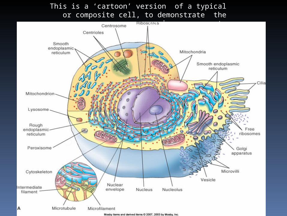
Slide 7
This is a ‘cartoon’ version of a typical or composite cell, to demonstrate the various components


‘actual’ cells are of varying shapes and sizes

Cell structures PLASMA MEMBRANE: separates the cell
from its surrounding environment
Primary structure of a cell membrane is a double layer of PHOSPHOLIPID MOLECULES
Heads are hydrophilic (“water loving”)
Tails are hydrophobic (“water fearing”)
Arrange themselves in BILAYERS in water
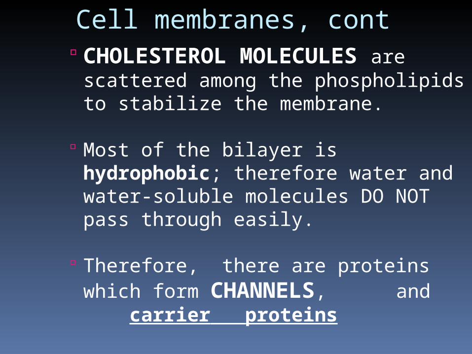
Cell membranes, cont CHOLESTEROL MOLECULES are
scattered among the phospholipids to stabilize the membrane.
Most of the bilayer is hydrophobic; therefore water and water-soluble molecules DO NOT pass through easily.
Therefore, there are proteins which form CHANNELS, and carrier proteins


The plasma (cell) membrane
39
40
41
From these choices, identify the structures:
A.Membrane channel protein E. GlycoproteinB.Hydrophobic tail F Phospholipid moleculeC.Cholesterol molecule G. Hydrophilic headD. Microtubule H. Lipoprotein

Membrane proteins A cell controls what moves through the
membrane by membrane proteins embedded in the phospholipid bilayer. ( carriers, channels)
Some membrane proteins have carbohydrates attached to them and, as a result, form GLYCOPROTEINS that act as identification markers
Some membrane proteins are RECEPTORS that react to specific chemicals, such as hormones

CYTOPLASM AND ORGANELLES
Cytoplasm: gel-like internal substance of cells that includes:
many organelles and cytoskeletal structures, and molecules of various types suspended in a watery intracellular
fluid also called CYTOSOL
The cytoplasm allows for movement of molecules, etc inside the cell

NUCLEUS
spherical body in center of cell; enclosed by an envelope with many pores
CONTAINS THE GENETIC MATERIAL – DNA MOLECULE within the
CHROMOSOME - ALSO SEEN AS CHROMATIN, when the cell is not dividing
(Interphase) Also inside the nucleus: NUCLEOLUS
- made up of RNA, it produces ribosomal subunits

NUCLEUS (cont.)
Structure (cont.) Contains DNA (heredity molecules), which
appear as: Chromatin threads or granules in nondividing
cells Chromosomes in early stages of cell division
Functions of the nucleus are functions of DNA molecules; DNA determines the structure and function of cells, as well as heredity.
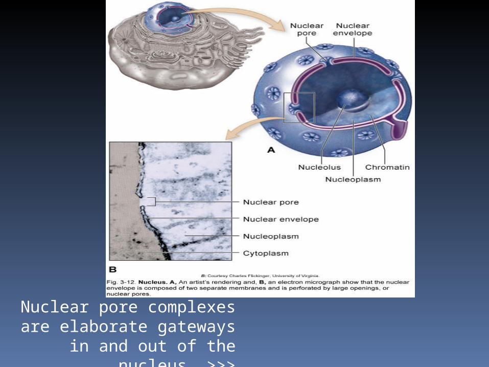
Nuclear pore complexes are elaborate gateways in and out
of the nucleus >>>


Mitochondria (Figure 3-11) wall composed of inner and outer membranes
separated by fluid; enzyme molecules are attached to both membranes
The “power plants” of cells; mitochondrial enzymes catalyze series of oxidation reactions that provide most of a cell’s energy supply
(CELLULAR RESPIRATION)
Each mitochondrion has a DNA molecule, which allows it to produce its own enzymes and replicate copies of itself
MITOCHONDRIA, the power houses of the cell


CYTOPLASM AND ORGANELLES (cont.) Endoplasmic reticulum (Figure 3-5)
Made of membranous, walled canals and flat, curving sacs arranged in parallel rows throughout the cytoplasm; extend from the plasma membrane to the nucleus
Proteins move through the canals, and are packaged

Endoplasmic reticulum

ENDOPLASMIC RETICULUM Two types of endoplasmic reticulum
Rough endoplasmic reticulum RIBOSOMES dot the outer surface of the membranous walls
Ribosomes synthesize proteins, which are moved toward the Golgi apparatus and then eventually leave the cell
Function in protein synthesis and intracellular transportation
Smooth endoplasmic reticulum No ribosomes border the membranous wall
Functions in packaging and storage - steroids and various ions :
Synthesizes certain lipids and carbohydrates and creates membranes for use throughout the cell
Removes and stores calcium ions from the cell’s interior

RIBOSOMES: make protein!
Many are attached to the rough endoplasmic reticulum
and many lie FREE, scattered throughout the cytoplasm
Each ribosome is a nonmembranous structure made of two pieces, a large subunit and a small subunit;
each subunit is composed of rRNA and protein

Ribosomes make protein
Ribosomes in the E.R. make proteins for “export,” or for the plasma membrane;
FREE ribosomes make proteins for the cell’s ‘domestic’, internal use

Ribosomes, two subunits
GOLGI APPARATUS
Membranous organelle consisting of
cisternae stacked on one another and located near the endoplasmic reticulums (Figure 3-7)
Processes protein molecules from the endoplasmic reticulum (Figure 3-8)
Processed proteins leave the final cisterna in a
vesicle; contents may then be secreted to outside the cell


(Janitors of the cells):Lysosomes, peroxisomes, and Proteosomes LYSOMOMES ; solid waste compactors and incinerators , for cellular debris and foreign invaders. ------ Abnormalities may lead to cellular injury and death
PEROXISOMES: chemical detoxifiers; TOXINS, such as ethanol
PROTEOSOMES : Recycle PROTEINS

LYSOSOMES
Lysosomes (Figure 3-9) Made of microscopic membranous
sacs that have “pinched off” from Golgi apparatus
THE CELL’S OWN DIGESTIVE SYSTEM; enzymes in lysosomes digest the protein structures of defective cell parts, including plasma membrane proteins, and particles that have become trapped in the cell


PEROXISOMES act as detoxifiers Peroxisomes
Small membranous sacs containing
enzymes that detoxify harmful substances that enter the cells
Often seen in kidney and liver cells

PROTEASOMES - BREAKDOWN DEFECTIVE PROTEINS Proteasomes (Figure 3-10)
Hollow protein cylinders found throughout the cytoplasm
Break down abnormal or misfolded proteins and normal proteins no longer needed by the cell (and that may cause disease)
Break down protein molecules one at a time by tagging each molecule, unfolding the protein as it enters the proteasome , and then breaking apart peptide bonds, RELEASING THE AMINO ACIDS ,
WHICH ARE THEN AVAILABLE FOR RECYCLING !!!!!!


CYTOSKELETON
The cell’s internal supporting framework;
made of rigid, rodlike pieces that provide support and allow movement and
mechanisms that can move the cell or its parts (Figure 3-14)


CYTOSKELETON (cont.)
Centrosome (Figure 3-16) near the nucleus coordinates the building and breaking apart
of microtubules in the cell Nonmembranous structure also called the
microtubule organizing center
Plays an important role during cell division
General location of the centrosome is identified by the centrioles


CYTOSKELETON (cont.)
Cell extensions Cytoskeleton forms projections that extend the plasma membrane outward to form tiny, fingerlike processes
Three types of these processes; each has specific functions (Figure 3-18)

CYTOSKELETON (cont.)Microvilli: found in epithelial cells that line the intestines and other areas where absorption is important; help increase the surface area manyfold


Cytoskelton, cell extensionsCilia and flagella: cell processes that have cylinders made of microtubules and molecular motors at their core
Cilia are shorter and more numerous than flagella; cilia have coordinated oarlike movements that brush material past the cell’s surface
FLAGELLA are found only on human sperm cells; flagella move with a tail-like movement that propels the sperm cell forward

Ciliated respiratory epithelium


Flagellated spermatazoa


CELL CONNECTIONS Cells are held together by fibrous nets
that surround groups of cells (e.g., muscle cells), or cells have direct connections to each
other
Three types of direct cell connections (Figure 3-20)


CELL CONNECTIONS: DIRECT DESMOSOME
Fibers on the outer surface of each desmosome interlock with each other; anchored internally by intermediate filaments of the cytoskeleton
Spot desmosomes are like “spot welds” at various points connecting adjacent membranes
Belt desmosomes encircle the entire cell Gap junctions: membrane channels of adjacent
plasma membranes adhere to each other; have two effects Form gaps or “tunnels” that join the cytoplasm of two
cells Fuse two plasma membranes into a single structure
TIGHT JUNCTIONS Occur in cells that are joined by “collars” of tightly
fused material Molecules cannot permeate the cracks of tight junctions Occur in the lining of the intestines and other parts of
the body where controlling what gets through a sheet of cells is important