
Fluorine assembly nanocluster breaks the shackles of ...
8
Fluorine assembly nanocluster breaks the shackles of immunosuppression to turn the cold tumor hot Zhaoting Li a,1 , Lianghan Zhu a,1 , Honghao Sun a , Yuexin Shen a , Dandan Hu a , Wenhao Wu a , Yixin Wang a , Chenggen Qian a , and Minjie Sun a,2 a State Key Laboratory of Natural Medicines, Department of Pharmaceutics, China Pharmaceutical University, Nanjing 210009, China Edited by Chad A. Mirkin, Northwestern University, Evanston, IL, and approved November 10, 2020 (received for review June 3, 2020) Clinical investigations have shown that a nonimmunogenic “cold” tumor is usually accompanied by few immunopositive cells and more immunosuppressive cells in the tumor microenvironment (TME), which is still the bottleneck of immune activation. Here, a fluorine assembly nanocluster was explored to break the shackles of immunosuppression, reawaken the immune system, and turn the cold tumor “hot.” Once under laser irradiation, FS@PMPt pro- duces sufficient reactive oxygen species (ROS) to fracture the ROS- sensitive linker, thus releasing the cisplatin conjugated PMPt to penetrate into the tumors and kill the regulatory T cells (Tregs) and myeloid-derived suppressor cells (MDSCs). Meanwhile, ROS will induce potent immunogenic cell death (ICD) and further pro- mote the accumulation of dendritic cells (DCs) and T cells, there- fore not only increasing the infiltration of immunopositive cells from the outside but also reducing the immunosuppressive cells from the inside to break through the bottleneck of immune acti- vation. The FS@PMPt nanocluster regulates the immune process in TME from negative to positive, from shallow to deep, to turn the cold tumor into a hot tumor and provoke a robust antitumor immune response. photodynamic immunotherapy | immunosuppressive tumor microenvironment | cold tumor | immunogenic cell death | nanocluster I n recent years, with the development of tumor immunology research, some scholars put forward the concept of “cold” and “hot” tumors to lead the future of antitumor immune research. In this concept, the number of tumor-infiltrating lymphocytes is taken as the scoring standard, and the tumor with more T cells and other positive immunoregulatory cells is called a hot tumor. On the contrary, a tumor with fewer or no positive immuno- regulatory cells and more immunosuppressive cells is called a cold tumor (1–3). Cold tumors are usually unrecognized by the immune system for various reasons and do not cause an effective immune response. In general, antigen-presenting cells (APCs) can no longer recognize such tumors, and T cells have been excluded from the tumor microenvironment (TME), which causes a huge obstacle for antitumor immunotherapy (4, 5). Clinical investigations point out that most tumors are cold, and regulating the immune process to turn a cold tumor into a hot tumor has become an urgent task of antitumor immunotherapy. Photodynamic therapy (PDT) could generate immunogenic cell death (ICD), accompanied by the release of high-mobility group box 1 protein (HMGB1) and adenosine triphosphate (ATP) and the exposure of calreticulin (CRT), sending the “eat- me” signal and promoting the antigen presentation and matu- ration of dendritic cells (DCs) (6, 7). ICD could recruit the DCs and antigen-specific cytotoxic T lymphocytes (CTLs) to the TME, thus turning the cold tumor into a hot tumor and acti- vating the antitumor immune response. Also, for the photody- namic immunotherapy, most researchers focus on developing more effective PDT strategies to provoke robust antitumor im- mune responses. Encouragingly, they have made remarkable progress in increasing the tumor infiltration of immunopositive regulatory cells, such as DCs and CTLs (8–10). However, the immunosuppressive TME of the cold tumor is known to impair the function of DCs and CTLs to greatly diminish the efficacy of the photodynamic immunotherapy. Notably, regulatory T cells (Tregs), supporting the establishment of an immunosuppressive TME, suppress T cell immune responses and activities of APCs, including DCs and macrophages, through several mechanisms. Tregs inhibit the expression of CD80 and CD86 via the cytotoxic T lymphocyte associated protein 4 (CTLA-4), which will directly compromise the ICD-induced APC maturation and activation. More lethally, Tregs can promote the apoptosis of effector T cells in the TME through cell to cell contact and interleukin-2 (IL-2) deprivation (11–13). In addition, Tregs could inhibit the immunostimulatory functions of DCs and infiltration of effector T cells by producing transforming growth factor-β (TGF-β) and IL-10. Interestingly, TGF-β can further lead to the differentia- tion of naive CD4 + T cells into Tregs and promote the Tregs accumulation in tumors (14–17). Similarly, myeloid-derived suppressor cells (MDSCs) can also recruit Tregs and promote their proliferation by secreting IL-10 and TGF-β. MDSCs, working hand in glove with Tregs, jointly maintain the immu- nosuppression of TME, prevent cold tumors from becoming hot, and greatly reduce the efficacy of photodynamic immunotherapy (18, 19). It is therefore of the utmost importance that we break the shackles of immunosuppression in the TME to turn a cold tumor into a hot tumor from the perspective of immune regulation. There is sufficient evidence to support that some chemother- apeutic drugs with a certain dosage, such as paclitaxel and cis- platin, are capable of decreasing the MDSCs and Tregs to regulate the immunosuppressive TME (20–24). Here, as shown Significance “Cold” tumors are good at camouflaging themselves, thus making it difficult for the immune system to recognize and to construct a great barrier for cancer immunotherapies. New methods that could awaken the immune system, enhance T cells and antigen-presenting cells (APCs) trafficking, and re- lieve immunosuppression to treat cold tumors are urgently in need. Here, we first report a chemically ingenious nanocluster FS@PMPt assembly by fluorine–fluorine interaction to regulate the immune process. The nanocluster not only increased the infiltration of immunopositive cells from the outside but also decreased the immunosuppressive cells from the inside to break the shackles of immunosuppression, which provides a promising para- digm for improving the anti –cold tumor immunotherapy. Author contributions: Z.L. and M.S. designed research; Z.L., L.Z., H.S., Y.S., D.H., W.W., and Y.W. performed research; Z.L. and L.Z. contributed new reagents/analytic tools; Z.L., L.Z., H.S., Y.S., Y.W., and C.Q. analyzed data; and Z.L. and M.S. wrote the paper. The authors declare no competing interest. This article is a PNAS Direct Submission. Published under the PNAS license. 1 Z.L. and L.Z. contributed equally to this work. 2 To whom correspondence may be addressed. Email: [email protected]. This article contains supporting information online at https://www.pnas.org/lookup/suppl/ doi:10.1073/pnas.2011297117/-/DCSupplemental. First published December 14, 2020. 32962–32969 | PNAS | December 29, 2020 | vol. 117 | no. 52 www.pnas.org/cgi/doi/10.1073/pnas.2011297117 Downloaded by guest on November 26, 2021
Transcript of Fluorine assembly nanocluster breaks the shackles of ...
Fluorine assembly nanocluster breaks the shackles ofimmunosuppression to turn the cold tumor hotZhaoting Lia,1, Lianghan Zhua,1, Honghao Suna
, Yuexin Shena, Dandan Hua, Wenhao Wua, Yixin Wanga,Chenggen Qiana
, and Minjie Suna,2
aState Key Laboratory of Natural Medicines, Department of Pharmaceutics, China Pharmaceutical University, Nanjing 210009, China
Edited by Chad A. Mirkin, Northwestern University, Evanston, IL, and approved November 10, 2020 (received for review June 3, 2020)
Clinical investigations have shown that a nonimmunogenic “cold”tumor is usually accompanied by few immunopositive cells andmore immunosuppressive cells in the tumor microenvironment(TME), which is still the bottleneck of immune activation. Here, afluorine assembly nanocluster was explored to break the shacklesof immunosuppression, reawaken the immune system, and turnthe cold tumor “hot.” Once under laser irradiation, FS@PMPt pro-duces sufficient reactive oxygen species (ROS) to fracture the ROS-sensitive linker, thus releasing the cisplatin conjugated PMPt topenetrate into the tumors and kill the regulatory T cells (Tregs)and myeloid-derived suppressor cells (MDSCs). Meanwhile, ROSwill induce potent immunogenic cell death (ICD) and further pro-mote the accumulation of dendritic cells (DCs) and T cells, there-fore not only increasing the infiltration of immunopositive cellsfrom the outside but also reducing the immunosuppressive cellsfrom the inside to break through the bottleneck of immune acti-vation. The FS@PMPt nanocluster regulates the immune process inTME from negative to positive, from shallow to deep, to turn thecold tumor into a hot tumor and provoke a robust antitumorimmune response.
photodynamic immunotherapy | immunosuppressive tumormicroenvironment | cold tumor | immunogenic cell death | nanocluster
In recent years, with the development of tumor immunologyresearch, some scholars put forward the concept of “cold” and
“hot” tumors to lead the future of antitumor immune research.In this concept, the number of tumor-infiltrating lymphocytes istaken as the scoring standard, and the tumor with more T cellsand other positive immunoregulatory cells is called a hot tumor.On the contrary, a tumor with fewer or no positive immuno-regulatory cells and more immunosuppressive cells is called acold tumor (1–3). Cold tumors are usually unrecognized by theimmune system for various reasons and do not cause an effectiveimmune response. In general, antigen-presenting cells (APCs)can no longer recognize such tumors, and T cells have beenexcluded from the tumor microenvironment (TME), whichcauses a huge obstacle for antitumor immunotherapy (4, 5).Clinical investigations point out that most tumors are cold, andregulating the immune process to turn a cold tumor into a hottumor has become an urgent task of antitumor immunotherapy.Photodynamic therapy (PDT) could generate immunogenic
cell death (ICD), accompanied by the release of high-mobilitygroup box 1 protein (HMGB1) and adenosine triphosphate(ATP) and the exposure of calreticulin (CRT), sending the “eat-me” signal and promoting the antigen presentation and matu-ration of dendritic cells (DCs) (6, 7). ICD could recruit the DCsand antigen-specific cytotoxic T lymphocytes (CTLs) to theTME, thus turning the cold tumor into a hot tumor and acti-vating the antitumor immune response. Also, for the photody-namic immunotherapy, most researchers focus on developingmore effective PDT strategies to provoke robust antitumor im-mune responses. Encouragingly, they have made remarkableprogress in increasing the tumor infiltration of immunopositiveregulatory cells, such as DCs and CTLs (8–10). However, theimmunosuppressive TME of the cold tumor is known to impair
the function of DCs and CTLs to greatly diminish the efficacy ofthe photodynamic immunotherapy. Notably, regulatory T cells(Tregs), supporting the establishment of an immunosuppressiveTME, suppress T cell immune responses and activities of APCs,including DCs and macrophages, through several mechanisms.Tregs inhibit the expression of CD80 and CD86 via the cytotoxicT lymphocyte associated protein 4 (CTLA-4), which will directlycompromise the ICD-induced APC maturation and activation.More lethally, Tregs can promote the apoptosis of effectorT cells in the TME through cell to cell contact and interleukin-2(IL-2) deprivation (11–13). In addition, Tregs could inhibit theimmunostimulatory functions of DCs and infiltration of effectorT cells by producing transforming growth factor-β (TGF-β) andIL-10. Interestingly, TGF-β can further lead to the differentia-tion of naive CD4+ T cells into Tregs and promote the Tregsaccumulation in tumors (14–17). Similarly, myeloid-derivedsuppressor cells (MDSCs) can also recruit Tregs and promotetheir proliferation by secreting IL-10 and TGF-β. MDSCs,working hand in glove with Tregs, jointly maintain the immu-nosuppression of TME, prevent cold tumors from becoming hot,and greatly reduce the efficacy of photodynamic immunotherapy(18, 19). It is therefore of the utmost importance that we breakthe shackles of immunosuppression in the TME to turn a cold tumorinto a hot tumor from the perspective of immune regulation.There is sufficient evidence to support that some chemother-
apeutic drugs with a certain dosage, such as paclitaxel and cis-platin, are capable of decreasing the MDSCs and Tregs toregulate the immunosuppressive TME (20–24). Here, as shown
Significance
“Cold” tumors are good at camouflaging themselves, thusmaking it difficult for the immune system to recognize and toconstruct a great barrier for cancer immunotherapies. Newmethods that could awaken the immune system, enhanceT cells and antigen-presenting cells (APCs) trafficking, and re-lieve immunosuppression to treat cold tumors are urgently inneed. Here, we first report a chemically ingenious nanoclusterFS@PMPt assembly by fluorine–fluorine interaction to regulatethe immune process. The nanocluster not only increased theinfiltration of immunopositive cells from the outside but alsodecreased the immunosuppressive cells from the inside to break theshackles of immunosuppression, which provides a promising para-digm for improving the anti–cold tumor immunotherapy.
Author contributions: Z.L. and M.S. designed research; Z.L., L.Z., H.S., Y.S., D.H., W.W., andY.W. performed research; Z.L. and L.Z. contributed new reagents/analytic tools; Z.L., L.Z.,H.S., Y.S., Y.W., and C.Q. analyzed data; and Z.L. and M.S. wrote the paper.
The authors declare no competing interest.
This article is a PNAS Direct Submission.
Published under the PNAS license.1Z.L. and L.Z. contributed equally to this work.2To whom correspondence may be addressed. Email: [email protected].
This article contains supporting information online at https://www.pnas.org/lookup/suppl/doi:10.1073/pnas.2011297117/-/DCSupplemental.
First published December 14, 2020.
32962–32969 | PNAS | December 29, 2020 | vol. 117 | no. 52 www.pnas.org/cgi/doi/10.1073/pnas.2011297117
Dow
nloa
ded
by g
uest
on
Nov
embe
r 26
, 202
1
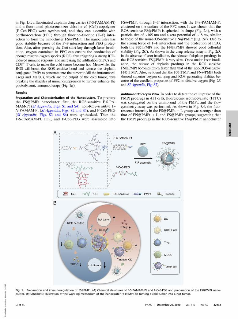
in Fig. 1A, a fluorinated cisplatin drug carrier (F-S-PAMAM-Pt)and a fluorinated photosensitizer chlorine e6 (Ce6) copolymer(F-Ce6-PEG) were synthesized, and they can assemble withperfluorocarbon (PFC) through fluorine–fluorine (F–F) inter-action to form the nanocluster FS@PMPt. The nanocluster hasgood stability because of the F–F interaction and PEG protec-tion. Also, after pressing the Ce6 start key through laser irradi-ation, oxygen contained in PFC can ensure the production ofenough reactive oxygen species (ROS), thus triggering a strong ICD-induced immune response and increasing the infiltration of DCs andCD8+ T cells to make the cold tumor become hot. Meanwhile, theROS will break the ROS-sensitive bond and release the cisplatinconjugated PMPt to penetrate into the tumor to kill the intratumoralTregs and MDSCs, which are the culprit of the cold tumor, thusbreaking the shackles of immunosuppression to further enhance thephotodynamic immunotherapy (Fig. 1B).
ResultsPreparation and Characterization of the Nanoclusters. To preparethe FS@PMPt nanocluster, first, the ROS-sensitive F-S-PA-MAM-Pt (SI Appendix, Figs. S1 and S4), non-ROS-sensitive F-N-PAMAM-Pt (SI Appendix, Figs. S2 and S5), and F-Ce6-PEG(SI Appendix, Figs. S3 and S6) were synthesized. Then theF-S-PAMAM-Pt, PFC, and F-Ce6-PEG were assembled into
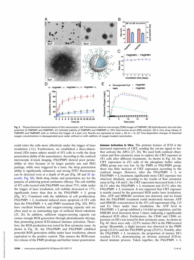
FS@PMPt through F–F interaction, with the F-S-PAMAM-Ptclustered on the surface of the PFC core. It was shown that theROS-sensitive FS@PMPt is spherical in shape (Fig. 2A), with aparticle size of ∼165 nm and a zeta potential of ∼18 mv, similarto those of the non-ROS-sensitive FN@PMPt (Fig. 2B). Due tothe strong force of F–F interaction and the protection of PEG,both the FS@PMPt and the FN@PMPt showed good colloidalstability (Fig. 2C). As shown in the drug release assay in Fig. 2D,in the absence of laser irradiation, the release of cisplatin prodrugs inthe ROS-sensitive FS@PMPt is very slow. Once under laser irradi-ation, the release of cisplatin prodrugs in the ROS sensitiveFS@PMPt becomes much faster than that of the non-ROS-sensitiveFN@PMPt. Also, we found that the FS@PMPt and FN@PMPt bothshowed superior oxygen carrying and ROS generating abilities be-cause of the excellent properties of PFC to dissolve oxygen (Fig. 2Eand SI Appendix, Fig. S7).
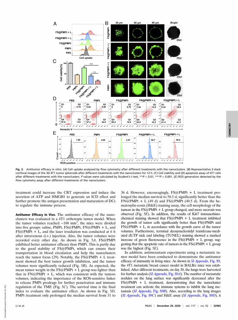
Antitumor Efficacy In Vitro. In order to detect the cell uptake of thePMPt prodrugs in 4T1 cells, fluoresceine isothiocyanate (FITC)was conjugated on the amino end of the PMPt, and the flowcytometry assay was performed. As shown in Fig. 3A, the fluo-rescence intensity in the FS@PMPt + L group was stronger thanthat of FN@PMPt + L and FS@PMPt groups, suggesting thatthe PMPt prodrugs in the ROS-sensitive FS@PMPt nanocluster
Fig. 1. Preparation and immunoregulation of FS@PMPt. (A) Chemical structures of F-S-PAMAM-Pt and F-Ce6-PEG and preparation of the FS@PMPt nano-cluster. (B) Schematic illustration of the working mechanism of the nanocluster FS@PMPt on turning a cold tumor into a hot tumor.
Li et al. PNAS | December 29, 2020 | vol. 117 | no. 52 | 32963
CHEM
ISTR
Y
Dow
nloa
ded
by g
uest
on
Nov
embe
r 26
, 202
1
could enter the cells more effectively under the trigger of laserirradiation (+L). Furthermore, we established a three-dimen-sional (3D) tumor sphere model of 4T1 cells to verify the deeppenetration ability of the nanoclusters. According to the confocalmicroscopic Z-stack imaging, FS@PMPt showed poor perme-ability in vitro because of its larger particle size and PEGpackage, while once triggered by a laser, the deep penetrationability is significantly enhanced, and strong FITC fluorescencecan be detected even at a depth of 60 μm (Fig. 3B and SI Ap-pendix, Fig. S8). Both drug intake and penetration are for thepurpose of achieving potent antitumor efficacy. The cell viabilityof 4T1 cells treated with FS@PMPt was about 75%, while underthe trigger of laser irradiation, cell viability decreased to 17%,significantly lower than that in the FN@PMPt + L group(Fig. 3C). Consistent with the inhibition of cell proliferation,FS@PMPt + L treatment induced more apoptosis of 4T1 cellsthan the FN@PMPt + L and PMPt treatment (Fig. 3D). PFCshave excellent biosafety and oxygen-carrying capacity and areoften used as an outstanding type of artificial blood substitute(25, 26). In addition, sufficient oxygen-carrying capacity canensure enough ROS generation through photodynamic therapy,thus promoting potent ICD-induced immune activation. There-fore, the ROS production was tested by flow cytometry assay. Asshown in Fig. 3E, the FN@PMPt and FS@PMPt exhibitedpowerful ROS generation ability under laser irradiation, almostequivalent to the positive control. This ensures the ROS-sensi-tive release of the PMPt prodrugs and further tumor penetration.
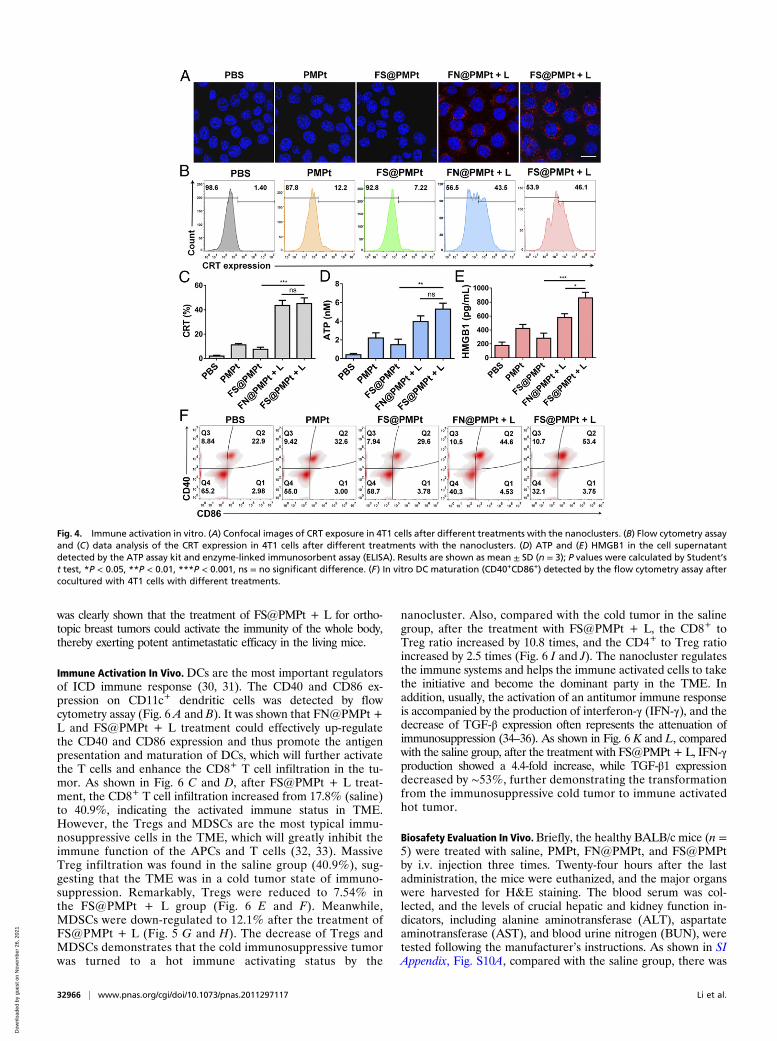
Immune Activation In Vitro. The primary feature of ICD is theincreased expression of CRT, sending the eat-me signal to fur-ther activate the APCs (27, 28). We used both confocal obser-vation and flow cytometry assay to explore the CRT exposure in4T1 cells after different treatments. As shown in Fig. 4A, theCRT expression in 4T1 cells of the phosphate buffer saline(PBS) group was very low. In the PMPt or FS@PMPt group,there was little increase of CRT expression according to theconfocal images. However, after the FN@PMPt + L orFS@PMPt + L treatment, significantly more CRT exposure wasobserved. Similarly, according to the results of flow cytometryassay in Fig. 4 B and C, the CRT expression increased from 1.4 to46.1% after the FS@PMPt + L treatment and 43.5% after theFN@PMPt + L treatment. It was supposed that CRT exposureis mainly caused by the generated ROS under laser irradiation.Also, ATP and HMGB1 secretion was detected, and we foundthat the FS@PMPt treatment could moderately increase ATPand HMGB1 concentration in the 4T1 cell supernatant (Fig. 4 Dand E). Once under laser irradiation, the ATP level inFS@PMPt + L group exhibited a 3.6-fold up-regulation, and theHMGB1 level increased about 3 times, indicating a significantlyenhanced ICD effect. Furthermore, the CD40 and CD86 ex-pression on DCs was tested by flow cytometry assay. As shown inFig. 4F, there were only ∼22.9% mature DCs in the PBS-treatedgroup. Meanwhile, a slight increase was observed in the PMPtgroup (32.6%) and the FS@PMPt group (29.6%). Notably, afterthe FS@PMPt + L treatment, the proportion of mature DCsreached 53.4%, demonstrating an obvious activated ICD-in-duced immune process. Taken together, the FS@PMPt + L
Fig. 2. Physicochemical characterization of the nanoclusters. (A) Transmission electron microscopy (TEM) images of FS@PMPt. (B) Hydrodynamic size and zetapotential of FS@PMPt and FN@PMPt. (C) Colloidal stability of FS@PMPt and FN@PMPt in 10% fetal bovine serum (FBS) solution. (D) In vitro drug release ofFS@PMPt and FN@PMPt with or without the trigger of a laser (+L). Results are expressed as mean ± SD (n = 3). (E) Time-dependent changes of dissolvedoxygen concentrations in deoxygenated pure water without or with addition of oxygen-loaded nanoclusters.
32964 | www.pnas.org/cgi/doi/10.1073/pnas.2011297117 Li et al.
Dow
nloa
ded
by g
uest
on
Nov
embe
r 26
, 202
1
treatment could increase the CRT expression and induce thesecretion of ATP and HMGB1 to generate an ICD effect andfurther promote the antigen presentation and maturation of DCsto regulate the immune process.
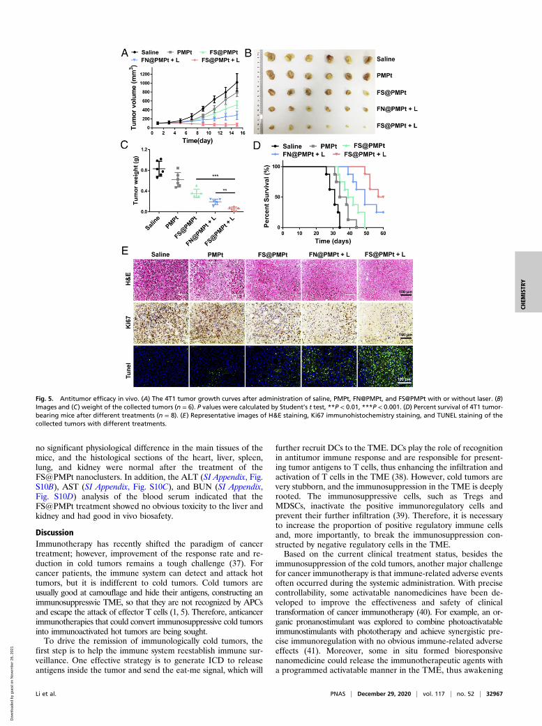
Antitumor Efficacy In Vivo. The antitumor efficacy of the nano-clusters was evaluated in a 4T1 orthotopic tumor model. Whenthe tumor volumes reached ∼100 mm3, the mice were dividedinto five groups: saline, PMPt, FS@PMPt, FN@PMPt + L, andFS@PMPt + L, and the laser irradiation was conducted at 6 hafter intravenous (i.v.) injection. Also, the tumor volumes wererecorded every other day. As shown in Fig. 5A, FS@PMPtexhibited better antitumor efficacy than PMPt. This is partly dueto the good stability of FS@PMPt, which can ensure theirtransportation in blood circulation and help the nanoclustersreach the tumor focus (29). Notably, the FS@PMPt + L treat-ment showed the best tumor growth inhibition, and the tumorvolumes were significantly reduced (Fig. 5B). As expected, themean tumor weight in the FS@PMPt + L group was lighter thanthat in FN@PMPt + L, which was consistent with the tumorsvolumes, indicating the importance of the ROS-sensitive linkerto release PMPt prodrugs for further penetration and immuneregulation of the TME (Fig. 5C). The survival time is the finalindex to evaluate the antitumor effect. As shown in Fig. 5D,PMPt treatment only prolonged the median survival from 31 to
36 d. However, encouragingly, FS@PMPt + L treatment pro-longed the median survival to 58.5 d, significantly better than theFN@PMPt + L (49 d) and FS@PMPt (40.5 d). From the he-matoxylin-eosin (H&E) staining assay, the cell morphology of thetumors in the FS@PMPt + L group changed, and more necrosis wasobserved (Fig. 5E). In addition, the results of Ki67 immunohisto-chemical staining showed that FS@PMPt + L treatment inhibitedthe growth of tumor cells significantly better than FS@PMPt andFN@PMPt + L, in accordance with the growth curve of the tumorvolumes. Furthermore, terminal deoxynucleotidyl transferase-medi-ated dUTP nick end labeling (TUNEL) staining showed a markedincrease of green fluorescence in the FS@PMPt + L group, sug-gesting that the apoptotic rate of tumors in the FS@PMPt + L groupwas the highest (Fig. 5E).In addition, antimetastasis experiments using a metastatic tu-
mor model have been conducted to demonstrate the antitumorefficacy of immunity in living mice. As shown in SI Appendix, Fig. S9,the 4T1 metastatic breast cancer model in BALB/c mice was estab-lished. After different treatments, on day 30, the lungs were harvestedfor further analysis (SI Appendix, Fig. S9A). The number of metastaticnodules on the lung surface was significantly decreased after theFS@PMPt + L treatment, demonstrating that the nanoclustertreatment can activate the immune systems to inhibit the lung me-tastasis (SI Appendix, Fig. S9B). Also, according to the lung images(SI Appendix, Fig. S9C) and H&E assay (SI Appendix, Fig. S9D), it
Fig. 3. Antitumor efficacy in vitro. (A) Cell uptake analyzed by flow cytometry after different treatments with the nanoclusters. (B) Representative Z-stackconfocal images of the 3D 4T1 tumor spheroids after different treatments with the nanoclusters for 12 h. (C) Cell viability and (D) apoptosis assay of 4T1 cellsafter different treatments with the nanoclusters. P values were calculated by Student’s t test, **P < 0.01, ***P < 0.001. (E) ROS generation detected by theflow cytometry assay after different treatments of the nanoclusters.
Li et al. PNAS | December 29, 2020 | vol. 117 | no. 52 | 32965
CHEM
ISTR
Y
Dow
nloa
ded
by g
uest
on
Nov
embe
r 26
, 202
1
was clearly shown that the treatment of FS@PMPt + L for ortho-topic breast tumors could activate the immunity of the whole body,thereby exerting potent antimetastatic efficacy in the living mice.
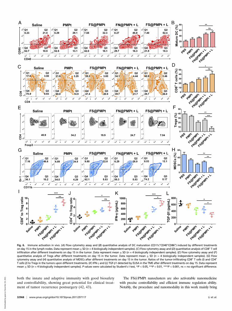
Immune Activation In Vivo.DCs are the most important regulatorsof ICD immune response (30, 31). The CD40 and CD86 ex-pression on CD11c+ dendritic cells was detected by flowcytometry assay (Fig. 6 A and B). It was shown that FN@PMPt +L and FS@PMPt + L treatment could effectively up-regulatethe CD40 and CD86 expression and thus promote the antigenpresentation and maturation of DCs, which will further activatethe T cells and enhance the CD8+ T cell infiltration in the tu-mor. As shown in Fig. 6 C and D, after FS@PMPt + L treat-ment, the CD8+ T cell infiltration increased from 17.8% (saline)to 40.9%, indicating the activated immune status in TME.However, the Tregs and MDSCs are the most typical immu-nosuppressive cells in the TME, which will greatly inhibit theimmune function of the APCs and T cells (32, 33). MassiveTreg infiltration was found in the saline group (40.9%), sug-gesting that the TME was in a cold tumor state of immuno-suppression. Remarkably, Tregs were reduced to 7.54% inthe FS@PMPt + L group (Fig. 6 E and F). Meanwhile,MDSCs were down-regulated to 12.1% after the treatment ofFS@PMPt + L (Fig. 5 G and H). The decrease of Tregs andMDSCs demonstrates that the cold immunosuppressive tumorwas turned to a hot immune activating status by the
nanocluster. Also, compared with the cold tumor in the salinegroup, after the treatment with FS@PMPt + L, the CD8+ toTreg ratio increased by 10.8 times, and the CD4+ to Treg ratioincreased by 2.5 times (Fig. 6 I and J). The nanocluster regulatesthe immune systems and helps the immune activated cells to takethe initiative and become the dominant party in the TME. Inaddition, usually, the activation of an antitumor immune responseis accompanied by the production of interferon-γ (IFN-γ), and thedecrease of TGF-β expression often represents the attenuation ofimmunosuppression (34–36). As shown in Fig. 6 K and L, comparedwith the saline group, after the treatment with FS@PMPt + L, IFN-γproduction showed a 4.4-fold increase, while TGF-β1 expressiondecreased by ∼53%, further demonstrating the transformationfrom the immunosuppressive cold tumor to immune activatedhot tumor.
Biosafety Evaluation In Vivo.Briefly, the healthy BALB/c mice (n =5) were treated with saline, PMPt, FN@PMPt, and FS@PMPtby i.v. injection three times. Twenty-four hours after the lastadministration, the mice were euthanized, and the major organswere harvested for H&E staining. The blood serum was col-lected, and the levels of crucial hepatic and kidney function in-dicators, including alanine aminotransferase (ALT), aspartateaminotransferase (AST), and blood urine nitrogen (BUN), weretested following the manufacturer’s instructions. As shown in SIAppendix, Fig. S10A, compared with the saline group, there was
Fig. 4. Immune activation in vitro. (A) Confocal images of CRT exposure in 4T1 cells after different treatments with the nanoclusters. (B) Flow cytometry assayand (C) data analysis of the CRT expression in 4T1 cells after different treatments with the nanoclusters. (D) ATP and (E) HMGB1 in the cell supernatantdetected by the ATP assay kit and enzyme-linked immunosorbent assay (ELISA). Results are shown as mean ± SD (n = 3); P values were calculated by Student’st test, *P < 0.05, **P < 0.01, ***P < 0.001, ns = no significant difference. (F) In vitro DC maturation (CD40+CD86+) detected by the flow cytometry assay aftercocultured with 4T1 cells with different treatments.
32966 | www.pnas.org/cgi/doi/10.1073/pnas.2011297117 Li et al.
Dow
nloa
ded
by g
uest
on
Nov
embe
r 26
, 202
1
no significant physiological difference in the main tissues of themice, and the histological sections of the heart, liver, spleen,lung, and kidney were normal after the treatment of theFS@PMPt nanoclusters. In addition, the ALT (SI Appendix, Fig.S10B), AST (SI Appendix, Fig. S10C), and BUN (SI Appendix,Fig. S10D) analysis of the blood serum indicated that theFS@PMPt treatment showed no obvious toxicity to the liver andkidney and had good in vivo biosafety.
DiscussionImmunotherapy has recently shifted the paradigm of cancertreatment; however, improvement of the response rate and re-duction in cold tumors remains a tough challenge (37). Forcancer patients, the immune system can detect and attack hottumors, but it is indifferent to cold tumors. Cold tumors areusually good at camouflage and hide their antigens, constructing animmunosuppressvie TME, so that they are not recognized by APCsand escape the attack of effector T cells (1, 5). Therefore, anticancerimmunotherapies that could convert immunosuppressive cold tumorsinto immunoactivated hot tumors are being sought.To drive the remission of immunologically cold tumors, the
first step is to help the immune system reestablish immune sur-veillance. One effective strategy is to generate ICD to releaseantigens inside the tumor and send the eat-me signal, which will
further recruit DCs to the TME. DCs play the role of recognitionin antitumor immune response and are responsible for present-ing tumor antigens to T cells, thus enhancing the infiltration andactivation of T cells in the TME (38). However, cold tumors arevery stubborn, and the immunosuppression in the TME is deeplyrooted. The immunosuppressive cells, such as Tregs andMDSCs, inactivate the positive immunoregulatory cells andprevent their further infiltration (39). Therefore, it is necessaryto increase the proportion of positive regulatory immune cellsand, more importantly, to break the immunosuppression con-structed by negative regulatory cells in the TME.Based on the current clinical treatment status, besides the
immunosuppression of the cold tumors, another major challengefor cancer immunotherapy is that immune-related adverse eventsoften occurred during the systemic administration. With precisecontrollability, some activatable nanomedicines have been de-veloped to improve the effectiveness and safety of clinicaltransformation of cancer immunotherapy (40). For example, an or-ganic pronanostimulant was explored to combine photoactivatableimmunostimulants with phototherapy and achieve synergistic pre-cise immunoregulation with no obvious immune-related adverseeffects (41). Moreover, some in situ formed bioresponsivenanomedicine could release the immunotherapeutic agents witha programmed activatable manner in the TME, thus awakening
Fig. 5. Antitumor efficacy in vivo. (A) The 4T1 tumor growth curves after administration of saline, PMPt, FN@PMPt, and FS@PMPt with or without laser. (B)Images and (C) weight of the collected tumors (n = 6). P values were calculated by Student’s t test, **P < 0.01, ***P < 0.001. (D) Percent survival of 4T1 tumor-bearing mice after different treatments (n = 8). (E) Representative images of H&E staining, Ki67 immunohistochemistry staining, and TUNEL staining of thecollected tumors with different treatments.
Li et al. PNAS | December 29, 2020 | vol. 117 | no. 52 | 32967
CHEM
ISTR
Y
Dow
nloa
ded
by g
uest
on
Nov
embe
r 26
, 202
1
both the innate and adaptive immunity with good biosafetyand controllability, showing great potential for clinical treat-ment of tumor recurrence postsurgery (42, 43).
The FS@PMPt nanoclusters are also activatable nanomedicinewith precise controllability and efficient immune regulation ability.Notably, the procedure and nanomodality in this work mainly bring
Fig. 6. Immune activation in vivo. (A) Flow cytometry assay and (B) quantitative analysis of DC maturation (CD11c+CD40+CD86+) induced by different treatmentson day 15 in the lymph nodes. Data represent mean ± SD (n = 4 biologically independent samples). (C) Flow cytometry assay and (D) quantitative analysis of CD8+ T cellinfiltration after different treatments on day 15 in the tumor. Data represent mean ± SD (n = 4 biologically independent samples). (E) Flow cytometry assay and (F)quantitative analysis of Tregs after different treatments on day 15 in the tumor. Data represent mean ± SD (n = 4 biologically independent samples). (G) Flowcytometry assay and (H) quantitative analysis of MDSCs after different treatments on day 15 in the tumor. Ratios of the tumor-infiltrating CD8+ T cells (I) and CD4+
T cells (J) to Tregs in the tumors upon different treatments. (K) IFN-γ and (L) TGF-β1 detected by ELISA in the TME after different treatments on day 15. Data representmean ± SD (n = 4 biologically independent samples). P values were calculated by Student’s t test, *P < 0.05, **P < 0.01, ***P < 0.001, ns = no significant difference.
32968 | www.pnas.org/cgi/doi/10.1073/pnas.2011297117 Li et al.
Dow
nloa
ded
by g
uest
on
Nov
embe
r 26
, 202
1
two inspirations to the clinical treatment of cancer immunotherapy.First, for the immunological cold tumors, drugs or treatments thatcould generate ICD should be combined with Tregs or MDSCs in-hibitors to achieve synergistic immunoregulation and reverse immu-nosuppression. Second, precise activatable nanomedicines ornanomodalities could be developed to incorporate clinical combina-tion strategies in a unified manner with superior biosafety andeffectiveness.Overall, in this study, we developed an immunomodulatory
nanocluster FS@PMPt to reverse the immunosuppressive TMEand turn a cold tumor into a hot tumor. FS@PMPt was assem-bled through F–F interaction with good stability and oxygen-carrying capacity to guarantee the production of sufficient ROSfor potent ICD induction. Hence, the ICD induced by thenanocluster, accompanied by the exposure of CRT and release ofATP and HMGB1, promotes the maturation and antigen presenta-tion of DCs both in vitro and in vivo. Also, significantly increasedCD8+ T cell infiltration was observed after the treatment of thenanocluster to further heat up the cold tumor and light the flames ofthe antitumor war. Meanwhile, the released PMPt prodrugs, workingsynergistically with the ICD induction, penetrated into the tumor tokill the Tregs and MDSCs, which are the main drivers of immuno-suppression in a cold tumor. This immunomodulatory nanocluster
FS@PMPt not only increased the infiltration of immunopositive cellsfrom the outside but also decreased the immunosuppressive cellsfrom the inside to break the shackles of immunosuppression in theTME, which provides a promising paradigm for improving theanti–cold tumor immunotherapy.
Materials and MethodsExperimental materials and methods for the synthesis, preparation, andcharacterization of the nanoclusters, immune activation, in vitro antitumorefficacy, and in vivo animal experiments are provided in SI Appendix. Allanimal experiments followed regulations of the Institutional Animal Careand Use Committee of China Pharmaceutical University, and protocols wereapproved by the Science and Technology Department of Jiangsu Province.
Data Availability. All study data are available in the article and SI Appendix.
ACKNOWLEDGMENTS. This work was supported by the National KeyResearch and Development Program of China (grant number:2017YFA0205402), the Science Fund for Distinguished Young Scholars ofJiangsu Province, China (grant number: BK20170028), the Natural ScienceFoundation of Jiangsu Province (BK20180557), the China National ScienceFoundation (grant numbers: 81573377, 81872817, and 81803477), and the“Double First-Class” University Project of China Pharmaceutical University(grant number: CPU2018GY07).
1. J. H. Newman et al., Intratumoral injection of the seasonal flu shot converts immu-
nologically cold tumors to hot and serves as an immunotherapy for cancer. Proc. Natl.
Acad. Sci. U.S.A. 117, 1119–1128 (2020).
2. N. Zaidi, E. M. Jaffee, Immune cells track hard-to-target brain tumours. Nature 565,
170–171 (2019).
3. J. Li et al., Tumor cell-intrinsic factors underlie heterogeneity of immune cell infil-
tration and response to immunotherapy. Immunity 49, 178–193.e7 (2018).
4. M. McLaughlin et al., Inflammatory microenvironment remodelling by tumour cells
after radiotherapy. Nat. Rev. Cancer 20, 203–217 (2020).
5. J. Galon, D. Bruni, Approaches to treat immune hot, altered and cold tumours with
combination immunotherapies. Nat. Rev. Drug Discov. 18, 197–218 (2019).
6. Z. Meng et al., Light-triggered in situ gelation to enable robust photodynamic-im-
munotherapy by repeated stimulations. Adv. Mater. 31, e1900927 (2019).
7. X. Feng et al., Immunomodulatory nanosystems. Adv. Sci. (Weinh.) 6, 1900101 (2019).
8. C. W. Ng, J. Li, K. Pu, Recent progresses in phototherapy-synergized cancer immu-
notherapy. Adv. Funct. Mater. 28, 1804688 (2018).
9. C. Zhang et al., Enzyme-driven membrane-targeted chimeric peptide for enhanced
tumor photodynamic immunotherapy. ACS Nano 13, 11249–11262 (2019).
10. R. Liang et al., Oxygen-boosted immunogenic photodynamic therapy with gold
nanocages@manganese dioxide to inhibit tumor growth and metastases. Biomate-
rials 177, 149–160 (2018).
11. S. Musetti, L. Huang, Nanoparticle-mediated remodeling of the tumor microenvi-
ronment to enhance immunotherapy. ACS Nano 12, 11740–11755 (2018).
12. M. P. Matheu et al., Imaging regulatory T cell dynamics and CTLA4-mediated sup-
pression of T cell priming. Nat. Commun. 6, 6219 (2015).
13. P. Pandiyan, L. Zheng, S. Ishihara, J. Reed, M. J. Lenardo, CD4+CD25+Foxp3+ regu-
latory T cells induce cytokine deprivation-mediated apoptosis of effector CD4+ T cells.
Nat. Immunol. 8, 1353–1362 (2007).
14. L. Wang et al., Connecting blood and intratumoral Treg cell activity in predicting fu-
ture relapse in breast cancer. Nat. Immunol. 20, 1220–1230 (2019).
15. H. Wang, F. Franco, P. C. Ho, Metabolic regulation of Tregs in cancer: Opportunities
for immunotherapy. Trends Cancer 3, 583–592 (2017).
16. Y. Mi, C. T. Hagan IV, B. G. Vincent, A. Z. Wang, Emerging nano-/microapproaches for
cancer immunotherapy. Adv. Sci. (Weinh.) 6, 1801847 (2019).
17. A. Facciabene, G. T. Motz, G. Coukos, T-regulatory cells: Key players in tumor immune
escape and angiogenesis. Cancer Res. 72, 2162–2171 (2012).
18. X. R. Ros, L. Vermeulen, Turning cold tumors hot by blocking TGF-β. Trends Cancer 4,335–337 (2018).
19. N. Kamran et al., Immunosuppressive myeloid cells’ blockade in the glioma micro-
environment enhances the efficacy of immune-stimulatory gene therapy. Mol. Ther.
25, 232–248 (2017).
20. A. R. de Biasi, J. Villena-Vargas, P. S. Adusumilli, Cisplatin-induced antitumor im-
munomodulation: A review of preclinical and clinical evidence. Clin. Cancer Res. 20,
5384–5391 (2014).
21. C. W. Tseng et al., Pretreatment with cisplatin enhances E7-specific CD8+ T-cell-me-
diated antitumor immunity induced by DNA vaccination. Clin. Cancer Res. 14,
3185–3192 (2008).
22. C. L. Chang et al., Dose-dense chemotherapy improves mechanisms of antitumor
immune response. Cancer Res. 73, 119–127 (2013).
23. J. Guo, Z. Yu, M. Das, L. Huang, Nano codelivery of oxaliplatin and folinic acid ach-
ieves synergistic chemo-immunotherapy with 5-fluorouracil for colorectal cancer and
liver metastasis. ACS Nano 14, 5075–5089 (2020).
24. Q. Song et al., Tumor microenvironment responsive nanogel for the combinatorial an-
titumor effect of chemotherapy and immunotherapy. Nano Lett. 17, 6366–6375 (2017).
25. P. Yu et al., Artificial red blood cells constructed by replacing heme with per-
fluorodecalin for hypoxia‐induced radioresistance. Adv. Ther. 2, 1900031 (2019).
26. Z. Zhou et al., Perfluorocarbon nanoparticle-mediated platelet inhibition promotes
intratumoral infiltration of T cells and boosts immunotherapy. Proc. Natl. Acad. Sci.
U.S.A. 116, 11972–11977 (2019).
27. W. L. Liu et al., Expandable immunotherapeutic nanoplatforms engineered from
cytomembranes of hybrid cells derived from cancer and dendritic cells. Adv. Mater.
31, e1900499 (2019).
28. Z. Wang et al., Janus nanobullets combine photodynamic therapy and magnetic hy-
perthermia to potentiate synergetic anti-metastatic immunotherapy. Adv. Sci.
(Weinh.) 6, 1901690 (2019).
29. G. Chen et al., Reversibly stabilized polycation nanoparticles for combination treatment
of early- and late-stage metastatic breast cancer. ACS Nano 12, 6620–6636 (2018).
30. R. Kuai et al., Elimination of established tumors with nanodisc-based combination
chemoimmunotherapy. Sci. Adv. 4, eaao1736 (2018).
31. N. Kanaya et al., Immune modulation by telomerase-specific oncolytic adenovirus
synergistically enhances antitumor efficacy with anti-PD1 antibody. Mol. Ther. 28,
794–804 (2020).
32. D. Ha et al., Differential control of human Treg and effector T cells in tumor immunity by
Fc-engineered anti-CTLA-4 antibody. Proc. Natl. Acad. Sci. U.S.A. 116, 609–618 (2019).
33. P. Zhang et al., Therapeutic targeting of tumor-associated myeloid cells synergizes with
radiation therapy for glioblastoma. Proc. Natl. Acad. Sci. U.S.A. 116, 23714–23723 (2019).
34. D. V. F. Tauriello et al., TGFβ drives immune evasion in genetically reconstituted colon
cancer metastasis. Nature 554, 538–543 (2018).
35. S. Mariathasan et al., TGFβ attenuates tumour response to PD-L1 blockade by con-
tributing to exclusion of T cells. Nature 554, 544–548 (2018).
36. Z. Li et al., Targeting pulmonary tumor microenvironment with CXCR4-inhibiting
nanocomplex to enhance anti-PD-L1 immunotherapy. Sci. Adv. 6, eaaz9240 (2020).
37. A. Mansurov et al., Collagen-binding IL-12 enhances tumour inflammation and drives
the complete remission of established immunologically cold mouse tumours. Nat.
Biomed. Eng. 4, 531–543 (2020).
38. A. D. Garg et al., Dendritic cell vaccines based on immunogenic cell death elicit
danger signals and T cell-driven rejection of high-grade glioma. Sci. Transl. Med. 8,
328ra27 (2016).
39. M. D. Sharma et al., The PTEN pathway in Tregs is a critical driver of the suppressive
tumor microenvironment. Sci. Adv. 1, e1500845 (2015).
40. C. Zhang, K. Pu, Molecular and nanoengineering approaches towards activatable
cancer immunotherapy. Chem. Soc. Rev. 49, 4234–4253 (2020).
41. J. Li et al., Organic semiconducting pro-nanostimulants for near-infrared photoactivatable
cancer immunotherapy. Angew. Chem. Int. Ed. Engl. 58, 12680–12687 (2019).
42. C. Wang et al., In situ formed reactive oxygen species-responsive scaffold with gemcitabine
and checkpoint inhibitor for combination therapy. Sci. Transl. Med. 10, eaan3682 (2018).
43. Q. Chen et al., In situ sprayed bioresponsive immunotherapeutic gel for post-surgical
cancer treatment. Nat. Nanotechnol. 14, 89–97 (2019).
Li et al. PNAS | December 29, 2020 | vol. 117 | no. 52 | 32969
CHEM
ISTR
Y
Dow
nloa
ded
by g
uest
on
Nov
embe
r 26
, 202
1


